Cardiac Resynchronization Therapy: Current Practice, Refining Implantation Methods, Effects on
Ventricular Arrhythmias and New Indications
Ph.D. Thesis
Valentina Kutyifa M.D.
M.Sc. in Health Service Management
Semmelweis University
Doctoral School of Basic Medical Sciences
Supervisor: Béla Merkely M.D., Ph.D., D.Sc.
Official Reviewers: Lívia Jánoskúti M.D., Ph.D.
Gábor Duray M.D., Ph.D.
Head of the Comprehensive Exam Committee: István Préda M.D., Ph.D.
Members of the Comprehensive Exam Committee:
Gábor Veress M.D., Ph.D.
András Zsáry M.D., Ph.D.
Budapest 2012
2
Preface
Invictus
Out of the night that covers me, Black as the Pit from pole to pole, I thank whatever gods may be For my unconquerable soul.
In the fell clutch of circumstance I have not winced nor cried aloud.
Under the bludgeonings of chance My head is bloody, but unbowed.
Beyond this place of wrath and tears Looms but the Horror of the shade, And yet the menace of the years Finds, and shall find, me unafraid.
It matters not how strait the gate,
How charged with punishments the scroll.
I am the master of my fate:
I am the captain of my soul.
William Ernest Henley
3
Table of Content
Preface ... 2
Table of Content ... 3
Abbreviations ... 7
1 Introduction ... 8
1.1 Current Practice of Cardiac Resynchronization Therapy ... 8
1.2 MADIT-CRT ... 10
1.3 Refining Implantation Methods ... 12
1.3.1 Prognostic Significance of Right Ventricular to Left Ventricular Interlead Sensed Electrical Delay in CRT Patients ... 12
1.3.2 Electroanatomical Mapping Guided Transseptal Endocardial LV Lead Implantation ... 12
1.4 Effects of CRT on Ventricular Arrhythmias ... 13
1.4.1 Left Ventricular Lead Location and the Risk of Ventricular Tachyarrhythmias ... 13
1.4.2 Left Ventricular Dyssynchrony and the Risk of Ventricular Tachyarrhythmias ... 14
1.5 New Indications of CRT ... 14
1.5.1 Chronic Right Ventricular Apical Pacing ... 14
1.5.2 Cardiac Resynchronization Therapy in Patients with Less Severe Ventricular Dysfunction ... 15
2 Aims ... 17
2.1 Current Practice of Cardiac Resynchronization Therapy ... 17
2.1.1 Evaluating the Effects of CRT-P versus CRT-D in a CRT Registry ... 17
2.2 Refining Implantation Methods ... 17
2.2.1 Prognostic significance of right ventricular to left ventricular interlead sensed electrical delay in CRT patients ... 17
2.2.2 Electroanatomical Mapping-Guided Transseptal Endocardial LV Lead Implantation ... 17
2.3 Effects of CRT on Ventricular Arrhythmias ... 18
2.3.1 Left Ventricular Lead Location and the Risk of Ventricular Tachyarrhythmias ... 18
2.3.2 Left Ventricular Dyssynchrony and the Risk of Ventricular Tachyarrhythmias ... 18
4
2.4 New indications of CRT ... 18
2.4.1 Chronic Right Ventricular Apical Pacing ... 18
2.4.2 Cardiac Resynchronization Therapy in Patients with Less Severe Ventricular Dysfunction ... 18
3 Methods ... 19
3.1 Current Practice of Cardiac Resynchronization Therapy ... 19
3.1.1 Evaluating the Effects of CRT-P versus CRT-D in a CRT Registry ... 19
3.2 Refining Implantation Methods ... 21
3.2.1 Prognostic Significance of Right Ventricular to Left Ventricular Interlead Sensed Electrical Delay in CRT Patients ... 21
3.2.2 Electroanatomical Mapping-Guided Transseptal Endocardial LV Lead Implantation ... 22
3.3 Effects of CRT on Ventricular Arrhythmias ... 26
3.3.1 Left Ventricular Lead Location and the Risk of Ventricular Tachyarrhythmias ... 26
3.3.2 Left Ventricular Dyssynchrony and the Risk of Ventricular Tachyarrhythmias ... 30
3.4 New indications of CRT ... 34
3.4.1 Chronic Right Ventricular Apical Pacing ... 34
3.4.2 Cardiac Resynchronization Therapy in Patients with Less Severe Ventricular Dysfunction ... 35
3.5 Statistical Considerations ... 37
3.5.1 Current Practice of Cardiac Resynchronization Therapy, Evaluating the effects of CRT-P versus CRT-D in the total patient population: Statistical considerations ... 37
3.5.2 Refining Implantation Methods: Statistical considerations ... 38
3.5.3 Left Ventricular Lead Location and the Risk of Ventricular Tachyarrhythmias: Statistical considerations ... 39
3.5.4 Left Ventricular Dyssynchrony and the Risk of Ventricular Tachyarrhythmias: Statistical considerations ... 40
3.5.5 Chronic Right Ventricular Apical Pacing: Statistical considerations ... 41
3.5.6 Cardiac Resynchronization Therapy in Patients with Less Severe Ventricular Dysfunction: Statistical considerations ... 42
4 Results ... 43
4.1 Current Practice of Cardiac Resynchronization Therapy ... 43
4.1.1 Evaluating the Effects of CRT-P versus CRT-D in a CRT Registry ... 43
4.2 Refining Implantation Methods ... 48
5
4.2.1 Prognostic Significance of Right Ventricular to Left Ventricular Interlead Sensed Electrical Delay
in CRT Patients ... 48
4.2.2 Electroanatomical Mapping-Guided Transseptal Endocardial LV Lead Implantation ... 51
4.3 Effects of CRT on Ventricular Arrhythmias ... 55
4.3.1 Left Ventricular Lead Location and the Risk of Ventricular Tachyarrhythmias ... 55
4.3.2 Left Ventricular Dyssynchrony and the Risk of Ventricular Tachyarrhythmias ... 64
4.4 New indications of CRT ... 78
4.4.1 Chronic Right Ventricular Apical Pacing ... 78
4.4.2 Cardiac Resynchronization Therapy in Patients with Less Severe Ventricular Dysfunction ... 87
5 Discussion ... 97
5.1 Current Practice of Cardiac Resynchronization Therapy ... 97
5.1.1 Evaluating the Effects of CRT-P versus CRT-D in a CRT Registry ... 97
5.2 Refining Implantation Methods ... 98
5.2.1 Prognostic Significance of Right Ventricular to Left Ventricular Interlead Sensed Electrical Delay in CRT Patients ... 98
5.2.2 Electroanatomical Mapping-Guided Transseptal Endocardial LV Lead Implantation ... 99
5.3 Effects of CRT on Ventricular Arrhythmias ... 100
5.3.1 Left Ventricular Lead Location and the Risk of Ventricular Tachyarrhythmias ... 100
5.3.2 Left Ventricular Dyssynchrony and the Risk of Ventricular Tachyarrhythmias ... 103
5.4 New indications of CRT ... 105
5.4.1 Chronic Right Ventricular Apical Pacing ... 105
5.4.2 Cardiac Resynchronization Therapy in Patients with Less Severe Ventricular Dysfunction ... 106
6 Conclusions ... 109
6.1 Current Practice of Cardiac Resynchronization Therapy ... 109
6.1.1 Evaluating the effects of CRT-P versus CRT-D in a CRT Registry ... 109
6.2 Refining Implantation Methods ... 109
6.2.1 Prognostic Significance of Right Ventricular to Left Ventricular Interlead Sensed Electrical Delay in CRT Patients ... 109
6.2.2 Electroanatomical Mapping-Guided Transseptal Endocardial LV Lead Implantation ... 109
6.3 Effects of CRT on Ventricular Arrhythmias ... 110
6.3.1 Left Ventricular Lead Location and the Risk of Ventricular Tachyarrhythmias ... 110
6
6.3.2 Left Ventricular Dyssynchrony and the Risk of Ventricular Tachyarrhythmias ... 110
6.4 New indications of CRT ... 110
6.4.1 Chronic Right Ventricular Apical Pacing ... 110
6.4.2 Cardiac Resynchronization Therapy in Patients with Less Severe Ventricular Dysfunction ... 111
7 Summary ... 112
8 Összefoglalás ... 113
9 References ... 114
10 Publications ... 128
10.1 Publications closely related to the present thesis ... 128
10.1.1 Original articles ... 128
10.2 Publications not related to the present thesis ... 129
10.2.1 Original articles ... 129
10.2.2 Articles in Hungarian ... 130
10.2.3 Book chapters ... 130
Acknowledgements ... 131
7
Abbreviations
CI Confidence Interval
CRT Cardiac Resynchronization Therapy
CRT-P Cardiac Resynchronization Therapy with Pacemaker Capabilities CRT-D Cardiac Resynchronization Therapy with Defibrillator
HF Heart Failure
ICD Implantable Cardioverter Defibrillator IVD Interventricular Delay
JTc JT Interval Corrected for Heart Rate LV Left Ventricle
LAV Left Atrial Volume
LBBB Left Bundle Branch Block
LVEF Left Ventricular Ejection Fraction LVEDD Left Ventricular End-Diastolic Diameter LVEDV Left Ventricular End-Diastolic Volume LVESD Left Ventricular End-Systolic Diameter LVESV Left Ventricular End-Systolic Volume NYHA New York Heart Association
PM Pacemaker
RV Right Ventricular
TDR Transmural Dispersion of Repolarization VT Ventricular Tachycardia
VF Ventricular Fibrillation
8
1 Introduction
1.1 Current Practice of Cardiac Resynchronization Therapy
Heart Failure (HF) is a growing public health problem worldwide with rising prevalence due to aging, improved medical treatment and successful prevention of cardiac events. The prevalence of heart failure is 2-3% in developed European countries, while it might reach 10-20% in the elderly.1
Cardiac resynchronization therapy (CRT) provides synchronization of the dyssynchronous left ventricular activation in patients with conduction abnormalities and severely reduced left ventricular function, resulting in an immediate decrease of left ventricular intra- and interventricular dyssynchrony, mitral regurgitation and an acute increase of LV dP/dt.2 During long-term follow-up, patients develop reduction in left ventricular end-diastolic (LVEDV) and left ventricular end-systolic volume (LVESV), and improvement in left ventricular ejection fraction (LVEF), this process is described as left ventricular reverse remodeling.3-12
Cardiac resynchronization therapy or a combination of CRT with an Implantable Cardioverter Defibrillator (CRT-D) was proven to reduce heart failure symptoms, hospitalizations and mortality in patients with severe heart failure (NYHA class III-IV), reduced left ventricular ejection fraction (LVEF≤35%) and a prolonged QRS (QRS width≥120 ms).10, 13
The Multicenter Automatic Defibrillator Implantation Trial – Cardiac Resynchronization Therapy (MADIT-CRT), the Resynchronization-Defibrillation in Ambulatory Heart Failure Trial (RAFT) and Resynchronization Reverses Remodeling in Systolic Left Ventricular Dysfunction (REVERSE) trials have further broadened CRT indication to patients with mild HF and NYHA class I and II.6, 7, 12 Table 1 is summarizing the main randomized clinical trials on CRT.3-12
9
Table 1. Randomized clinical trials on CRT
Trials
Patients (n)
Female
(%) Primary endpoints Secondary endpoints
Etiology (isch. %)
LVEF (%)
QRS (ms)
PATH-CHF 41 50% 6MWT, peak VO2
NHYA class, QOL,
Hospitalizations 29% 21±7 175
MUSTIC-SR 58 26% 6MWT
NYHA, QOL, Peak VO2,
MR, LV, Hosp, Mortality 37% 23±7 174
MIRACLE 453 32%
6MWT, NHYA, QOL
Peak VO2, LVEF, LVEDD, MR, Clin
Response 54% 22±6 166
MIRACLE
ICD 555 23%
6MWT, NYHA, QOL
Peak VO2, LVEF, LV volumes, MR, Clinical
Response 70% 24±6 164
COMPANION 1520 22%
All-cause mortality or hospitalization
All-cause mortality and
cardiac mortality 56% 21 159
CARE-HF 814 26% All-cause mortality
NYHA, QOL, LVEF, LVESV, Hospitalization
for heart failure 38% 25 160
REVERSE 610 21%
HF clinical
composite score LVESVi 54% 27±7 153
MADIT-CRT 1820 25% HF or death
LVESV, LVEDV change,
multiple HF events 57% 24±5 162
RAFT 1798 17%
All-cause mortality
or HF
hospitalization
All-cause mortality, cardiac mortality, HF
hospitalization 67% 23±5 158
6MWT, 6-min walk test; CARE-HF, Cardiac Resynchronization-Heart Failure; COMPANION, Comparison of Medical Therapy, Pacing and Defibrillation in Heart Failure; HF, heart failure; LV, left ventricular; LVEDD, left ventricular end-diastolic dimension; LVEF, left ventricular ejection fraction; LVESV, left ventricular end-systolic volume; MADITCRT, Multicenter Automatic Defibrillator Implantation Trial–Cardiac Resynchronization Therapy;
MIRACLE, Multicenter InSync Randomized Clinical Evaluation; MIRACLE ICD, Multicenter InSync Implantable Cardioverter Defibrillator trial; MR, mitral regurgitation; MUSTIC, Multisite Simulation in Cardiomyopathies;
NYHA, New York Heart Association; PATH-CHF, Pacing Therapies in Congestive Heart Failure trial; QOL, quality-of-life score; RAFT, Resynchronization-Defibrillation for Ambulatory Heart Failure; REVERSE, Resynchronization Reverses Remodeling in Systolic Left Ventricular Dysfunction; VO2, volume of oxygen.
10
Besides multicenter randomized clinical trials, multi- and single-center registries are also important sources for providing real-world information on CRT. It is well-known, that patients with NYHA class IV and inotropes, those with severe renal dysfunction, or on dialysis, those who are having coexisting malignant diseases, or previously implanted pacemakers are excluded from randomized clinical studies. However, evaluating the effects of CRT in registry patients might refine treatment delivery, and potentially expand the use of CRT in patients who may not have been included in clinical trials.14
1.2 MADIT-CRT
Patients with heart failure and reduced left ventricular function are at increased risk for arrhythmia-related sudden cardiac death. Implantation of an implantable cardioverter–
defibrillator (ICD) reduces mortality and the risk of sudden death in selected patients with cardiac disease.15 However, life-prolonging defibrillator therapy might be associated with an increased risk of first and recurrent heart-failure events.16
The MADIT-CRT trial was designed to investigate whether CRT–D therapy would prevent death or nonfatal heart-failure events (whichever came first) in mild heart failure patients as compared with ICD-only treatment.17
From December 22, 2004, through April 23, 2008, a total of 1820 patients who had ischemic or non-ischemic cardiomyopathy, an ejection fraction (LVEF) less than 30%, prolonged intraventricular conduction with a QRS > 130 ms were randomized to receive CRT-D or ICD therapy in a 3:2 ratio in 110 hospital centers: 1271 patients at 88 centers in the United States, 22 patients at 2 centers in Canada, and 527 patients at 20 centers in Europe.
Patients were excluded if they had an existing indication for CRT, if they received a
11
pacemaker, had NYHA class III/IV less than 90 days before enrolment, underwent coronary artery bypass graft surgery or percutaneous coronary intervention, or had myocardial infarction within the past 90 days prior to enrolment.
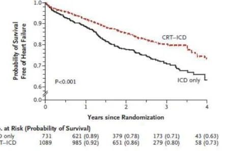
Figure 1. MADIT-CRT trial. Kaplan-Meier Cumulative Probability of Survival Free of Heart Failure Stratified by Treatment Arm
During the average follow-up of 29 months, the primary end point occurred in 187 of 1089 patients in the CRT–D group (17.2%) and 185 of 731 patients in the ICD-only group (25.3%). This indicates a 34% reduction in the risk of death or heart failure events (whichever came first) in the CRT–D group, as compared to the ICD-only treated patients. This effect was driven by a 41% reduction in the risk of heart failure in CRT-D patients as compared to those who received ICD-only (Figure 1). During the study, 127 deaths occurred of any cause representing a low 3% annual mortality rate. CRT-D treatment was associated with significant reduction in left ventricular (LV) volumes and improvement in left ventricular ejection fraction.18
12
1.3 Refining Implantation Methods
1.3.1 Prognostic Significance of Right Ventricular to Left Ventricular Interlead Sensed Electrical Delay in CRT Patients
Cardiac resynchronization therapy is most commonly achieved using a standard approach to implant the LV lead in a lateral or postero-lateral coronary sinus branch.19, 20 However, recent studies suggested that implanting the LV lead at the latest LV mechanical activation segment might provide a better resynchronization effect with CRT.21, 22
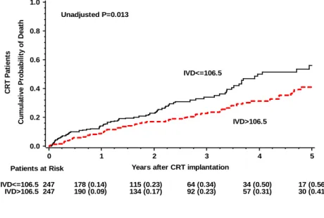
Right ventricular to left ventricular interlead sensed electrical delay, interventricular delay (IVD) is the time delay on the sensed electrogram between RV and LV sensing signals.
This parameter is measured during CRT implantation, after positioning the right and left ventricular leads. Essentially, this measure is representing the electrical time delay between the right and left ventricle at the right and left ventricular lead position.
Previous studies failed to predict left ventricular reverse remodeling using the right to left ventricular interlead electrical delay.23-25 However, it was shown to be correlated with intraventricular dyssynchrony,24, 25 a well-known powerful predictor of response to CRT.26-28
1.3.2 Electroanatomical Mapping Guided Transseptal Endocardial LV Lead Implantation The left ventricular lead is usually implanted using a transvenous approach. However, even with innovative lead technology, LV lead placement fails in 4-8%.29, 30 The most frequent reasons are coronary sinus occlusion or dissection, abnormal ostium of the coronary sinus, coronary vein stenosis, lead instability, high threshold or phrenic nerve stimulation.29, 31, 32
Epicardial lead placement is an alternative method, which includes minimal-invasive thoracoscopy or lateral thoracotomy and usually requires general anaesthesia.33 When epicardial LV lead implantation is
13
contraindicated or at higher risk, LV endocardial lead implantation might be considered.34
CRT is typically delivered aiming a lateral or posterolateral LV lead position,19, 29, 35 and the LV lead is usually placed in the middle or distal portion of the side-branch to ensure stable position. In contrast, when using endocardial transseptal approach, the LV lead position is independent of the coronary vein anatomy. The major difficulty of this method is to relocate the transfemoral transseptal puncture site performed from the subclavian access and to find the optimal LV lead position within the LV cavity.
Electroanatomical mapping is visualizing cardiac structures and gathering data of electrical activation of the heart. It is widely used to guide ablation procedures in the left atrium, in the right ventricle and in the left ventricle.36 Electroanatomical mapping might be a useful tool to guide endocardial LV lead implantation for CRT.
1.4 Effects of CRT on Ventricular Arrhythmias
1.4.1 Left Ventricular Lead Location and the Risk of Ventricular Tachyarrhythmias
Heart failure patients are at higher risk of ventricular tachycardia (VT) or ventricular fibrillation (VF),37 even after receiving CRT.
The Cardiac Resynchronization in Heart Failure (CARE-HF) extension trial reported 7.8% of sudden cardiac death during the mean follow-up of 29.4 months in patients receiving CRT.38 In the Cardiac-Resynchronization Therapy with or without an Implantable Defibrillator in Advanced Heart Failure (COMPANION) study, 19.3% of the CRT-D patients experienced appropriate ICD therapy by the second year after device implantation. ICD shock therapy is associated with worse outcome.29, 39
There are several risk factors contributing to the occurrence of ventricular
14
tachyarrhythmias, ischemic events, depressed left ventricular function, increased ventricular wall stress, renal dysfunction and atrial fibrillation.29, 40 Some data also indicate that there is a potential pro-arrhythmic risk of biventricular pacing itself.41-45 However, other studies demonstrated anti-arrhythmic effects of CRT, explained by the improved hemodynamic status and left ventricular reverse remodeling.22, 46-48
It is currently unknown, whether left ventricular lead location might play a role in the development of VT/VF, possibly by enhancing electrical heterogeneity.
1.4.2 Left Ventricular Dyssynchrony and the Risk of Ventricular Tachyarrhythmias
Intraventricular mechanical dyssynchrony might also play an important role in the development of VT/VF by abnormal mechanical and subsequent electrical activation inducing electrical heterogeneity.
The effects of LV lead location and LV dyssynchrony on ventricular arrhythmias have not yet been investigated in mild heart failure patients with implanted CRT-D or ICD.
1.5 New Indications of CRT
1.5.1 Chronic Right Ventricular Apical Pacing
Large randomized trials have demonstrated the adverse effects of chronic right ventricular (RV) apical pacing associated with increased risk of atrial fibrillation and heart failure.49, 50 Up to 40%
of patients with chronic RV apical pacing develop heart failure during long-term follow-up.49, 50 These detrimental effects might be attributed to the altered electrical and mechanical activation
15
of the ventricles resulting in left ventricular (LV) dyssynchrony, impaired LV filling, perfusion defects and myofibrillar disarrays.51
Randomized clinical trials showing beneficial effects of CRT were performed only in patients with de novo implantations4, 8, 10, 11
however, according to registry data more than one quarter of CRT implantations are upgrades of implanted pacemaker devices.52
Previous smaller studies demonstrated the efficacy, feasibility and safety of CRT upgrade in patients with RV apical pacing compared to de novo CRT implantation.53-57 However, we have no data on the outcome of patients with implantable cardioverter-defibrillator (ICD) upgraded to CRT. Additionally, predictors of long-term outcome have not yet been investigated in this patient cohort.
1.5.2 Cardiac Resynchronization Therapy in Patients with Less Severe Ventricular Dysfunction
Using pre-selected cut-off point for left ventricular ejection fraction as an inclusion criterion for CRT is considered an arbitrary method, as patients develop heart failure across the spectrum of left ventricular ejection fraction. Depressed LVEF was shown to be a surrogate marker of heart failure status and is associated with increased risk of adverse events, all-cause mortality and sudden cardiac death.58-60 However, the risk associated with LVEF was shown to be a continuum until the range of 45%.59 Therefore, there is a rationale for CRT in patients with less depressed LVEF.
In MADIT-CRT, the inclusion criteria comprised patients with LVEF below or equal to 30%, as evaluated by the enrolling centers prior to enrollment. All patients additionally underwent central echocardiographic analysis of LVEF in the study core laboratory of Brigham
16
and Women’s Hospital, Harvard Medical School in Boston, Massachusetts, where a substantial proportion of patients were identified to have an LVEF of greater than 30%, beyond the eligibility criteria. This provides unique opportunity to evaluate the efficacy of CRT-D in patients with less decreased cardiac function.
17
2 Aims
2.1 Current Practice of Cardiac Resynchronization Therapy
2.1.1 Evaluating the Effects of CRT-P versus CRT-D in a CRT Registry
In this analysis, we sought to evaluate the long-term echocardiographic and clinical outcome of CRT patients in a single-center high-volume registry, and to assess the all-cause mortality of patients with an implanted CRT-P or CRT-D device.
2.2 Refining Implantation Methods
2.2.1 Prognostic significance of right ventricular to left ventricular interlead sensed electrical delay in CRT patients
We aimed to determine the prognostic significance of right to left ventricular interlead sensed electrical delay on the end point of all-cause mortality in CRT patients of the single-center, high- volume registry.
2.2.2 Electroanatomical Mapping-Guided Transseptal Endocardial LV Lead Implantation We sought to evaluate the feasibility and safety of transseptal endocardial left ventricular lead implantation in a small patient cohort of the single-center, high volume CRT registry.
Furthermore, we aimed to determine whether electroanatomic mapping guided left ventricular lead targeting might be associated with better clinical and echocardiographic improvement after CRT implantation.
18
2.3 Effects of CRT on Ventricular Arrhythmias
2.3.1 Left Ventricular Lead Location and the Risk of Ventricular Tachyarrhythmias
We aimed to analyze the association between LV lead position and the risk of VT/VF/Death or VT/VF in patients enrolled in the MADIT-CRT trial.
2.3.2 Left Ventricular Dyssynchrony and the Risk of Ventricular Tachyarrhythmias
We sought to investigate the association between left ventricular dyssynchrony, CRT-induced change in LV dyssynchrony and the risk of VT/VF/Death or VT/VF events in LBBB and non- LBBB patients enrolled in MADIT-CRT.
2.4 New indications of CRT
2.4.1 Chronic Right Ventricular Apical Pacing
The aim of this analysis was to evaluate the effects of CRT upgrade in ICD patients with chronic RV pacing compared to PM patients with chronic RV pacing, and to identify predictors of long- term outcome in this patient population.
2.4.2 Cardiac Resynchronization Therapy in Patients with Less Severe Ventricular Dysfunction
We aimed to evaluate the relationship between LVEF and the clinical outcome of mild heart failure patients enrolled in MADIT-CRT; the echocardiographic response to CRT-D in the trial;
and the clinical benefit of CRT-D, with a specific focus on the subset of patients with more preserved LVEF enrolled in the trial.
19
3 Methods
3.1 Current Practice of Cardiac Resynchronization Therapy
3.1.1 Evaluating the Effects of CRT-P versus CRT-D in a CRT Registry 3.1.1.1 Patient Population
From June 2000 to April 2011, 1122 consecutive patients had undergone CRT device implantation at the Semmelweis University Heart Center, Budapest, Hungary. Patients met the guideline criteria for CRT, including New York Heart Association (NYHA) class II, III or IV, QRS ≥ 120 ms, LVEF ≤ 35% and optimal medical treatment including beta-blocker, ACE- inhibitor or ARB therapy, diuretics and aldosterone antagonist, unless contraindicated or not tolerated by the patient. Optimization of the medical therapy was performed according to current guidelines.61 All patients gave written informed consent before the procedure.
3.1.1.2 Pre-implant Assessment
Diagnostic coronary angiography and revascularization was performed if indicated. Baseline clinical characteristics were recorded prior CRT implantation. Two-dimensional transthoracal echocardiography was performed before CRT implantation and during follow-up using commercially available systems (Toshiba Aplio, Toshiba Medical Systems Co, Ltd, Tokyo, Japan, and Philips iE33, Andover, Massachusetts, USA). Left ventricular end-diastolic and end- systolic diameters (LVEDD, LVESD) and left ventricular ejection fraction were measured according to standard methods.62
20 3.1.1.3 Device implantation
CRT device implantation was performed using transvenous, epicardial or transseptal approach.
Patients in sinus rhythm or those with paroxysmal atrial fibrillation were implanted a right atrial lead and right ventricular lead, while patients in permanent atrial fibrillation received right and left ventricular leads only. During the implantation procedure after cannulating the coronary sinus, balloon catheter was used to perform coronary sinus venogram and to identify the target vein for CRT therapy, preferably the lateral or postero-lateral vein. Left ventricular pacing, sensing and impedance were measured. Phrenic nerve stimulation was tested in supine body position using 10 V at 0.5 ms pacing of the LV lead at the end of CRT implantation.
Commercially available LV leads and CRT devices were used. If the patient received a CRT device with ICD capabilities, ventricular fibrillation (VF) testing was performed at implantation according to current standards to achieve a safety margin of at least 10 J.
3.1.1.4 Post-implant Assessment
All patients were scheduled for outpatient visit one month after the implantation and every 6- month thereafter. Clinical status assessment and device follow-up was performed at each follow- up visit or at any meaningful clinical event. Two-dimensional echocardiography was performed 6 months after CRT upgrade and every 12-month thereafter or in case of heart failure progression. Echocardiographic data available at last follow up (median 20 months, IQR: 10-38 months) were analyzed.
3.1.1.5 Study End Points
The primary end point of this analysis was all-cause mortality. Secondary end points included
21
improvement in NYHA functional class, increase in left ventricular ejection fraction and decrease in left ventricular end-diastolic and end-systolic volumes. Mortality data were collected from medical records, phone follow-up, and using the mortality database of the Hungarian National Health Fund.
3.2 Refining Implantation Methods
3.2.1 Prognostic Significance of Right Ventricular to Left Ventricular Interlead Sensed Electrical Delay in CRT Patients
3.2.1.1 Patient Population
From June 2000 to April 2011, 494 of 1122 patients (44%) undergoing CRT implantation at the Semmelweis University Heart Center, Budapest, Hungary and had measurements of right to left ventricular interlead sensed electrical delay. Patients met the guideline criteria for CRT, including New York Heart Association (NYHA) class II, III or IV, QRS ≥ 120 ms, LVEF ≤ 35%
and optimal medical treatment including beta-blocker, ACE-inhibitor or ARB therapy, diuretics and aldosterone antagonist, unless contraindicated or not tolerated by the patient. Optimization of the medical therapy was performed according to current guidelines.61 All patients gave written informed consent before the procedure.
3.2.1.2 Pre-, Post-implant Assessment
Pre- and post-implant assessment was performed as explained in section 3.1.1.2. and 3.1.1.4.
3.2.1.3 Device Implantation
CRT device implantation was performed according to standard methods as described in section
22
3.1.1.3. (Current practice of cardiac resynchronization therapy-methods).
During implantation, after positioning the right and left ventricular leads we connected the right and left ventricular leads to an electrophysiology system (Biotronik, Berlin, Germany) and measured the right to left interventricular sensed delay by the time delay of the peak activation in the right and left ventricular sensed signals (ms).
3.2.1.4 Study End Points
The end point of this analysis was death of any cause. Mortality data were collected from hospital records, using follow-up of the patients and the mortality database of the Hungarian National Health Fund.
3.2.2 Electroanatomical Mapping-Guided Transseptal Endocardial LV Lead Implantation 3.2.2.1 Study Population
Four patients had undergone endocardial LV lead implantation at the Semmelweis University Heart Center, Budapest, Hungary between November 2007 and May 2010, guided by electroanatomical mapping. Patients met the indication criteria for CRT according to current guidelines.63 All patients had left bundle branch block (LBBB) or paced rhythm with LBBB- morphology.
CRT was attempted or performed either via a transvenous or an epicardial approach.
Patient 1 had epicardial LV lead dysfunction, a repeated surgery was contraindicated therefore the patient was referred for endocardial LV lead implantation. Patient 2 and 4 had unsuccessful transvenous LV lead implantation. Patient 3 had LV lead dysfunction after successful transvenous LV lead implantation. In Patient 2 and 3, mini-thoracotomy was contraindicated
23
because of multiple comorbidities and higher risk of surgical intervention. Patient 1 and 4 did not give the consent for epicardial surgical LV lead implantation. All patients had given informed consent prior to procedure.
3.2.2.2 LV Lead Implantation Procedure
Left ventricular endocardial lead implantation was performed using a combined femoral and subclavian approach guided by electroanatomical mapping. The first step of the procedure was to introduce the CARTO Quick Star catheter (Biosense Webster, Diamond Bar, CA) through the right femoral vein to capture the anatomical map of the right atrium and the right ventricle. The transseptal puncture was performed with the guidance of both fluoroscopy and intracardiac echocardiography, including continuous monitoring of the arterial pressure. Intravenous heparin was given after the transseptal puncture (5000 IU), in case of long-lasting procedures it was administered repeatedly to maintain an ACT level of 250 msec.
The puncture point of the septum was marked on the CARTO map (Biosense Webster, Diamond Bar, CA) (Figure 2). A guide wire (0.035 inch*260 cm) was inserted into the left atrium (LA) and advanced into the left upper pulmonary vein. The dilator of the transseptal sheath was removed and an angioplasty balloon (6mm*20mm Maverick, Boston Scientific, Natick, MA, USA) was inserted into the LA. The transseptal sheath was withdrawn into the right atrium and the balloon was positioned across the septal puncture site. It was inflated 3 times with 12 atm for 5 seconds before its removal. The transseptal sheath was then positioned into the LA cavity.
Figure 2. Patient 1. CARTO image, AP projection. The location of the transseptal puncture is indicated with a single white arrow on the CARTO map.
24
The Quick Star deflectable catheter was inserted into the LA and advanced in the LV cavity via the right femoral vein. LV activation map was recorded (Figure 3).
LA
RA
25
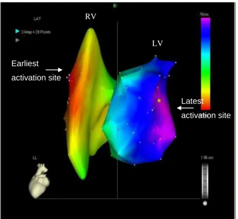
Figure 3. Patient 1. CARTO image, left lateral projection. Right and the left ventricular activation map: the earliest activation site is the right ventricular anteroseptal region, the latest one is the mid-basal part of the posterolateral wall.
An 11 F long sheath (SCOUT Pro 8 Fr, Biotronik GmbH&Co, Berlin, Germany) was introduced via the left subclavian vein. The Quick Star catheter was advanced into the sheath and guided to the location of the transseptal puncture by CARTO location guidance. At this time, a second angioplasty balloon (6mm*20mm Maverick, Boston Scientific, Natick, MA, USA) was positioned to the puncture site through the previously applied guide-wire from the femoral access to facilitate the manipulation of the Quick Star catheter across the septum. The long sheath was pushed over the deflectable catheter through the interatrial septum into the LA and further into
Earliest activation site
RV
LV
Latest
activation site
26
the LV. The Quick Star catheter was used to relocate the LV segment with the latest activation.
At this step, the Quick Star catheter was withdrawn into the sheath and the sheath was pushed against the left ventricular wall to ensure stable position. Active fixation LV leads were implanted at the most delayed area of the LV via the sheath. The leads were fixed at the basal or mid-basal portion of the left ventricle in all patients where the latest activation was detected on the activation map. The sheath was pushed against the LV wall to have a stable support and facilitate to position the LV lead close to the mitral valve. Standard unipolar srew-in leads were used in all patients (Medtronic- 5076-52cm, Medtronic, Minneapolis, MN, n=1; Medtronic 5076- 65cm, Medtronic, Minneapolis, MN, n=2 and Vitatron, ICQ09B-58 cm, Medtronic, Minneapolis, MN, n=1). The LV lead was connected to the CRT device placed in the left pectoral area.
3.3 Effects of CRT on Ventricular Arrhythmias
3.3.1 Left Ventricular Lead Location and the Risk of Ventricular Tachyarrhythmias 3.3.1.1 Evaluation of LV Lead Locations
LV lead position was evaluated by biplane coronary venograms and anterior/posterior, lateral chest X-rays in patients enrolled in the Multicenter Automatic Defibrillator Implantation Trial - Cardiac Resynchronization Therapy. At the time of CRT implantation, coronary venous angiograms were obtained in at least 2 orthogonal views (Right Anterior Oblique- RAO and Left Anterior Oblique- LAO) as well as fluoroscopic images in the same views after definitive LV lead placement. Anterior-posterior and lateral chest X-rays were performed after the procedure or prior to discharge. The stored images were copied onto a CD-ROM and sent to the core laboratory at the University of Rochester Medical Center for central reading. The study protocol recommended positioning the LV lead in the lateral or postero-lateral side-branch of the
27
coronary sinus if possible.
The final LV lead position was assessed in the longitudinal axis view (RAO 20°-40°) and the short axis view (LAO 20°-40°) together with the anterior/posterior and lateral chest-X ray. In case the LV lead images were not available in both angle views, stored at completely different angles or showed poor quality making lead assessment impossible, the lateral chest-X rays were used to define the final lead position.
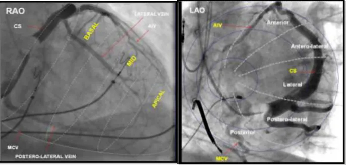
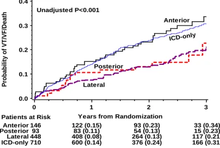
The LAO view, representing the short-axis view of the heart was used to classify the left ventricular wall into 3 equal parts; anterior, lateral (antero-lateral, lateral, postero-lateral) and posterior. The RAO view, representing the long axis of the heart, was used to distinct the lead position to be basal, mid-ventricular or apical.36, 64 We defined an anterior, lateral and posterior LV lead location along the short axis, including all basal and mid-ventricular lead locations. We also grouped patients with apical versus non-apical (basal and mid-ventricular) LV lead location along the short axis (Figure 4).
28
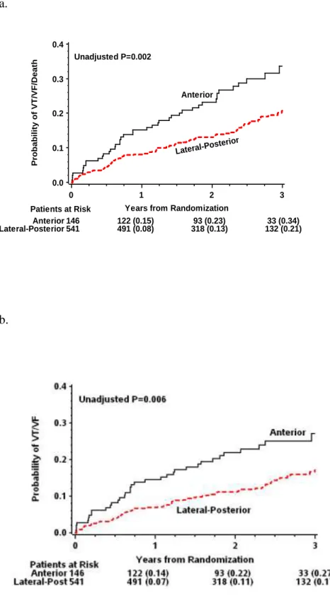
Figure 4. Classification of LV lead location. Sinus venograms in RAO (left panel) and LAO view (right panel) representing the segments of the left ventricle along the long- and the short axis of the heart. In this analysis, the antero-lateral, lateral and postero-lateral segments were grouped as lateral lead location. Lateral and posterior locations were grouped together as lateral- posterior. Reproduced with permission (license number: 2884921460326, Circulation).
We compared anterior, lateral, posterior and apical LV-lead locations; anterior versus lateral-posterior and apical lead location along short axis as well as the apical versus non-apical lead positions along the long-axis of the heart.
3.3.1.2 Study Population
We were able to analyze LV lead location in 797 of 1089 (73%) patients who received CRT-D devices and were followed over a mean of 30.6 (±10.9) months. The following patients were not included in the analysis: those who needed a cross-over to ICD only (n=66, 6.1%) or to CRT-D (n=2, 0.2%), who were withdrawn prior to device implantation (n=56, 5.1%), who underwent LV
29
lead repositioning more than one week after initial CRT device implantation because of lead dislodgement (n=54, 5%), those, who had epicardial LV lead placement (n=36, 3.3%) or cases with incomplete data-sets of device implantation venograms and X-rays (n=78, 7.2%).
3.3.1.3 Device Programming and Interrogation
Commercially available transvenous ICD and CRT-D devices (Boston Scientific) were used in the trial. Standard techniques were used to implant the devices. Device testing and programming were performed as reported in the study protocol.65 Devices were programmed to monitor + therapy, with protocol recommendation to a setting of ventricular tachycardia (VT) zone at 180 bpm, and ventricular fibrillation (VF) zone at 250 bpm. Sensitivity was programmed according to physician discretion. Detection was 1.0 second for the VF zone and 2.5 seconds for the VT zone. The protocol recommended to program VT zone first therapy to burst-type antitachycardia pacing (ATP), then shock therapy; second therapy should be shock at defibrillation threshold plus at least 10 J. The remaining therapies should be maximal energy shocks. All device interrogation disks were sent to an independent core laboratory for categorization and final evaluation of detected arrhythmias.
3.3.1.4 Patient Follow-Up
Patients had outpatient follow-up 1-month after CRT-D or ICD implantation and every 3 months thereafter until the termination of the trial. The mean follow-up of the enrolled patients was 29.4 months. All patients had clinical evaluation and ICD interrogation with retrieval of stored electrograms at each follow up visit or at any meaningful clinical events.
30 3.3.1.5 End Points
The primary end point of the current study was the first occurrence of appropriate therapy for ventricular tachycardia (VT) or ventricular fibrillation (VF) or death assessed as the cumulative probability of first events or risk of events. All ICD interrogations were adjudicated by an independent, blinded core laboratory reviewing the electrograms of the episodes for categorization and final evaluation of the detected arrhythmias. Definition of VT was set to a rate from 180 bpm (recommended programming) up to 250 bpm, V rate ≥ A rate if 1:1 A:V, V-V changes drive AA changes. VF was defined as ventricular rate faster, than 250 beats/min with disorganized ventricular electrograms. Only appropriate therapy, antitachycardia pacing (ATP) or shock delivery for VT or VF was considered in the present analysis.
We analyzed VT/VF and rapid VT/VF episodes (rate ≥ 200 bpm) as separate end points.
We evaluated VT/VF events requiring ICD shock or death, as well as recurrent VT/VF events (≥2 VT/VF episodes in one patient). We also analyzed all-cause mortality of the subgroups.
3.3.2 Left Ventricular Dyssynchrony and the Risk of Ventricular Tachyarrhythmias 3.3.2.1 Echocardiographic Methods
Echocardiography investigators and sonographers from each enrolling sites were qualified to perform echocardiography according to the approved echocardiography protocol. Recordings were analyzed off-line at the Brigham and Women's Hospital, Boston, Massachusetts as an independent echocardiography core laboratory.
LV mechanical dyssynchrony was measured using B-mode speckle tracking software (TomTec Imaging Systems, Unterschleissheim, Germany) as reported previously.66 Briefly, endocardial borders were traced in the end-systolic frame of the 2D images from the apical four-
31
and two-chamber views. Speckles were tracked frame-by-frame using two or more cardiac cycles. Segments if needed were manually adjusted. If we had at least two segments which could not be tracked, the study was excluded from the analysis. Transverse strain is a measure of myocardial thickening (like radial strain from parasternal view) but the nomenclature is different as in this case the apical view is utilized for data analysis (Figure 5 a, b).
Figure 5. Assessment of LV dyssynchrony before and after CRT implantation. This Figure is showing two-dimensional speckle-tracking imaging from the apical four-chamber view before (A) and after CRT-D implantation (B). Upper curves represent transverse strain and left ventricular dyssynchrony was measured by assessing the standard deviation of time-to-peak transverse strain. Panel A represents heterogeneous LV activation and significant LV dyssynchrony before CRT implantation, while Panel B shows synchronized LV activation after CRT implantation in the same patient.
A. B.
Tracings in each view were performed by a single investigator blinded to treatment assignment, clinical/demographical data and clinical outcomes. LV mechanical dyssynchrony
32
was determined as the standard deviation of regional time-to-peak transverse strain, measured during systole in the 12 anatomic wall segments of the ventricle from the apical 4- and 2- chamber views (septum, lateral, anterior and inferior walls; all of them subdivided into basal, mid and apical segments). The intra- and inter-observer variability for LV dyssynchrony was 13.8% and 15.4% for time-to-peak transverse strain, respectively as reported elsewhere.66, 67
3.3.2.2 Study Population
One-thousand seventy-seven patients had digital echocardiograms of sufficient image quality to allow for 2D speckle tracking analysis,66 after excluding 607 patients with non-DICOM images and 136 patients with poor image quality. Therefore, we analyzed 764 patients (42%) with LBBB and 312 (17%) patients with non-LBBB at baseline. One patient whose ECG pattern was unknown was excluded from the analysis.
Paired echocardiograms from baseline and at 12 months eligible for 2D speckle-tracking were available in 761 of 1077 patients. 361 patients had either poor image quality or their CRT device OFF at the time of the echocardiographic analysis. Out of the 761 patients with paired echocardiograms, 288 patients received ICD device, 473 patients received CRT-D. Patients who needed a cross-over to ICD therapy (n=45) were excluded from this analysis. Those patients with CRT-D had either LBBB ECG pattern (n=303) or non-LBBB ECG pattern (n=125).
3.3.2.3 Device Programming, Patient Follow-up, Device interrogation
Device programming, patient follow-up, device interrogation was identical as reported in the section of Left Ventricular Dyssynchrony and the Risk of Ventricular Tachyarrhythmias, Device Programming (3.3.1.3. and 3.3.1.4.).
33 3.3.2.4 Definitions and Study End Points
The relationship between baseline LV dyssynchrony and study end points was analyzed in the total patient population with LV dyssynchrony data regardless of treatment assignment, split up by LBBB and non-LBBB ECG pattern, as significant differences were demonstrated earlier in clinical outcome and ventricular arrhythmia rate in these patient subgroups.68 Patients were divided into quartiles of baseline LV dyssynchrony as suggested earlier.67
The change in LV dyssynchrony was analyzed in CRT-D patients only, since ICD patients did not show improvement in LV dyssynchrony.66 Again, LBBB and non-LBBB patients were analyzed separately. The change of dyssynchrony was calculated as the difference between LV dyssynchrony from baseline to the 12-month recording. CRT-D patients were categorized into 2 groups based on the change, improving or unchanged/worsening LV dyssynchrony.
Improving LV dyssynchrony was defined as negative change, decrease in LV dyssynchrony, while unchanged/worsening dyssynchrony included no change (difference=0) and any positive change indicating more LV dyssynchrony. We also evaluated the effects of LV dyssynchrony improvement at one-year using 5% or 15% LV dyssynchrony percent change cut-offs.
Arrhythmia episodes were defined as descrived in the Left Ventricular Lead Location analysis, in the section of End points.
The end point of the baseline analysis was the first episode of VT/VF or death and first VT/VF events. When analyzing the effects of LV dyssynchrony change at the 12-month follow- up, first VT/VF events after one-year assessment or death and first VT/VF after one-year were considered as end points, excluding 25 LBBB patients who had VT/VF or death in the first year.
34
3.4 New indications of CRT
3.4.1 Chronic Right Ventricular Apical Pacing 3.4.1.1 Patient Population
From December 2001 to September 2011, 198 consecutive patients had undergone CRT upgrade procedure at the Semmelweis University Heart Center, Budapest. Patients met the guideline criteria for CRT, New York Heart Association (NYHA) class II, III or IV, QRS ≥ 120 ms, LVEF
≤ 35% and optimal medical treatment including beta-blocker, ACE-inhibitor or ARB therapy, diuretics and aldosterone antagonist, unless contraindicated or not tolerated by the patient.
Optimization of the medical therapy was performed according to current guidelines.61 All patients gave written informed consent before the procedure.
3.4.1.2 Pre-, Post-implant Assessment
Pre- and post-implant assessment was performed as explained in section 3.1.1.2. and 3.1.1.4.
3.4.1.3 Device Implantation
Implantation of CRT devices was performed using transvenous, epicardial or transseptal approach. Patients with single chamber devices (PM or ICD) were implanted right atrial lead and LV lead, while patients with dual-chamber devices received LV lead only. Patients with chronic atrial fibrillation have not received right atrial lead. During the implantation procedure, after cannulation of the coronary sinus, balloon catheter was used to perform coronary sinus venogram and to identify the target vein, preferably lateral or postero-lateral vein for CRT therapy. Left ventricular pacing, LV sensing, LV impedance were measured and phrenic nerve stimulation was
35
tested during the implantation procedure. Commercially available LV leads and CRT devices were used. If the patient received CRT device with ICD capabilities, VF testing was performed at implantation to provide a safety margin of at least 10 J.
3.4.1.4 Study End Points
The primary end point of the present analysis was all-cause mortality. Secondary endpoints included improvement in NYHA functional class, in left ventricular ejection fraction (LVEF) and in quality of life assessed by EQ-5D questionnaires. Mortality data were collected from medical records, patient follow-ups, and using the mortality database of the Hungarian National Health Fund.
3.4.2 Cardiac Resynchronization Therapy in Patients with Less Severe Ventricular Dysfunction
3.4.2.1 Study Population
The design, protocol and results of the MADIT-CRT study have been described previously.11, 65 This analysis included all patients enrolled in MADIT-CRT. Patients were excluded if baseline LVEF measurement was not available due to missing images or poor quality of echocardiographic images. Accordingly, the present study sample comprised 1809 of the 1820 patients enrolled in MADIT-CRT (99%) patients; of whom 1074 (60%) were randomized to CRT-D therapy. The baseline analysis was performed on an intention-to-treat basis.
36 3.4.2.2 Data Acquisition and Patient Follow-Up
The MADIT-CRT trial was carried out from December 22, 2004 through June 22, 2009. After the device implantation, patients had an ambulatory follow-up at one-month and every three months thereafter until the termination of the trial. The mean follow-up of the enrolled patients was 29.4 months. All patients had clinical evaluation at each follow up visit or at any meaningful clinical event.
3.4.2.3 Echocardiographic Methods
Left ventricular (LV) volumes were measured by Simpson’s disk method in the apical 4- and 2-chamber views and LVEF was calculated according to the established American Society of Echocardiography protocols.62 The coefficients of variation for end-diastolic volume, end- systolic volume and LVEF were 5.2%, 6.2%, and 5.5%, respectively, as reported previously.18
3.4.2.4 Definitions and End Points
Patients with baseline LVEF measurements were divided into three pre-specified groups based on the echocardiography core laboratory assessment, LVEF ≤ 25%, LVEF 26-30% (classical criterion) and LVEF > 30% (beyond the eligibility criteria).
The primary endpoint of the current study was the first occurrence of a heart failure episode or death from any cause. The diagnosis of heart failure was made by physicians un- blinded to treatment assignment, if patients were exhibiting signs and symptoms consistent with congestive HF that resulted in intravenous decongestive treatment in an outpatient setting or augmented decongestive therapy with oral or parenteral medications during an in-hospital stay.
Adjudication of the end points was carried out by an independent mortality committee and by a
37
heart-failure committee unaware of treatment assignments, according to pre-specified criteria, as described previously.65
When analyzing the echocardiographic response to CRT, we evaluated the left ventricular end-diastolic volume (LVEDV), left ventricular end-systolic volume (LVESV) and left atrial volume (LAV) percent changes at the 12-month follow-up in all three LVEF groups.
The left ventricular remodeling effect of CRT-D was defined as percent reduction in LVEDV between enrollment and 1-year echocardiogram, calculated as the difference between 1- year and baseline LVEDV, divided by baseline LVEDV. The left atrial remodeling effect of CRT-D was defined as percent reduction in left atrial volume between enrollment and 1-year echocardiogram, calculated as the difference between 1-year volume and baseline volume, divided by baseline volume.
3.5 Statistical Considerations
3.5.1 Current Practice of Cardiac Resynchronization Therapy, Evaluating the effects of CRT-P versus CRT-D in the total patient population: Statistical considerations
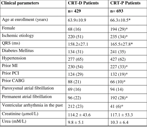
Continuous variables are expressed as mean ± SD. Categorical data are summarized as frequencies and percentages. Baseline clinical characteristics were compared between the subgroups, stratified by implanted CRT-D or CRT-P and using nonparametric Wilcoxon or Kruskal-Wallis tests for continuous variables and the 2 - test for dichotomous variables, as appropriate.
Cumulative probability of survival was determined according to the Kaplan-Meier method in subgroups of CRT-D and CRT-P patients, with comparisons of cumulative event rates by the log-rank test. Multivariate Cox proportional hazards regression analysis was used to
38
evaluate the effect of implanted CRT-D or CRT-P device on the risk of mortality after adjustment for all relevant clinical covariates showing potential imbalance at baseline.
Adjusted hazards ratios (HR) with their 95% confidence intervals (CI) are reported. A p- value < 0.05 was considered statistically significant. Analyses were carried out with SAS software (version 9.3, SAS institute, Cary, North Carolina).
3.5.2 Refining Implantation Methods: Statistical considerations
3.5.2.1 Prognostic Significance of Right Ventricular to Left Ventricular Interlead Sensed Electrical Delay in CRT Patients
Continuous variables are expressed as mean ± SD. Categorical data are summarized as frequencies and percentages. Baseline clinical characteristics were compared between the subgroups, stratified by median interlead sensed electrical delay (106.5 ms) and using nonparametric Wilcoxon or Kruskal-Wallis tests for continuous variables and the 2 - test for dichotomous variables, as appropriate.
Cumulative probability of survival was determined according to the Kaplan-Meier method in subgroups of low vs. high interlead sensed electrical delay, with comparisons of cumulative event rates by the log-rank test. Multivariate Cox proportional hazards regression analysis was used to evaluate the effect of interlead sensed electrical delay on mortality after adjustment for relevant clinical covariates at baseline.
Adjusted hazards ratio (HR) with 95% confidence intervals (CI) is reported. A p-value <
0.05 was considered statistically significant. Analyses were carried out with SAS software (version 9.3, SAS institute, Cary, North Carolina).
39
3.5.2.2 Electroanatomical Mapping-Guided Transseptal Endocardial Left Ventricular Lead Implantation
Data are presented as mean ± standard deviation. Changes in the LV pacing threshold, LV pacing impedance at implantation and at last patient visit were analyzed using paired t-test.
NYHA functional class and mitral regurgitation were analyzed using the Wilcoxon’s signed rank test, as appropriate. Statistical significance was considered at p<0.05. Statistical analyses were performed using GraphPad Prism software (GraphPad Software Inc., La Jolla, CA, USA).
3.5.3 Left Ventricular Lead Location and the Risk of Ventricular Tachyarrhythmias:
Statistical considerations
Continuous variables are expressed as mean ± SD. Categorical data are summarized as frequencies and percentages. Baseline clinical characteristics were compared between the pre- specified subgroups, stratified by implanted device and LV lead position, using nonparametric Wilcoxon or Kruskal-Wallis tests for continuous variables and 2 - test for dichotomous variables, as appropriate. Baseline, 12-month and change in LV dyssynchrony were evaluated among the subgroups using the Kruskal-Wallis test.
Cumulative probability of first VT/VF or death episodes was displayed according to the Kaplan-Meier method, with comparisons of cumulative event rates by the log-rank test.
Multivariate Cox proportional hazards regression analysis was used to identify and evaluate the impact of LV lead location on the end point of first VT/VF or death, whichever occurred first.
The Cox model was adjusted for the following covariates: female gender, etiology of cardiomyopathy, left ventricular ejection fraction, apical LV lead location, QRS duration and morphology (left bundle branch block - LBBB and right bundle branch block - RBBB). Crude event rates were reported as counts of events. As these are composite descriptive measures of
40
risk, ignoring risk variation across patients, no statistical analyses were done.
Propensity analysis was additionally performed to evaluate the robustness of our findings.
The propensity score was developed using logistic regression, which showed RBBB, LVEF and BUN to be statistically significant predictors of lead position.
Adjusted hazards ratios (HR) with their 95% confidence intervals (CI) are reported. A p- value of <0.05 was considered statistically significant and all statistical tests were two-sided.
Analyses were conducted with SAS software (version 9.2, SAS institute, Cary, North Carolina).
3.5.4 Left Ventricular Dyssynchrony and the Risk of Ventricular Tachyarrhythmias:
Statistical considerations
Continuous variables are expressed as mean ± SD. Categorical data are summarized as frequencies and percentages. Baseline clinical characteristics were compared between pre- specified non-LBBB and LBBB subgroups, stratified by baseline LV dyssynchrony quartiles or by changes over one-year in LV dyssynchrony, using nonparametric Wilcoxon or Kruskal-Wallis tests for continuous variables and Chi squarred test or Fisher test for dichotomous variables, as appropriate. When analyzing the effects of LV dyssynchrony change, only first VT/VF events after the 12-month visit were considered as end points.
The correlation of baseline LV dyssynchrony and baseline QRS duration was analyzed using Pearson’s correlation method. Paired comparisons of baseline LV dyssynchrony and change in LV dyssynchrony at 12-months in LBBB and non-LBBB patients were analyzed using nonparametric Wilcoxon test.
Cumulative probability of first VT/VF/Death and VT/VF episodes was determined according to the Kaplan-Meier method with comparisons of cumulative event rates by the log- rank test in non-LBBB and LBBB patients separate. Multivariate Cox proportional hazards