European Heart Rhythm Association (EHRA) international consensus document on how to prevent, diagnose, and treat cardiac
implantable electronic device infections—
endorsed by the Heart Rhythm Society (HRS), the Asia Pacific Heart Rhythm Society
(APHRS), the Latin American Heart Rhythm Society (LAHRS), International Society for Cardiovascular Infectious Diseases (ISCVID), and the European Society of Clinical
Microbiology and Infectious Diseases (ESCMID) in collaboration with the European Association for Cardio-Thoracic Surgery (EACTS)
Carina Blomstro ¨ m-Lundqvist
1* (Chair), Vassil Traykov
2(Co-Chair), Paola Anna Erba
3,4, Haran Burri
5, Jens Cosedis Nielsen
6,
Maria Grazia Bongiorni
7, Jeanne Poole
8(HRS representative), Giuseppe Boriani
9, Roberto Costa
10(LAHRS representative), Jean-Claude Deharo
11,
Laurence M. Epstein
12(HRS representative), La´szlo ´ Sa´ghy
13, Ulrika Snygg-Martin
14(ESCMID and ISCVID representative), Christoph Starck
15(EACTS representative), Carlo Tascini
16(ESCMID representative), and Neil Strathmore
17(APHRS
representative)
1Department of Medical Science and Cardiology, Uppsala University, S-751 85 Uppsala, Sweden;2Department of Invasive Electrophysiology and Cardiac Pacing, Acibadem City Clinic Tokuda Hospital, Nikola Vaptsarov blvd 51 B, 1 407 Sofia, Bulgaria;3Department of Translational Research and New Technology in Medicine, University of Pisa-AOUP, Lungarno Antonio Pacinotti, 43, 56126 Pisa PI, Italy;4Department of Nuclear Medicine & Molecular Imaging University Medical Center Groningen, University of Groningen, 9712 CP Groningen, Netherlands;5Department of Cardiology, University Hospital of Geneva, Rue Gabrielle-Perret-Gentil 4, 1205 Geneva, Switzerland;6Department of Cardiology, Aarhus University Hospital, Palle Juul-Jensens Blvd. 161, 8200 Aarhus, Denmark;7CardioThoracic and Vascular Department, University Hospital of Pisa, Via Paradisa 2, 56125 Pisa PI, Italy;8Department of Cardiology, University of Washington, Roosevelt Way NE, Seattle, WA 98115, USA;9Cardiology Division, Department of Biomedical, Metabolic and Neural Sciences, University of Modena and Reggio Emilia, Policlinico di Modena, Largo del Pozzo, 71, 41125 Modena, Italy;10Department of Cardiovascular Surgery, Heart Institute (InCor) of the University of S~ao Paulo, Butanta, S~ao Paulo - State of S~ao Paulo, Brazil;11Department of Cardiology, Aix Marseille Universite´, CHU la Timone, 278 Rue Saint-Pierre, 13005 Marseille, France;12Electrophysiology, Northwell Health, Hofstra/Northwell School of Medicine, 300 Community Drive, Manhasset, NY 11030, USA;
The opinions expressed in this article are not necessarily those of the Editors of theEuropean Heart Journalor of the European Society of Cardiology.
* Corresponding author. Tel:þ46 18 611 3113, Email:carina.blomstrom.lundqvist@akademiska.se
Published on behalf of the European Society of Cardiology. All rights reserved.VCThe Author(s) 2020. For permissions, please email: journals.permissions@oup.com.
doi:10.1093/eurheartj/ehaa010
Downloaded from https://academic.oup.com/eurheartj/article/41/21/2012/5760353 by 81728827 user on 19 January 2022
.. ..
.. ..
.. ..
.. ..
.. ..
.. ..
.. ..
.. ..
.. ..
.. ..
.. ..
.. ..
.. ..
.. ..
.. ..
.. ..
.. ..
.. ..
.. ..
.. ..
.. ..
.. ..
.. ..
.. ..
.. .
13Electrophysiology Division, 2nd Department of Medicine and Cardiology Centre, University of Szeged, Aradi ve´rtanu´k tere 1, 6720 Szeged, Hungary;14Department of Infectious Diseases, Sahlgrenska Academy, University of Gothenburg, 405 30 Gothenburg, Sweden;15Department of Cardiothoracic and Vascular Surgery, German Heart Center Berlin, Augustenburger Pl. 1, 13353 Berlin, Germany;16First Division of Infectious Diseases, Cotugno Hospital, Azienda ospedaliera dei Colli, Via Gaetano Quagliariello, 54, 80131 Napoli NA, Italy; and17Department of Cardiology, Royal Melbourne Hospital, 300 Grattan St, Parkville VIC 3050, Melbourne, Australia
Received 26 August 2019; revised 7 October 2019; editorial decision 18 December 2019; accepted 10 January 2020; online publish-ahead-of-print 26 February 2020
Pacemakers, implantable cardiac defibrillators, and cardiac resynchronization therapy devices are potentially lifesaving treatments for a number of cardiac conditions but are not without risk. Most concerning is the risk of a cardiac implantable electronic device (CIED) infection, which is associated with significant morbidity, increased hospitalizations, reduced survival, and increased health care costs. Recommended preventive strategies such as administration of intravenous antibiotics before implantation are well-recognized. Uncertainties have remained about the role of various preventive, diagnostic, and treatment measures such as skin antiseptics, pocket antibiotic solutions, antibacterial envelopes, prolonged antibiotics post-implantation, and others. When compared with previous guidelines or consensus statements, the present consen- sus document gives guidance on the use of novel device alternatives, novel oral anticoagulants, antibacterial envelopes, prolonged antibiotics post-implantation, as well as definitions on minimum quality requirements for centres and operators and volumes. The recognition that an international consensus document focused on management of CIED infections is lacking, the dissemination of results from new important randomized trials focusing on prevention of CIED infections, and observed divergences in managing device-related infections as found in an European Heart Rhythm Association worldwide survey, provided a strong incentive for a Novel 2019 International State-of-the-art Consensus document on risk assessment, prevention, diagnosis, and treatment of CIED infections.
䊏 䊏 䊏 䊏 䊏 䊏 䊏 䊏 䊏 䊏 䊏 䊏 䊏 䊏 䊏 䊏 䊏 䊏 䊏 䊏 䊏 䊏 䊏 䊏 䊏 䊏 䊏 䊏 䊏 䊏 䊏 䊏 䊏 䊏 䊏 䊏 䊏 䊏 䊏 䊏 䊏 䊏 䊏 䊏 䊏 䊏 䊏 䊏 䊏 䊏 䊏 䊏 䊏 䊏 䊏 䊏 䊏 䊏 䊏 䊏 䊏 䊏 䊏 䊏 䊏 䊏 䊏 䊏 䊏 䊏 䊏 䊏 䊏 䊏 䊏 䊏 䊏 䊏 䊏 䊏 䊏 䊏 䊏 䊏 䊏 䊏 䊏 䊏 䊏 䊏 䊏 䊏 䊏 䊏 䊏 䊏 䊏 䊏 䊏 䊏 䊏 䊏 䊏 䊏 䊏 䊏 䊏 䊏 䊏 䊏 䊏 䊏 䊏 䊏 䊏 䊏 䊏 䊏 䊏 䊏 䊏 䊏 䊏 䊏 䊏 䊏 䊏 䊏 䊏 䊏 䊏 䊏 䊏 䊏 䊏 䊏 䊏 䊏 䊏 䊏 䊏 䊏 䊏 䊏 䊏 䊏 䊏 䊏 䊏 䊏 䊏 䊏 䊏 䊏 䊏 䊏 䊏 䊏 䊏 䊏 䊏 䊏 䊏 䊏 䊏 䊏 䊏 䊏 䊏 䊏 䊏 䊏 䊏 䊏 䊏 䊏 䊏 䊏 䊏 䊏 䊏 䊏 䊏 䊏 䊏 䊏 䊏 䊏 䊏 䊏 䊏 䊏 䊏 䊏 䊏 䊏 䊏 䊏 䊏 䊏 䊏 䊏 䊏 䊏 䊏 䊏 䊏 䊏 䊏 䊏 䊏 䊏
Keywords Infection • Device • Extraction • Defibrillator • Pacemaker • Leads
Table of contents
Introduction . . . 2013
Background and epidemiology . . . 2014
Pathogenesis and microbiology of cardiac implantable electronic device infections . . . 2014
Risk factors for cardiac implantable electronic device infection . . . 2015
Prevention. . . 2015
Pre-procedural measures . . . 2015
Patient selection . . . 2015
Lead management. . . 2016
Patient factors . . . 2016
Anticoagulation and antiplatelet drugs . . . 2016
Appropriate environment . . . 2018
Staff training. . . 2018
Nasal swabs/Staphylococcus aureus decolonization of patients . . 2018
Pre-procedure skin preparation. . . 2018
Pre-procedure antibiotic therapy . . . 2018
Periprocedural measures. . . 2019
Patient surgical preparation . . . 2019
Good surgical technique. . . 2019
Antibiotic envelope . . . 2019
Local instillation of antibiotics or antiseptics . . . 2019
Capsulectomy . . . 2020
Closure. . . 2020
Post-procedural measures . . . 2020
Post-procedure antibiotic therapy . . . 2020
Wound care . . . 2020
Reintervention . . . 2020
Diagnosis of cardiac implantable electronic device infections and related complications . . . 2020
Clinical findings . . . 2020
Identification of the causative microorganisms. . . 2020
Imaging. . . 2020
Echocardiography . . . 2020
Radiolabelled leucocyte scintigraphy, positron emission tomography and computerized tomography. . . 2021
Management of cardiac implantable electronic device infections . . . . 2023
Cardiac implantable electronic device removal . . . 2023
Antimicrobial therapy including long-term suppressive therapy . . 2025
Preventive strategies after cardiac implantable electronic device implantations, reimplantations, and alternative novel devices . . . 2028
Prognosis, outcomes, and complications of cardiac implantable electronic device infections . . . 2029
Special considerations to prevent device-related infections (elderly, paediatrics, adult with congenital heart disease) . . . 2029
Minimum quality requirements concerning centres and operator experience and volume. . . 2030
Health economics for cardiac implantable electronic devices infections and strategies to reduce costs. . . 2030
Conclusion . . . 2031
Introduction
Pacemakers, implantable cardiac defibrillators (ICDs), and cardiac resynchronization therapy (CRT) devices are lifesaving treatments for a number of cardiac conditions. Device-related infection is, how- ever, one of the most serious complications of cardiac implantable electronic device (CIED) therapy. The recognition of gaps in know- ledge, reports of new important randomized trials, observed diver- gences in managing device-related infections,
1and the lack of international consensus documents specifically focusing on CIED infections provided a strong incentive for a 2019 State-of-the-art Consensus document on management of CIED infections.
This consensus document is an international collaboration among seven professional societies/associations with a writing group consist- ing of cardiologists with varying subspecialties, infectious disease spe- cialists, imaging specialist, and thoracic surgeon, from 11 countries in
Downloaded from https://academic.oup.com/eurheartj/article/41/21/2012/5760353 by 81728827 user on 19 January 2022
.. ..
.. ..
.. ..
.. ..
.. ..
.. ..
.. ..
.. ..
.. ..
.. ..
.. ..
.. ..
.. ..
.. ..
.. ..
.. ..
.. ..
.. ..
.. ..
.. ..
.. ..
.. ..
.. ..
.. ..
.. ..
4 continents. A detailed literature search until May 2019 and system- atic reviews of published evidence related to CIED infection topics were performed. Results of the international survey on CIED infec- tions conducted for this purpose
1and of previous registries
2were considered. Consensus statements were evidence-based, derived pri- marily from published data and by consensus opinion after thorough deliberations, requiring at least 80% predefined consensus.
The European Heart Rhythm Association (EHRA) ranking system for consensus documents, with ‘coloured hearts’ providing the cur- rent status of the evidence and consequent guidance, was used for the coding of the scientific evidence for statements made (Table 1).
The document was peer-reviewed by official external reviewers representing EHRA, the participating societies, and European Society of Cardiology (ESC) Committee for Practice Guidelines (CPG). All members of the writing group as well as reviewers have disclosed po- tential conflicts of interest, at the end of this document.
The medical approaches discussed may include drugs or devices that are not approved by governmental regulatory agencies in all countries. The ultimate decision on management must be made by the health care provider and the patient in light of individual factors presented.
Background and epidemiology
Infection is one of the most serious complication of CIED therapy and is associated with significant mortality, morbidity, and financial health care burden. In the Danish registry of pacemaker implantation between 1982 and 2007, the incidence of infection was 4.82/1000 device-years after a primary implantation, and 12.12/1000 device- years after replacement.
3The incidence of CIED infection in the USA increased from 1.53% in 2004 to 2.41% in 2008
4and a National Inpatient Sample database study showed an increase from 1.45% to
3.41% (P < 0.001) from 2000 to 2012.
5Infection rates in prospective observational studies,
6,7registries,
8and recent cross-over cluster PADIT- and randomized WRAP-IT trials
9,10were only 0.6–1.3%, when compared with retrospective studies
11,12reporting significantly higher rates (2.3–3.4%) in the first year after implantation.
Pathogenesis and microbiology of cardiac implantable electronic device infections
Cardiac implantable electronic device infections occur via two major mechanisms. The most common is contamination of leads and/or pulse generator during implantation or subsequent manipulation.
13Device erosions late after interventions may either be due to or re- sult in pocket infection. Contamination and subsequent bacterial col- onization result in pocket infection which can spread along the intravascular parts of the leads and progress to systemic infection.
The second mechanism is a bloodstream infection.
14Direct lead seeding can occur during bacteraemia caused by a distant infectious focus or bacterial entry via the skin, mouth, gastrointestinal, or urin- ary tract.
The pathogenesis of CIED infections can be related to the host, the device, or the microorganism. The patient’s own skin flora can be introduced into the wound at the time of skin incision.
Contamination may also occur before implantation via the air in the operating room or via the hands of anyone handling the device.
Device-related factors are those affecting bacterial adherence to the generator or lead and the biofilm formation on these surfaces.
15Normally non-pathogenic microorganisms such as Coagulase-nega- tive Staphylococci (CoNS) may adhere to the CIED and establish a focus of infection. The microorganisms most frequently isolated have ...
Table 1 Scientific rationale of recommendations
Consensus state-ment related to a treatment or procedure
Definitions of consensus statement Statement
class
Scientific evidence coding
References
Recommended/indi- cated or ‘should do this’
Scientific evidence that a treatment or procedure is beneficial and effective. Requires at least one randomized trial or is supported by large observational studies and authors’ consensus
R
May be used or recommended
General agreement and/or scientific evidence favour the usefulness/
efficacy of a treatment or procedure. May be supported by randomized trials based on small number of patients or not wide- ly applicable
O
Should NOT be used or recommended
Scientific evidence or general agreement not to use or recommend a treatment or procedure
E
This categorization for the consensus document should not be considered as being directly similar to that used for official society guideline recommendations which apply a classification (I–III) and level of evidence (A, B, and C) to recommendations. The grading does not have separate levels of evidence, which instead are defined in each of the col- oured heart grades.
The ‘ROME’ coding was applied for each consensus statement, defining existing scientific evidence; R for randomized trials, O for observational studies, M for meta-analyses, and E for expert opinion.
Downloaded from https://academic.oup.com/eurheartj/article/41/21/2012/5760353 by 81728827 user on 19 January 2022
.. ..
.. ..
.. ..
.. ..
.. ..
.. ..
.. ..
.. ..
.. ..
.. ..
.. ..
.. ..
.. ..
.. ..
.. ..
.. ..
.. ..
.. ..
.. ..
.. ..
.. ..
.. ..
been Gram-positive bacteria (70–90%), especially CoNS (37.6% of .
the isolates) and Staphylococcus aureus (30.8%), which are far more prone to adhere to non-biological material than others (Table 2).
16,17,19Staphylococcus aureus is the most common cause of bacteraemia and early pocket infections. Altogether, methicillin- resistant staphylococci were isolated in 33.8% of CIED infections (49.4% of all staphylococcal infections).
16,18,20Risk factors for cardiac
implantable electronic device infection
Identification of modifiable risk factors is important because it may allow for preventive measures to reduce the risk. In patients with non-modifiable risks, alternative approaches may be an option to lower the overall risk.
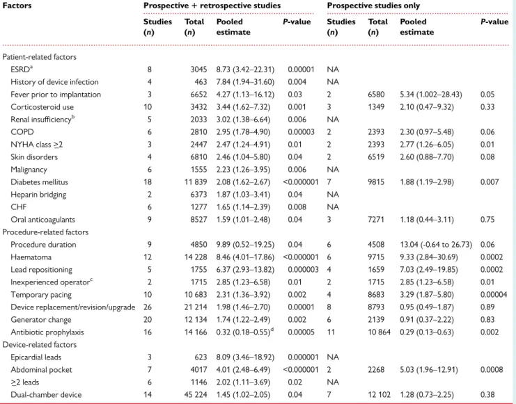
A meta-analysis
21of pooled data including 206 176 patients sum- marizes the most important risk factors in Table 3. In large device registry-, heath care database-, and device-cohort studies,
5,22,23the importance of risk factors varied from study to study and findings were in some cases contradictory (age as an example).
Of the patient-related factors, end-stage renal disease was consist- ently associated with the highest risk, but not age or gender.
21Younger age, along with prior device infection were identified as sig- nificant risks in the Danish device-cohort study.
23Others identified malnutrition [odds ratio (OR) 2.44, P < 0.001] as a strong risk factor.
5Regarding procedure-related factors, the presence of a haematoma was associated with an approximately nine-fold increased risk of
infection, later confirmed by the prospective BRUISE-CONTROL study.
24Early reoperation for haematoma or lead dislodgement was identified as strongest risk factors for CIED infection in a device regis- try data.
5,22Procedure duration was associated with a multifold increased risk of infection,
21,23as were implantation of CRT and reoperations. Experience has an impact on outcome,
25and risk of in- fection may be increased by allocating generator changes to inexperi- enced operators.
There are fewer device-related factors for CIED infection.
Device complexity and the numbers of leads were significantly associated with increased infection risk on multivariate analysis [hazard ratio (HR) 1.26, 1.67, and 2.22 for ICD, CRT-P, and CRT-D systems, respectively vs. pacemakers, P < _ 0.002 for all comparisons].
23Risk stratification with risk score calculations could potentially play a role in better identifying patients at risk than individual factors
26,27but can currently not be recommended because the evidence of their benefit remains weak.
Prevention
Recommended preventive measures are summarized in Table 4 and Figure 1.
Pre-procedural measures
Patient selection
For patients undergoing device removal for infection, up to one half may not require device reimplantation.
39Implanting an epicardial ...
...
Table 2 Pathogens isolated in patients undergoing interventions for device infection from three large patient cohorts in North America, Europe, and Asia
Pathogens Percentage of isolates
North America16 Europe17 Asia18
Coagulase-negative Staphylococci 69 45.2
Methicillin-resistant 18.8
Methicillin-sensitive 18.8
Staphylococcus aureus 13.8 4.1
Methicillin-sensitive 15.8
Methicillin-resistant 15.0
Streptococcusspp. 2.5
Enterococcusspp.
Vancomycin-sensitive 2.8
Vancomycin-resistant 1.4
Cutibacteriumspp. (previouslyPropionibacteriumspp.) 2.5
Corynebacterium 5
Gram-negative bacteria 8.9 6.1 9.1
Enterobacteriaceae 3 3.2
Non-fermentative bacilli, incl.Pseudomonasspp. 1.5 5.9
Anaerobes 1.6
Fungi 0.9 1 0.9
Mycobacteria 0.2
Downloaded from https://academic.oup.com/eurheartj/article/41/21/2012/5760353 by 81728827 user on 19 January 2022
.. ..
.. ..
.. ..
.. ..
.. ..
.. ..
.. ..
.. ..
..
system may be preferential in high-risk patients.
40‘Leadless’ pace- makers may be less prone to infection and can be used in high-risk patients.
41,42Subcutaneous ICDs (S-ICDs) are an option in patients requiring sudden death protection.
Lead management
The number of leads and the presence of abandoned leads are associ- ated with increased risk for infection. The decision to abandon or ex- tract a lead must be made on an individual basis weighing all known risks and benefits.
43,44Patient factors
A procedure should be delayed until a patient has been afebrile for at least 24 h.
28Better glycaemic control in the periprocedural period may reduce infections in surgical patients.
45Anticoagulation and antiplatelet drugs
A ‘bridging’ approach with heparin is not recommended.
30In patients with CHA
2DS
2VASc score <4, holding anticoagulation for the pro- cedure and restarting when the bleeding risk is reduced seems pru- dent. In higher-risk patients (prior embolic event or mechanical valve) continuing anticoagulation with Warfarin is recommended.
... ...
...
Table 3 Pooled effect estimates for potential risk factors predisposing to CIED infection
Factors Prospective1retrospective studies Prospective studies only Studies
(n)
Total (n)
Pooled estimate
P-value Studies (n)
Total (n)
Pooled estimate
P-value
Patient-related factors
ESRDa 8 3045 8.73 (3.42–22.31) 0.00001 NA
History of device infection 4 463 7.84 (1.94–31.60) 0.004 NA
Fever prior to implantation 3 6652 4.27 (1.13–16.12) 0.03 2 6580 5.34 (1.002–28.43) 0.05
Corticosteroid use 10 3432 3.44 (1.62–7.32) 0.001 3 1349 2.10 (0.47–9.32) 0.33
Renal insufficiencyb 5 2033 3.02 (1.38–6.64) 0.006 NA
COPD 6 2810 2.95 (1.78–4.90) 0.00003 2 2393 2.30 (0.97–5.48) 0.06
NYHA class >_2 3 2447 2.47 (1.24–4.91) 0.01 2 2393 2.77 (1.26–6.05) 0.01
Skin disorders 4 6810 2.46 (1.04–5.80) 0.04 2 6519 2.60 (0.88–7.70) 0.08
Malignancy 6 1555 2.23 (1.26–3.95) 0.006 NA
Diabetes mellitus 18 11 839 2.08 (1.62–2.67) <0.000001 7 9815 1.88 (1.19–2.98) 0.007
Heparin bridging 2 6373 1.87 (1.03–3.41) 0.04 NA
CHF 6 1277 1.65 (1.14–2.39) 0.008 NA
Oral anticoagulants 9 8527 1.59 (1.01–2.48) 0.04 3 7271 1.18 (0.44–3.11) 0.75
Procedure-related factors
Procedure duration 9 4850 9.89 (0.52–19.25) 0.04 6 4508 13.04 (-0.64 to 26.73) 0.06
Haematoma 12 14 228 8.46 (4.01–17.86) <0.000001 6 9715 9.33 (2.84–30.69) 0.0002
Lead repositioning 5 1755 6.37 (2.93–13.82) 0.000003 4 1659 7.03 (2.49–19.85) 0.0002
Inexperienced operatorc 2 1715 2.85 (1.23–6.58) 0.01 2 1715 2.85 (1.23–6.58) 0.01
Temporary pacing 10 10 683 2.31 (1.36–3.92) 0.002 4 8683 3.29 (1.87–5.80) 0.00004
Device replacement/revision/upgrade 26 21 214 1.98 (1.46–2.70) 0.00001 8 8793 0.95 (0.49–1.87) 0.89
Generator change 20 12 134 1.74 (1.22–2.49) 0.002 6 2139 0.91 (0.37–2.22) 0.83
Antibiotic prophylaxis 16 14 166 0.32 (0.18–0.55)d 0.00005 11 10 864 0.29 (0.13–0.63) 0.002
Device-related factors
Epicardial leads 3 623 8.09 (3.46–18.92) 0.000001 NA
Abdominal pocket 7 4017 4.01 (2.48–6.49) <0.000001 2 2268 5.03 (1.96–12.91) 0.0008
>_2 leads 6 1146 2.02 (1.11–3.69) 0.02 NA
Dual-chamber device 14 45 224 1.45 (1.02–2.05) 0.04 7 12 102 1.28 (0.73–2.25) 0.38
Adapted from Polyzoset al.21
Risk parameters, which were statistically significant for retrospective and prospective data are shown. Analyses restricted to prospective data only for the same parameters (if available) are also shown.
CHF, congestive heart failure; COPD, chronic obstructive pulmonary disease; ESRD, end-stage renal disease; NA, not available; NYHA, New York Heart Association.
aGlomerular filtration rate <_15 mL/min or haemodialysis or peritoneal dialysis.
bGlomerular filtration rate <60 mL/min or creatinine clearance <60 mL/min.
c<100 previous procedures.
dThe pooled effect estimate from randomized studies was 0.26 (0.13–0.52).
Downloaded from https://academic.oup.com/eurheartj/article/41/21/2012/5760353 by 81728827 user on 19 January 2022
...
Table 4 List of recommended preventive measures for CIED infections
Consensus statement Statement class Scientific evidence
coding
References
Pre-procedural measures
Confirm indication for CIED E
Delay CIED implantation in patients with infection E 28
Avoid temporary transvenous pacing and central venous lines, which should ideally be removed prior to introducing new hardware, whenever possible
O, M 21
Measures to avoid pocket haematoma are recommended (avoid heparin bridg- ing, discontinue antiplatelets if possible)
R 21,29–31
Periprocedural use of therapeutic low-molecular-weight heparin R, M, O 30,32,33
Perform the CIED procedure in an operating room/suite with complete sterile environment as required for other surgical implant procedures
E 34
Procedure should be performed or supervised by an operator with sufficient training and experience (Table12)
O 35
TopicalStaphylococcus aureusdecolonization may be performed E
Pre-procedural skin wash may be performed E
Hair removal with electric clippers (not razors) is recommended O 36
Antibiotic prophylaxis is recommended within 1 h of incision for cefazolin and flucloxacilline, within 90–120 min for vancomycin
R, M 21
A continuous surveillance programme of infection rates and associated micro- biology should be set-up at the level of each implanting centre
E —
Periprocedural measures
Surgical preparation with alcoholic chlorhexidine should be used rather than povidone–iodine
R 37,38
Allow sufficient time for the antiseptic preparation to dry E
Adhesive iodophor-impregnated incise drapes may be used E
Perform the procedure with adequate surgical technique—minimize tissue damage, haemostasis, adequate wound closure
E
Continued
Downloaded from https://academic.oup.com/eurheartj/article/41/21/2012/5760353 by 81728827 user on 19 January 2022
.. ..
.. ..
.. ..
.. ..
.. ..
.. ..
.. ..
.. ..
.. ..
.. ..
.. ..
.. .
Preliminary data suggest the same for non-vitamin K antagonist oral anticoagulants.
29Therapeutic low-molecular-weight heparin should be avoided.
30,32,33Antiplatelet agents, especially P2Y12 inhibitors should preferably be discontinued for 5–10 days before the intervention.
31Appropriate environment
The standards for sterile procedures must be met as for other surgi- cal procedures associated with implants.
34Staff training
All staff involved in CIED implantation must be trained in appropriate strict sterile techniques and behaviour in an operating room setting.
Operators should be adequately trained.
35Nasal swabs/Staphylococcus aureus decolonization of patients
For elective procedures, S. aureus colonization can be detected by nasal swabs. Nasal treatment with mupirocin and chlorhexidine skin washing has been shown in some surgical studies to reduce the risk for infection.
46Pre-procedure skin preparation
Routine pre-surgical washing with an antimicrobial agent cannot be strongly supported.
47Electric clippers with a single-use head (not razors) should be used for chest hair removal.
36Pre-procedure antibiotic therapy
Prophylactic systemic antibiotics are the standard of care
21,48,49and should at least cover S. aureus species. Randomized trials have used ...
Table 4 Continued
Consensus statement Statement class Scientific evidence
coding
References
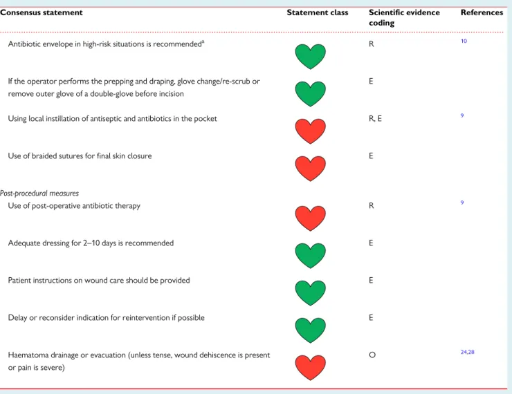
Antibiotic envelope in high-risk situations is recommendeda R 10
If the operator performs the prepping and draping, glove change/re-scrub or remove outer glove of a double-glove before incision
E
Using local instillation of antiseptic and antibiotics in the pocket R, E 9
Use of braided sutures for final skin closure E
Post-procedural measures
Use of post-operative antibiotic therapy R 9
Adequate dressing for 2–10 days is recommended E
Patient instructions on wound care should be provided E
Delay or reconsider indication for reintervention if possible E
Haematoma drainage or evacuation (unless tense, wound dehiscence is present or pain is severe)
O 24,28
CIED, cardiac implantable electronic device; E, expert opinion; M, meta-analysis; O, observational studies; R, randomized trials.
aCandidates are those as defined in the WRAP-IT study population10(patients undergoing pocket or lead revision, generator replacement, system upgrade, or an initial CRT-D implantation) and patients with other high-risk factors as outlined inTable3, considering also the local incidence of CIED infections.
Downloaded from https://academic.oup.com/eurheartj/article/41/21/2012/5760353 by 81728827 user on 19 January 2022
.. ..
.. ..
.. ..
.. ..
.. ..
.. ..
.. ..
.. ..
.. ..
.. ..
.. ..
.. ..
.. ..
.. ..
.. ..
.. ..
.. ..
i.v. flucloxacillin (1–2 g) and first-generation cephalosporins such as .
cefazolin (1–2 g).
9,48,49Vancomycin (15 mg/kg) may be used in case of allergy to cephalosporins.
Periprocedural measures
Patient surgical preparation
Alcoholic 2% chlorhexidine was superior to povidone–iodine for skin preparation prior to surgery
37or intravascular catheter insertion
38but no randomized data exist regarding CIED implantation. There is no evidence that adhesive incise drapes reduces infection rates (may increase risk of infection when non-iodophor incise drapes are used
50).
Good surgical technique
Non-powdered gloves may reduce the risk of infection by reducing local inflammation.
51Vigorous pocket irrigation is important to re- move devitalized tissue as well as dilute any contaminants.
52Diagnostic or therapeutic aspiration of a haematoma is contraindi- cated given the risk of ‘inoculating’ the pocket and causing an infec- tion.
24,28Haematoma evacuation should only be undertaken if pain is unmanageable or wound closure is threatened, ideally performed in an operating room.
24,28Antibiotic envelope
An antibacterial mesh envelope (TYRX
TM, Medtronic, MN, USA), which locally releases minocycline and rifampin, significantly reduced the incidence of CIED infection in high-risk patients (WRAP-IT trial
10) without a higher incidence of complications. The incidence of primary endpoints (infection resulting in system extraction or revi- sion, long-term antibiotic therapy, or death) within 12 months after the CIED implantation was lower in patients who received the enve- lope (0.7%) vs. controls (1.2%) (HR 0.60, 95% confidence interval (CI) 0.36–0.98; P = 0.04).
10The number of patients needed to treat to prevent one infection was high. The exclusion of higher-risk patients (immunosuppressive treatments, with vascular access, or on dialysis) may have contributed to a lower-than-expected rate of infections (1.2%) also observed in other prospective studies.
6,7,9Higher infection rates (2.3–3.4%), as observed in less selected retro- spective studies
11,12would improve the overall cost-effectiveness of the envelope. Recommendation for the use of the antibacterial enve- lope is outlined in Table 4.
Local instillation of antibiotics or antiseptics
Local instillation of antibiotics or antiseptics is not recommended.
The recent PADIT trial demonstrated no benefit.
9Device type: CRT or ICD More than 2 leads Abandoned / complex route leads Dual chamber device Presence of epicardial leads
Evaluaon of risk factors for CIED infecon
Modifiable Non-modifiable
Paent-related factors Procedure-related factors Device-lead-related factors
Device replacement/upgrade
Paent-related factors Procedure-related factors Device-lead-related factors
Lead reposioning
Postpone procedure if
fever or infecon
Treat any comorbidity
OAC uninterrupted Anplatelets
paused 1 w prior surgery if possible
Experienced operator (shortens procedure duraon &
reduces lead dislodgement risk)
Limit number of persons in operang room
Follow outlined surgical field preparaon /techniques
Limit number of
IV lines Consider epicardial pacing, leadless
pacing, subcutaneous ICD Fever prior to implantaon
Skin disorders Heparin bridging Oral ancoagulants
Prolonged procedure Hematoma Prior procedure(s) Inexperienced implanter Temporary pacing wire
Abdominal pocket
Reassess indicaons for primary implantaon, reoperaon or re-implantaon of a new device following lead extracon
Replace temporary pacing by external pacing or drugs in non-
dependent paent
Evaluate need to use anbacterial envelopes
Administer preprocedural anbioc prophylaxis as
recommended
Reduce risk by taking acon on modifiable risk factors
End-stage renal disease Corcosteroid use Renal failure History of device infecon COPD
Heart Failure NYHA > II Malignancy Diabetes mellitus
Figure 1 A flowchart indicating how device-related infections can be minimized by targeting modifiable risk factors on various levels. Risk factors ranked in order of strength from top to bottom. CIED, cardiac implantable electronic device; COPD, chronic obstructive pulmonary disease; CRT, cardiac resynchronization therapy; ICD, implantable cardiac defibrillator; NYHA, New York Heart Association; OAC, oral anticoagulation; w, week.
Downloaded from https://academic.oup.com/eurheartj/article/41/21/2012/5760353 by 81728827 user on 19 January 2022
.. ..
.. ..
.. ..
.. ..
.. ..
.. ..
.. ..
.. ..
.. ..
.. ..
.. ..
.. ..
.. ..
.. ..
.. ..
.. ..
.. ..
.. ..
.. ..
.. ..
.. ..
.. ..
.. ..
.. ..
.. ..
.. ..
.. ..
.. ..
.. ..
.. ..
.. ..
.. ..
.. ..
.. ..
.. ..
.. ..
.. ..
.. ..
.. ..
.. ..
.. ..
.. .
Capsulectomy
Even in the absence of signs of clinical infections, cultures taken at the time of generator change demonstrate a significant incidence of col- onization.
53The fibrous capsule inhibits the body’s normal defence mechanisms and antibiotic penetration but ‘capsulectomy’ could also result in more pocket bleeding/haematoma and cannot be recom- mended as routine practice.
54Closure
Closure in layers minimize wound tension and reduces the risk of de- hiscence and infection.
55Post-procedural measures
Post-procedure antibiotic therapy
The recent PADIT trial,
9tested the clinical effectiveness of incremen- tal perioperative antibiotics but the primary outcome of 1-year hospi- talization for device infection in the high-risk group was not statistically significant. It is therefore not recommended to administer post-operative antibiotic therapy.
Wound care
Pressure dressing may be used for the first 24 h to avoid haematoma.
Reintervention
All measures must be taken to avoid the need of early reintervention which dramatically increases the risk of infection.
19,21,28Diagnosis of cardiac implantable electronic device infections and related complications
Clinical findings
A superficial incisional infection involves only the skin and the subcuta- neous tissue without communication with the pocket.
56,57Close monitoring of the patient must be pursued in order to recognize a significant pocket infection.
Pocket infection is defined as an infection limited to the generator pocket. Local signs of inflammation may be mild (erythema, warmth, and fluctuation).
14,57Deformation of the pocket, adherence or threatened erosion are often signs of low grade, indolent infection.
Once a wound dehiscence occurs, a purulent drainage or a sinus is established, and a pocket infection is clearly present. If the generator or proximal leads are exposed, the device should be considered infected, irrespective of the results of the microbiology. Material from the pocket may be used for culture, recognizing the potential for contamination. Pocket infections may be associated with lead infections and CIED systemic infections and/or infective endocarditis.
58The diagnosis of CIED systemic infection and infective endocarditis with- out local infection may be more challenging (Table 5). Symptoms may be non-specific (fever, chills, and night sweats). Patients with CIED in- fection may present with embolic involvement of lungs and pleural
space
60,61or with vertebral osteomyelitis and discitis. Procalcitonin test may be of value, especially if positive (> _0.05).
62,63The modified Duke criteria
64and the ESC 2015 criteria
59for the diagnosis of infective endocarditis are the only available framework for CIED endocarditis diagnosis. In order to increase sensitivity for CIED infection diagnosis, this panel developed the 2019 International CIED Infection Criteria (Table 5).
Identification of the causative microorganisms
Every effort should be made to obtain cultures prior to the institution of antibiotic therapy. Blood cultures should be repeated in patients with CIED and fever without clear signs of local infections and infect- ive endocarditis (Table 6). In unstable patients with sepsis or septic shock, early empiric antibiotic therapy should be administered fol- lowing two sets of blood cultures. Blood bottles must be filled prop- erly in order to increase the sensitivity.
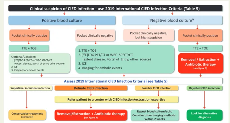
17,65Every positive blood culture, including a single bottle with CoNS or other Gram-positive organisms, should prompt active exclusion of CIED infection with other diagnostic techniques employed (Figure 2).
71In case of negative blood cultures (usually 5 days), the use of biomolecular methods (DNA amplification and/or gene sequencing) to detect fastidious or atypical pathogenes
19may be considered for CIED endocarditis (Table 6).
67Some Gram-positive microorganisms species may re- quire longer period of incubation, such as Cutibacterium (previously Propionibacterium) acnes.
19Tissue or fluid collected from the pocket via an adjacent intact por- tion of the skin (via a sterile needle or syringe) should only be used to make a bacterial diagnosis, not to determine the presence of a pocket infection. Entering an intact pocket should be avoided to avoid inocu- lation with bacteria.
During an extraction procedure, distal and proximal lead frag- ments, lead vegetation and generator pocket tissue should be sent for culture (Table 6).
71Culture media suggested are chocolate agar incubated in 5% CO
2, MacConkey agar, blood agar in anaerobic con- dition, and Sabouraud agar.
72,73In case of pus, but no growth after 3 days, consider slow growing microorganisms including C. acnes and increase incubation duration. Tissue samples and sonication for the recovery of bacteria from CIED leads and tissue may be useful.
68–70Imaging
Echocardiography
Transthoracic echocardiography (TTE) and transoesophageal echo- cardiography (TEE) are both recommended to identify lead vegeta- tions and valvular involvement in suspected CIED infections.
59Transoesophageal echocardiography is superior for the detection and sizing of vegetations.
74Lead masses in asymptomatic CIED car- riers may be observed on TTE/TEE and do not predict CIED-related infective endocarditis over long-term follow-up.
75,76Once a lead mass is identified, careful clinical assessment to rule out either infec- tion or non-bacterial lead-thrombotic endocarditis is needed, includ- ing serial TTE/TEE or additional imaging tests.
Intracardiac echocardiography (ICE) has a high sensitivity for the detection of vegetations in cardiac devices.
77,78Therefore, a vegeta- tion seen with ICE may be considered a major criterion for diagnosis
Downloaded from https://academic.oup.com/eurheartj/article/41/21/2012/5760353 by 81728827 user on 19 January 2022
.. ..
.. ..
.. ..
.. ..
(Table 5). Transvenous biopsy, guided by TEE, was shown to be useful ..
to differentiate vegetation from thrombus.
79A TEE should be considered after percutaneous lead extraction in order to detect infected material, ghosts,
80and potential tricuspid valve complications (Table 7). A normal echocardiography does not rule out CIED-related infective endocarditis.
Radiolabelled leucocyte scintigraphy, positron emission tomography and computerized tomography
Fluorine-18-fludeoxyglucose ([
18F]FDG) positron emission tomog- raphy/computerized tomography (PET/CT) scanning and radiola- belled leucocyte (white blood cell, WBC) scintigraphy are complementary tools for the diagnosis of CIED-related infections ...
Table 5 Recommendations for diagnosis of CIED infections and/or infective endocarditis: the Novel 2019 International CIED Infection Criteria
aConsensus statement Statement class Scientific evidence coding
Reference
‘Definite’ CIED clinical pocket/generator infection = generator pocket shows swelling, erythema, warmth, pain, and purulent discharge/sinus formation OR deformation of pocket, adherence, and threatened erosion OR exposed generator or proximal leads.
‘Definite’ CIED/IE = presence of either two major criteria or one majorþthree minor criteria
‘Possible’ CIED/IE = presence of either one majorþone minor criteria or three minor criteria
‘Rejected’ CIED/IE diagnosis = patients who did not meet the aforementioned criteria for IE Major criteria
E
59
Microbiology A. Blood cultures positive for typical microorganisms found in CIED infection and/or IE (Coagulase-negative Staphylococci,Staphylococcus aureus)
B. Microorganisms consistent with IE from two separate blood cultures:
a. Viridans streptococci,Streptococcus gallolyticus(Streptococcus bovis), HACEK group,S. aureusor b. Community-acquired enterococci, in the absence of a primary focus.
C. Microorganisms consistent with IE from persistently positive blood cultures:
a. >_2 positive blood cultures of blood samples drawn >12 h apart; or
b. All of three or a majority of >_4 separate cultures of blood (first and last samples drawn >_1 h apart); or c. Single positive blood culture forCoxiella burnetiior phase I IgG antibody titre >1:800
Imaging positive for CIED infections and/or IE
D. Echocardiogram (including ICE) positive for:
a. CIED infection:
i. Clinical pocket/generator infection ii. Lead vegetation
b. Valve IE i. Vegetations
ii. Abscess, pseudoaneurysm, intracardiac fistula;
iii. Valvular perforation or aneurysm;
iv. New partial dehiscence of prosthetic valve
E. [18F]FDG PET/CT (caution should be taken in case of recent implants) or radiolabelled WBC SPECT/CT de- tection of abnormal activityat pocket/generator site, along leadsor at valve site
F. Definite paravalvular leakage by cardiac CT Minor criteria
E
59
a. Predisposition such as predisposing heart condition (e.g. new onset tricuspid valve regurgitation) or injection drug use b. Fever (temperature >38C)
c. Vascular phenomena (including those detected only by imaging): major arterial emboli, septic pulmonary embolisms, infectious (mycotic) aneurysm, intracranial haemorrhage, conjunctival haemorrhages, and Janeway’s lesions
d. Microbiological evidence: positive blood culture which does not meet a major criterion as noted above or serological evidence of active infection with organism consistent with IE orpocket culture or leads culture (extracted by non-infected pocket)
Green text refers to CIED-related infection criteria.
CIED, cardiac implantable electronic device; E, expert opinion; ICE, intracardiac echocardiography; IE, infective endocarditis; M, meta-analysis; O, observational studies;
R, randomized trials.
aBased on merging of the modified Duke- and ESC 2015 Guidelines criteria, see text.63,64
Downloaded from https://academic.oup.com/eurheartj/article/41/21/2012/5760353 by 81728827 user on 19 January 2022
.. ..
.. ..
.. ..
.. ..
.. ..
.. ..
.. ..
.. ..
.. ..
.. ..
.. ..
.. ..
.. ..
..
and related complications, particularly in the subset of possible CIED infections, and may distinguish between early-onset superficial surgical site infection and a true generator pocket infection (Figure 2). When patients present with systemic infection without local findings at the generator pocket a PET/CT is useful for the diagnosis of local infection [pooled specificity and sensitivity of 93% (95% CI 84–98%) and 98%
(95% CI 88–100%), respectively, and AUC of 0.98 at ROC analysis].
85,99White blood cell count scintigraphy including single-photon emission tomography/computerized tomography (SPECT/CT) has high sensitiv- ity and specificity for the detection and localization of CIED-related infections (94% and 100%, respectively).
89In case of CIED-related in- fective endocarditis, PET/CT and WBC are very specific when tracer uptake is visualized (only if applied late after implantation), although a negative result does not completely exclude the presence of small veg- etations with low metabolic activity (i.e. limited sensitivity and negative predictive value). Therefore, the diagnostic accuracy for lead infections is lower,
99,100with overall pooled sensitivity of 65% (95% CI 53–76%), specificity of 88% (95% CI 77–94%), and AUC of 0.861.
Positron emission tomography/computerized tomography is par- ticularly useful for the identification of unexpected embolic localiza- tions and metastatic infections.
84,88The identification of the infection entry site by PET/CT and WBC imaging is critical for the prevention of infective endocarditis relapse.
90Positron emission tomography/
computerized tomography imaging may also contribute to mortality risk stratification after lead extraction, as patients with definite CIED infection without pocket involvement on PET/CT had unfavourable outcome.
101The addition of contrast-enhanced CT to standard PET/CT proto- col resulted in a high rate of reclassifications from ‘possible’ to ‘defin- ite’ infective endocarditis in patients with suspected pulmonary embolism or CIED infections.
84Pulmonary CT angiography may be useful in patients with recurrent pneumonia.
91The technical aspects and the interpretation criteria for multimodality imaging has recently been published.
85Multidisciplinary team evaluations of imaging results significantly reduced the 1-year mortality
98from 18.5% to 8.2%.
...
Table 6 Recommendations for diagnosis of CIED infections by clinical findings and microbiology
Consensus statement Statement class Scientific evidence coding References
At least three sets of blood cultures should be acquired in case of clinically suspected CIED endocarditis
E, O 19,65
Samples from the pocket should be cultured but only if acquired during removal and not passing through the sinus
E, O 19,65
Suspect CIED infections in case of vertebral osteomyelitis and/or embolic pneumonia (clinical signs and symptoms of CIED systemic infections may be difficult to recognize as only fever may be present)
E, O 60,65
Cultures of extracted CIED should be performed E, O 66
PCT may be useful in case of infective endocarditis and em- bolism and/or in case ofStaphylococcus aureusCIED- related infective endocarditis
E, O 63
Increased incubation time (10–14 days) for slowly growing microorganism may be considered in case of CIED- related infective endocarditis and persistent negative blood cultures
E 67
The usefulness of sonication of CIED to enhance microbial detection during removal/extraction is still under evalu- ation but may be used with caution when interpreting results
E, O 68–70
Cultures from the sinus of the CIED pocket or from parts of the device exposed
E 19
CIED, cardiac implantable electronic device; E, expert opinion; M, meta-analysis; O, observational studies; PCT, Procalcitonin; R, randomized trials.
Downloaded from https://academic.oup.com/eurheartj/article/41/21/2012/5760353 by 81728827 user on 19 January 2022
.. ..
.. ..
.. ..
.. ..
.. ..
.. ..
.. ..
.. ..
.. ..
.. ..
.. ..
.. ..
.. ..
.. ..
.. ..
.. ..
.. .
Management of cardiac
implantable electronic device infections
Cardiac implantable electronic device removal
Successful treatment of definite CIED infections (systemic and local- ized) requires complete removal of all parts of the system and trans- venous hardware, including vascular ports or permanent haemodialysis catheter.
81,102,103Antibiotic therapy without device re- moval was associated with a seven-fold increase in 30-day mortality in multivariate analysis.
104The timing of the extraction procedure should be without unnecessary delay after the diagnosis of CIED in- fection (Figures 2 and 3, Table 8). Transvenous lead extraction within 3 days after hospitalization results in significantly lower in-hospital mortality and shorter hospitalizations in patients with CIED infec- tions.
114Systemic infection was a predictor for increased all-cause mortality (OR 4.93, 95% CI 2.72–8.93; P < 0.0001) in the ELECTRa registry.
2Percutaneous transvenous extraction is the method of first choice (Table 8) since major complications and mortality are significantly
lower compared with open surgical approaches.
105,115Transvenous extraction procedures are even preferred in the presence of lead vegetations with a diameter of >10 mm (Table 8)
116,117In patients with systemic CIED infection and vegetations larger than approxi- mately 20 mm, open surgical extraction
59,81or percutaneous aspir- ation with a veno-venous extracorporeal circuit with an in-line filter may be considered.
106,107The goal is to reduce the overall ‘vegeta- tive’ burden and the risk of embolization of infectious material into the pulmonary circulation.
In case of infections of CIED systems with epicardial leads, com- plete lead removal is recommended in case of definite involvement based on individual risk-risk-analysis.
115For localized pocket infection without definite involvement of the distal epicardial lead, it is reason- able to leave the distal portion by cutting the lead through a separate incision away from the device pocket.
81A PET/CT scan may prove helpful.
In cases of occult bacteraemia or fungaemia the results of micro- biological examination influence further therapy (Table 8). Complete CIED removal is indicated in bacteraemia or fungaemia with S. aureus, CoNS, Cutibacterium spp. and Candida spp., whereas it may be carried out as a second step for other bacteraemia in case of recurrent/con- tinued bacteraemia despite appropriate antibiotic therapy Posive blood culture
Definite CIED infecon
Negave blood culture
aLook for alternave diagnosis Repeat blood culture/echo
Consider other imaging methods Within 2 weeks Pocket clinically negave
Pocket clinically posive Pocket clinically posive
Removal/Extracon + Anbioc therapy
(see figure 3) Oponal/Consider:
1. [18F]FDG PET/CT or WBC SPECT/CT (extent disease, portal of entry, other source) 2. ICE
3. Imaging for embolic events
TTE + TOE
Clinical suspicion of CIED infecon - use 2019 Internaonal CIED Infecon Criteria (Table 5)
Superficial incisional infecon
Conservave treatment
(see figure 3)
Assess 2019 Internaonal CIED Infecon Criteria (see Table 5)
Pocket clinically negave,but high suspicion
Rejected CIED infecon TTE + TOE
Removal / Extracon + Anbioc therapy
(see figure 3)
Refer paent to a center with CIED infecon/extracon experse Possible CIED infecon 1. TTE + TOE
2. [18F]FDG PET/CT or WBC SPECT/CT
(extent disease, Portal of Entry, other source) 3. ICE
4. Imaging for embolic events
Figure 2 Diagnostic algorithm for diagnosis of suspected cardiac implantable electronic device infections.
aEnsure sufficient number of blood cul- tures collected and absence of confounding antibiotic therapy prior to cultures. CIED, cardiac implantable electronic device; [
18F]FDG PET/CT, fluo- rodeoxyglucose positron emission tomography-computed tomography; ICE, intracardiac echocardiography; IE, infective endocarditis; TTE, transthoracic echocardiography; TEE, transoesophageal echocardiography; WBC SPECT/CT, white blood cell single-photon emission computed tomography-computed tomography.
Downloaded from https://academic.oup.com/eurheartj/article/41/21/2012/5760353 by 81728827 user on 19 January 2022
...
Table 7 Recommendations for diagnosis of CIED infections by imaging
59Consensus statement Statement class Scientific evidence
coding
Reference
TTE is recommended as the first-line imaging modality in patients with suspected CIED-related IE
O 81
A chest X-ray should be performed in all patients with sus- pected CIED infection
E
TEE is recommended in suspected CIED infection with positive or negative blood cultures, independent of TTE results be- fore an extraction, to evaluate CIED infection and IE
O 74
Repeat TTE and/or TEE within 5–7 days is recommended in case of initially negative examination when clinical suspicion of CIED-related IE remains high
O 81
TEE should be performed in CIED patients withStaphylococcus aureusbacteraemia
O 82,83
ICE may be considered if suspected CIED-related IE, with posi- tive blood cultures and negative TTE and TEE results
O, E 77,78
[18F]FDG PET/CT scanning or radiolabelled WBC scintigraphy or contrast-enhanced CT are recommended if suspected CIED-related IE, positive blood cultures and negative echo- cardiography (attention in imaging interpretation early after device implant)
O, M 84,85
[18F]FDG PET/CT should be performed in case ofS. aureus bacteraemia in CIED patients
O, E 86,87
[18F]FDG PET/CT, radiolabelled WBC scintigraphy and/or contrast-enhanced CT is recommended for identification of unexpected embolic localizations (i.e. lung embolism) and metastatic infections
O, M 84,88,89
The identification of the infection portal of entry may be con- sidered by [18F]FDG PET/CT and WBC imaging in order to prevent IE relapse
O, E 84,90
Pulmonary CT angiography is recommended in patients with recurrent pneumonia
O, E 91
In patients with CIED infection treated with percutaneous lead extraction, TTE/TEE before hospital discharge are recom- mended to detect presence of retained segments of pace- maker lead, and to assess tricuspid valve function, RV function, and pulmonary hypertension
O 80,92,93
In case of persistent sepsis after device extraction:
•
TEE is recommended to identify residual insulation mater- ial and local complications•
[18F]FDG PET/CT, radiolabelled WBC scintigraphy and/or contrast-enhanced CT for better assessment of local ex- tension of the infection and whole-body assessmentO, M 84,94–97
A multidisciplinary team (the Endocarditis Team) is recom- mended for evaluation of imaging results
E 98
[18F]FDG PET/CT, fluorine-18-fludeoxyglucose positron emission tomography/computerized tomography scanning; CIED, cardiac implantable electronic device; E, expert opin- ion; ICE, intracardiac echocardiography; IE, infective endocarditis; M, meta-analysis; O, observational studies; R, randomized trials; RV, right ventricular; TEE, transoesophageal echocardiography; TTE, transthoracic echocardiography; WBC, white blood cell count.
Downloaded from https://academic.oup.com/eurheartj/article/41/21/2012/5760353 by 81728827 user on 19 January 2022
.. ..
.. ..
.. ..
.. ..
.. ..
.. ..
.. ..
.. ..
.. ..
.. ..
.. ..
.. ..
.. ..
.. ..
.. ..
.. ..
.. ..
.. ..
.. ..
.. ..
.. ..
.. ..
.. ..
(Table 8).
14,72,102,111,112Complete CIED removal is indicated in patients with infective endocarditis without definite involvement of the CIED system.
113After device and lead removal a meticulous debridement of the device pocket with complete excision of the fibrotic capsule, removal of all non-absorbable suture material and subsequent wound irriga- tion with sterile saline solution, is crucial.
108Cardiac implantable electronic device patients with superficial wound infections early after implantation, device exchange or revi- sion surgery should not undergo device and lead removal.
Antimicrobial therapy including long- term suppressive therapy
Definitive treatment of CIED infection is early and complete removal of all parts of the system and antibiotic therapy is to be seen as a com- plement.
103Randomized studies to guide antibiotic choice in CIED infections are lacking.
19,59,65Successful salvage therapy
103and long- term suppressive antibiotic therapy have been used in selected cases not candidates for device removal.
118Antibiotic treatment recommendations are summarized in Table 9.
Systemic infections are further divided depending on presence of positive blood cultures and vegetations on leads and/or valves.
65For superficial incisional infection, a wound culture before initiation of antibiotic treatment is recommended (Table 9).
For isolated pocket infections empirical i.v. therapy is recom- mended after blood cultures have been obtained (Table 9, Figure 3). A switch to oral treatment after device removal is reasonable, but evidence-based recommendations are lacking. In pocket erosion with minimal inflammation, delayed antibiotic
therapy until after device removal and pocket cultures should be considered.
For pocket infection with positive blood culture but without vegetation on leads or valves, the definite treatment follows recommendations given above but the systemic involvement makes a switch to an oral antibiotic regimen inappropriate (Table 9, Figure 3). Shorter post- extraction treatment duration is considered possible by some experts.
19For blood culture positive CIED endocarditis with vegetation on lead or valve, the recommendations follow guidelines for infective endocardi- tis (Table 9).
59Total treatment duration should always be at least 4 weeks. If the TEE performed after device removal shows no signs of valve vegetation, the follow-up blood cultures are negative, the clinic- al improvement is good and there are no pulmonary abscesses, a 2 weeks treatment duration post-device extraction can be sufficient (Figure 3).
For bacteraemia in a CIED patient without signs of pocket infec- tion or echocardiographic evidence of lead or valve involvement, the antibiotic treatment follows general recommendations. Device removal should be considered even in the absence of vegeta- tions, in case of infection with specific pathogens or relapsing bacteraemia without other source, but randomized studies are lacking.
119The addition of rifampicin is not recommended in patients with S. aureus bacteraemia but can be considered in the presence of concomitant non-removable foreign body.
120,121For S. aureus, CoNS, Cutibacterium spp. and Candida spp., CIED re- moval is generally recommended. With viridans group and beta- haemolytic Streptococcus spp. or Enterococcus spp., device re- moval should be considered as well as prolonged i.v. treatment (4 weeks). Even though Gram-negative bacteria are capable of
Definite CIED infecon
Isolated pocket infecon (negave blood culture)
Without vegetaon on leads or valves + pocket infecon
CIED endocardis with vegetaon on leads and/or
valves + embolism
Removal /Extracon +
Anbioc therapy 10-14 days
Removal /Extracon +
Anbioc therapy 4 weeks
(2 weeks if negaveblood culture)
Removal /Extracon +
Anbioc therapy 4-6 weeks
+ oral anbioc therapy FUIf indicated by secondary infecous focus Superficial incisional infecon