Nitric oxide-related cardioprotection during the development of myocardial ischaemia and
athletic heart
Doctoral thesis
Dr. Árpád Lux, MD
Semmelweis University Doctoral School of Basic Medicine
Supervisor: Dr. Zsolt Szelid, MD, PhD
Official reviewers: Dr. József Kaszaki, MD, PhD, med. Habil.
Dr. Tímea Kováts, MD, PhD Examination committee:
President: Prof. Dr. László Rosivall, MD, DSc Members: Dr. Noémi Nyolczas, MD, PhD
Dr. Zsuzsanna Miklós, MD, PhD
Budapest 2016
1. INTRODUCTION
Research presented in this work aimed to investigate nitric oxide signalling during both pathologic and physiologic cardiovascular adaptations.
More than 30 years have passed since the recognition, that the mysterious endothelium-derived relaxing factor (EDRF) is a gaseous molecule, nitric oxide (NO). It became widely known that this inorganic, highly diffusible gas is endogenously synthetized from L-arginine by nitric oxide synthase (NOS) enzymes or is reduced from nitrates or nitrites.
Three NOS isoforms – endothelial, neuronal and inducible - were discovered, and the strong influence of endothelial NOS on cardiovascular regulation was recognized.
Nitric oxide exerts its effects either via cGMP-dependent, or via cGMP-independent, mainly nitrosation and S- nitrosylation pathways. The potent second messenger cGMP is produced by soluble and particulate guanylate cyclases (sGC, pGC) upon binding with NO or natriuretic peptides. Direct targets of cGMP and participants of cGMP signalling are protein kinase G (PKG), cyclic nucleotide-gated ion channels (CNG) and cGMP hydrolysing phosphodiesterases (PDEs).
Some PDEs are regulated via allosteric cGMP binding sites and their activity may be modulated by phosphorylation or other protein modifications.
Cardiovascular NOS /sGC signalling is transduced mainly by one member of the PKG family, the PKGIα. This enzyme influences among others calcium levels and signalling, cytoskeleton organization, vascular smooth muscle contraction, gene function, fibroblast motility, neutrophil migration, endothelial function, its own catabolism by phosphodiesterases and exerts anti-apoptotic effects.
Phosphodiesterases are catabolic enzymes in charge of cGMP and cAMP degradation. Five enzymes out of this 21-
member family have known activities in the cardiovascular system, and due to the available selective inhibitors, PDE5 has been the most widely studied.
Ischemia-reperfusion (I/R) injury results in marked and potentially lethal loss of cardiac function, and is suspected to be responsible for about 40-50% of all myocardial damage suffered after an ischemic event. After the initial damage delivered by the ischemic cascade, reperfusion and rapid normalization of pH leads to additional ROS formation, calcium oscillations, the uncoupling of oxidative respiration, cytokine triggered neutrophil infiltration and NOS uncoupling.
Both NO supplementation (inhaled NO) and inhibition of cGMP catabolism were proven to effectively mitigate ischemia reperfusion injury. After inhalation, NO metabolites formed in the blood are transferred to the ischemic region where low oxygen concentration and acidic pH facilitate NO release and consequently stimulate the NO/sGC signalling pathway. A more selective restoration of cGMP signalling by PDE-5 inhibition has been proven to protect against cardiac hypertrophy, ischemia induced cardiomyopathy and I/R injury.
Nitric oxide signalling also takes part in exercise induced cardiac adaptation. Intensive physical conditioning is associated with the development of enlarged myocardial mass, left- and right ventricular hypertrophy and increased stroke volumes at rest, generally referred to as athlete’s heart. Main facilitators of these changes are trophic hormones (e.g. insulin like growth factor). In addition exercise induced endothelial NOS activity is known to substantially influence cardiac contraction force and to mobilize and recruit cardiac and vascular progenitor cells.
Genetic variants of the endothelial NOS gene have already been reported to change NO bioavailability, influence athletic performance and to some extent alter left ventricular remodelling.
2. OBJECTIVES
My work addresses two distinct and special aspects of NO mediated cardioprotection: reduction of ischemia reperfusion injury and adverse ischemic remodelling, and athletic adaptation of the human heart.
A murine ischemia-reperfusion study was conducted to assess the potential benefit of concomitant cGMP generation using inhaled NO (iNO) and inhibition of its degradation by PDE5 inhibition using tadalafil (TAD). My objectives were to:
- Establish a reproducible murine I/R model.
- Evaluate the effect of iNO and TAD therapies on functional and structural changes after ischemia reperfusion injury.
Effects of genetically determined changes of NO bioavailability on exercise induced cardiac adaptation were studied in elite athletes with single gene approach. My objectives were:
- To establish a DNA extraction and single nucleotide polymorphism (SNP) analysis protocol for local screening programs.
- To assess the influence of the Glu298Asp polymorphism in NOS3 gene on athletic adaptation
3. MATERIALS AND METHODS
3.1 Reduction of ischemia-reperfusion injury 3.1.1. Induction of I/R and experimental design
Age matched, adult, male C57Bl6J mice were anesthetized (sodium pentobarbital, 40-60mg/kg), and ventilated. Ischemia
was induced by 60 min transient ligation of the left anterior descending artery (LAD). Post-operative pain suppression was administered for two days.
Mice were randomized into four treatment groups and followed either for three days (3d) or four weeks (4w):
untreated control (CON), inhaled nitric oxide (iNO), tadalafil (TAD) and combination treatment (iNO+TAD). Inhalation was started 30 minutes prior to and continued for 20 minutes after reperfusion. Tadalafil was administered via gastric gavage (4mg/kg) one hour prior to I/R. cGMP levels were measured in an additional subset of animals.
3.1.2. Echocardiography
Transthoracic echocardiography (TTE) was performed using a 18-38 MHz transducer in anesthetized (1.5% isoflurane in oxygen), temperature-controlled mice. Left ventricular dimensions and wall thicknesses were measured at end-diastole (LVIDd), end-systole (LVIDs) and interventricular septum and posterior wall thickness at end-diastole and end-systole (IVSd, IVSs, LVPWd, LVPWs) were measured and fractional shortening (FS) was calculated.
3.1.3. Invasive hemodynamic measurements
Invasive blood pressure measurement was performed at day 3. After 4 weeks invasive pressure-conductance hemodynamic recordings were performed. Steady-state LV pressure-volumes and occlusion loops with progressively lowering preload were acquired and parallel conductance was recorded. Recorded measurements were evaluated and indices of systolic and diastolic function were calculated.
3.1.4. Measurement of infarct size and myocardial necrosis Three days after I/R blue tissue marking dye was injected via the right carotid artery (following repeated ligature of the
LAD). Excised, agarose embedded specimens were cut into 500-µm thick slices and stained with triphenyl tetrazolium chloride (TTC). Images were taken and evaluated using planimetry. Circulating troponin I (TnI) was measured using ELISA before I/R (Baseline) and 4, and 24 hours after I/R.
3.1.5. Measurement of cGMP
Blood samples were collected into 3-isobutyl-1- methylxanthine-containing tubes. LVs were pulverized using liquid nitrogen and extracted. Plasmatic and cardiac cGMP levels were determined using competitive EIA assay.
3.1.6. Measurement of nitric oxide-derived end products Nitric oxide-derived end products in the blood were measured by ozone-based chemiluminescence. Cardiac tissue collected 20 min after reperfusion was homogenized, extracted and protein concentration was measured by bicinchoninic acid assay and adjusted to 5 mg/mL. 3-Nitrotyrosine content was determined in cardiac extracts and plasma using ELISA.
3.1.7. Determination of collagen deposition and myeloperoxidase-positive cell infiltration
Myeloperoxidase (MPO) staining was performed after 3 days. Mosaic scans were used to count the number of cells infiltrating the LV septal and LV free wall. Collagen deposition was measured in a semi-quantitative manner on Sirius red- stained myocardial sections, using colour thresholding.
3.1.9. Statistical analysis
All data are expressed as mean ± SEM. One-way ANOVA with Bonferroni’s post-hoc test, two-way ANOVA with Dunnett’s test and Kruskal-Wallis method with Dunn’s post- hoc test was used when appropriate. Probability value of p<0.05 was considered statistically significant.
3.2. The NOS3 Glu298Asp polymorphism and athletic cardiac adaptation
3.2.1. Selection of candidate individuals
Hungarian athletes with high level qualification (n=145) and age and sex matched non-athletes (n=162) with identical ethnical, medical and social background were screened.
3.2.2. Cardiopulmonary stress test
A continuous ramp test to exhaustion was performed on an electromagnetically braked bicycle ergometer. Gas exchange parameters and respiratory variables were recorded breath-by- breath. Vital parameters, blood lactate levels were measured and rating of perceived exertion (BORG scale) was collected.
Athletes performing under and non-athletes performing above a VO2 maximum consumption level of 50ml/kg/min were excluded from further analysis.
3.2.3. Cardiac magnetic resonance imaging (cMRI)
Cardiac MRI scans were performed to acquire volumetric parameters, ejection fractions, maximal end-diastolic wall thickness, and ventricular end-diastolic and end-systolic volume index ratios were determined. Body height and weight were measured and body surface area was calculated using the Mosteller formula.
3.2.4. DNA extraction and genotyping
Genomic DNA was isolated from whole peripheral blood with a protease-based technique. Estimation of the DNA yield and quality control was done by spectrophotometry.
Genotyping of the Glu298Asp single nucleotide polymorphism was done with real time quantitative polymerase chain reaction (RT-qPCR) using pre-designed primers (ThermoFisher).
3.2.5 Statistical analysis
Data are presented as mean ± SD for continuous variables, or n (%) for categorical variables. Comparisons between two groups were performed using Student’s t-test and chi-square test. Interactions were evaluated with ANOVA (post-hoc test:
Tukey HSD), linear regression and multivariate analysis. P values less than 0.05 were considered significant. Calculations were performed using the SPSS 22.0 program package.
4. RESULTS
4.1 Reduction of ischemia reperfusion injury
4.1.1. Combined therapy with iNO and TAD confers synergistic myocardial protection after I/R
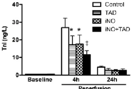
Reduction of peak plasma TnI levels at 4h by iNO (n=9;
17.6±5.1 ng/l) and TAD (n=7; 17.3±5.0 ng/l) was further amplified by the combined therapy (n=9; 11.4±2.4 ng/l), when compared to CON animals (n=9; 24.6±5.3 ng/l, Figure 1.).
Figure 1. Troponin I (TnI) plasma levels after I/R. Biomarker release peaked at 4h and diminished after 24 hours. TAD (n=7), iNO (n=9) and iNO+TAD (n=9) treatments all significantly reduced TnI levels compared to CON (n=9). * P<0.05, ** P<0.001 vs CON.
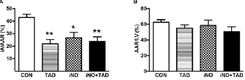
Risk area (AAR), based on TTC staining, encompassed
~57% of the LV area in all groups but did not compromise hemodynamic status or early survival at 3 days.
Non-viable area within the AAR was significantly smaller in TAD (n=8), iNO (n=5) and iNO-TAD (n=5) animals (27±4%, 22±3% and 24±4%, respectively, versus 43±2% in CON (n=7), P<0.05 for all; Figure 2.).
Figure 2. Planimetric analysis of TTC-stained heart sections. All treatment strategies (CON n=7, TAD n=8, iNO n=5, iNO+TAD n=5) reduced infarcted area (IA) expressed relative to the area at risk (AAR) when compared to CON animals (Panel A). The AAR/LV ratio was comparable in all groups (Panel B). CON = untreated mice, TAD = Tadalafil, iNO = inhaled nitric oxide, ino+TAD = combined treatment. * P<0.05, ** P<0.01 vs CON
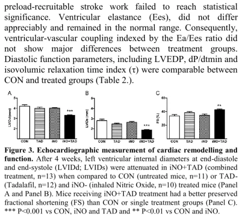
Subsequent long term (4w) LV structural and functional remodelling was evaluated with transthoracic echocardiography and pressure-conductance catheter analysis.
Combined intervention significantly reduced adverse remodeling and improved fractional shortening, whereas either therapy alone failed to do so (Figure 3.). Peri-infarct wall thickness measurements revealed less hypertrophy and better- preserved wall thickening in all treatment groups (Table 1.).
Pressure-volume analysis showed a significantly greater stroke volume in iNO+TAD treated mice, resulting in a proportionately higher cardiac output at comparable heart rates.
Mice, which inhaled NO had an intermediate response with higher LV end-systolic pressure and stroke work. Increased
preload-recruitable stroke work failed to reach statistical significance. Ventricular elastance (Ees), did not differ appreciably and remained in the normal range. Consequently, ventricular-vascular coupling indexed by the Ea/Ees ratio did not show major differences between treatment groups.
Diastolic function parameters, including LVEDP, dP/dtmin and isovolumic relaxation time index (τ) were comparable between CON and treated groups (Table 2.).
Figure 3. Echocardiographic measurement of cardiac remodelling and function. After 4 weeks, left ventricular internal diameters at end-diastole and end-systole (LVIDd; LVIDs) were attenuated in iNO+TAD (combined treatment, n=13) when compared to CON (untreated mice, n=11) or TAD- (Tadalafil, n=12) and iNO- (inhaled Nitric Oxide, n=10) treated mice (Panel A and Panel B). Mice receiving iNO+TAD treatment had a better preserved fractional shortening (FS) than CON or single treatment groups (Panel C).
*** P<0.001 vs CON, iNO and TAD and ** P<0.01 vs CON and iNO.
Treatment N IVSd WTIVS LVPWd LVPWs WTPW
(mm) (%) (mm) (mm) (%)
CON 11 1.07±0.05 24±6 1.05±0.03 1.23±0.004 18±4 TAD 12 0.93±0.02 ** 40±3 0.99±0.01 1.23±0.005 25±1 iNO 10 0.90±0.03 ** 48±5 ** 0.94±0.03 * 1.27±0.015 ** 35±5 **
iNO+TAD 13 0.87±0.02 ** 52±3 *** 0.93±0.02 ** 1.23±0.003 32±2 * Table 1. Echocardiographic measurements at 4 weeks. IVSd and IVSs – diastolic and systolic interventricular septal thickness, LVPWd and LVPWs – diastolic and systolic left ventricular posterior wall thickness, WTIVS and WTPW – percentage of interventricular septal and posterior wall thickening. CON = untreated mice, TAD = Tadalafil, iNO = inhaled nitric oxide, iNO+TAD = combination treatment. * P<0.05; ** P<0.01; **
P<0.001 versus CON
CON TAD iNO iNO-TAD
(n=11) (n=13) (n=12) (n=13)
HR (BPM) 601±12 604±14 603±15 609±13
LVESP (mmHg) 78±3 85±4 89±2* 82±3
LVEDP (mmHg) 2.4±0.6 2.1±0.3 3.5±0.7 2.3±0.5 SV (µL) 10.2±0,9 11.0±1.1 13.6±1.1 14.9±1.2*
CO (µL/min) 6129±566 6637±713 8277±707 9156±773*
EF (%) 50.6±4 54±4 54±4 59±3
SW (mmHg x µL) 771±85 932±91 1205±97* 1134±119
PRSW 69±8 71±6 75±10 94±8
Ea (mmHg/µL) 7.8±1.1 7.0±0.5 6.3±0.7 5.7±0.3 Ees (mmHg/µL) 10.1±2.8 10.3±1.5 6.7±1.1 9.1±2.2 Ea / Ees 1.01±0.16 0.79±0.09 1.10±0.14 0.83±0.13 dP/dtmax (mmHg/s) 8795±946 9956±646 11899±879 10617±969 dP/dtmin (mmHg/s) -7498±538 -8248±586 -8930±822 -7837±396
(ms) 5.2±0,2 5.2±0.2 5.0±0.3 5.1±0.2
Table 2. Pressure-volume analysis at 4 weeks. HR- Heart rate in beats per minute (BPM); LVESP and LVEDP - left ventricular end systolic and end diastolic pressures; SV-stroke volume, CO- cardiac output; EF- ejection fraction; SW- stroke work; PRSW-preload-recruitable stroke work; Ea- arterial elastance; Ees – left ventricular end-systolic elastance; Ea/Ees ratio - ventricular-arterial coupling; dP/dtmax and dP/dtmin- maximum and minimum of systolic pressure change over time; - tau time constant of isovolumic relaxation according to Weiss’ method. All data are presented as mean±SEM. CON = untreated mice, TAD = Tadalafil, iNO = inhaled nitric oxide, iNO+TAD = combination therapy. * P<0.05 versus CON
4.1.2. iNO and tadalafil treatment modulate NO-cGMP signaling and cardiac nitrosative stress
Inhaled nitric oxide therapy resulted in a 5- to 6-fold increase in plasma NOx levels, comprising nitrate, nitrite and S-nitroso compounds (P<0.0001 for iNO and iNO+TAD).
Similarly, plasma nitrite (NO2-)concentrations were more than 3-fold higher than in CON and TAD animals (P<0.0001 for both). Of interest, nitrotyrosine content in the immediate reperfusion period trended to be lower in reperfused cardiac tissue after iNO and iNO+TAD treatment. (P=0.10 and 0.08, respectively).
Plasma cGMP levels showed a non-significant increase after iNO and TAD treatments (48±3 pmol/mL [n=8] and 51±7
Figure 3. NO-derived oxidation products, 3-Nitrotyrosine and cGMP levels in plasma and in cardiac tissue at 20 min after reperfusion.
Plasma levels of NOx, comprising nitrates, nitrites and S-nitroso compounds (Panel A) and nitrite (NO2-, Panel B) are measured in CON (n=8), TAD (n=7), iNO (n=7), and iNO+TAD (n=8) groups. Nitrosative stress is evaluated by measuring proteins containing 3-nitrotyrosine residues (3-NT) using ELISA (Panel C-D). Plasma cGMP increased after iNO+TAD (n=7; P<0.01 vs CON and P<0,05 vs iNO) treatment, while TAD (n=6) and iNO (n=8) was not different from CON (n=7). Similarly iNO+TAD (n=7), but not TAD (n=8) or iNO (n=6), increased tissue cGMP levels vs CON (P<0.01, panel (Panels E-F). CON = untreated mice, TAD = Tadalafil, iNO
= inhaled nitric oxide, iNO+TAD = combination therapy. *** P<0.0001 vs CON and TAD, ** P<0.01 vs CON, † P<0.05 vs iNO
pmol/mL [n=6], respectively versus 38±6 pmol/mL [n=7] in CON), but nearly doubled after iNO-TAD (80±12 pmol/mL [n=7], P<0.01 vs CON and iNO, P<0.05 vs TAD). Cardiac tissue cGMP level increased significantly only after iNO+TAD
treatment (0.15±0.02 pmol/mg [n=7] vs 0.05±0.01 pmol/mg in CON [n=7], P<0.01). The increase with either iNO (0.07±0.01 pmol/mg [n=6]), or TAD (0.10±0.02 pmol/mg [n=8]) alone did not reach statistical significance (Figure 3.).
4.1.3. iNO+TAD therapy attenuates leukocyte infiltration.
After 3 days, the number of MPO-positive cells relative to mid-ventricular transversal tissue area was significantly lower in iNO+TAD treated mice (P=0.02 vs CON, Figure 4.).
Four weeks after I/R, collagen fibre deposition encompassed
~7% of the ischemic LV area, and was not influenced by treatment protocols.
Figure 4. Myeloperoxidase-positive (MPO) cell infiltration three days after ischemia-reperfusion. iNO+TAD (n=6) significantly reduced MPO- positive cell infiltration compared to CON mice (n=5), while iNO (n=6) or TAD (n=7) had only a modest effect. CON = untreated mice, TAD = Tadalafil, iNO = inhaled nitric oxide, iNO+TAD = combination therapy. * P<0.05 vs CON
4.2. The NOS3 Glu298Asp polymorphism and athletic cardiac adaptation
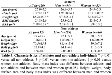
4.2.1. Characteristics of athletes and control individuals Age distribution and gender ratio (25.4% and 29.7% female in athletes and non-athletes, respectively) were not different between the study groups. Height, weight, body mass index and body surface area were higher in men versus women and in athletes versus non-athletes. (Table 3.)
Athletes
All (n=126) Men (n=94) Women (n=32)
Age (years) 25.9±5.5 26.9±5.7 24.9±5.3
Height (m) 1.87±0.11 * 1.95±0.04 † 1.77±0.07 ‡ Weight (kg) 85.2±15.6 * 97.5±8.3 † 72.3±10.2 ‡
BMI (kg/m2) 24.8±2.6 25.8±2.2 23.4±2.5
BSA (m2) 2.18±0.28 * 2.38±0.1 † 1.89±0.3 ‡
Non-athletes
All (n=155) Men (n=109) Women (n=46)
Age (years) 27.4±5.2 27.1±5.1 28.0±5.7
Height (m) 1.76±0.11 1.83±0.07 1.67±0.09
Weight (kg) 70.5±15.3 81.4±13.6 59.9±7.2
BMI (kg/m2) 22.8±4.3 24.1±4.6 21.6±3.9
BSA (m2) 1.94±0.3 2.08±0.3 1.74±0.2
Table 3. Characteristics of athletes and non-athlete individuals * p<0.01 versus all non-athletes, † p<0.01 versus men non-athletes, ‡ p<0.01 versus women non-athletes. Body mass index was different between athletes and non-athletes and between men and women (p<0.05). Height, weight, body surface area and body mass index was different between men and women irrespective of athletic status (p<0.05).
4.2.2. Cardiac morphology and function
Left and right ventricular volume and myocardial mass indexes were significantly higher in athletes, while resting ejection fractions (LVEF % and RVEF%) were similar in athletes and non-athletes. Significant gender-related differences were noted, irrespective of athletic activity (Table 4.). At exhaustion both VO2 maximum (60±7 versus 40±7 ml/kg/min, p<0.0001) and minute ventilation (VE, 150±15 versus 84±39 l/min, p<0.0001) were significantly higher in athletes and within each group significantly higher in men than in women.
4.2.3. Genotype distribution
Allelic distributions were similar, with a minor allelic frequency of 0.27 in athletes vs. 0.26 in controls. Correlation with athletic status was not found. The investigated genotype distributions were in Hardy-Weinberg equilibrium.
Athletes Non-athletes
All Men Women All Men Women
N 126 94 32 155 109 46
LVEF (%) 58.5
±6.3 57.8
±4.3 60.4
±9.7 59.4
±4.3 59.5
±4.4 58.4
±4.2 LVEDVi (ml/m2) 116.2
±17.4 * 121.4
±14.9 ‡ 102.4
±16.2 92.9
±12.9 97.3
±11.1 ‡ 84.4
±13.2 LVESVi (ml/m2) 48.8
±11.2 * 51.4
±9.6 ‡ 41.9
±12.1 37.8
±7.6 39.5
±7.3 ‡ 35.4
±8.0 LVMi ( g/m2) 81.1
±19.6 * 88.1
±15.9 ‡ 62.4
±16.0 61.3
±13.9 68.1
±10.4 ‡ 47.0
±10.1 LVSVi (ml/m2) 67.6
±8.3 * 69.8
±8.1 ‡ 61.6
±5.6 54.8
±7.8 57.8
±6.8 ‡ 49.0
±6.8 RVEF (%) 57.9
±6.2
57.1
±4.0
60.2
±9.9 58.5
±4.8 58.2
±4.9
58.2
±4.4 RVEDVi (ml/m2) 121.5
±19.6 * 127.8
±17.4 ‡ 106.0
±17.3 95.5
±15.2 100.7
±14.1 ‡ 85.7
±13.4 RVESVi (ml/m2) 53.2
±11.3 * 55.8
±10.9 ‡ 46.6
±9.9 39.8
±9.6 42.6
±9.9 ‡ 35.3
±7.3 RVSVi (ml/m2) 68.2
±10.2 * 70.7
±9.5 ‡ 61.5
±9.2 56.7
±7.0 58.3
±6.4 ‡ 52.4
±7.7 RVMi ( g/m2) 29.9
±6.1 * 32.5
±4.6 ‡ 23.5
±5.4 24.4
±4.3 25.3
±3.5 ‡ 20.1
±3.0 Table 4. Characteristics of athlete and non-athlete men and women. n – number of individuals; LV – left ventricular, LVEF – LV ejection fraction;
LVEDVi - LV end-diastolic volume index; LVESVi – LV end-systolic ventricular index; LVMi – LV mass index; LVSVi – LV stroke volume index; RV – right ventricular, RVEF – RV ejection fraction; RVEDVi – RV end-diastolic volume index; RVESVi – RV end-systolic volume index;
RVSVi – RV stroke volume index; RVMi – RV mass index
* p<0.01 versus all non-athletes, ‡ p<0.01 versus women. Age was not different between study groups.
4.2.4. Association of cardiac structure and function with the NOS3 298 genotype
In Asp carriers compared to Glu homozygous participants (irrespective of athletic status, n=133 vs. n=148) resting RVSVi and RVMi were higher (60±9 versus 62±12 ml/m2, p=0.047 and 26±6 versus 27±6 g/m2 respectively, p=0.019). Athletes carrying the Asp allelic variant had higher RVSVi and RVMi, than those homozygous for the Glu allelic variant (71.1±9.6 versus 64.3±9.8 ml/m2 and 31.7±5.5 versus
27.4±6.0 g/m2, p<0.01 respectively, p<0.001). Associations were not found among non-athletic individuals, nor between genotype and left ventricular anatomy, nor O2 consumption at exhaustion and genotype. (Table 5.)
Athletes (VO2 max
>50ml/kg/min)
Non-athletes (VO2 max
<50ml/kg/min) Glu/Glu Glu/Asp
+ Asp/Asp Glu/Glu Glu/Asp + Asp/Asp P
N 64 62 84 71 -
LVEF (%) 58.3±8.1 58.7±4.5 59.1±2.4 60.0±6.7 0.688 LVEDVi (ml/m2) 115.1±20.5 * 117.0±14.9 * 92.1±14.6 94.4±9.3 <0.001 LVESVi (ml/m2) 49.2±13.3 * 48.5±9.3 * 37.8±7.8 37.9±7.6 <0.001 LVMi ( g/m2) 77.9±23.1 * 83.5±16.2 * 60.3±15.3 63.2±11.2 <0.001 LVSVi (ml/m2) 66.4±7.6 * 68.5±8.7 * 54.1±7.6 56.1±8.2 <0.001 RVEF (%) 57.7±8.1 58.1±4.4 58.3±4.1 58.4±5.8 0.427 RVEDVi (ml/m2) 117.8±19.9 * 124.4±19.1 * 95.6±17.6 95.4±9.8 <0.001 RVESVi (ml/m2) 53.0±10.5 * 53.4±12.1 * 40.4±11.0 38.7±6.5 <0.001 RVSVi (ml/m2) 64.3±9.8 #* 71.1±9.6 * 56.6±6.7 56.8±7.8 <0.001 RVMi ( g/m2) 27.4±6.0 #* 31.7±5.5 * 25.3±4.7 23.3±3.4 <0.001 Table 5. Characteristics of different genotypes within athletes and non- athletes (irrespective of their gender). n – number of individuals; LV – left ventricular; LVEF – LV ejection fraction; LVEDVi – LV end-diastolic volume index; LVESVi – LV end-systolic ventricular index; LVMi – LV mass index; LVSVi – LV stroke volume index; RV – right ventricular;
RVEF – RV ejection fraction; RVEDVi – RV end-diastolic volume index;
RVESVi – RV end-systolic volume index; RVSVi – RV stroke volume index; RVMi – RV mass index
ANOVA showed significant differences among all four inspected groups.
Post hoc tests revealed a significant influence of the genotype (# p<0.001 vs Asp carriers) on resting RVSVi and RVMi in athletes. Athletic status had significant influence on all parameters with the exception of LVEF and RVEF (* p<0.001 vs non-athletes irrespective of genotype).
5. CONCLUSIONS
5.1. Concomitant TAD treatment enhances iNO dependent cardioprotection
Combination of iNO with oral administration of TAD is safe and confers incremental myocardial protection during I/R injury in mice. It results in a greater reduction in troponin
release during the acute phase, and less inflammatory cell infiltration when compared to either treatment alone. This early benefit translated in improved functional and structural remodelling after 4 weeks. LV end-systolic dimensions following combination treatment were reduced and associated with a better-preserved regional LV function. Invasive pressure-volume analysis using conductance catheter technology confirmed improved contractile performance in mice treated with iNO and TAD with significantly higher stroke volumes. Finally, combination therapy was associated with significantly greater nitrite plasma concentration, a trend for lower cardiac nitrosative stress levels and significantly higher cGMP bioavailability in the heart and in the circulation.
Increase in cGMP-signalling after the combined treatment suggests the importance of this pathway for beneficial long- term cardiac structural and functional remodelling. This approach may represent a promising strategy for translational research to attenuate ischemia-reperfusion injury in patients.
5.2. The NOS3 Glu298Asp polymorphism and athletic cardiac adaptation
In this study cardiac function and structure of elite athletes was determined and a previously unknown correlation was found between load-dependent right ventricular adaptation (RVSVi and RVMi) and the NOS3 Glu298Asp polymorphism.
Importance of these observations lies in their potential influence on athletic performance and, perhaps even more importantly, on the development of exercise induced right ventricular cardiomyopathy. To evaluate its exact clinical significance long-term follow up and gene interaction studies are indispensable.
6. LIST OF RELATED PUBLICATIONS Full papers
Lux A, Pokreisz P, Swinnen M, Caluwe E, Gillijns H, Szelid Z, Merkely B, Janssens SP. (2016) Concomitant Phosphodiesterase 5 Inhibition Enhances Myocardial Protection by Inhaled Nitric Oxide in Ischemia-Reperfusion Injury. J Pharmacol Exp Ther. 356(2):284-92. IF: 3.972
Szelid Z, Lux A, Kolossváry M, Tóth A, Vágó H, Lendvai Z, Kiss L, Maurovich-Horvat P, Bagyura Z, Merkely B. (2015) Right Ventricular Adaptation Is Associated with the Glu298Asp Variant of the NOS3 Gene in Elite Athletes. PLoS One.10(10):e0141680. IF: 3.234
Other papers, not related to the dissertation
Oláh A, Németh BT, Mátyás Cs, Hidi L, Lux A, Ruppert M, Kellermayer D, Sayour AA, Szabo L, Torok M, Meltzer A, Gellér L, Merkely B, Radovits T (2014) Physiological and pathological left ventricular hypertrophy of comparable degree is associated with characteristic differences of in vivo hemodynamics. Am J Physiol Heart Circ Physiol.
310(5):H587-97. IF: 3,838
Szilveszter B., Elzomor H, Károlyi M, Kolossváry M, Raaijmakers R, Benke K, Celeng Cs, Bartykowszki A, Bagyura Zs, Lux A, Merkely B, Maurovich-Horvat P. (2016) The effect of iterative model reconstruction on coronary artery calcium quantification. Int J Cardiovasc Imaging. 2016, 32(1):153-60. IF: 1,810
Kovacs A, Oláh A, Lux A, Mátyás M, Németh BT, Kellermayer D, Ruppert M, Torok M, Szabo L, Meltzer A, Assabiny A, Birtalan E, Merkely B, Radovits T (2014) Strain and strain rate by speckle tracking echocardiography correlate with pressure-volume loop derived contractility indices in a rat model of athlete's heart. AJP Heart and Circulatory Physiology 01/2015 IF: 3,838
Bagyura Zs, Kiss L, Edes E, Lux A, Polgár L, Soós P, Szenczi O, Szelid Zs, Vadas R, Józan P, Bagdy Gy, Merkely B (2014) Cardiovascular screening programme in the Central Hungarian region. The Budakalász Study. Orv Hetil. 24;155(34):1344-52.
Kellermayer D, Oláh A, Lux A, Németh BT, Hidi L, Birtalan E, Ruppert M, Mátyás Cs, Merkely B, Radovits T (2014) Detailed Hemodynamic Characterization of Athlete's Heart using Left Ventricular Pressure-Volume Analysis in a Rat Model. Biophysical Journal 106(2):344A. IF: 3.97
Radovits T, Oláh A, Lux A, Németh BT, Hidi L, Birtalan E, Kellermayer D, Mátyás Cs, Szabó G, Merkely B (2013) Rat model of exercise-induced cardiac hypertrophy - hemodynamic characterization using left ventricular pressure-volume analysis. AJP Heart and Circulatory Physiology; IF: 4.012 Szelid Zs, Kerecsen G, Maurovich-Horvát P, Lux A, Marosi E, Kovács A, Kiss RG, Préda I, Merkely B (2010) Determination of coronary in-stent restenosis using dual source computed tomography angiography. Interventional Medicine and Applied Science; 2(1): 5-9.
Szelid ZsL, Lux A (2009) Génalapú lehetőségek a cardiovascularis prevencióban és kezelésben. Orvosképzés; S4:
201-366.
Bagyura Zs., Kiss L, Édes E.,Lux Á., Polgár L., Soós P., Szenczi O., Szelid Zs., Vadas R., Józan P., Bagdy Gy., Merkely B. (2014) Cardiovascularis szűrőprogram a közép- magyarországi régióban. Orv. Hetil., 155(34), 1344–1352.