Nitric oxide-related cardioprotection during the development of myocardial ischaemia and athletic
heart
Doctoral Thesis Dr. Árpád Lux, MD
Semmelweis University Doctoral School of Basic Medicine
Supervisor: Dr. Zsolt Szelid, MD, PhD
Official reviewers: Dr. József Kaszaki, MD, PhD, med. habil Dr. Tímea Kováts, MD, PhD
Examination committee:
President: Prof. Dr. László Rosivall, MD, DSc Members: Dr. Noémi Nyolczas, MD, PhD
Dr. Zsuzsanna Miklós, MD, PhD
Budapest, 2016
2
Table of contents
Abbreviations ... 6
1. Introduction ... 12
1.1. Nitric oxide (NO) signaling in cardiovascular physiology and pathophysiology 12 1.1.1. Identification of NO ... 12
1.1.2. General chemical and biological properties of NO ... 12
1.1.3. General behaviour of NO in biological systems: toxicity and safety issues . 13 1.1.4. Natural sources and protective effects of nitric oxide ... 13
1.1.5. Nitric oxide synthesis ... 14
1.1.5.1. Endothelial nitric oxide synthase (eNOS) ... 15
1.1.5.2. Neuronal nitric oxide synthase (nNOS)... 16
1.1.5.3. Inducible nitric oxide synthase (iNOS) ... 16
1.1.6. NOS uncoupling: superoxide production ... 17
1.1.7. Endogenous NOS inhibitors ... 17
1.1.8. Nitric Oxide signaling ... 18
1.1.8.1 cGMP dependent NO signaling ... 18
1.1.8.1.1. Cyclic guanosine 3’,5’-monophosphate ... 18
1.1.8.1.2. Soluble guanylate cyclase (sGC) ... 19
1.1.8.1.3. Protein kinase G (PKG) ... 19
1.1.8.1.3.1. PKG regulation of calcium levels and signaling ... 20
1.1.8.1.3.2. PKGI-mediated inactivation of the Ras homolog gene family member A (RhoA) ... 20
1.1.8.1.3.3. Gene regulation ... 21
1.1.8.1.3.4. Phosphorylation of Vasodilator-Stimulated Phosphoprotein .... 21
1.1.8.1.3.5. Phosphodiesterase 5 regulation ... 21
1.1.8.1.3.6. Cytoprotection ... 21
1.1.8.1.3.7. Pro-apoptotic effects ... 22
1.1.8.1.4. Cyclic nucleotide regulated ion channels ... 22
1.1.8.1.4.2. Cyclic nucleotide-gated channels (CNG):... 22
1.1.8.1.4.3. Hyperpolarization-activated cyclic nucleotide-gated ion channels (HCN): ... 22
3
1.1.8.1.5. cGMP catabolism - phosphodiesterases ... 23
1.1.8.1.5.1. PDE regulation ... 24
1.1.8.1.5.2. PDE5 expression and regulation ... 26
1.1.8.2. cGMP independent pathway ... 26
1.2. Ischemia-reperfusion injury ... 27
1.2.1. Biochemical and metabolic changes during ischemia reperfusion ... 27
1.2.1.1. The ischemic cascade ... 28
1.2.1.2. pH normalization ... 28
1.2.1.3. ROS generation ... 28
1.2.1.4. Calcium oscillation ... 29
1.2.2.5. Targeting the mitochondria ... 29
1.2.2.6. Neutrophil involvement in lethal myocardial reperfusion injury ... 30
1.2.2.7. NOS uncoupling ... 31
1.3. Modulation of the nitric oxide mediated signaling pathway – therapeutic applications ... 31
1.3.1 Inhaled nitric oxide... 32
1.3.1.1. Taking advantage of pulmonary effects ... 32
1.3.1.2. Extrapulmonary effects of inhaled NO ... 32
1.3.1.2.3. Cardiopulmonary resuscitation ... 33
1.3.1.2.6. Myocardial ischemia-reperfusion injury: ... 33
1.3.1.3. Theories behind extrapulmonary effects ... 33
1.3.2. Other inhaled drugs to modify NO signaling ... 34
1.3.3. Phosphodiesterase 5 inhibition ... 34
1.3.3.1. Protection against ischemia-reperfusion injury ... 36
1.3.3.2. Protection against ischemic and doxorubicin induced cardiomyopathy 36 1.3.3.4. Protection against cardiac hypertrophy: ... 37
1.3.4. sGC stimulators and activators ... 37
1.3.4.1 sGC activators ... 37
1.3.4.2. sGC stimulators ... 38
1.4. The role of NOS in physical conditioning ... 38
1.4.1. Structural changes in the left and right ventricles among athletes ... 38
1.4.2. Functional changes in the left and right ventricles among athletes... 39
1.4.3. Mechanisms of athletic cardiac remodelling ... 40
4
1.4.4. Possible interactions of NO and NOS3 polymorphisms with athletic cardiac
remodelling... 40
1.4.5 Exercise induced right ventricular dysfunction ... 41
2. Objective: to identify new aspects of NOS signaling under physiologic and pathophysiologic conditions ... 42
2.1 Murine experiment on potentiation of NO derived cardioprotection ... 42
2.2. Clinical study on the role of NOS3 polymorphisms in physiologic adaptation in elite athletes ... 42
3. Methods ... 43
3.1. Murine experiment on potentiation of the NO mediated cardioprotection with PDE5 inhibition ... 43
3.1.1. Drugs used in the experiments ... 43
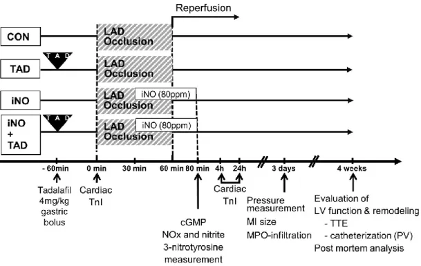
3.1.2. Induction of I/R and experimental design ... 43
3.1.3. Echocardiography... 45
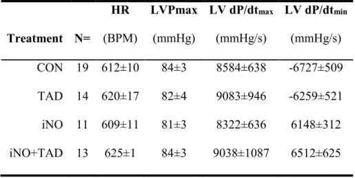
3.1.4 Invasive hemodynamic measurements ... 45
3.1.5. Measurement of infarct size and myocardial necrosis ... 46
3.1.6. Measurement of tissue and circulating cGMP levels ... 47
3.1.7. Measurement of nitric oxide-derived oxidative end products ... 47
3.1.8. Histological and immune-histochemical determination of collagen deposition and myeloperoxidase-positive cell infiltration ... 48
3.1.9. Statistical analysis ... 48
3.2. Clinical study on the role of NOS3 polymorphisms in physiologic adaptation in elite athletes ... 48
3.2.1. Selection of candidate individuals and study protocol ... 48
3.2.2. Screening protocol... 49
3.2.3. Cardiopulmonary stress test ... 50
3.2.3. Cardiac magnetic resonance imaging (cMRI) ... 51
3.2.4. DNA extraction and genotyping ... 52
3.2.5. Statistical analysis ... 52
4. Results ... 53
4.1. Murine experiment on potentiation of NO mediated cardioprotection with PDE5 inhibition ... 53
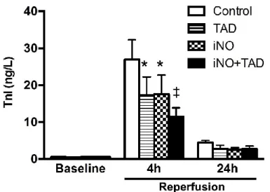
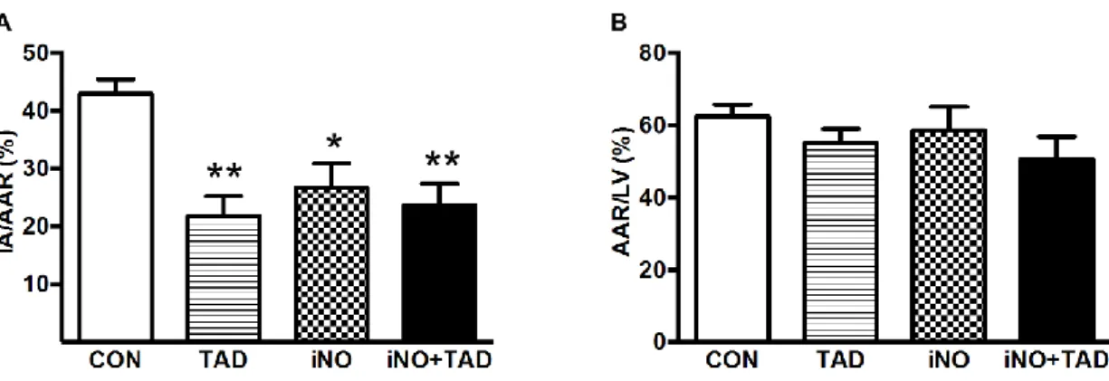
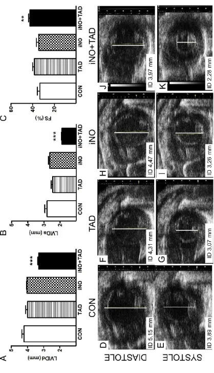
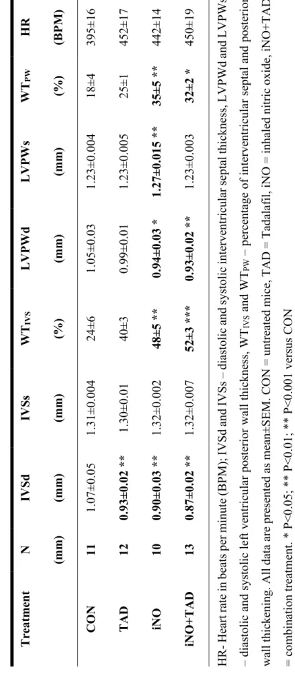
4.1.1. Combined therapy with iNO and TAD confers synergistic myocardial protection after I/R ... 53
10.3±1.5 ... 58
5
4.1.2. iNO and tadalafil treatment modulate NO-cGMP signaling and cardiac
nitrosative stress ... 60
4.1.3. Combined iNO and TAD therapy attenuates myocardial leukocyte infiltration, but not myocardial fibrosis... 62
4.2. Clinical study on the role of NOS3 in physiologic adaptation in elite athlethes . 64 4.2.1. Characteristics of athletes and control individuals ... 64
4.2.3. Cardiac morphology and function in athletes and controls ... 64
4.2.4. Genotype distribution ... 67
4.2.5. Association of cardiac structure and function with the NOS3 298 genotype ... 67
5. Discussion ... 70
5.1. Murine experiment on potentiation of cardioprotection from NO with PDE5 inhibition ... 70
5.1.1. Limitations ... 74
5.1.2. Conclusion ... 74
5.1.3. Potential influence on human ischemia reperfusion therapy ... 74
5.2. Clinical study on the role of NOS3 in physiologic adaptation in elite athletes ... 75
5.2.1. Limitations ... 78
5.2.1 Conclusion ... 78
6. Future directions ... 79
7. Summary ... 80
8. Összefoglalás ... 81
9. Bibliography ... 82
10. List of publications ... 100
10.1. Publications related to the doctoral work ... 100
10.2. Other publications ... 100
11. Acknowledgements ... 102
6
Abbreviations
AAR - Area at risk
ADMA - Asymmetric dimethylarginine ANOVA - Analysis of variance
ANP - atrial type natriuretic peptide AP-1 - Activator protein 1
Arg - Arginine
Asp - Aspartic acid
ATP - Adenosine triphosphate
AV - Atrioventricular
Bad - Bcl-2-associated death promoter
BH2 - Dihydropteridin
BH4 - Tetrahydrobiopterin (2-amino-6-(1,2-dihydroxypropyl)-5,6,7,8- tetrahydro-1H-pteridin-4-one)
BNP - Brain type natriuretic peptide b-TFE - balanced turbo field echo
cAMP - Cyclic adenosine monophosphate
Cav1.2 - Calcium channel, voltage-dependent, L type, alpha 1C subunit cGMP - Cyclic guanosine monophosphate
CHD - Congenital heart disease
cMRI - Cardiac magnetic resonance imaging CNBD - Cyclic nucleotide binding domain CNG - Cyclic nucleotide-gated ion channels
cNOS - Constitutive nitric oxide synthase (also known as eNOS, NOS3)
CO - Carbon monoxide
CO - Cardiac output
CON - control, untreated mice
CSE - Cystathionine-γ-lyase
DDAH - Dimethylarginine dimethylaminohydrolases DHFR - Dihydrofolate reductase
DNA - Deoxyribonucleic acid
dP/dtmax - Maximum rates of systolic pressure rise
7
dP/dtmax - Minimum rates of systolic pressure decline
Ea - Arterial elastance
Ea/Ees ratio - ventricular-arterial coupling
ECMO - Extracorporeal membrane oxygenation
ECG - Electrocardiogram
eCSC - Endogenous cardiac stem and progenitor cells EDRF - Endothelium-derived relaxing factor
Ees - ventricular elastance
EF - Ejection fraction
ELISA - enzyme-linked immunosorbent assay
eNOS - Endothelial nitric oxide synthase (also known as cNOS, NOS3) ERK - Extracellular signal-regulated kinase
ETC - Mitochondrial electron transport chain
ETT-TUKEB - Hungarian Scientific Council National Ethics Committee for Scientific Research
FDA - US Food and Drug Administration
FS - Fractional shortening
GAF domain - This name derives from the first proteins it was found in: cGMP-specific phosphodiesterases, adenylyl cyclases and FhlA
Glu - Glutamic acid
GSK3β - Glycogen synthase kinase 3β
GTP - Guanosine triphosphate
H-NOX - Heme-nitric oxide and oxygen binding
H2S - Hydrogen sulfide
HCN - Hyperpolarization-activated cyclic nucleotide-gated ion channels
HIF-1α - Hypoxia-inducible factor-1α HSP70 - 70 kilodalton heat shock proteins
IA - Infarcted area
I/R - Ischemia reperfusion
ICAM-1 - Intercellular Adhesion Molecule 1
8
If - funny current
IGF-1 - Insulin-like growth factor 1
IL-1 - Interleukin 1
iNO - Inhaled nitric oxide
iNOS - Inducible nitric oxide synthase
iNO+TAD - Combination of inhaled nitric oxide and tadalafil
IP - Intraperitoneal
IP3 - Inositol 1,4,5-trisphosphate
IRAG - Inositol 1,4,5-trisphosphate (IP3) receptor-associated PKG substrate
iv - intra venous
IVSd - interventricular septum thickness at end-diastole IVSs - interventricular septum thickness at end-systole JAK - Janus-activated kinase
JNK1 - c-Jun NH2-terminal kinase 1
k-t BLAST - Broad-use Linear Acquisition Speed-up Technique LAD - Left anterior descending artery
L-NMMA - NG-monomethyl-L-arginine LTCC - L-type Ca2+ channels
LV - Left ventricle
LVAD - Left ventricular assist device
LVEDVi - Left ventricular end-diastolic volume index LVEF - Left ventricular ejection fraction
LVESVi - Left ventricular end-systolic ventricular index LVH - Left ventricular hypertrophy
LVIDd - Left ventricular dimensions at end-diastole LVIDs - Left ventricular dimensions at end-systole LVMi - Left ventricular mass index
LVPWd - Posterior wall thickness at end-diastole LVPWs - Posterior wall thickness at end-systole LVSVi - Left ventricular stroke volume index MAPK - Mitogen-activated protein kinases
9
MEKK1 - Mitogen-activated protein kinase kinase
MI - Myocardial infarction
MIP-1 /-2 - Macrophage Inflammatory Proteins mitoKATP - Mitochondrial ATP-sensitive K+ channel mitochondrial PTP - Mitochondrial Permeability Transition Pore
MPO - myeloperoxidase
NADPH - Nicotinamide adenine dinucleotide phosphate
NF-κB - nuclear factor kappa-light-chain-enhancer of activated B cells NFAT - Nuclear factor of activated T cells
NIH - National Institutes of Health nNOS - Neural nitric oxide synthase
NO - Nitrogen oxide
NO2 - Nitrogen dioxide
NOHLA - Nω-hydroxy-L-arginine
NOMI - Nitric oxide for inhalation to reduce reperfusion injury in STEMI
NOS - Nitric oxide synthase
NOS1 - nitric oxide synthase 1 (also known as nNOS) NOS2 - nitric oxide synthase 2 (also known as iNOS)
NOS3 - nitric oxide synthase 3 (also known as cNOS, eNOS)
Nox -NADPH oxidase
p53 - cellular tumor antigen p53
p70S6K - extracellular regulated kinase system with downstream p70 ribosomal protein S6 kinase
PAP - Pulmonary artery pressure
PAS - Protein fold named for its association with the Per, ARNT, and Sim proteins
PDE - Phosphodiesterase
pGC - Particulate guanylate cyclase PI3K(p110α) - Phosphoinositide 3-kinase (p110α) PIP3 - Phosphatidylinositol 3,4,5-trisphosphate PKA - cAMP dependent protein kinase
10
PKG - cGMP dependent proteinkinase
PPARα - peroxisome proliferator-activated receptor alpha PPHN - Persistent pulmonary hypertension of the new-born
ppm - Parts-per-million, 10−6
PRSW - Preload-recruited stroke work
PV - pressure-volume
PVR - Pulmonary vascular resistance RBBB - Right Bundle Branch Block
RhoA - Ras homolog gene family member A RISK - Reperfusion Injury Salvage Kinase
ROS - Reactive oxygen species
RV - Right ventricle
RVEDVi - Right ventricular end-diastolic volume index RVEF - Right ventricular ejection fraction
RVESVi - Right ventricular end-systolic volume index RVMi - Right ventricular mass index
RVSVi - Right ventricular stroke volume index
RyR - Ryanodine receptor
SADMA - Symmetric dimethylarginine
SAFE - Survival Activating Factor Enhancement SAP - Systemic arterial pressure
SEK1 - Stress-activated protein/ERK kinase 1
SEM - Standard error of mean
SERCA - Sarco/endoplasmic reticulum Ca2+-ATPase sGC - Soluble guanylate cyclase
STAT - Signal transducer and activator of transcription
SV - Stroke volume
SW - Stroke work
Τ - isovolumic relaxation
T2w-SPIR - Triggered blood suppressed T2-weighted spectral inversion recovery
TAD - Tadalafil
11 TNF-α - Tumor necrosis factor α
TnI - Troponin I
TTC - Triphenyl tetrazolium chloride TTE - Transthoracic echocardiography
VASP - Vasodilator-Stimulated Phosphoprotein
VO2 - Maximal oxygen uptake
VCAM-1 - Vascular cell adhesion protein 1
XO - Xanthine oxidase
XOR - Xanthine oxidoreductase
YC-1 - 3-[5′-hydroxymethyl-2′-furyl]-1-benzylindazole
12
1. Introduction
1.1. Nitric oxide (NO) signaling in cardiovascular physiology and pathophysiology
1.1.1. Identification of NO
Nitrogen oxide (NO) is not only an important intermediate in chemical industry, but an important signaling molecule as well. This free radical is a byproduct of combustion and is naturally produced during electrical discharges of lightning in thunderstorms (National Center for Biotechnology Information and U. S. National Library of Medicine, n.d.).
It was first described in the 18th century by Joseph Priestly – an English polymath – in the first volume of his collection of his most important scientific texts: Experiments and Observations on Different Kinds of Air (Johnson et al., 1775). It took almost three centuries for this “nitrous air” to amaze the scientific world. Search for the mysterious endothelium-derived relaxing factor (EDRF) led to the recognition that despite being a gaseous molecule and a free radical NO is a key vertebrate biological messenger (Ignarro et al., 1987; Palmer et al., 1987). It became Molecule of the year in 1992 and in 1998 Robert F. Furchgott, Louis C. Ignarro and Ferid Murad were awarded the Nobel Prize in Physiology or Medicine for their discoveries concerning nitric oxide as a signaling molecule in the cardiovascular system (“The Nobel Prize in Physiology or Medicine 1998,” n.d.).
1.1.2. General chemical and biological properties of NO
Nitric oxide is an inorganic, odorless, colorless and only slightly water-soluble gas in radical form in statu nascendi, with rapid dimerization capacity. It is non- flammable, but it will accelerate combustion and increase the risk of fire and explosion in combustible and flammable materials (Bhatraju et al., 2015; Bloch et al., 2007;
National Center for Biotechnology Information and U. S. National Library of Medicine, n.d.). As a byproduct of combustion NO may reach significant concentrations in ambient or indoor air. When exposed to oxygen NO is readily and rapidly oxidized to nitrogen dioxide (National Center for Biotechnologyr Information and U. S. National Library of Medicine, n.d.).
13
2 NO + O2 → 2 NO2
1.1.3. General behaviour of NO in biological systems: toxicity and safety issues
Nitric oxide has a half-life of seconds in biological fluids and is highly diffusible (Lei et al., 2013). It can get oxidized to nitrate and nitrite, react with superoxide to form peroxynitrite, nitrosylate thiol containing amino acids and react with heme containing proteins (Bhatraju et al., 2015; Bryan and Loscalzo, 2011; National Center for Biotechnology Information and U. S. National Library of Medicine, n.d.).
Under normal circumstances absorption occurs through the eyes and lungs. (Nitric oxide levels can reach a concentration up to 1000ppm in tobacco smoke (Bryan and Loscalzo, 2011)!) After reaching the lower respiratory tract NO diffuses through the alveoli into the microvasculature and reacts with the hemoglobin in the erythrocytes.
Methemoglobin is formed, which does not bind oxygen and impairs oxygen delivery (Bloch et al., 2007). Methemoglobin is rapidly reduced to ferrous hemoglobin and in the end nitrite and nitrate will be formed. The greater part of the nitrate, and thus the inhaled nitric oxide, is excreted into the urine and eliminated from the body (Kapil et al., 2010).
In the presence of moisture and oxygen nitric and nitrous acid are formed and corrosive conditions develop, making NO a skin, eye and a mucous membrane irritant (National Center for Biotechnology Information and U. S. National Library of Medicine, n.d.). Clinical experience states that prolonged exposure to NO will result in rebound pulmonary hypertension after weaning of exposure (Bloch et al., 2007; Bryan and Loscalzo, 2011).
When exposed to oxygen NO will form the pulmonary irritant nitrogen dioxide (NO2). The amount of NO2 formed depends on gas concentrations and the time reacting gasses are mixed (National Center for Biotechnology Information and U. S. National Library of Medicine, n.d.).
1.1.4. Natural sources and protective effects of nitric oxide
Nitric oxide is a known byproduct in almost all type of organisms. It is endogenously synthetized from L-arginine, oxygen and NADPH by various nitric oxide synthase enzymes (NOS) (Knowles and Moncada, 1994). NO production changes with
14
altitude changes (acute and chronic) and has heavy influence on altitude adaptation (Beall et al., 2012).
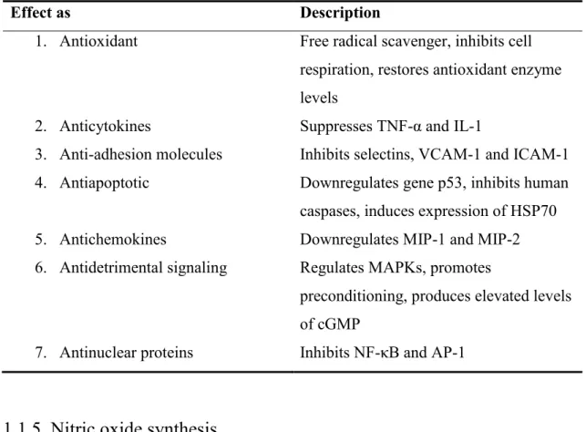
An NOS independent way, the reduction of inorganic nitrate (nitrate → nitrite → nitric oxide) can also elevate nitric oxide levels. After consumption of nitrate rich vegetables facultative anaerobe bacteria reduce it to nitrite, which is then swallowed and absorbed (Kapil et al., 2010; Lundberg et al., 2011). Some enzymes, e.g. xanthine oxidase may contribute to the endogenous conversion of nitrate to nitrite (Lei et al., 2013; Zweier and Talukder, 2006). Nitrate elevation can lead to protection on both molecular (Table 1.) and systematic levels resulting among others in cardo-protection, blood pressure reduction, beneficial modulation of hypercholesterolemia and increased exercise tolerance (Kapil et al., 2010; Lundberg et al., 2011; Phillips et al., 2009).
Table 1. Chemical and molecular protective effects of nitric oxide.(Phillips et al., 2009)
Effect as Description
1. Antioxidant Free radical scavenger, inhibits cell respiration, restores antioxidant enzyme levels
2. Anticytokines Suppresses TNF-α and IL-1
3. Anti-adhesion molecules Inhibits selectins, VCAM-1 and ICAM-1 4. Antiapoptotic Downregulates gene p53, inhibits human
caspases, induces expression of HSP70 5. Antichemokines Downregulates MIP-1 and MIP-2 6. Antidetrimental signaling Regulates MAPKs, promotes
preconditioning, produces elevated levels of cGMP
7. Antinuclear proteins Inhibits NF-κB and AP-1
1.1.5. Nitric oxide synthesis
Arginine derived NO synthesis has been best studied in mammals and three isoforms of the NOS enzyme were identified (Knowles and Moncada, 1994). NOS oxidizes the guanidine group of L-arginine in a process that consumes five electrons and
15
results in the formation of NO with stoichiometric formation of L-citrulline. The process involves the oxidation of NADPH and the reduction of molecular oxygen. The transformation occurs at a catalytic site adjacent to a specific binding site of L-arginine.
(Figure 1.)
1.1.5.1. Endothelial nitric oxide synthase (eNOS)
This isoform is also known as nitric oxide synthase 3 (NOS3) or constitutive NOS (cNOS) and is functionally similar to the nNOS, as both are clearly Ca2+- Calmodulin dependent. In humans it is encoded by the NOS3 gene and is partially membrane associated. Endothelial NOS generates NO in the blood vessels and is involved with vascular tone and platelet regulation (Knowles and Moncada, 1994). Huang et al. proved the elementary protective role of eNOS against hypertension and maintaining baseline bloodpressures (Huang et al., 1995).
In myocytes eNOS binds with Caveolin-1 and Caveolin-3 and is localized in caveolae and both increased Ca2+ concentration and phosphorylation can activate it.
NOS3 has his own unique localization and signaling pathway, which among others will influence myocyte contraction and action potential development (Tang et al., 2014).
Several mutations of the eNOS gene are known that could display significant differences under different clinical conditions. The three most widely studied NOS-3 polymorphisms in humans include the G894CT (Glu298Asp) in exon 7, the 4a/5b 27- Figure 1. NO synthesis. Oxidation of L-Arg to L-citrulline occurs via two successive mono-oxygenation reactions producing Nω-hydroxy-L-arginine (NOHLA) as an intermediate. 2 mol of O2 and 1.5 mol of NADPH are consumed per mole of NO formed.
(Based on the work of Prof. Moncada)8
16
basepair variable number of tandem repeats in intron 4, and the T-786C variant in the promoter region (Pereira et al., 2007). These polymorphisms are associated with a variety of clinical conditions, e.g. atherosclerosis, hypertension, heart failure and adaptation to physical exercise (Lembo et al., 2001; Matsa et al., 2013; Pereira et al., 2007, p. 3;
Wolfarth et al., 2008).
Cardiomyocyte specific NOS3 overexpression preserved LV function and endothelial NOS3 overexpression improved survival and decreased pulmonary edema after myocardial infarction (Janssens et al., 2004; Jones et al., 2003). Potential therapeutic benefits of NOS3 overexpression and increased NO bioavailability were proven in gene transfer models. In chronic ischemia clinically relevant neovascularization was induced and it attenuated acute ischemia reperfusion injury and ischemic remodeling (Kupatt et al., 2007; Szelid et al., 2009). Overexpression of NOS3 in cardiomyocytes was also suggested to be protective against arrhythmias by restoring sympatho-vagal balance (Massion et al., 2004).
1.1.5.2. Neuronal nitric oxide synthase (nNOS)
Neuronal nitric oxide was the first isoform to be purified and cloned, is also known as NOS1 and is encoded in humans by the NOS1 gene. It is Ca2+- Calmodulin dependent and is constitutively expressed at high activity in the central and peripheral nervous system and in the human skeletal muscle as well. nNOS is responsible for the largest proportion of constitutive NOS activity in humans (Knowles and Moncada, 1994). In cardiomyocytes NOS1 is predominantly localized in the sarcoplasmic reticulum, signals through the cGMP independent pathway and acts as a major regulator of excitation contraction coupling and myocyte Ca2+ handling. NOS1 is necessary for the positive force-frequency response and β-adrenergic induced positive inotropy as well (Tang et al., 2014).
1.1.5.3. Inducible nitric oxide synthase (iNOS)
Also known as type II NOS (NOS2), this isoform was identified in activated macrophages. From the first studies on it was evident, that this one differs from the others:
it is Ca2+-independent (Knowles and Moncada, 1994). It is expressed in response to
17
inflammatory stimuli and the NO produced is important in protection against pathogens (Francis et al., 2010, p. -).
1.1.6. NOS uncoupling: superoxide production
NOS enzymatic activity depends on substrate and cofactor availability and on the rate of electron transfer (Thomas et al., 2008). Substrate or cofactor shortage result in uncoupling: electrons removed from NADPH and normally travelling through the enzyme are diverted from L-arginine to a molecular oxygen and superoxide is produced (Rochette et al., 2013; Roe and Ren, 2012). The resulting oxidative stress is accompanied by reduced NO bioavailability (Roe and Ren, 2012; Schäfer et al., 2004).
Substrate (arginine) availability is influenced by competition with arginase, arginine uptake and methylation, and proteolysis to form symmetric and asymmetric dimethylarginine (SADMA and ADMA) (Rochette et al., 2013; Thomas et al., 2008).
The critical regulator of NOS function is one of its cofactors, tetrahydrobiopterin (BH4). BH4 is either synthetized from GTP or regenerated from dihydrobiopterin (BH2) and its loss either by oxidation or reduced expression of the recycling enzyme dihydrofolate reductase (DHFR) is the most prominent cause of NOS uncoupling (Roe and Ren, 2012). Intracellular ROS will accelerate BH4 oxidation and in turn the reduced BH4 form will trigger further uncoupling. In addition paradox eNOS expression during oxidative stress further amplifies ROS production (Kim and Park, 2010; Rochette et al., 2013).
Oxidized BH4 and NOS uncoupling contribute e.g. to vascular dysfunction, hypertension, ischemia/reperfusion injury, diabetes and other cardiovascular diseases (Rochette et al., 2013).
1.1.7. Endogenous NOS inhibitors
Symmetric and asymmetric methyl-arginines - e.g. ADMA, SDMA or NG- monomethyl-L-arginine (L-NMMA) - inhibit NO synthesis in vivo by competing with L- arginine at the active site of NO synthases (Leone et al., 1992). ADMA and SDMA are the major circulating forms of methylarginines in humans. (Leiper and Vallance, 1999).
ADMA is continuously formed during intracellular protein turnover and its concentration is very sensitive to changes in expression or activity of its degrading enzymes, the dimethylarginine dimethyl-amino-hydrolases (DDAH) (Rochette et al.,
18
2013). ROS increase may reduce DDAH activity and the resulting ADMA generation works as a second ROS-dependent, amplifying, feed-forward cellular loop that further stimulates ROS production (Rochette et al., 2013). Increasing bioactive NO availability can prevent ROS formation and is able to restore normal levels of ADMA (Carlström et al., 2011).
SDMA does not have the same biological activity as ADMA and is not hydrolyzed by DDAH, but it can interrupt the transportation of arginine and other cationic amino acids as well (Huang et al., 2004; Leiper and Vallance, 1999).
1.1.8. Nitric Oxide signaling
Nitric oxide exerts its effects either via the cGMP dependent, or via the cGMP independent signaling pathway. Balance between these two pathways depends upon redox status that influences net NO chemistry and NO and cGMP synthetic capacity by NOS and soluble guanylate cyclase (sGC) (Kass et al., 2007b).
1.1.8.1 cGMP dependent NO signaling
1.1.8.1.1. Cyclic guanosine 3’,5’-monophosphate
cGMP is a second messenger signaling molecule and is generated from guanosine triphosphate (GTP) by two distinct enzymes: the cytoplasmic heterodimeric haemoprotein sGC activated by NO and carbon monoxide (CO), and the transmembrane receptor particulate guanylate cyclases (pGC), activated upon binding of atrial and brain type natriuretic peptides (ANP and BNP), respectively (Burley et al., 2007; Garcia- Dorado et al., 2009; Tsai and Kass, 2009). Since the discussed experimental work focuses on the role of the NO mediated signaling, the second, receptor linked GC class will not be discussed in detail.
Direct targets of cGMP and participants of cGMP signaling are PKGs, cyclic nucleotide-gated ion channels (CNG), cGMP hydrolyzing phosphodiesterases (PDEs) and PDEs that contain allosteric cGMP binding sites. cGMP affinity among its targets varies and is modulated by phosphorylation or other modifications (Biel, 2009; Francis et al., 2010; Tsai and Kass, 2009).
19 1.1.8.1.2. Soluble guanylate cyclase (sGC)
sGC consists of two homologous subunits and each subunit consists of a heme- nitric oxide and oxygen binding (H-NOX), PAS (protein fold named for its association with the Per, ARNT, and Sim proteins), coiled-coil, and catalytic domain (Derbyshire and Marletta, 2012). Out of the two known active sGC isoforms (sGCα1β1 and sGCα2β1) sGCα1β1 is the principal isoform found in the heart and vasculature. NO mediated cardioprotection is lost without the α1 subunit and without the β1 subunit functional heterodimers are not formed (Derbyshire and Marletta, 2012; Nagasaka et al., 2008).
NO binding to the soluble guanylate cyclase (sGC) can increases its activation 100- 200 fold (others mention 200-400 fold increase) (Francis et al., 2010; Tsai and Kass, 2009). Two different states of sGC activation – basal and high activity – make sure that only an acute increase of NO levels, and the binding of a second NO to the enzyme is capable of fully activating the sGC (Cary et al., 2006; Tsai and Kass, 2009).
sGC response to its ligands diminishes upon repeated exposure. S-nitrosation induced desensitization is rapid and has major influence on therapeutic applications (Derbyshire and Marletta, 2012).
Since sGC is soluble, intracellular cGMP pool formation depends on the site of sGC activation and restriction of cGMP diffusion by PDE. This functional compartmentalization accounts for different intracellular effects (Burley et al., 2007;
Francis et al., 2010).
1.1.8.1.3. Protein kinase G (PKG)
PKG was first identified in lobster tail muscle and so far two genes and families are known (PKGI and PKGII) (Burley et al., 2007). The members of the PKGI family (PKGIα and PKGIβ) are more commonly involved with NO mediated cGMP signaling.
PKGIα transduces NOS / sGC signaling in the cardiovascular system and it requires one tenth of cGMP concentration for activation than does PKGIβ (Burley et al., 2007; Francis et al., 2010; Tsai and Kass, 2009).
Oxidative processes can provide a NO / cGMP independent mechanism for PKGIα activation and auto-phosphorylation can change isozyme affinities and functions and further fine tune their regulatory role (Francis et al., 2010).
20
1.1.8.1.3.1. PKG regulation of calcium levels and signaling
A modest NO availability will result in lower intracellular Ca2+ levels and interact with smooth muscle contraction, myocyte contractility, β-adrenergic response and attenuate cardiac hypertrophy (Francis et al., 2010).
Modulation of L-type calcium channels (LTCC): LTCC phosphorylation is restricted to the caveolar microdomain by PDE5 and appears to be gender dependent.
This regulatory pathway is believed to blunt contractile response to β-adrenergic stimulation, and attenuate calcineurin activation to prohibit NFAT (nuclear factor of activated T cells) translocation and hypertrophy (Tang et al., 2014).
In addition direct phosphorylation of the Cav1.2 channel (Calcium channel, voltage-dependent, L type, alpha 1C subunit) also reduces Cav1.2. current and limits contractility (Yang et al., 2007).
Troponin, regulating cardiac muscle contraction: troponin I phosphorylation reduces myofilament Ca2+ sensitivity (Layland et al., 2002).
Phosphorylation of G-protein-activated phospholipase-Cβ3: phosphorylation will inhibit this enzyme and inhibit agonist stimulated intracellular calcium release (Xia et al., 2001).
Phosphorylation of inositol 1,4,5-trisphosphate (IP3) receptor-associated PKG substrate (IRAG): PKGIβ converts IRAG into an inhibitor of IP3-receptor activity and suppresses calcium release (Francis et al., 2010).
Phospholamban phosphorylation: canceling its inhibition of the sarcoplasmic reticulum calcium/ATPase pump will increase calcium sequestration. Phospholamban phosphorylation can also occur via a cGMP independent pathway, namely PKA up- regulation with S-nitrosylation (Francis et al., 2010; Tang et al., 2014).
Ryanodine receptor (RyR) phosphorylation: phosphorylation will influence the function of the cardiac sarcoplasmic reticulum Ca2+ release channel (Burley et al., 2007;
Zhang et al., 2005).
1.1.8.1.3.2. PKGI-mediated inactivation of the Ras homolog gene family member A (RhoA)
Rho kinase regulates actin cytoskeleton organization, stress fiber formation and vascular smooth muscle contraction and is inactivated and blocked by serine phosphorylation (Ellerbroek et al., 2003; Murthy et al., 2003; Surma et al., 2011). RhoA
21
kinase inhibitors have demonstrated beneficial effects in the treatment of various cardiovascular diseases including arterial hypertension, atherosclerosis, I/R injuries, hypertrophic remodeling, cardiac dysfunction and heart failure (Surma et al., 2011).
1.1.8.1.3.3. Gene regulation
PKGI activity influences the expression of smooth muscle specific contractile proteins, proteins in the NO sGC signaling pathway, proteins regulating smooth muscle migration and differentiation, proteins influencing neuronal plasticity and learning and proteins in pathological conditions (Alzheimer’s and schizophrenia) (Francis et al., 2010).
1.1.8.1.3.4. Phosphorylation of Vasodilator-Stimulated Phosphoprotein
Vasodilator-Stimulated Phosphoprotein (VASP) is phosphorylated by both protein kinase G and protein kinase A (PKA). VASP signaling influences fibroblast motility, neutrophil migration, neuronal migration and endothelial function and could be potentiating metastasis formation (Krause et al., 2003).
1.1.8.1.3.5. Phosphodiesterase 5 regulation
PKGI mediated activation of the cGMP specific PDE5 is a powerful negative feedback on the NO / sGC / cGMP signaling pathway (Das et al., 2015a; Kass et al., 2007b).
1.1.8.1.3.6. Cytoprotection
Cytoprotection was observed in myocytes, neurons and glia and epithelial cells.
Opening of mitochondrial K+/ATP channels: cGMP can activate PKG localized at the cytosolic surface of the mitochondrial outer membrane. This may lead to the phosphorylation of a target protein that carries the cardio-protective signal to a protein kinase C isozyme (PKC-ε) residing in the intermembrane space of mitochondria and increase opening of mitochondrial K+/ATP channels (mitoKATP) (Costa et al., 2005;
Francis et al., 2010).
MitoKATP independent anti-apoptotic pathway: Activation of extracellular signal- regulated kinase (ERK) and ERK dependent inactivation of glycogen synthase kinase 3β (GSK3β) by PKG phosphorylation also attenuates ischemia reperfusion injury and prevents cardio-myocyte apoptosis (Das et al., 2009; Francis et al., 2010).
22
Blockade of p38 mitogen-activated protein kinase is also reported to reduce cell damage, although these protective effects were not observed after sildenafil induced cGMP increase (Das et al., 2009; Francis et al., 2010).
It was also suggested that the Bcl-2-associated death promoter (Bad) is likely to be an important downstream substrate of PKG-I, and its phosphorylation may contribute to the cGMP mediated survival in neural cells (Johlfs and Fiscus, 2010).
1.1.8.1.3.7. Pro-apoptotic effects
PKGI activity in colon cancer cells promotes apoptosis by phosphorylation and activation of the mitogen-activated protein kinase kinase (MEKK1), activation of the stress-activated protein/ERK kinase 1 (SEK1), and activation of the c-Jun NH2-terminal kinase 1 (JNK1) pathway (Deguchi et al., 2004; Soh et al., 2001). It also suppresses growth and promotes apoptosis in some endothelial cells and human colon tumor HT29 cells (Francis et al., 2010).
1.1.8.1.4. Cyclic nucleotide regulated ion channels
Cyclic nucleotide regulated ion channels belong to the superfamily of voltage gated cation channels and are directly regulated by the binding of cAMP or cGMP. Channel activation is directly coupled to extracellular cation influx and plasma membrane depolarization. Two families can be distinguished: cyclic nucleotide gated (CNG) channels and hyperpolarization-activated cyclic nucleotide gated channels (HCN) (Biel, 2009; Roubille and Tardif, 2013). CNG channels prefer cGMP over cAMP, while HCN channels have 10 fold higher affinity for cAMP (Biel, 2009; Podda and Grassi, 2013).
1.1.8.1.4.2. Cyclic nucleotide-gated channels (CNG):
These channels play a key role in visual and olfactory signal transduction, but their role in other cell types remains unclear. CNG channels pass monovalent cations without discrimination. CNGs do not desensitize or inactivate after prolonged exposure to their ligands and they are influenced by divalent ion currents and several other factors (Biel, 2009; Podda and Grassi, 2013).
1.1.8.1.4.3. Hyperpolarization-activated cyclic nucleotide-gated ion channels (HCN):
All four known HCN channel isoforms were identified in the myocardium and have a preference for K+. They activate on hyperpolarisation, which was the reason why the
23
current resulting from their activation was named funny (If). Regulation of these channels is complex, but modulation through their cyclic nucleotide binding domain region appears to be a common mechanism. Selective blockade of these channels is getting more and more attention in the treatment of heart failure patients (Roubille and Tardif, 2013).
1.1.8.1.5. cGMP catabolism - phosphodiesterases
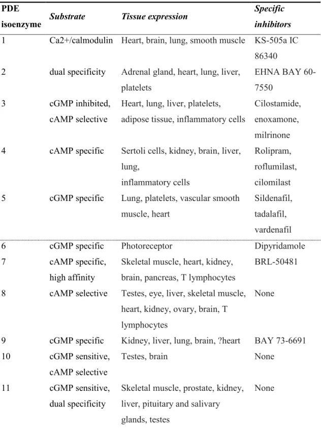
Phosphodiesterases are catabolic enzymes in charge of the fate of cGMPs and cAMPs. This 21 gene family of proteins was grouped into 11 different isozymes (Table 2.) and 48 isoforms. Due to all these variants any organism is estimated to have more than fifty cGMP hydrolyzing enzymes at the same time. Cyclic nucleotide affinity varies, PDE5 / PDE6 / PDE9 are highly cGMP specific, PDE1 / PDE2 / PDE 11 have dual substrate affinity, and PDE3 / PDE 10 are cGMP sensitive but cAMP selective.
Phosphodiesterases can be found in all tissues, but their distribution varies (Das et al., 2015a; Francis et al., 2010; Kass et al., 2007b). cGMP-phosphodiesterases with known activity in the cardiovascular system are:
PDE1 is a Ca2+/calmodulin dependent enzyme,
PDE2, a cGMP-stimulated cAMP esterase that can also hydrolyze cGMP,
PDE3 although it is cAMP specific, it is inhibited by cGMP
PDE5 the first identified selective cGMP esterase
PDE9A could work as a “housekeeping” PDE to maintain low intracellular cGMP levels and controls nitric oxide independent cGMP and hypertrophic heart disease (Francis et al., 2010; Lee et al., 2015, p. 9).
A large number of factors influence the catalytic function from PDEs, including processes that affect protein expression and breakdown, enzyme localization and substrate binding. For most PDEs their catalytic rate is high, thus absolute PDE concentration within a cell is low. In addition the Km value of different PDEs is also in a broad range of concentrations. This diversity together with enzyme and substrate compartmentalization enables phosphodiesterases to take part in a large variety of signaling processes (Francis et al., 2010).
Under pathologic conditions PDE regulation contributes to the progression of several diseases. PDE3 down regulation influences heart failure progression, PDE1 influences smooth muscle hypertrophy, sustained stimulation of PDE1 and PDE5 results in NO tolerance, PDE1 and PDE5 induction can be behind exacerbated hypertension and
24
PDE5 up-regulation is part of pulmonary hypertension and congestive heart failure (Kass et al., 2007b).
1.1.8.1.5.1. PDE regulation
Phosphodiesterases can undergo complex post-translational modifications. Among others, activation by Ca2+/calmodulin binding, or allosteric binding of cGMP may mediate PDE and protein interactions. (Das et al., 2015a).
Allosteric cGMP binding to tandem regulatory GAF domains (its name derives from the first proteins it was found in: cGMP-specific phosphodiesterases, adenylyl cyclases and FhlA) can act as an auto feedback mechanism (increasing catabolic activity for cGMP) or cross-regulation mechanism (activating catabolism of cAMP). Five of the known PDEs are equipped with a GAF domain: PDE2, PDE5, PDE6, PDE10 and PDE11.
Binding affinities for the allosteric sites are much higher than for the catalytic sites, thus before reaching the catalytic site cGMP first induces a more active catalytic state and sustains an activated cGMP breakdown for a significant time. In addition ligand occupation of the catalytic site, phosphorylation of the regulatory domain, oxidation/reduction and other processes enhance GAF binding affinity. Allosteric binding of cGMP to a phosphodiesterase might as well serve as an intracellular storage and protection till later release (Francis et al., 2010; Kass et al., 2007b).
Four of the PDEs (PDE1, PDE3, PDE4, PDE5) contain phosphorylation sites for various kinases and PDE phosphorylation by PKG is another mechanism to induce positive feedback. Phosphorylation of PDE5 by PKG may serve to increase its cGMP affinity and catalytic activity and represents an alternative mode of regulatory feedback inhibition to normalize cGMP levels (Das et al., 2015a; Kass et al., 2007b).
Another type of modulation can happen at the catalytic site. In the case of PDE3 catalytic site affinity is similar for both cyclic nucleotides, but a higher Vmax for cAMP confers specificity, while cGMP becomes a competitive inhibitor of PDE3 hydrolysis (Kass et al., 2007b).
In the case of PDE1 an auto-inhibitory domain is known to maintain low activity in the absence of Ca2+, and neighboring calmodulin binding domains were found to reactivate the enzyme in the presence of Ca2+-Calmodulin (Kass et al., 2007b).
25
Table 2. PDE isozymes, their specificity and selective inhibitors (Kass et al., 2007b).
PDE
isoenzyme Substrate Tissue expression Specific inhibitors 1 Ca2+/calmodulin Heart, brain, lung, smooth muscle KS-505a IC
86340 2 dual specificity Adrenal gland, heart, lung, liver,
platelets
EHNA BAY 60- 7550
3 cGMP inhibited,
cAMP selective
Heart, lung, liver, platelets, adipose tissue, inflammatory cells
Cilostamide, enoxamone, milrinone 4 cAMP specific Sertoli cells, kidney, brain, liver,
lung,
inflammatory cells
Rolipram, roflumilast, cilomilast 5 cGMP specific Lung, platelets, vascular smooth
muscle, heart
Sildenafil, tadalafil, vardenafil
6 cGMP specific Photoreceptor Dipyridamole
7 cAMP specific,
high affinity
Skeletal muscle, heart, kidney, brain, pancreas, T lymphocytes
BRL-50481
8 cAMP selective Testes, eye, liver, skeletal muscle, heart, kidney, ovary, brain, T lymphocytes
None
9 cGMP specific Kidney, liver, lung, brain, ?heart BAY 73-6691 10 cGMP sensitive,
cAMP selective
Testes, brain None
11 cGMP sensitive, dual specificity
Skeletal muscle, prostate, kidney, liver, pituitary and salivary glands, testes
None
26 1.1.8.1.5.2. PDE5 expression and regulation
One PDE5 coding gene (PDE5A) and three gene expression variants are known. It is assumed that different promoters for the PDE5 isoforms allow physiologically relevant differential control of PDE5 gene expression and provide a long-term feedback mechanism for PDE5 activation (Kass et al., 2007a). High PDE5 expression rate in smooth muscle cells make determination of exact localization in vascularized tissues difficult. Intracellular localization is different among cellular compartments and seems to be at least partially dependent on the presence of eNOS (Kass et al., 2007a).
PDE5 has two highly homologous GAF domains (A and B), but high affinity cGMP binding occurs only to the GAF-A domain. This domain is very similar to the PDE2 GAF- B and PDE6 GAF A domains and stimulates substrate catalysis to its ten-fold. Although the enzyme is largely inactive in the absence of GAF ligand binding, under usual circumstances it is very likely to be occupied by cGMP and maintain full enzyme activity.
Binding site phosphorylation either by PKG or PKA enhances cGMP binding affinity and stabilizes the increased catabolic activity. This is the way, by which sGC activators can promote a feed-forward enzyme activation and limit cGMP rise. The very same processes ensure easy and lasting binding of PDE5 inhibitors (Das et al., 2015a; Kass et al., 2007a).
1.1.8.2. cGMP independent pathway
NO directly regulates protein function via posttranslational modification of S- nitrosylation (addition of a nitrosyl group to a free thiol on a cysteine residue of the target protein). S-nitrosylation is a redox based signal and it not only takes part in mammalian signaling, but can be used against the invasion of microbes or cancer cells as well (Stamler et al., 2001; Tang et al., 2014).
Nitrosylation and nitrosative chemistry can lead to secondary oxidative modifications and production of reactive nitrogen species. These reactive nitrogen species can interact with reactive oxygen species, which can either impair its signaling and increase cytotoxicity (irreversibly oxidizing and nitrating proteins) or open up new signaling pathways. NO may as well promote cytotoxicity by mobilization of free iron or by the production of other oxidants, radicals and reactive aldehydes (Stamler et al., 2001).
Recently it was proven that one of the main reperfusion injury quenching mechanisms of NO signaling is the reversible S-nitrosation of a mitochondrial protein
27
that will decelerate the mitochondrial electron transport chain (ETC) and inhibit extensive reactive oxygen species production during reperfusion (Schumacker, 2013).
NOS1 produced NO takes advantage of the cGMP independent signaling pathway and modulates myocyte Ca2+ handling. Co-localization of NOS1 with the SR Ca2+ release channel, ryanodine receptor and xanthine oxidoreductase (XOR) on the sarcoplasmic reticulum enables their S-nitrosylation (Tang et al., 2014).
1.2. Ischemia-reperfusion injury
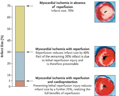
In case of an acute cardiac ischemic event, besides restoration of blood flow in ischemic areas, reduction of pre- and post-procedural damage is indispensable (Garcia- Dorado et al., 2009). Revascularization of the culprit lesion is accompanied by ischemia- reperfusion (I/R) injury, which is responsible for about 40-50% percent of myocardial damage suffered (Figure 2.) (Yellon and Hausenloy, 2007).
Ischemia reperfusion can cause four types of cardiac dysfunction: the mechanical dysfunction known as myocardial stunning, the disruption of microvascular circulation in the reperfused tissue (“no reflow”), reperfusion arrhythmias and lethal reperfusion injury (Yellon and Hausenloy, 2007).
During lethal reperfusion injury myocardial cell death occurs mainly as necrosis:
histological analysis of reperfused infarcts showed composed areas of contraction band necrosis and ultrastructure showed sarcolemmal rupture and massive calcium deposits.
Contraction band necrosis, when myocytes show characteristic disruption of their architecture and disorganization of their sarcomeres, is usually referred to as hypercontraction or round-up (Garcia-Dorado et al., 2009).
1.2.1. Biochemical and metabolic changes during ischemia reperfusion
Ischemia-reperfusion subjects the myocardium to several biochemical and metabolic changes. During prolonged ischemia intra- and extracellular acidosis, energy depletion, Na+ and Ca2+ overload and hyper-osmolality develops. Upon reperfusion mitochondrial re-energization, generation of reactive oxygen species (ROS), intracellular Ca2+ overload, rapid restoration of physiologic pH, and inflammation interact and induce cell death.
28 1.2.1.1. The ischemic cascade
A switch from aerobic to anaerobic metabolism result in lactic acid accumulation.
The resulting acidosis together with the fall in ATP concentration and consequent failure of ATP dependent processes triggers an uncontrolled rise in intracellular ion levels. ROS production increases and disrupts sarcolemmal and mitochondrial function, facilitating cell death (McAlindon et al., 2015).
1.2.1.2. pH normalization
The rapid wash-out of lactic acid and the activation of the sodium-hydrogen exchanger and the sodium–bicarbonate symporter results in quick pH restoration and may lead to potassium ion overload, which in turn results in a reversed Na+/Ca2+ exchanger activity and additional calcium influx (Bond et al., 1991; Garcia-Dorado et al., 2009;
Yellon and Hausenloy, 2007). After restoration of pH mitochondrial ROS formation is initiated and mitochondrial PTP (Permeability Transition Pore) channels activate (Kim et al., 2006).
1.2.1.3. ROS generation
Xanthine oxidase (XO), the mitochondrial electron transport chain, NADPH oxidase (Nox) and NOS uncoupling are the known sources of ROS (Zweier and Talukder, 2006). Nox is thought to be the major source of O2- and H2O2 in the heart and it is upregulated during ischemia reperfusion injury. Nox is a double edged sword as minimal activity is needed to maintain peroxisome proliferator-activated receptor alpha (PPARα) and hypoxia-inducible factor-1α (HIF-1α) conferred cardioprotection. The consumption of NAD(P)+/NAD(P)H by Nox is an alternative regulatory mechanism of redox state and thus mitochondrial electron transport (Chen and Zweier, 2014; Matsushima et al., 2014).
ROS induce mitochondrial PTP opening, attract neutrophils, mediate sarcoplasmic reticulum dysfunction and contribute to intracellular Ca2+ overload, damage the cell membrane by lipid peroxidation, induce enzyme denaturation, and cause direct oxidative damage to DNA.(Yellon and Hausenloy, 2007; Zweier and Talukder, 2006).
29 1.2.1.4. Calcium oscillation
Parallel restoration of respiration to pH normalization allows mitochondrial repolarization and ATP synthesis. This favors calcium uptake into the mitochondria and sarcoplasmic reticulum and despite the higher calcium influx cytosolic calcium levels fall. Increased calcium concentration in the sarcoplasmic reticulum interacts with the ryanodine receptor and calcium oscillations (release/reuptake) propagate across the cell (Garcia-Dorado et al., 2009). This can lead to hyper-contraction and disorganization of the cell structure, but to damage the sarcolemmal membrane the proteolysis of the subsarcolemmal cytoskeleton by calpains (calcium dependent and calcium activated proteases) is necessary as well(Garcia-Dorado et al., 2009; Inserte et al., 2004).
1.2.2.5. Targeting the mitochondria
The opening of the mitochondrial PTP channel during the first few minutes of reperfusion collapses mitochondrial membrane potential and uncouples oxidative phosphorylation: ATP depletion and cell death occur (Yellon and Hausenloy, 2007).
Direct or PI3K / AKT / NOS / GC mediated PKG and PKC activation will induce the mitochondrial ATP-dependent K channel (KATP) and ROS formation resulting in p38
Figure 2. Components of final myocardial injury. This scheme taken from Yellon and Hausenloy depicts the contribution of both ischemia and reperfusion injury to final myocardial injury.52
30
mitogen-activated kinase and PKC activation and priming of mitochondrial PTP for opening (Heusch et al., 2008).
Two endogenous cardioprotective pathways, the Reperfusion Injury Salvage Kinase (RISK) and the Survival Activating Factor Enhancement (SAFE) pathways, are known to inhibit mitochondrial PTP, activate mitochondrial connexin-43 channels and open mitochondrial ATP dependent potassium channels (Hausenloy et al., 2013).
Activation of sarcolemmal G-protein–coupled receptors or of receptors for growth factors result in activation of the RISK program. The RISK pathway includes the pro- survival kinase cascades MEK1/2-Erk1/2 and involves PI3K/Akt/NO signaling, p70S6K (extracellular regulated kinase system with downstream p70 ribosomal protein S6 kinase) and GSK3β (glycogen synthase kinase 3β) activation (Heusch et al., 2008).
The SAFE pathway is made up by the TNF-α (tumor necrosis factor-α) receptor and STAT3 (Hausenloy et al., 2013). After activation of gp130 (sarcolemmal glycoprotein 130) receptors or TNF α receptors, the janus-activated kinase (JAK) and signal transducer and activator of transcription (STAT) pathway is activated with projection to the nucleus and possibly to mitochondria (Heusch et al., 2008). As an upstream regulator of TNF-α NO influences this pathway as well (Thielmann et al., 2002).
1.2.2.6. Neutrophil involvement in lethal myocardial reperfusion injury
Neutrophil response to reperfusion is triggered by cytokines, the complement system, ROS and various lipid mediators. Compared to ischemia only, reperfusion accelerates neutrophil infiltration and focuses their accumulation in the sub-endocardium (Chatelain et al., 1987; Vinten-Johansen, 2004). Initial, rapid neutrophil adhesion to the endothelium is accompanied by a reduced endothelial function (Vinten-Johansen, 2004).
For the first 6 hours cells stay in the intravascular space and after the 6th hour start to migrate to the parenchyma (Zhao et al., 2000).
Neutrophils can form aggregates with platelets, plug capillaries and mechanically stop blood flow (Engler et al., 1983; Hataishi et al., 2006; Wong et al., 2013). Together with platelets and damaged endothelial cells they release vasoconstrictors and contribute microvascular constriction (Niccoli et al., 2009; Wong et al., 2013). Both processes contribute actively to the “No reflow” phenomenon.
31
Neutrophils can induce endothelial dysfunction and by impairing NO generation contract arteries (Vinten-Johansen, 2004). Endothelial dysfunction is nearly restored after 72 hours following reperfusion (Zhao et al., 2000).
Neutrophils release more than 20 different enzymes which primary target is the extracellular matrix. While these enzymes have a relatively long half-life and catalyze specific reactions, ROS produced by neutrophil is responsible for fast, aspecific and widespread destruction (Vinten-Johansen, 2004).
The reduction of neutrophil infiltration is associated with reduced infarct size and attenuated adverse cardiac remodeling while after the 3-7th post-ischemic day this could possibly assist repair mechanisms (Carbone et al., 2013; Huang et al., 2007). Limitation of neutrophil recruitment is one of main cardio-protective mechanisms attributed to inhaled NO (Hataishi et al., 2006).
1.2.2.7. NOS uncoupling
Nitric oxide signaling, previously described in detail, can interact with lethal myocardial injury on several levels. Ischemia reperfusion will induce NOS uncoupling, reduce NO bioavailability and produce ROS. Most probably due to the decreased GC activity under acidic conditions cGMP levels will drop and the activity of the cGMP dependent pathway will decrease. The very same pathway that directly effects contractility, calcium handling, activates protective cascades, regulates mitochondrial PTP, inhibits platelet activation, reduces endothelial adhesion protein expression, increases endothelial permeability and induces vasorelaxation (Garcia-Dorado et al., 2009). In addition the cGMP independent pathway reversibly S-nitrosylates one of the electron transport chain components (a subunit of mitochondrial complex I) and thus slows it down (Schumacker, 2013). A study with selective sGC/NO/cGMP signaling inhibition even suggested, that nitrosylation might play a more important role in cardioprotection (Sun et al., 2013).
1.3. Modulation of the nitric oxide mediated signaling pathway – therapeutic applications
Nitric oxide generating drugs have long been used by physicians and are still in the first line of medical therapies (e.g. nitro-glycerine) while other fields of use are severely
32
limited by systemic vasodilatation. To avoid this unwanted side effect novel NO donor compounds and agents to sensitize or to directly activate NO signaling were developed and are being tested (Bryan and Loscalzo, 2011).
1.3.1 Inhaled nitric oxide
Before 1990 NO was considered poison. Zapol and Frostel in their publication from 1991 proved that inhalation of low concentrations of nitric oxide (up to 80 ppm) is safe and effectively relaxes pulmonary vascular smooth muscle, without systemic vasodilatation (Frostell et al., 1991). Short gas transit time and low concentrations ensure that during inhaled NO administration NO2 concentrations remain under 5 ppm and measuring met-hemoglobin levels establishes adequate therapy-monitoring (Bhatraju et al., 2015; Frostell et al., 1991) (Figure 3.).
1.3.1.1. Taking advantage of pulmonary effects
Nitric oxide/oxygen blends are used in critical care to promote capillary and pulmonary dilation to treat a variety of diseases. These are often last-resort gas mixtures before the use of extracorporeal membrane oxygenation (ECMO) (Bryan and Loscalzo, 2011).
Nitric oxide inhalation is an approved therapy for persistent pulmonary hypertension of the new-born (PPHN), for chronic obstructive pulmonary disease, in patients with pulmonary hypertension and undergoing cardiac surgery, in patients undergoing left ventricular assist device (LVAD) implantation in patients with right ventricular infarction or pulmonary embolism (Adhikari et al., 2014; Argenziano et al., 1998; Bhatraju et al., 2015; Bloch et al., 2007; Bryan and Loscalzo, 2011).
1.3.1.2. Extrapulmonary effects of inhaled NO
Identifying inhaled NO related platelet inhibition broke the paradigm that its effects are limited to the lungs (Frostell et al., 1991; Högman et al., 1993). A number of extra- pulmonary effects were described ranging from reducing neointima formation and inflammatory modulation in malaria to reduction of ischemia-reperfusion injury and protection of the central nervous system. Cardiovascular applications were studied and
33
verified as well (Bergmark et al., 2012; Bryan and Loscalzo, 2011; Charriaut-Marlangue et al., 2013; Mathru et al., 2007; Phillips et al., 2009).
1.3.1.2.3. Cardiopulmonary resuscitation
In mice it was proven that NO inhalation has protective effects on the outcomes of cardiopulmonary resuscitation. The neuro-protective effects largely depend on sGC- mediated mechanisms, although the role of sGC independent and NO metabolite mediated arteriole dilatation could not be excluded (Ichinose, 2013).
1.3.1.2.6. Myocardial ischemia-reperfusion injury:
Several experiments were conducted to prove the effectiveness of inhaled NO therapy in attenuating myocardial reperfusion injury (Bhatraju et al., 2015; Bloch et al., 2007). Pre-clinical studies agree on the infarct size limitative and cardiac function preserving effects of this therapy (Hataishi et al., 2006; Liu et al., 2007; Nagasaka et al., 2008; Neye et al., 2012). A larger scale international, randomized, double-blind clinical study was conducted recently, the NOMI trial – as this work strongly connects to it - will be discussed separately.
1.3.1.3. Theories behind extrapulmonary effects
Although therapeutic systemic applications are known and were proven by several groups, the question remains: how can NO, with its short half-life in biological fluids, reach the periphery and elicit its effects?
First extrapulmonary effects were attributed to changes in platelet and leukocyte function and only after discarding this turned the scientific interest towards stable NO metabolites. Nitric oxide can react with heme in hemoglobin forming nitrosyl hemoglobin and can nitrosylate the cysteine in the hemoglobin to form SNO-hemoglobin. This reaction will occur in the presence of high oxygen concentrations and NO will be released under low oxygen concentrations (Bryan and Loscalzo, 2011; McMahon and Doctor, 2006).
Nitrite and nitrate are also candidates for the cardio protective effects, as they could be major stable reservoirs of NO. Nitrite was proven to dramatically reduce infarct size in a canine model, and was suggested as potent adjunctive therapy in myocardial reperfusion injury (Gonzalez et al., 2008).
34
Nitric oxide also reacts with a variety of plasma components - proteins, peptides and lipids – and forms intravascular agents capable of endocrine vasodilatation: S- nitrosthiols, N-nitrosamines, iron-nitrosyls and nitrated lipids (Bryan and Loscalzo, 2011;
Gonzalez et al., 2008).
1.3.2. Other inhaled drugs to modify NO signaling
Inhalation is an effective way to administer various NO donor agents: nitroglycerin relieves angina pectoris, and sodium nitroprusside may improve oxygenisation in hypoxic neonates and reduce PVP and PVR in children with congenital heart disease (CHD). O- nitrosoethanol (an S-nitrosothiol) was reported to improve oxygenation and systemic hemodynamics in hypoxemic newborns. Nitrite can also be converted to NO by a variety of enzymes (e.g. hemoglobin and xanthine oxidoreductase) (Bryan and Loscalzo, 2011).
PDE5 inhibitors (e.g. sildenafil) and sGC stimulators potentiate the pulmonary vasodilator effects of NO (Evgenov et al., 2007; Ichinose et al., 2001).
1.3.3. Phosphodiesterase 5 inhibition
After the commercial success of Viagra it took almost a decade for PDE5 inhibition to make its comeback into more direct cardiovascular applications. The recognition that it is responsible for ~22-43% of cytosolic cGMP hydrolytic activity and that under Figure 3. Schematic image of an alveolar capillary unit. NO dilates pulmonary arterioles and reduces pulmonary artery pressure (PAP). Inhaled NO does not dilate systemic arterioles or alter systemic arterial pressure (SAP) under normal conditions.6
35
pathologic conditions its expression is upregulated 2 to 6 fold opened new frontiers in cardiovascular research (Das et al., 2015a; Kass et al., 2007a).
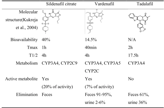
PDE5 inhibitors provide protection from ischemia-reperfusion injury and preserve left ventricular function, improve survival rate, reduce apoptosis in the infarct border zone and reduce heart failure progression (Das et al., 2015a; Salloum et al., 2008). Extracardial uses include dampening of systemic inflammation in diabetic patients, protection of stem cells from I/R injury, reducing I/R injury in skin island flaps, neuroprotection and anticancer effects (Das et al., 2015a; Gulati and Singh, 2014a, 2014b; Kayiran et al., 2013). The most studied compounds are sildenafil, vardenafil and tadalafil. (Table 3.)
PDE5 inhibitors interact with gaso-transmitters (e.g. NO and H2S), as it was seen after blocking the H2S producing enzyme: cardio-protective effects of tadalafil were abolished (Das et al., 2015a).
Table 3. PDE5 inhibitors for cardiovascular use. Data on metabolism and pharmacokinetics in humans was taken from http://toxnet.nlm.nih.gov/ (US National Library of Medicine, n.d.).
Sildenafil citrate Vardenafil Tadalafil Molecular
structure(Kukreja et al., 2004)
Bioavailability 40% 14.5% N/A
Tmax 1h 40min 2h
T1/2 4h 4h 17.5h
Metabolism CYP3A4, CYP2C9 CYP3A4, CYP3A5 CYP2C
CYP3A4
Active metabolite Yes
(20% of activity)
Yes
(7% of activity)
No
Elimination Feces Feces 91-95%,
urine 2-6%
Feces 61%, urine 36%
36
Recently some researchers questioned the role of PDE5 in the cardiac cGMP-PKG signaling, as they failed to detect PDE5 in tissue lysates (mouse, canine and human) and to increase hypertrophy by creating cardiac specific PDE5 knock out animals (Degen et al., 2015; Lukowski et al., 2010).
1.3.3.1. Protection against ischemia-reperfusion injury
Both sildenafil and vardenafil were effective not only when administered prior ischemia, but when given at the time of reperfusion as well. Preclinical data on I/R injury and ischemic cardiomyopathy showed beneficial effects of PDE5 inhibition on collagen deposition and verified functional therapeutic benefits with both echocardiography and invasive pressure volume analysis of the left ventricle (Pokreisz et al., 2009; Salloum et al., 2014; Sesti et al., 2007).
To exert cardio-protection these agents influence mitochondrial KATP and mitochondrial Ca2+ channels, and the cGMP/PKG pathway also confers protection.
Beside PKG activation enhanced eNOS and iNOS expression and an increased Bcl-2/Bax ratio also mediate cardioprotection (Chau et al., 2011; Salloum et al., 2014, 2008).
Tadalafil also attenuated ischemia-reperfusion injury via PKG dependent generation of hydrogen sulfide (Salloum et al., 2009). In vitro studies proved that these anti-necrotic and apoptotic effects are independent from the vasculature and hemodynamics (Das et al., 2015a).
Myocardial infarct size reduction was also observed in diabetic models, which are known to be refractory to some cardio-protective modalities. Chronic tadalafil treatment improves cardiac function, NO bioavailability and also promotes antioxidant like effects (suppresses ROS production, NADPH oxidase activity, lipid peroxidation and oxidizes glutathione) (Das et al., 2015a; Koka et al., 2014, 2013).
1.3.3.2. Protection against ischemic and doxorubicin induced cardiomyopathy
Sildenafil and tadalafil attenuated ischemic cardiomyopathy and slowed down heart failure progression. Beneficial effects are associated among others with the activation of Rho kinases and the production of hydrogen sulfide (Das et al., 2015a; Salloum et al., 2014, 2009).