t h e C ro s s ro a d s B e t w e e n
t h e L e f t a n d R i g h t H e a r t i n S y s t e m i c S c l e ro s i s
A Clinical Challenge for Cardiologists and Rheumatologists
Luna Gargani,
MD, PhDa,*, Damien Voilliot,
MDb, Michele D ’ Alto,
MD, PhDc, Gergely Agoston,
MD, PhDd, Antonella Moreo,
MDe, Walter Serra,
MD, PhDf, Francesco Pieri,
MDg, Fabio Mori,
MDg,
Karina Wierzbowska-Drabik,
MD, PhDh,
Marco Matucci-Cerinic,
MD, PhDi, Alberto Moggi-Pignone,
MD, PhDiINTRODUCTION
Systemic sclerosis (SSc) is a complex multiorgan immune-mediated disease characterized by fibrosis of the skin and internal organs and by
vasculopathy.1,2 Pulmonary hypertension (PH) is defined as an increase in mean pulmonary arterial pressure (mPAP) greater than or equal to 25 mm Hg at rest, as assessed by right heart catheterization
Disclosure: This article has been partially funded by the Italian Ministry of Health (Ricerca Finalizzata 2011-2012).
a Institute of Clinical Physiology, National Research Council, Via Moruzzi, 1, Pisa 56124, Italy;bDepartment of Cardiology, University Hospital of Nancy, Institut Lorrain du Cœur et des Vaisseaux, 5 Rue du Morvan, 54500 Vandœuvre-le`s-Nancy, France;c Department of Cardiology, Second University of Naples, Monaldi Hospital, Piazzale E. Ruggieri 1, Naples 80131, Italy; dDepartment of Family Medicine, University of Szeged, Tisza Lajos krt. 109, 6725 Szeged, Hungary;eCardiovascular Department, Niguarda Hospital, Piazza dell’Ospedale Maggiore, 3, 20162 Milano MI, Italy;f Cardiology Unit, University Hospital of Parma, Via Gramsci, 14, 43126 Parma, Italy;gDepartment of Heart and Vessels, Azienda Ospedaliero-Universitaria Careggi, Largo Brambilla, 3, 50134 Florence, Italy;h Department of Cardiology, Medical University of Lodz, aleja Tadeusza Kosciuszki 4, 90-419qo´dz, Poland;i Department of Experimental and Clinical Medicine, Azienda Ospedaliera Universitaria Careggi, Largo Brambilla, 3, 50134 Florence, Italy
* Corresponding author.
E-mail address:gargani@ifc.cnr.it
KEYWORDS
Pulmonary hypertensionSystemic sclerosisRight heartPulmonary circulation
KEY POINTS
Pulmonary hypertension is frequent in systemic sclerosis and is associated with poor prognosis.
Pulmonary hypertension occurs as a result of a pulmonary arteriopathy but also can be a conse- quence of interstitial lung disease and/or left heart involvement.
These phenotypes may be difficult to differentiate and often overlap, complicating both the diag- nosis and the follow-up.
An integrated multidisciplinary approach, including a rheumatologist, cardiologist, and pulmonolo- gist, is mandatory to improve patients’ management.
Heart Failure Clin 14 (2018) 271–281 https://doi.org/10.1016/j.hfc.2018.02.004
1551-7136/18/Ó2018 Elsevier Inc. All rights reserved.
heartfailure.theclinics.com
(RHC).3In patients with SSc, PH can be the result of an isolated pulmonary arteriopathy, determining a condition of pulmonary arterial hypertension (PAH), a relevant cause of morbidity in SSc.4It is included in the first group of the new clinical classi- fication of PH, characterized by precapillary PH with pulmonary artery wedge pressure (PAWP) less than or equal to 15 mm Hg.3
Elevated pulmonary artery pressure (PAP) in SSc also may occur, however, as a consequence of interstitial lung disease (ILD) or left ventricular (LV) systolic and/or diastolic dysfunction.5In these situations, the term PAH is not correct and the more generic term PH should be used. It is also true that an overlap between the different etiol- ogies of PH is possible and likely frequent in SSc patients; therefore, it is important to distinguish the hemodynamic contribution of the diverse mechanisms, which are linked to different thera- peutic and prognostic correlates.
DIFFERENT ETIOLOGIES OF PULMONARY HYPERTENSION IN SYSTEMIC SCLEROSIS The pathophysiology of the mechanisms leading to the onset of PH is complex, with interplay between inflammation process, autoimmunity, and systemic vasculopathy. Some overlap within different sub- types of PH may exist, because this condition shows a pathophysiologic continuum,6 which is particularly evident in SSc patients, who can pre- sent with several forms of PH during the course of the disease. The most typical form was tradition- ally believed PAH, group I, according to the most recent European and American guidelines.3,7 Group II (PH due to left heart disease) and group III (PH due to lung disease and/or hypoxia), how- ever, also can be present in SSc patients. In the Pulmonary Hypertension Assessment of Recogni- tion of Outcomes Registry of Scleroderma (PHAROS), SSc patients with PH were classified as group I PAH in 69% of cases, group II PH in 10% of cases, and group III PH in 21% of patients.8 Rarely, pulmonary veno-occlusive disease (PVOD) may also be present in SSc patients.9
Pulmonary Arterial Hypertension
According to the 2015 European Guidelines3on PH, PAH is defined by a mean PAP (mPAP) of greater than or equal to 25 mm Hg with a PAWP of less than or equal to 15 mm Hg at RHC and a pulmonary vascular resistance (PVR) of greater than 3 Wood units with either normal or reduced cardiac output (CO)10 in absence of other forms of precapillary PH. The prevalence of PAH in SSc is reported as 8% to 12% in the European League Against Rheu- matism (EULAR) Scleroderma Trials and Research
Group database.2 Nevertheless, a recent study confirms a lower prevalence of PH in Italy compared with Anglo-Saxon cohorts.11Moreover, it ranges from 0.5% to 15% based on RHC diag- nosis in different studies.12–14PAH greatly affects morbidity and mortality in these patients, respon- sible for almost 30% of SSc-related deaths.2SSc patients with PAH have a significantly worse 3- year survival compared with SSc patients without PAH.15 It is debated whether SSc-PAH is less responsive to specific vasoactive therapies than patients with idiopathic PAH,16–18 because data from randomized trials indicate that more intensive treatments—especially combination therapy—
would gain similar benefits in SSc-associated PAH compared with other forms of PAH.19–23One of the reasons given to explain the suboptimal effi- cacy of PAH treatment, highlighted in some studies, is that drugs are started too late in the course of the disease, due to delay in diagnosis. Signs and symptoms of PAH are generally nonspecific and underestimated, because they are often not discriminated from general SSc symptoms, post- poning the diagnosis to more advanced phases of the disease, characterized by structural and irre- versible damage of the pulmonary vasculature. It has been shown that patients identified with PAH via an active screening program have a better prog- nosis than those diagnosed in the course of routine clinical practice,24underlining the potential benefit of early diagnosis and early intervention in the course of the pathologic process.
PVOD is a rare form of PH, with a prevalence of 0.1 to 0.2 per million persons per year. From a his- tologic point of view it is characterized by fibrotic occlusion of postcapillary venules. In the 2015 Eu- ropean Society of Cardiology Guidelines3, PVOD has been classified, together with pulmonary capillary hemangiomatosis, in a specific subgroup next to PAH, because of the similar pathologic, ge- netic and clinical features.3PVOD may complicate SSc,25,26 although a recent study showed that radiological signs of PVOD seem less common in SSc-PAH than previous reports suggest. They correlate, however, with a worse prognosis, and clinicians should be aware of the risk of noncardio- genic pulmonary edema induced by PAH-specific therapy.9Portal hypertension can also occur in pa- tients with hepatobiliary involvement, which is not infrequent in SSc.5,27
Pulmonary Hypertension Due to Lung Disease
ILD is common in both diffuse and limited cuta- neous SSc, with clinical manifestations in ap- proximately 40% of patients.28 When ILD is
complicated by PH, the prognosis of patients worsens significantly.15,29–31 Mathai and col- leagues30 showed that PH associated with ILD (PH-ILD) in SSc patients was linked to a 5-fold increased risk of death compared with SSc-PAH.
These data were confirmed in another recent large study by Condliffe and colleagues,15where the 3- year survival was shown significantly worse in SSc patients with PH-ILD, compared with patients with isolated SSc-PAH. The pathogenic basis of PH-ILD is multifactorial, including fibrotic destruc- tion of the pulmonary vasculature and parenchyma, vascular remodeling due to chronic hypoxia, and diffuse specific vasculopathy similarly to that observed in isolated SSc-PAH.31,32
An article by Launay and colleagues33published in 2011 shed some light on the clinical and prog- nostic characteristics of PH-ILD in SSc. Patients with PH-ILD were more likely to be younger male patients, with the diffuse cutaneous form of the dis- ease, more frequent with antitopoisomerase, and less frequent anticentromere antibodies, with a lower PaO2and a worse prognosis compared with SSc-PAH. Pericardial effusion and diffusion ca- pacity for carbon monoxide (DLCO), with a cutoff of 30%, were the only 2 prognostic determinants in the PH-ILD group, whereas mPAP was not, consistent with previous data.30 Usually, in pa- tients with COPD or ILD unrelated to SSc, PH is generally mild, and when mPAP is greater than 35 mm Hg, it is considered too high to be entirely due to ILD. The prognosis for patients with mild PH-ILD in this study was as poor as for patients with moderate to severe PH-ILD.33Because SSc
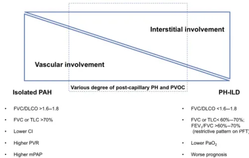
is frequently associated with ILD, in a patient with SSc with both PH and ILD, it can be difficult to firmly establish whether the patient has a PAH in- dependent from ILD, a PH-ILD, or the combination of PH-ILD and a pulmonary vasculopathy (Fig. 1).
Combined pulmonary fibrosis and emphysema syndrome (CPFE) can also be a cause of PH in SSc34and is associated with poor prognosis. It is characterized by combined emphysema of the up- per lobes and fibrosis of the lower lobes on chest CT, with preserved lung volumes, impaired DLCO, and hypoxemia at exercise and at rest in advanced cases. Whereas in the general population CPFE is usually observed in smokers, in SSc patients this condition is also present in nonsmokers.35
Pulmonary Hypertension Due to Left Heart Disease
Cardiac involvement in SSc is frequent and rele- vant from a prognostic point of view.36Although the real incidence is highly variable because it de- pends on the definition of cardiac involvement and on the diagnostic tools used to detect it, cardiac magnetic resonance37,38 and autoptic studies39 report percentages up to 75%. Therefore, it is diffi- cult to exclude patients with any kind of left heart abnormality when assessing PH in SSc.40
In a retrospective population of 107 SSc patients, Fox and colleagues41 evaluated all subjects with suspected PH by right and left heart catheterization, assessing LV end-diastolic pressure (LVEDP) mea- surement prefluid and postfluid challenge. The study found a high prevalence of postcapillary
Fig. 1.The spectrum of PH phenotype in SSc patients. CI, cardiac index; FEV1, forced expiratory volume in 1 sec- ond; PaO2, partial pressure of O2in arterial blood; TLC, total lung capacity.
PH in this population (mPAP 25 mm Hg, PAWP >15 mm Hg, and normal or reduced CO), including a significant number of occult postcapil- lary PH (mPAP25 mm Hg, PAWP15 mm Hg, LVEDP >15 mm Hg before or after a 500-mL fluid challenge administered over 5–10 min). Although RHC is the gold standard for assessment of intra- cardiac and pulmonary pressures, some controver- sial issues remain. In a large cohort of 11,523 patients undergoing simultaneous right heart and left heart catheterization, Halpern and Taichman42 found a high percentage of patients with a signifi- cant discrepancy between PAWP and LVEDP (PAWP <15 mm Hg and LVEDP >15 mm Hg). There- fore, approximately half of the patients presumed to have PAH based on PAWP were found to have post- capillary PH based on LVEDP. It is known that PAWP and LVEDP are not identical: a compliant left atrium can protect the pulmonary vasculature from elevated LVEDP, whereas a stiff left atrium can result in postcapillary PH in the setting of a normal LVEDP.40,43 Moreover, filling pressures vary over time, as shown in the Registry to Evaluate Early and Long-term Pulmonary Arterial Hyperten- sion Disease Management (REVEAL) database, where 10% of patients with an initial PAWP less than or equal to 12 mm Hg had a follow-up PAWP of greater than or equal to 16 mm Hg, whereas 50% of patients with an initial PAWP greater than or equal to 16 mm Hg had a follow-up PAWP less than or equal to 12 mm Hg.44 Altogether, these data highlight the discrepancies that may occur be- tween PAWP and LVEDP, which reflect the complexity of diagnosing pulmonary vascular dis- ease in the presence of left heart abnormalities40; a significant overlap between PAH and PH due to left heart disease—which is frequent in SSc—
makes a straightforward differentiation between the 2 conditions not always feasible6and the man- agement uncertain.
THE NEED FOR EARLY DIAGNOSIS—A MULTIPARAMETRIC APPROACH
Many attempts have been made in the past few years to establish a reliable way to identify the subgroup of SSc patients prone to developing PAH early, given the availability of more effective PAH-specific therapies and the evidence that patients identified early through an active screening program have better survival than pa- tients identified during routine clinical care.24 The European Guidelines recommend resting echocardiography as a screening test in asymp- tomatic SSc patients, followed by annual screening with echocardiography, DLCO, and biomarkers.3
Echocardiography
Echocardiography is the routine imaging tool to noninvasively assess the right heart and pulmonary circulation unit.45Although RHC remains the gold standard for confirming diagnosis and supporting treatment decisions, echocardiography has the advantage of being widely available, cost- effective, and well tolerated. A thorough cardiac ul- trasound examination should include not only the indirect estimation of systolic PAP (sPAP), which is essential in symptomatic patients with a clinical suspicion of PH to establish the probability of this condition, but also a detailed evaluation of the right and left heart dimensions and function as well as pulmonary artery diameter and inferior vena cava size and collapsibility. It is only by an accurate description of the 4 chambers’ anatomic and func- tional characteristics that it is possible to attempt a prediction of precapillary versus postcapillary PH46 (Table 1). A careful assessment of the right heart is often neglected in routine echocardiograms, despite its relevance, not only in connective tissue disease (CTD).45 An adequate echocardiogram should include a right ventricular (RV)-focused api- cal 4-chamber view, which would reduce the vari- ability in how the right heart is sectioned and, consequently, in RV dimensions and areas.47For the right atrium (RA), as well for the left atrium, vol- umes or at least areas, are more accurate to deter- mine the chamber size compared with linear dimensions. The European Guidelines include an end-systolic RA area greater than 18 cm2as one of the echocardiographic signs suggestive of PH, to be used to assess the probability of PH in addi- tion to tricuspid regurgitation (TR) velocity.3The ac- celeration time (ACT) of the RV outflow tract (RVOT) is another simple measurement that should be assessed: when less than 105 milliseconds and/
or showing a midsystolic notch in the Doppler pro- file, it is considered a suggestive sign of PH,3as an indirect marker of increased PVR.48,49
It is now well established that there is a poor RV adaptation to overload in SSc compared with other CTDs,50which is also linked to a complex physio- pathology with possible diastolic and/or systolic dysfunction. Huez and colleagues51 pointed out RV diastolic dysfunction in SSc patients as well as a decrease in pulmonary arterial compliance. Over- beek and colleagues52showed that for the same level of PAP, SSc patients had lower RV systolic function compared with patients with idiopathic PAH; they also demonstrated that the RV systolic response to an increase in PAP was poorest in SSc patients. These studies highlight the impor- tance of assessing RV systolic function in SSc pa- tients, which unfortunately is often missing in
echocardiographic reports.53 Echocardiographic RV diastolic parameters can also be easily assessed54 and have been shown significantly different compared with control subjects.55 New techniques for the assessment of myocardial defor- mation have also been used to assess RV and RA function in SSc, with significant results56–59; howev- er, their use in routine clinical practice is still limited.
The addition of lung ultrasound to a standard echocardiogram, adding only a few minutes to the examination, may reveal the presence
of sonographic signs of pulmonary intersti- tial involvement (sonographic B-lines) which, when associated with an irregular pleural line, are highly suggestive for ILD and may have a role in the screening algorithm60,61(seeTable 1).
Exercise Echocardiography
There is increasing awareness of the clinical rele- vance of an abnormal pulmonary hemodynamic response during exercise,62but several questions Table 1
Typical echocardiographic features in different pulmonary hypertension phenotypes in systemic sclerosis
Pulmonary Arterial Hypertension
Interstitial Lung Disease Left Cardiac Involvement LV dimensions Normal to
reduced
Usually normal Usually increased Left atrial
dimensions
Normal Normal Usually increased
RV-RA dimensions
Increased Normal/increased Normal
Eccentricity index
1–2 Usually <1.2 1
LV systolic function
Normal Normal Reduced (ejection fraction
can be normal until later stages)
LV diastolic function
Normal, grade I Normal, grade I Grade II–-III E/e’ usually <10 E/e’ usually <10 E/e’ usually >10
RV function Reduced Usually normal Usually normal (reduced in
biventricular involvement) Mitral
regurgitation
Trivial–mild Trivial–mild Mild–moderate
TR Moderate–severe Mild–moderate Usually mild
sPAP 111 11 1
Inferior vena cava
Dilated and fixed Usually normal and collapsible
Normal and collapsible
PVR 111 1/11 Normal
Other signs RV forming heart apex
Reduced ACT notch of RVOT Doppler spectrum Pericardial effusion
Multiple diffuse B lines with irregular pleural line at lung ultrasound
Pericardial effusion
Abbreviations:E/e’, early mitral inflow velocity and mitral annular early diastolic velocity;1, slightly increased;11, moderately increased;111, highly increased.
remain to be elucidated; therefore, exercise PH is an entity that has not been endorsed by the latest European Guidelines, where its definition, even when estimated by RHC, has been considered un- supported due to insufficient data.3More recently, exercise PH has been defined as the presence of resting mPAP less than 25 mm Hg and mPAP greater than 30 mm Hg during exercise with total pulmonary resistance greater than 3 Wood units, during RHC.62 Exercise PH seems to represent the hemodynamic manifestation of early pulmo- nary vascular disease, left heart disease, lung dis- ease, or a combination of these conditions,62 acting as a possible transitional phase anticipating resting PH.
Exercise echocardiography is a noninvasive tool to estimate pulmonary hemodynamics during ex- ercise and is useful to assess abnormalities of pul- monary vascular function as well as the state of the right heart, although it does not have an estab- lished role in the management of SSc patients. A main issue in SSc is the high percentage of pa- tients showing exercise PH during exercise stress echocardiography, which clearly overestimates the subset of SSc patients who will develop PAH.63–66 PAP is dependent, however, not only on PVR, which is abnormally increased in PAH, but also on left atrial pressure and CO, as shown both in healthy subjects67and in SSc.51It is, there- fore, crucial to define the relative hemodynamic contribution of each parameter to better under- stand the main determinants of increased PAP.68,69Exercise echocardiography may identify a subset of SSc patients without PH with an inap- propriate exercise-induced increase in pulmonary arterial systolic pressure (PASP) and early signs of RV dysfunction. A study68enrolling 172 consec- utive SSc patients in New York Heart Association class I/II showed a higher exercise-induced sPAP (36.9 8.7 vs 25.9 3.3 mm Hg;P<.0001) and a lower cardiac index increase (2.8 1.2 vs 4.62.3 L/min/m2;P<.0001) than controls.
In a population of 164 SSc patients, the authors demonstrated that exercise PH (defined as an ex- ercise sPAP50 mm Hg and exercise PVR 3 Wood units during echocardiography) was present in approximately half of the patients with normal resting sPAP and was affected by age, ILD, and RV and LV diastolic dysfunction, whereas only a minority (5%) of these patients had an increase in PVR during exercise, suggesting high heteroge- neity of the pathophysiologic background.69These data were further confirmed in a smaller popula- tion of 45 patients, where exercise PH was present in 21 patients, with a positive correlation between exercise sPAP and both exercise left atrial pres- sure and exercise PVR (respectively, r2 5 0.61
and r250.57;P<.05), again suggesting that exer- cise PH was related to both increased exercise LV filling pressure and exercise PVR.70Thereby, exer- cise echocardiography allows identification of those patients with an abnormal increase in PAP as well as a better understanding of the mecha- nism leading to abnormal pulmonary hemody- namic response during exercise.
Exercise echocardiography may also help distinguish patients at risk of developing further resting PH. Codullo and colleagues71 found that aDsPAP cutoff of greater than 18 mm Hg, identi- fied by receiver operating characteristic curve analysis, had a sensitivity of 50% and a specificity of 90% for the development of resting PH during follow-up. In another study, exercise PH has been found useful to predict the onset of resting PH at echocardiography during follow-up, in addi- tion to nailfold videocapillaroscopy.72Exercise PH with normal resting sPAP was present in 43% of patients; after a mean follow-up of 24 months, 11 patients developed resting PH (as defined by echocardiography), and all of them belonged to the exercise PH group. Patients who did not have exercise PH never developed a resting sPAP greater than 35 mm Hg during the follow-up.72
Kusunose and colleagues73 prospectively enrolled 78 patients with CTD (including 70% of SSc) with a baseline resting and postexercise echocardiographic evaluation. During a median follow-up of 32 months, 16 patients developed resting PAH. The slope of mPAP/CO had an incre- mental value over a 6-minute walking test distance to predict PH at follow-up. Even though exercise echocardiography is not included in the current recommendations for screening patients at risk of resting PH, it remains an interesting tool to assess the physiopathology of the hemodynamic behavior during stress, with a promising role in the early detection of abnormal vascular response.
Moreover, an abnormal exercise-induced increase in PASP may explain an otherwise inexplicable effort dyspnea in SSc patients with normal base- line hemodynamics.
Nonechocardiographic Parameters
In the past few years, many studies have addressed the complex issue of early diagnosis in patients with SSc and in patients with CTD, underlining the importance of a multiparametric approach that should not be limited to transtho- racic echocardiography as the sole instrumental examination for establishing the likelihood of devel- oping PAH,74because other noninvasive screening tests, such as pulmonary function tests (PFTs),
and measurement of serum biomarkers, such as N- terminal pro brain natriuretic peptide (NT-proBNP), have been shown with PAH in SSc patients.75–77In particular, the Evidence-based detection of pulmo- nary arterial hypertension in systemic sclerosis (DETECT) study enrolled patients with more than 3 years’ disease duration from the first non–
Raynaud phenomenon symptom and a predicted DLCO less than 60%, thus representing a high- risk population. This was the first study on PAH screening to undertake systematic RHC in all pa- tients to develop an evidence-based algorithm for earlier identification of PAH in a mildly symptomatic population. In this study, 466 patients underwent noninvasive testing and RHC: results showed that 87 patients (19%) had RHC-confirmed PAH, a higher prevalence compared with previous studies.78The DETECT algorithm showed a signif- icantly higher sensitivity in identifying patients with PAH, missing only 4% of patients as false negative.
Longitudinal data from this cohort have demon- strated that 44% of the PAH patients who received an early diagnosis through the DETECT algorithm had disease progression during a relatively short follow-up time, again underlining the clinical rele- vance of early detection of PAH.79The DETECT al- gorithm has been successfully applied also to other populations of high-risk patients.80The DETECT al- gorithm, however, is not applicable to patients with a predicted DLCO greater than 60%. The PHAROS also confirmed that a low DLCO less than 55% and a high forced vital capacity % predicted to DLCO
%predicted ratio (FVC/DLCO) greater than 1.6 are good screening parameters in addition to echo-derived sPAP in selecting those patients who are at risk to develop SSc-PAH.81 More recently, a study comparing the DETECT algorithm with the screening models suggested by the 2009 and 2015 European Guidelines found that referring patients to RHC according to the DETECT algo- rithm yielded a high number of false-negative cases but was useful especially to identify patients with borderline PAP (mPAP 21–24 mm Hg),82which seems to be an intermediate stage on the contin- uum between normal pulmonary hemodynamics and PAH.83,84
Some recommendations for screening and detection of CTD-associated PAH were published in 2013, after a systematic review of the literature by an international expert panel.74This article con- tains the first evidence-based and consensus- based recommendations for screening and early detection of CTD-associated PAH with the aim of identifying patients with asymptomatic/preclinical disease and those with mild symptoms to prevent or delay progression of disease through early management. Box 1 summarizes these general
recommendations. It must be underlined that the quality of evidence, which was assessed accord- ing to the Grading of Recommendations Assess- ment, Development and Evaluation Working Group from very low to high, varies between the different statements.
The recommendations established specific criteria to recommend RHC in SSc and sclero- derma spectrum disorders, which is advised in pa- tients with (1) a TR jet velocity of 2.5 m/s–2.8 m/s with signs and/or symptoms consistent with PH;
(2) a TR jet velocity of greater than 2.8 m/s with
Box 1
Summary of general recommendations for early detection of connective tissue disease–
associated pulmonary arterial hypertension
General recommendations
All patients with SSc should be screened for PAH.
Patients with MCTD/CTD with scleroderma features should be screened similarly to pa- tients with SSc.
Screening is not recommended for asymp- tomatic patients with MCTD/CTD without scleroderma features.
All patients with SSc and MCTD/CTD with scleroderma features with positive screening results should be referred for RHC.
RHC is mandatory for diagnosis of PAH.
Initial screening evaluation PFT with DLCO
Transthoracic echocardiography NT-proBNP
DETECT algorithm if DLCO less than 60% and disease duration greater than 3 years Frequency of noninvasive tests
Transthoracic echocardiography annually as a screening test
Transthoracic echocardiography if new signs or symptoms develop
PFT with DLCO annually as a screening test PFT with DLCO if new signs or symptoms
develop
NT-proBNP if new signs or symptoms develop Abbreviation:MCTD, mixed CTD.
Adapted fromKhanna D, Gladue H, Channick R, et al.
Recommendations for screening and detection of connective-tissue disease associated pulmonary arterial hypertension. Arthritis Rheum 2013;65(12):3196; with permission.
or without signs and/or symptoms of PH; (3) RA or RV enlargement (RA major dimension >53 mm and RV midcavity dimension >35 mm), irrespective of TR jet velocity (including nonmeasurable or <2.5 m/s); or (4) signs or symptoms of PH and an FVC/DLCO greater than 1.6 and/or a predicted DLCO of less than 60%, without an overt systolic dysfunction, a greater than grade I diastolic dysfunction, a greater than mild mitral or aortic valve disease, or evidence of PH. The expert panel did not recommend acute vasodilator testing dur- ing RHC as part of the evaluation of PAH. This is supported by the small number of patients in this subset with both a positive vasodilator test result (defined as a reduction in mPAP by at least 10 mm Hg to an mPAP of <40 mm Hg in the setting of a normal CO) and a long-term response to calcium-channel blockers.3,85
No clear recommendations are provided on borderline mean PAP (21–24 mm Hg) or on exer- cise PH, due to lack of long-term outcome data and variability in exercise testing.86,87
The role of RV and RA measurements underlines the importance of referring these patients to specialized centers, where echocardiography is performed by certified personnel, who will include a thorough evaluation of the right heart.
SUMMARY
Involvement of the right heart-pulmonary circula- tion system is crucial in SSc and represents a main prognostic determinant. PH may respond to multiple and partially overlapping mechanisms of precapillary and postcapillary etiologies. An early diagnosis is mandatory to improve outcomes, and a multidisciplinary and multiparametric approach is required to fully understand the diverse mechanisms leading to abnormal pulmo- nary hemodynamics.
Recommendations on how to screen SSc- related PAH have been established and may help clinicians in this complex management, although they are not meant to substitute a clinically driven individualized assessment of the patient.
REFERENCES
1. Denton CP, Khanna D. Systemic sclerosis. Lancet 2017;390(10103):1685–99.
2. Walker UA, Tyndall A, Czirjak L, et al. Clinical risk assessment of organ manifestations in systemic sclerosis: a report from the EULAR Scleroderma Tri- als and Research group database. Ann Rheum Dis 2007;66(6):754–63.
3. Galie N, Humbert M, Vachiery JL, et al. 2015 ESC/
ERS guidelines for the diagnosis and treatment of
pulmonary hypertension: the joint task force for the diagnosis and treatment of pulmonary hypertension of the European Society of Cardiology (ESC) and the European Respiratory Society (ERS): endorsed by:
Association for European Paediatric and Congenital Cardiology (AEPC), International Society for Heart and Lung Transplantation (ISHLT). Eur Heart J 2016;37(1):67–119.
4. McLaughlin V, Humbert M, Coghlan G, et al. Pulmo- nary arterial hypertension: the most devastating vascular complication of systemic sclerosis. Rheu- matology (Oxford) 2009;48(Suppl 3):iii25–31.
5. Launay D, Sobanski V, Hachulla E, et al. Pulmo- nary hypertension in systemic sclerosis: different phenotypes. Eur Respir Rev 2017;26(145) [pii:
170056].
6. Opitz CF, Hoeper MM, Gibbs JS, et al. Pre-capillary, combined, and post-capillary pulmonary hyperten- sion: a pathophysiological continuum. J Am Coll Cardiol 2016;68(4):368–78.
7. Simonneau G, Gatzoulis MA, Adatia I, et al. Updated clinical classification of pulmonary hypertension.
J Am Coll Cardiol 2013;62(25 Suppl):D34–41.
8. Hinchcliff M, Fischer A, Schiopu E, Steen VD. PHAROS Investigators. Pulmonary Hypertension Assessment and Recognition of Outcomes in Scleroderma (PHAROS): baseline characteristics and description of study population. J Rheumatol 2011;38(10):2172–9.
9. Connolly MJ, Abdullah S, Ridout DA, et al. Prog- nostic significance of computed tomography criteria for pulmonary veno-occlusive disease in systemic sclerosis-pulmonary arterial hypertension. Rheuma- tology (Oxford) 2017;56(12):2197–203.
10. McLaughlin VV, Archer SL, Badesch DB, et al.
ACCF/AHA 2009 Expert consensus document on pulmonary hypertension: a report of the American College of Cardiology Foundation Task Force on Expert Consensus Documents and the American Heart Association Developed in Collaboration with the American College of Chest Physicians; Amer- ican Thoracic Society, Inc.; and the Pulmonary Hy- pertension Association. J Am Coll Cardiol 2009;
53(17):1573–619.
11. Iudici M, Codullo V, Giuggioli D, et al. Pulmonary hyper- tension in systemic sclerosis: prevalence, incidence and predictive factors in a large multicentric Italian cohort. Clin Exp Rheumatol 2013;31(2 Suppl 76):31–6.
12. Vandecasteele E, Melsens K, Thevissen K, et al.
Prevalence and incidence of pulmonary arterial hy- pertension: 10-year follow-up of an unselected sys- temic sclerosis cohort. J scleroderma Relat Disord 2017;2(3):196–202.
13. Launay D, Mouthon L, Hachulla E, et al. Prevalence and characteristics of moderate to severe pulmo- nary hypertension in systemic sclerosis with and without interstitial lung disease. J Rheumatol 2007;
34(5):1005–11.
14.Avouac J, Airo P, Meune C, et al. Prevalence of pul- monary hypertension in systemic sclerosis in Euro- pean Caucasians and metaanalysis of 5 studies.
J Rheumatol 2010;37(11):2290–8.
15.Condliffe R, Kiely DG, Peacock AJ, et al. Connective tissue disease-associated pulmonary arterial hyper- tension in the modern treatment era. Am J Respir Crit Care Med 2009;179(2):151–7.
16.Launay D, Sitbon O, Hachulla E, et al. Survival in systemic sclerosis-associated pulmonary arterial hy- pertension in the modern management era. Ann Rheum Dis 2013;72(12):1940–6.
17.Rubenfire M, Huffman MD, Krishnan S, et al. Survival in systemic sclerosis with pulmonary arterial hyper- tension has not improved in the modern era. Chest 2013;144(4):1282–90.
18.Lefevre G, Dauchet L, Hachulla E, et al. Survival and prognostic factors in systemic sclerosis-associated pulmonary hypertension: a systematic review and meta-analysis. Arthritis Rheum 2013;65(9):2412–23.
19.Sitbon O, Channick R, Chin KM, et al. Selexipag for the treatment of pulmonary arterial hypertension.
N Engl J Med 2015;373(26):2522–33.
20.Galie N, Barbera JA, Frost AE, et al. Initial use of am- brisentan plus tadalafil in pulmonary arterial hyper- tension. N Engl J Med 2015;373(9):834–44.
21.Pulido T, Adzerikho I, Channick RN, et al. Macitentan and morbidity and mortality in pulmonary arterial hy- pertension. N Engl J Med 2013;369(9):809–18.
22.Coghlan JG, Galie N, Barbera JA, et al. Initial com- bination therapy with ambrisentan and tadalafil in connective tissue disease-associated pulmonary arterial hypertension (CTD-PAH): subgroup analysis from the AMBITION trial. Ann Rheum Dis 2017;76(7):
1219–27.
23.Hassoun PM, Zamanian RT, Damico R, et al. Ambri- sentan and tadalafil up-front combination therapy in scleroderma-associated pulmonary arterial hyper- tension. Am J Respir Crit Care Med 2015;192(9):
1102–10.
24.Humbert M, Yaici A, de Groote P, et al. Screening for pulmonary arterial hypertension in patients with sys- temic sclerosis: clinical characteristics at diagnosis and long-term survival. Arthritis Rheum 2011;
63(11):3522–30.
25.Dorfmuller P, Humbert M, Perros F, et al. Fibrous remodeling of the pulmonary venous system in pul- monary arterial hypertension associated with con- nective tissue diseases. Hum Pathol 2007;38(6):
893–902.
26.Gunther S, Jais X, Maitre S, et al. Computed tomog- raphy findings of pulmonary venoocclusive disease in scleroderma patients presenting with precapillary pulmonary hypertension. Arthritis Rheum 2012;
64(9):2995–3005.
27.Mari-Alfonso B, Simeon-Aznar CP, Guillen-Del Castillo A, et al. Hepatobiliary involvement in
systemic sclerosis and the cutaneous subsets:
characteristics and survival of patients from the Spanish RESCLE Registry. Semin Arthritis Rheum 2017 [pii:S0049-0172(17)30288-3].
28.Steen VD, Medsger TA. Changes in causes of death in systemic sclerosis, 1972-2002. Ann Rheum Dis 2007;66(7):940–4.
29.Chang B, Wigley FM, White B, et al. Scleroderma patients with combined pulmonary hypertension and interstitial lung disease. J Rheumatol 2003;
30(11):2398–405.
30.Mathai SC, Hummers LK, Champion HC, et al. Sur- vival in pulmonary hypertension associated with the scleroderma spectrum of diseases: impact of interstitial lung disease. Arthritis Rheum 2009;
60(2):569–77.
31.Altman RD, Medsger TA Jr, Bloch DA, et al. Predic- tors of survival in systemic sclerosis (scleroderma).
Arthritis Rheum 1991;34(4):403–13.
32.Le Pavec J, Girgis RE, Lechtzin N, et al. Systemic sclerosis-related pulmonary hypertension associ- ated with interstitial lung disease: impact of pulmo- nary arterial hypertension therapies. Arthritis Rheum 2011;63(8):2456–64.
33.Launay D, Humbert M, Berezne A, et al. Clinical characteristics and survival in systemic sclerosis- related pulmonary hypertension associated with interstitial lung disease. Chest 2011;140(4):
1016–24.
34.Cottin V, Cordier JF. Combined pulmonary fibrosis and emphysema in connective tissue disease. Curr Opin Pulm Med 2012;18(5):418–27.
35.Antoniou KM, Margaritopoulos GA, Goh NS, et al.
Combined pulmonary fibrosis and emphysema in scleroderma-related lung disease has a major con- founding effect on lung physiology and screening for pulmonary hypertension. Arthritis Rheumatol 2016;68(4):1004–12.
36.Ferri C, Valentini G, Cozzi F, et al. Systemic scle- rosis: demographic, clinical, and serologic features and survival in 1,012 Italian patients. Medicine 2002;81(2):139–53.
37.Hachulla AL, Launay D, Gaxotte V, et al. Cardiac magnetic resonance imaging in systemic sclerosis:
a cross-sectional observational study of 52 patients.
Ann Rheum Dis 2009;68(12):1878–84.
38.Mavrogeni SI, Kitas GD, Dimitroulas T, et al. Cardio- vascular magnetic resonance in rheumatology: cur- rent status and recommendations for use. Int J Cardiol 2016;217:135–48.
39.Follansbee WP, Miller TR, Curtiss EI, et al. A controlled clinicopathologic study of myocardial fibrosis in sys- temic sclerosis (scleroderma). J Rheumatol 1990;
17(5):656–62.
40.Coghlan G. Does left heart disease cause most sys- temic sclerosis associated pulmonary hypertension?
Eur Respir J 2013;42(4):888–90.
41.Fox BD, Shimony A, Langleben D, et al. High preva- lence of occult left heart disease in scleroderma- pulmonary hypertension. Eur Respir J 2013;42(4):
1083–91.
42.Halpern SD, Taichman DB. Misclassification of pul- monary hypertension due to reliance on pulmonary capillary wedge pressure rather than left ventricular end-diastolic pressure. Chest 2009;136(1):37–43.
43.Frost AE, Farber HW, Barst RJ, et al. Demographics and outcomes of patients diagnosed with pulmonary hypertension with pulmonary capillary wedge pres- sures 16 to 18 mm Hg: insights from the REVEAL registry. Chest 2013;143(1):185–95.
44.Shirai Y, Kuwana M. Complex Pathophysiology of Pulmonary Hypertension Associated with Systemic Sclerosis: Potential Unfavorable Effects of Vasodila- tors. J scleroderma Relat Disord 2017;2(2):92–9.
45.Ferrara F, Gargani L, Ostenfeld E, et al. Imaging the right heart pulmonary circulation unit: insights from advanced ultrasound techniques. Echocardiogra- phy 2017;34(8):1216–31.
46.D’Alto M, Romeo E, Argiento P, et al. Echocardio- graphic prediction of pre- versus postcapillary pul- monary hypertension. J Am Soc Echocardiogr 2015;28(1):108–15.
47.Lang RM, Badano LP, Mor-avi V, et al. Recommen- dations for cardiac chamber quantification by echo- cardiography in adults: an update from the American Society of Echocardiography and the Eu- ropean Association of Cardiovascular Imaging. Eur Heart J Cardiovasc Imaging 2015;16(3):233–70.
48.Serra W, Chetta A, Santilli D, et al. Echocardiogra- phy may help detect pulmonary vasculopathy in the early stages of pulmonary artery hypertension associated with systemic sclerosis. Cardiovasc Ul- trasound 2010;8:25.
49.Granstam SO, Bjorklund E, Wikstrom G, et al. Use of echocardiographic pulmonary acceleration time and estimated vascular resistance for the evaluation of possible pulmonary hypertension. Cardiovasc Ultra- sound 2013;11:7.
50.Vonk Noordegraaf A, Naeije R. Right ventricular func- tion in scleroderma-related pulmonary hypertension.
Rheumatology (Oxford) 2008;47(Suppl 5):v42–3.
51.Huez S, Roufosse F, Vachiery JL, et al. Isolated right ventricular dysfunction in systemic sclerosis: latent pulmonary hypertension? Eur Respir J 2007;30(5):
928–36.
52.Overbeek MJ, Lankhaar JW, Westerhof N, et al.
Right ventricular contractility in systemic sclerosis- associated and idiopathic pulmonary arterial hyper- tension. Eur Respir J 2008;31(6):1160–6.
53.Galderisi M, Cosyns B, Edvardsen T, et al. Standard- ization of adult transthoracic echocardiography re- porting in agreement with recent chamber quantification, diastolic function, and heart valve dis- ease recommendations: an expert consensus
document of the European Association of Cardio- vascular Imaging. Eur Heart J Cardiovasc Imaging 2017;18(12):1301–10.
54. Rudski LG, Lai WW, Afilalo J, et al. Guidelines for the echocardiographic assessment of the right heart in adults: a report from the American Society of Echo- cardiography endorsed by the European Associa- tion of Echocardiography, a registered branch of the European Society of Cardiology, and the Cana- dian Society of Echocardiography. J Am Soc Echo- cardiogr 2010;23(7):685–713.
55. D’Alto M, Riccardi A, Argiento P, et al. Cardiac involvement in undifferentiated connective tissue disease at risk for systemic sclerosis (otherwise referred to as very early-early systemic sclerosis):
a TDI study. Clin Exp Med 2017. [Epub ahead of print].
56. Saito M, Wright L, Negishi K, et al. Mechanics and prognostic value of left and right ventricular dysfunc- tion in patients with systemic sclerosis. Eur Heart J Cardiovasc Imaging 2017. [Epub ahead of print].
57. Mukherjee M, Chung SE, Ton VK, et al. Unique ab- normalities in right ventricular longitudinal strain in systemic sclerosis patients. Circ Cardiovasc Imag- ing 2016;9(6) [pii:e003792].
58. D’Andrea A, D’Alto M, Di Maio M, et al. Right atrial morphology and function in patients with systemic sclerosis compared to healthy controls: a two- dimensional strain study. Clin Rheumatol 2016;
35(7):1733–42.
59. Schattke S, Knebel F, Grohmann A, et al. Early right ventricular systolic dysfunction in patients with sys- temic sclerosis without pulmonary hypertension: a Doppler Tissue and Speckle Tracking echocardiog- raphy study. Cardiovasc Ultrasound 2010;8(1):3.
60. Barskova T, Gargani L, Guiducci S, et al. Lung ultra- sound for the screening of interstitial lung disease in very early systemic sclerosis. Ann Rheum Dis 2013;
72(3):390–5.
61. Wang Y, Gargani L, Barskova T, et al. Usefulness of lung ultrasound B-lines in connective tissue disease- associated interstitial lung disease: a literature re- view. Arthritis Res Ther 2017;19(1):206.
62. Kovacs G, Herve P, Barbera JA, et al. An official Eu- ropean Respiratory Society statement: pulmonary haemodynamics during exercise. Eur Respir J 2017;50(5) [pii:1700578].
63. Collins N, Bastian B, Quiqueree L, et al. Abnormal pulmonary vascular responses in patients registered with a systemic autoimmunity database: pulmonary hypertension assessment and screening evaluation using stress echocardiography (PHASE-I). Eur J Echocardiogr 2006;7(6):439–46.
64. Alkotob ML, Soltani P, Sheatt MA, et al. Reduced ex- ercise capacity and stress-induced pulmonary hy- pertension in patients with scleroderma. Chest 2006;130(1):176–81.
65.Callejas-Rubio JL, Moreno-Escobar E, de la Fuente PM, et al. Prevalence of exercise pulmonary arterial hypertension in scleroderma. J Rheumatol 2008;35(9):1812–6.
66.Pignone A, Mori F, Pieri F, et al. Exercise Doppler echocardiography identifies preclinic asymptomatic pulmonary hypertension in systemic sclerosis. Ann N Y Acad Sci 2007;1108:291–304.
67.Argiento P, Chesler N, Mule M, et al. Exercise stress echocardiography for the study of the pulmonary cir- culation. Eur Respir J 2010;35(6):1273–8.
68.D’Alto M, Ghio S, D’Andrea A, et al. Inappropriate exercise-induced increase in pulmonary artery pres- sure in patients with systemic sclerosis. Heart 2011;
97(2):112–7.
69.Gargani L, Pignone A, Agoston G, et al. Clinical and echocardiographic correlations of exercise-induced pulmonary hypertension in systemic sclerosis: a multicenter study. Am Heart J 2013;165(2):200–7.
70.Voilliot D, Magne J, Dulgheru R, et al. Determinants of exercise-induced pulmonary arterial hypertension in systemic sclerosis. Int J Cardiol 2014;173(3):
373–9.
71.Codullo V, Caporali R, Cuomo G, et al. Stress Doppler echocardiography in systemic sclerosis:
evidence for a role in the prediction of pulmonary hy- pertension. Arthritis Rheum 2013;65(9):2403–11.
72.Voilliot D, Magne J, Dulgheru R, et al. Prediction of new onset of resting pulmonary arterial hypertension in systemic sclerosis. Arch Cardiovasc Dis 2016;
109(4):268–77.
73.Kusunose K, Yamada H, Hotchi J, et al. Prediction of future overt pulmonary hypertension by 6-min walk stress echocardiography in patients with connective tissue disease. J Am Coll Cardiol 2015;66(4):
376–84.
74.Khanna D, Gladue H, Channick R, et al. Recommen- dations for screening and detection of connective- tissue disease associated pulmonary arterial hyper- tension. Arthritis Rheum 2013;65(12):3194–201.
75.Allanore Y, Borderie D, Avouac J, et al. High N-termi- nal pro-brain natriuretic peptide levels and low diffusing capacity for carbon monoxide as indepen- dent predictors of the occurrence of precapillary pulmonary arterial hypertension in patients with sys- temic sclerosis. Arthritis Rheum 2008;58(1):284–91.
76.Thakkar V, Stevens WM, Prior D, et al. N-terminal pro-brain natriuretic peptide in a novel screening al- gorithm for pulmonary arterial hypertension in sys- temic sclerosis: a case-control study. Arthritis Res Ther 2012;14(3):R143.
77.Hachulla E, Gressin V, Guillevin L, et al. Early detec- tion of pulmonary arterial hypertension in systemic sclerosis: a French nationwide prospective multi- center study. Arthritis Rheum 2005;52(12):
3792–800.
78.Coghlan JG, Denton CP, Grunig E, et al. Evidence- based detection of pulmonary arterial hypertension in systemic sclerosis: the DETECT study. Ann Rheum Dis 2014;73(7):1340–9.
79.Mihai C, Antic M, Dobrota R, et al. Factors associ- ated with disease progression in early-diagnosed pulmonary arterial hypertension associated with systemic sclerosis: longitudinal data from the DETECT cohort. Ann Rheum Dis 2018;77(1):128–32.
80.Hao Y, Thakkar V, Stevens W, et al. A comparison of the predictive accuracy of three screening models for pulmonary arterial hypertension in systemic scle- rosis. Arthritis Res Ther 2015;17:7.
81.Hsu VM, Chung L, Hummers LK, et al. Development of pulmonary hypertension in a high-risk population with systemic sclerosis in the pulmonary hyperten- sion assessment and recognition of outcomes in scleroderma (PHAROS) cohort study. Semin Arthritis Rheum 2014;44(1):55–62.
82.Vandecasteele E, Drieghe B, Melsens K, et al.
Screening for pulmonary arterial hypertension in an unselected prospective systemic sclerosis cohort.
Eur Respir J 2017;49(5) [pii:1602275].
83.Visovatti SH, Distler O, Coghlan JG, et al. Borderline pulmonary arterial pressure in systemic sclerosis patients: a post-hoc analysis of the DETECT study.
Arthritis Res Ther 2014;16(6):493.
84.Hoffmann-Vold AM, Fretheim H, Midtvedt O, et al.
Frequencies of borderline pulmonary hypertension before and after the DETECT algorithm: results from a prospective systemic sclerosis cohort. Rheu- matology (Oxford) 2018;57(3):480–7.
85.Montani D, Savale L, Natali D, et al. Long-term response to calcium-channel blockers in non- idiopathic pulmonary arterial hypertension. Eur Heart J 2010;31(15):1898–907.
86.Saggar R, Khanna D, Furst DE, et al. Exercise- induced pulmonary hypertension associated with systemic sclerosis: four distinct entities. Arthritis Rheum 2010;62(12):3741–50.
87.Bae S, Saggar R, Bolster MB, et al. Baseline charac- teristics and follow-up in patients with normal hae- modynamics versus borderline mean pulmonary arterial pressure in systemic sclerosis: results from the PHAROS registry. Ann Rheum Dis 2012;71(8):
1335–42.
View publication stats View publication stats