Mitochondrial Substrate-Level
Phosphorylation as Energy Source for Glioblastoma: Review and Hypothesis
Christos Chinopoulos1 and Thomas N. Seyfried2
Abstract
Glioblastoma multiforme (GBM) is the most common and malignant of the primary adult brain cancers. Ultrastructural and biochemical evidence shows that GBM cells exhibit mitochondrial abnormalities incompatible with energy production through oxidative phosphorylation (OxPhos). Under such conditions, the mitochondrial F0-F1 ATP synthase operates in reverse at the expense of ATP hydrolysis to maintain a moderate membrane potential. Moreover, expression of the dimeric M2 isoform of pyruvate kinase in GBM results in diminished ATP output, precluding a significant ATP production from glycolysis. If ATP synthesis through both glycolysis and OxPhos was impeded, then where would GBM cells obtain high- energy phosphates for growth and invasion? Literature is reviewed suggesting that the succinate-CoA ligase reaction in the tricarboxylic acid cycle can substantiate sufficient ATP through mitochondrial substrate-level phosphorylation (mSLP) to maintain GBM growth when OxPhos is impaired. Production of high-energy phosphates would be supported by glutami- nolysis—a hallmark of GBM metabolism—through the sequential conversion of glutamine ! glutamate ! alpha- ketoglutarate!succinyl CoA!succinate. Equally important, provision of ATP through mSLP would maintain the adenine nucleotide translocase inforward mode, thus preventing the reverse-operating F0-F1 ATP synthase from depleting cytosolic ATP reserves. Because glucose and glutamine are the primary fuels driving the rapid growth of GBM and most tumors for that matter, simultaneous restriction of these two substrates or inhibition of mSLP should diminish cancer viability, growth, and invasion.
Keywords
therapies, gliomas, bioenergetics, Warburg
Received July 17, 2018; Received revised November 15, 2018; Accepted for publication November 16, 2018
Introduction
Glioblastoma multiforme (GBM) carries the highest mor- tality rate among primary brain tumors and remains large- ly unmanageable. Life expectancy following diagnosis is only about 12 to 14 months. This has changed little for decades despite continuing research (Fisher and Buffler, 2005; Krex et al., 2007; Stupp et al., 2009; Lawrence et al., 2011; Ahmadloo et al., 2013; Alexander and Cloughesy, 2017). Indeed, a recent reevaluation found that overall survival for GBM is woefully similar to that reported by Cushing almost a century ago (Fatehi et al., 2018). A defining characteristic of GBM is thesecondary structures of Scherer, which include diffuse parenchymal growth invasion over the subpial surface, along white matter tracks, and through the Virchow–Robin spaces (Scherer, 1940; Laws et al., 1993; Kleihues and Ohgaki, 1999;
Zagzag et al., 2008; Shelton et al., 2010c). The highly inva- sive nature of GBM makes most current therapies ineffec- tive (Chang et al., 2005; Krex et al., 2007; Cuddapah et al., 2014; Perry et al., 2017). Although the introduction of the toxic alkylating agent, temozolomide, has improved progression-free survival slightly, quality of life has also remained poor for most GBM patients especially for those
1Department of Medical Biochemistry, Semmelweis University, Budapest, Hungary
2Biology Department, Boston College, Chestnut Hill, MA, USA Corresponding Author:
Thomas N. Seyfried, Biology Department, Boston College, Chestnut Hill, MA 02467, USA.
Email: thomas.seyfried@bc.edu
Volume 10: 1–27
!The Author(s) 2018 Article reuse guidelines:
sagepub.com/journals-permissions DOI: 10.1177/1759091418818261 journals.sagepub.com/home/asn
Creative Commons Non Commercial CC BY-NC: This article is distributed under the terms of the Creative Commons Attribution- NonCommercial 4.0 License (http://www.creativecommons.org/licenses/by-nc/4.0/) which permits non-commercial use, reproduction and dis- tribution of the work without further permission provided the original work is attributed as specified on the SAGE and Open Access pages (https://us.
sagepub.com/en-us/nam/open-access-at-sage).
receiving radiation alone (Taphoorn et al., 2005; Stupp et al., 2009; Flechl et al., 2012).
GBM contains a plethora of morphologically diverse neoplastic cell types that express neural, glial, and mye- loid/mesenchymal markers (Rubinstein, 1972; Morantz et al., 1979; Wood and Morantz, 1979; Roggendorf et al., 1996; Seyfried, 2001; Yuan et al., 2004;
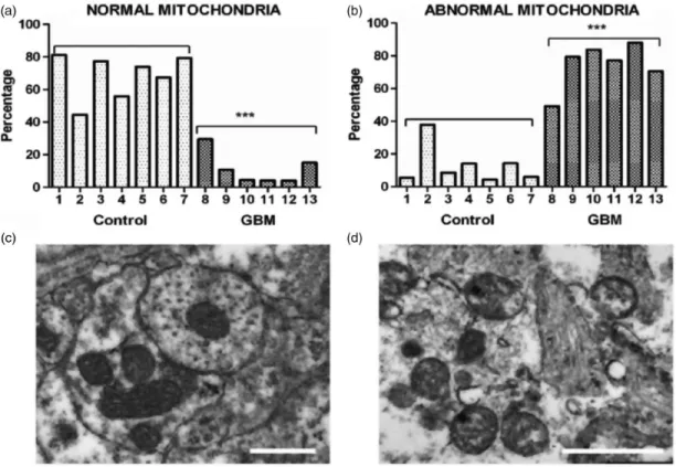
Huysentruyt et al., 2011; Karsy et al., 2012). Also recog- nized are abnormalities in the number, structure, and function of mitochondria in GBM cells (Figure 1).
Several groups have documented GBM cells with reduced or increased numbers of morphologically abnor- mal mitochondria with laminations and aberrant or absent cristae (Sipe et al., 1973; Scheithauer and Bruner, 1987; Oudard et al., 1997; Arismendi-Morillo and Castellano-Ramirez, 2008; Katsetos et al., 2013;
Deighton et al., 2014; Feichtinger et al., 2014; Seyfried et al., 2017). Some mitochondria in human GBM cells contain few, if any, cristae (Arismendi-Morillo and Castellano-Ramirez, 2008). In murine GBM, the content
and composition of cardiolipin, the signature lipid of the mitochondrial inner membrane that regulates oxidative phosphorylation (OxPhos), exhibit considerable devia- tions from the normal (Kiebish et al., 2008; Claypool and Koehler, 2012). A multitude of findings support the notion that OxPhos is defective in GBM (Lichtor and Dohrmann, 1986; Feichtinger et al., 2014;
Bartesaghi et al., 2015; Dahlberg et al., 2017). Based on the concept that mitochondrial structure determines mitochondrial function (Lehninger, 1964), these multiple mitochondrial abnormalities, which are of genetic or environmental origin, would be expected to compromise effective energy production through OxPhos (Seyfried and Shelton, 2010; Seyfried et al., 2014, 2015).
In view of the documented abnormalities in GBM mitochondria, alternative energy source(s) to OxPhos must be in place to maintain cell viability.
Accumulating evidence indicates that glucose and gluta- mine are the primary fuels used for driving the rapid growth of most tumors, including GBM (DeBerardinis
Figure 1. Morphological abnormalities in GBM mitochondria; (a) to (d) (distributed under a Creative Commons license) were repro- duced from Deighton et al. (2014). The morphology of 150 mitochondria was assessed in six GBM samples and in seven peritumoural control samples using electron microscopy. (a) Percentage of normal mitochondria where cristae were visible throughout the mito- chondria in peritumoural control and GBM samples (each bar represents one sample; ***pvalue¼.0001); (b) Percentage of abnormal mitochondria where cristae were sparse and abnormal in peritumoural control and GBM samples (***pvalue¼.0001). (c and d) Representative electron microscopy images of normal and abnormal mitochondria, respectively. The scale bars represent 0.5mm.
Cristolysis was significantly greater in mitochondria from GBM than from control brain. The possibility cannot be excluded that the abnormal GBM mitochondria shown in panel (d) originate from areas close to necrotic palisades and might not be representative of mitochondria in all types GBM cells.
GBM¼glioblastoma multiforme.
et al., 2007; Yang et al., 2009). Abundant levels of glu- cose and glutamine are also available in cyst fluid that is often present in GBM and could be used as fuel for the growing tumor cells (Dahlberg et al., 2017). Other poten- tial metabolic fuels in the tumor microenvironment, for example, acetate and branched-chain amino acids, are either not present in sufficient quantities to drive growth through fermentation or exert nonmetabolic effects that are yet to be understood (Jaworski et al., 2016; Seyfried et al., 2017). While amino acids other than glutamine could also be fuels, they would require energy for preparation prior to their catabolism (see later). Glucose drives tumor growth through aerobic fer- mentation (Warburg effect), whereas glutamine drives tumor growth through glutaminolysis (Rhodes et al., 1983; DeBerardinis and Cheng, 2010; Flavahan et al., 2013; Yang et al., 2017). Metabolism of glucose and glu- tamine is also responsible for the high antioxidant capac- ity of the tumor cells, thus making them resistant to chemo- and radiotherapies (Amores-Sanchez and Medina, 1999; Xu et al., 2005; Seyfried et al., 2017). It remains unclear, however, whether glutamine is used pri- marily as a substrate for citric acid cycle anaplerosis and lipid synthesis or if it is also used as a major substrate for energy production through mitochondrial substrate-level phosphorylation (mSLP). This review describes a frame- work for how provision of high-energy phosphates through mSLP could compensate for the loss of energy production through both glycolysis and OxPhos in GBM. The information reviewed is derived from a broad range of experimental materials including human glioma tissue, validated preclinical models of GBM, and cultured glioma cell lines. Advantages and disadvantages associated with some of the models systems were pre- sented previously (Seyfried, 2012c; Seyfried et al., 2014).
Role of Glucose and Glycolysis in GBM Energy Metabolism
Warburg originally proposed that cancer originated from an irreversible damage to cellular respiration that was fol- lowed by a gradual increase in fermentation, that is, glu- cose metabolized through glycolysis, leading to excessive formation of lactate (Warburg, 1956a). The increased fer- mentation, which was present even in saturating partial pressures of oxygen, was necessary to compensate for a loss of energy through OxPhos. Aerobic fermentation is also a metabolic hallmark of GBM (Kirsch et al., 1972;
Rhodes et al., 1983; Lichtor and Dohrmann, 1986;
Oudard et al., 1997; Roslin et al., 2003). Despite the afore- mentioned evidence showing that mitochondria are abnormal in GBM tumor tissue, some investigators have claimed that mitochondria and OxPhos are normal in GBM and other brain tumor types based mostly,
but not exclusively, onin vitrostudies of oxygen consump- tion rates in tumor cells (Elstrom et al., 2004; Wise et al., 2008; Pike et al., 2011; Zhou et al., 2011; Janiszewska et al., 2012; Marin-Valencia et al., 2012; Ward and Thompson, 2012; Yang et al., 2014; Maximchik et al., 2016). It is not clear how these findings can be reconciled in light of the abnormalities reported in the number, struc- ture, and function of GBM mitochondria obtained from GBM tissue sections (referenced earlier). Discrepancies betweenin vivo andin vitrostudies related to mitochon- drial structure and function were not discussed in most of these studies. Warburg also proposed that oxygen con- sumption could be similar in tumor cells and normal cells but that ATP output from respiration would be less in the former than in the latter.
Despite the widely held view that oxygen consumption in cultured cells is a biomarker for OxPhos activity, many studies have since established that oxygen consumption is not linked to OxPhos ATP output in various tumor cell lines (Arcos et al., 1969; Ramanathan et al., 2005; Hall et al., 2013; Leznev et al., 2013; Pacini and Borziani, 2016). It is not possible, however, to exclude a complete absence of OxPhos in tumor cells, as Hall et al. (2013) showed that coupling was reduced by about 50% even in an extreme case of V600EBRAF-induced OxPhos dys- function in melanoma cells. It is important to emphasize, however, that a50% reduction in OxPhos would dissi- pate the protonmotive force and cause reversal of the F0-F1 ATP synthase (Chinopoulos, 2011a, 2011b). The F0-F1 ATP synthase generally operates in forward mode (i.e., generating ATP) only when mitochondria are suffi- ciently polarized. It would not be possible for the F0-F1 ATP synthase to generate ATP under a loss of electron transport chain (ETC) operation on the order of 45% to 50% and would actually hydrolyze ATP, pumping pro- tons out of the matrix. Reversal of the ATP synthase is what affords mSLP the critical role of providing ATP directly within the matrix when OxPhos becomes inhib- ited or impaired. These considerations are further addressed in the section below on, Role of Adenine Nucleotide Translocase and F0-F1 ATP synthase in GBM. Furthermore, the possibility of an inverse rela- tionship between OxPhos efficiency and tumor aggres- sion has been reported (Solaini et al., 2011). Warburg and Burk also described a similar phenomenon with respect to the degree of fermentation and tumor growth, that is, the more aggressive is the cancer, the greater is the fermentation (Warburg, 1956a; Burk et al., 1967). It is also documented that estimates of mito- chondrial OxPhos efficiency can differ between in vivo and in vitro environments (Suarez et al., 1991; Kiebish et al., 2009). Viewed collectively, these studies indicate that oxygen consumption alone cannot be used as a mea- sure of normal respiration in cultured cells including GBM cells.
Another misconception comes from findings that aer- obic glycolysis is required not only for GBM cell prolif- eration but also for proliferation of nontransformed cells grown in vitro, thus giving the impression that aerobic glycolysis is a normal phenomenon of cell proliferation (Vander Heiden et al., 2009). This phenomenon does not, however, occur in proliferating nontransformed cells grown in vivo, for example, regenerating liver cells and normal colon cells, which use fatty acids and butyrate as fuel, respectively (Warburg, 1956a; Simek and Sedlacek, 1965; Hague et al., 1997; Thevananther, 2010). We also showed that the in vitrogrowth environment suppresses Complex I activity and forces normal astrocytes to fer- ment through a Crabtree-linked effect (Kiebish et al., 2009). This in vitro effect would obscure differences in energy metabolism between normal brain cells and GBM cells. Indeed, Warburg and Burk cautioned against generalizations made on cellular energy metabolism based solely onin vitrostudies (Burk and Schade, 1956;
Warburg, 1956a, 1956b). It is glucose and not lactate that primarily drives brain energy metabolism (Allen et al., 2005; Dienel, 2012; Nortley and Attwell, 2017), making it unlikely that lactate could serve as a major energy metabolite for neoplastic GBM cells with diminished OxPhos capacity. A continued production of lactate in the presence of saturating partial pressures of oxygen is indicative of an impaired Pasteur effect, which itself could be linked to respiratory impairment (Racker, 1974). Proliferation is the default state of metazoan cells and, together with fermentation, was the dominant phenotype of all cells in the alpha period before oxygen entered the atmosphere (Szent-Gyorgyi, 1977;
Sonnenschein and Soto, 2000). Respiratory impairment or inhibition, arising from either genetic or environmen- tal causes, could thus force cells into a fermentation metabolism with consequent proliferation (Seyfried and Shelton, 2010).
While it is well recognized that biochemical pathways are reprogramed in GBM and in other cancers (Vander Heiden et al., 2009; Ward and Thompson, 2012), it remains unclear how GBM cells would generate sufficient energy with the documented abnormalities in their mito- chondria. A reanalysis of how tumor cells obtain energy and metabolites for growth can help elucidate the origin of energy synthesis in GBM. The rate of glycolysis is increased in GBM, which is necessary for supporting growth under hypoxic conditions that commonly occur in the interior of the tumor mass (Roslin et al., 2003), shuttling glycolytic intermediates for building blocks of new cells. The upregulation of this pathway is due in part to hypoxia-induced stabilization of hypoxia-inducing factor 1a (HIF-1a), which amplifies the transcription of genes encoding glucose transporters and glycolytic enzymes (Semenza, 2010). Oncogenic signaling pathways, including phosphoinositide 3-kinase, can activate HIF-1a
under normoxic conditions (Plas and Thompson, 2005).
Mutations in tumor suppressor proteins such as the von Hippel–Lindau tumor suppressor gene product (Kaelin, 2008), succinate dehydrogenase (SDH; Selak et al., 2005), and fumarate hydratase (FH) can also activate HIF-1a (King et al., 2006). Due to the cellular heterogeneity of GBM, it is likely that individual cells harbor one or more of these mutations (Soeda et al., 2015). It should also be recognized that the upregulation of oncogenes in GBM and most other cancers is necessary to facilitate substrate-level phosphorylation when OxPhos becomes unable to maintain the differentiated state (Seyfried et al., 2014). Relevant to this, dysregulated tumor cell growth and oncogene expression are often abolished when the cancer nucleus is placed in a cytoplasm contain- ing normal mitochondria or when normal mitochondria replace tumor mitochondria in the cytoplasm (Seyfried, 2012b, 2012e; Kaipparettu et al., 2013; Seyfried, 2015).
These findings indicate that normal mitochondrial func- tions can downregulate oncogenic signaling pathways restoring normal metabolic programing regardless of the genetic abnormalities present in the tumor cells.
Indeed, recent findings of cancer driver mutations in normal cells seriously challenge the role of somatic muta- tions in the origin of cancer (Martincorena et al., 2018) including in the brain (Nishioka et al., 2018 and referen- ces therein).
The enhanced use of glucose is thought to provide essential intermediates for tumor cell growth and prolif- eration by diverting metabolites into the pentose phos- phate pathway (Icard and Lincet, 2012), as well as providing intermediates for lipid synthesis, the hexos- amine pathway, and the synthesis of uridine diphosphate glucose (Vander Heiden et al., 2009). More recently, it was shown that breast cancer cells could maintain rapid growth and proliferation by diverting 3-phosphoglycer- ate from glycolysis to generate serine and glycine through phosphoglycerate dehydrogenase (PHGDH; Possemato et al., 2011). Interestingly, suppression of PHGDH expression decreased cell proliferation but did not affect intracellular serine levels. On the other hand, reduced PHGDH expression was associated with reduced levels of a-ketoglutarate (Possemato et al., 2011), the main precursor for mSLP (see later). This mechanism also appears to operate in GBM (Liu et al., 2013).
It should also be recognized that blood glucose levels are directly correlated with GBM growth, that is, the higher the blood glucose, the faster the growth and less the survival, while the lower the blood glucose, the slower the growth and the greater the survival (Seyfried et al., 2003; McGirt et al., 2008; Derr et al., 2009; Mayer et al., 2014; Tieu et al., 2015).
In tumor cells, glycolytic intermediates escape leak- down toward pyruvate formation and subsequent entry into the mitochondria, being shunted toward the
aforementioned subsidiary pathways by the preferential expression of the M2 isoform of pyruvate kinase (PKM2). Pyruvate kinase (PK) catalyzes the rate- limiting, ATP-generating payoff step of glycolysis in which phosphoenolpyruvate (PEP) is converted to pyru- vate (Mazurek et al., 2005). Multiple isoenzymes of PK exist in mammals. The type L is found in liver and kid- neys, the type R in erythrocytes, type M1 in muscle and brain, and type M2 in embryonic and adult stem cells (Mazurek et al., 2005). GBM cells appear to switch from the PKM1 to PKM2 isoform (Desai et al., 2014).
Unlike PKM1, which promotes glycolysis and rapid energy generation, PKM2 may attain a dimeric form that exhibits low affinity to its substrate PEP rendering it inactive at physiological PEP concentrations (Christofk et al., 2008). The high GBM cellular heterogeneity makes it difficult to deduce with certainty, which cells exhibit the dimeric versus tetrameric PKM2 form (Marin-Valencia et al., 2012; Desai et al., 2014; Soeda et al., 2015).
Preferential expression of PKM2 over PKM1 is mediated by modulating exon splicing, promoted by the oncopro- tein Myc (David et al., 2010). HIF-1aalso induces tran- scription of PKM2, but not PKM1, and PKM2 serves as a cotranscriptional activator of HIF-1a, mediated by the proline hydroxylase PHD3 (Luo et al., 2011). A major paradox regarding PKM2 is that cells expressing it pro- duce more glucose-derived pyruvate than PKM1- expressing cells, despite the diminished activity of the M2 isoenzyme compared with M1. One way to achieve this is through a proposed alternative glycolytic pathway involving an enzymatic activity not yet purified that dephosphorylates PEP to pyruvate, without generating ATP (Vander Heiden et al., 2010). Alternatively, the decreased enzymatic activity of PKM2 promoting accu- mulation of 3-phosphoglycerate may serve as an entry point for serine synthesis. In this pathway, serine dehy- dratase can directly convert serine to pyruvate (Ward and Thompson, 2012). Most recently though, PKM2 was reported to localize to the nucleus activating transcrip- tion of MEK5, a protein kinase that belongs to theMAP kinase kinasefamily involved in growth factor-stimulated cell proliferation, using PEP as a phosphate donor (Gao et al., 2012). The latter study highlighted an important link between altered metabolism and gene expression in carcinogenesis and created a precedent for considering roles of metabolic intermediates other than those partic- ipating in classical biochemical pathways.
The fate of pyruvate in GBM is mostly conversion to lactate, by lactate dehydrogenase (LDH), rather than entry into mitochondria (see later). LDH is a tetramer composed of two different subunits, LDHA and LDHB, which can assemble into five different combinations (Markert et al., 1975). Augmented expression of Myc- mediated LDHA is a hallmark of many tumors, the majority of which are highly glycolytic and are associated
with a poor prognosis (Dang et al., 2008; Koukourakis et al., 2009). LDHA is the predominant isoform in GBM and most other malignant cancers (Moreno-Sanchez et al., 2009; Li et al., 2016). The association of LDHB with cancer is more complex (Doherty and Cleveland, 2013) and is not likely to play a major role in GBM growth. LDHA catabolizes pyruvate to lactate producing NADþ, which is necessary to drive glycolysis by GAPDH (Dawson et al., 1964; Li et al., 2016). The reaction cata- lyzed by the cytosolic malate dehydrogenase (MDH1) was also recently shown to contribute to NADþ provi- sion (Gaude et al., 2018). Furthermore, lactate acidifies the tumor microenvironment (Fantin et al., 2006; Gillies et al., 2008), which enhances the activity of several proin- vasive factors (Gimenez-Roqueplo et al., 2008; Parks et al., 2013). Recent studies show, however, that it is carbonic anhydrase, and not LDH, that is mostly respon- sible for tumor-induced acidification (Swietach et al., 2014). Nonetheless, lactate efflux provokes a local inflam- matory response attracting macrophages that secrete cytokines and growth factors promoting tumor cell growth and metastasis (Seyfried, 2001; Estrella et al., 2013). The importance of LDHA activity in acidifying the microenvironment and creating a local inflammatory response, together with the provision of NADþ for GAPDH regarding tumor survival, is reflected by the fact that LDHA inhibitors are becoming promising anti- tumor agents (Billiard et al., 2013; Doherty and Cleveland, 2013; Li et al., 2016). Relevant to this, down- regulation of LDHA in gliomas with mutations in isoci- trate dehydrogenase (IDH) appeared responsible for slow growth and better prognosis (Chesnelong et al., 2014).
This was probably mediated by increased methylation of the LDHA promoter because silencing of LDHA in brain tumor stem cells exhibiting mutations in IDH was associated with slower growth (Chesnelong et al., 2014).
Fantin et al. (2006) and Li et al. (2016) also found that silencing LDHA could reduce tumor progression.
Furthermore, Zhang et al. (2016) showed that additional epigenetic reprogramming, including immune-related genes, is likely to be involved in the pathogenesis of gli- omas exhibiting mutations in IDH. IDH mutant glioma cells acquire resistance to natural killer (NK) cells through epigenetic silencing of NKG2D ligands.
Role of Pyruvate Dehydrogenase Complex and Pyruvate Carboxylase in GBM
Pyruvate has three fates following entry into the mitochondria in normal cells, (a) conversion to oxaloac- etate by pyruvate carboxylase (PC), (b) conversion to acetyl-CoA by the pyruvate dehydrogenase complex (PDHC), and (c) transamination with glutamate to a-ketoglutarate and alanine by a mitochondrial alanine
aminotransferase. In GBM cells, however, both PDHC and PC are usually downregulated (Cheng et al., 2011;
Yuen et al., 2016). HIF-1a, Myc, and other tyrosine kin- ases activate pyruvate dehydrogenase kinases (PDKs), which phosphorylate and thus inactivate the mitochon- drial PDHC (Papandreou et al., 2006). PC activity is suppressed in several cancer cell types (Chang and Morris, 1973), including glioma cells (Portais et al., 1993). Accordingly, PC is induced only when tumors grow in a glutamine-independent manner (Cheng et al., 2011). Inhibition of mitochondrial pyruvate catabolism is critical for tumor survival. Inhibition of PDK1 activates PDHC, with dichloroacetate, a drug used for the treat- ment of hereditary lactic acidosis. This then results in suppression of tumor growth in vitro and in vivo (Michelakis et al., 2008). In GBM, due to the high cellu- lar heterogeneity, pyruvate metabolism through PDHC and PC exhibits cell-dependent variability (Marin- Valencia et al., 2012). However, the consensus is that PDHC, an NADþ and CoASH-requiring enzyme com- plex, and PC, an ATP-hydrolyzing enzyme, are down- regulated in cancer mitochondria. We discuss in the following sections how downregulation of PDHC free up CoASH for a-ketoglutarate dehydrogenase complex (KGDHC), a critical reaction by which mSLP produces high-energy phosphates through glutaminolysis.
From the earlier considerations, it is evident that pyru- vate metabolism by mitochondria is limited in GBM and that the high glycolytic rate may not generate adequate amounts of ATP due to a hindered payoff phase. This concept is depicted in Figure 2. Accordingly, the high glycolytic rate seen in GBM is needed more for rapid synthesis of growth metabolites than for ATP synthesis.
Relevant to this, no alternative cytosolic pathways are known that could generate sufficient ATP to replace energy loss through glycolysis and OxPhos.
Role of Citrate Metabolism in GBM
Acetyl-CoA is required for the cytosolic synthesis of fatty acids, cholesterol, and isoprenoid synthesis (Wakil et al., 1957). Acetyl-CoA cannot be directly exported to the cytoplasm but instead must first condense with oxaloac- etate to form citrate through citrate synthase. Citrate is then converted to either isocitrate by aconitase (ACO2) or is exported to the cytosol where it is catabolized to acetyl-CoA and oxaloacetate by ATP-citrate lyase (ACL), at the expense of a high-energy phosphate bond provided by ATP. Acetyl-CoA is subsequently carboxyl- ated to malonyl CoA also at the expense of a high-energy phosphate bond provided by ATP, initiating lipogenesis (Stryer, 1995). The importance of ACL is not only to catabolize citrate for the purpose of yielding acetyl- CoA, the building block for lipid synthesis in the cyto- plasm, but also to decrease the concentration of citrate,
which is a major negative allosteric modulator of phos- phofructokinase, a rate-limiting step of glycolysis (Stryer, 1995). Akt facilitates the appearance of acetyl-CoA in the cytosol by phosphorylating and activating ACL (Berwick et al., 2002). Pharmacologic inhibition or silencing of ACL or acetyl-CoA carboxylase or fatty acid synthase decreases proliferation of cancer cells (Swinnen et al., 2006), and especially GBM (Lin et al., 2010; Lee et al., 2012; Poteet et al., 2013). It is important to mention, however, that ACL is likely to have little importance in GBM, as GBM cells can acquire fatty acids from the microenvironment, thus bypassing an essential need for fatty acid synthesis (Ta and Seyfried, 2015). However, many cancer cells exhibit a downregulation in the activity of PDHC, which results in diminished provision of acetyl-CoA in the mitochondrial matrix. In aggregate, it can be deduced that in GBM mitochondria, upregula- tions of ACL and acetyl-CoA carboxylase demand an adequate pool of cytosolic ATP.
Role of IDH in GBM
IDH exhibits three isoforms IDH1, NADPþ-dependent isocitrate dehydrogenase (IDH2), and IDH3. IDH1 and IDH2 are homodimeric enzymes found in the cytoplasm and mitochondria, respectively, and produce NADPH.
IDH1 and IDH2 are highly homologous but structurally and functionally distinct from the intramitochondrial IDH3, which produces NADH (Stryer, 1995).
Many GBM cells exhibit reductive carboxylation of a-ketoglutarate to isocitrate by reversal of IDH and fur- ther isomerisation to citrate by ACO2, a pathway that was described in the early metabolic literature (Ochoa, 1948) and has been investigated as a way to produce cit- rate for the purpose of lipogenesis from a-ketoglutarate derived from glutamine (Wise et al., 2011). Furthermore, specific mutations in IDH1 and IDH2 are linked to tumorigenesis, in particular, GBM and acute myeloid leukemia (Yan et al., 2009), but not other tumors (Bleeker et al., 2009). It was shown that these mutations confer to IDH1 and IDH2 the ability to converta-keto- glutarate to 2-hydroxyglutarate (2-HG) in a NADPH- dependent manner (Dang et al., 2009; Ohka et al., 2014). Conversion of a-ketoglutarate to 2-HG could lower ATP production, as a-ketoglutarate would be needed as a substrate for mSLP (see later). As also dis- cussed earlier, reduceda-ketoglutarate levels and LDHA silencing could explain in part the more favorable prog- nosis for those GBM patients harboring IDH mutations (Chesnelong et al., 2014).
Role of Glutamine Metabolism in GBM Substantial evidence exists documenting an important role for glutaminolysis and glutamine in cancer and
specifically in GBM (Newsholme and Board, 1991;
Medina, 2001; Yuneva, 2008; DeBerardinis and Cheng, 2010; Zhang et al., 2014; Liu et al., 2017; Maus and Peters, 2017; Oizel et al., 2017; Lemberg et al., 2018).
Since the 1950s, it was recognized that tumors require large amounts of glutamine for growth and survival (hence the inclusion of glutamine in most culture media). The high-affinity glutamine transporter Slc1a5 (ASCT2) is upregulated in multiple types of cancer including GBM and has been implicated in mediating
net glutamine uptake (Sidoryk et al., 2004; Fuchs and Bode, 2005). Even in the early studies, it was deduced that the high rate of glutamine consumption could not be fully accounted by protein synthesis because it exceeded the need for essential amino acids by an order of magnitude (Eagle et al., 1956). Several decades later, it was recognized that glutamine is a major energy source in tumor cells including GBM (Reitzer et al., 1979;
Rossignol et al., 2004). The interconversion of glutamine and glutamate is bidirectional in normal cells, with Figure 2. Energetics of glycolysis and OxPhos in normal tissue (a) versus GBM (b). In GBM, the diminished activity of dimeric PKM2 to generate ATP yields a net0 ATP output from glycolysis. Also, because GBM mitochondria exhibit a dysfunctional ETC and a reverse- operating F0-F1ATP synthase, they are ATP consumers (0 ATP output). OxPhos in GBM mitochondria may operate at a diminished rate, and this is depicted by the gray-dashed arrows traversing complexes I, III, and IV implying diminished ability for proton expulsion.
FADH¼reduced form of flavin adenine dinucleotide; GBM¼glioblastoma multiforme; GPI¼phospohexose isomerase; HK¼hexokinase (any isoform); LDH¼lactate dehydrogenase; PEP¼phosphoenolpyruvate; PFK¼phosphofructokinase; PGK¼phosphoglycerate kinase;
PK¼pyruvate kinase; PKM2¼pyruvate kinase M2 isoform, dimeric; BPG¼biphosphoglycerate; 3-PG¼3-phosphoglycerate.
glutamine synthetase catalyzing glutamine formation. In tumors, however, overexpression of glutaminases and suppression of glutamine synthetase favor the forward reaction toward glutamate (Perez-Gomez et al., 2005).
Glutaminase activity correlates well with tumor growth rates in vivo (Knox et al., 1969), especially for GBM (Shelton et al., 2010b; Panosyan et al., 2016).
Glutaminase activity is critically important for GBM, as glutaminase and glutamate levels are elevated follow- ing treatment with mTOR kinase inhibitor (Perez-Gomez et al., 2005). Glutamine-derived glutamate also facilitates GBM invasion (Takano et al., 2001). We also proposed that the most invasive cells of GBM are derived from neoplastic microglia/macrophages, which depend heavily on glutamine for growth (Huysentruyt et al., 2011).
Moreover, a decrease in glutaminase activity diminishes growth rates of many tumor cells including the metastatic VM-M3 GBM model (Shelton et al., 2010b).
Implantation of tumor tissue triggers a rapid increase in muscle glutamine output and a drop in muscle gluta- mine stores in rodents (Parry-Billings et al., 1991).
A similar burden has been suggested to occur in human tumors (Souba, 1993). The consistency of high glutamine uptake in many tumors led to the development of gluta- mine PET tracers showing promise for the imaging of glutaminolysis (Wang et al., 2010; Qu et al., 2012).
Although glutamine, like glucose, is a major energy metabolite for GBM, the mechanism by which glutamine generates energy remains unclear especially in tumor cells with abnormalities in the number, structure, and function of their mitochondria.
Some have suggested that GBM cells predominantly oxidize acetate rather than glutamine for growth (Marin- Valencia et al., 2012; Mashimo et al., 2014). As both acetate and glutamine pass through the common KGDHC!succinate-CoA ligase (SUCL)!SDH path- way (see later), it remains to be resolved how acetate, but not glutamine, is catabolized to citric acid cycle inter- mediates. Relevant to this, acetate has been implicated as an epigenetic regulator of posttranslational protein modification and might actually have therapeutic poten- tial in cancer management (Jaworski et al., 2016). Thus, the role of acetate in GBM metabolism remains unclear.
The fate of glutamine in normal and tumor cells varies widely. An active synthesis of nitrogen-containing com- pounds, specifically nucleotides and nonessential amino acids (NEAAs), is required for tumor growth.
Glutamine is the obligate nitrogen donor in at least three independent steps for purine synthesis including phosphoribosylpyrophosphate amidotranferase, phos- phoribosylformylglycinamidine synthetase, and GMP synthetase. Glutamine is also important in two indepen- dent enzymatic steps for pyrimidine synthesis, carbamoyl phosphate synthetase II and CTP synthetase (Ahluwalia et al., 1990; Young and Ajami, 2001). Glutamine-derived
glutamate is the primary nitrogen donor for the synthesis of NEAAs including asparagine (Ahluwalia et al., 1990).
This is interesting because asparagine is considered a growth metabolite for GBM and other tumors (Panosyan et al., 2017; Knott et al., 2018). The glutamine:fructose-6-phosphate amidotransferase reac- tion transfers the amide nitrogen of glutamine to form glucosamine-6-phosphate, a precursor for N-linked and O-linked glycosylation needed for hexosamine synthesis (DeBerardinis and Cheng, 2010). Although some have considered that glutamine can be metabolized to lactate through the malic enzyme to produce NADPH for lipid biosynthesis (Qu et al., 2012), other findings indicate that little glutamine is metabolized to lactate in GBM and in other tumor cells (Ta and Seyfried, 2015).
In addition to the previous reactions, glutamine also participates in the synthesis of glutathione (GSH) through GSH cysteine ligase, thus playing a major role in the maintenance of cellular redox homeostasis (Amores-Sanchez and Medina, 1999; Le et al., 2012).
GSH comprises glutamate, cysteine, and glycine.
Glutamate contributes to the uptake of cystine (Sato et al., 1999). Cystine can then be converted to cysteine inside the cell and used in GSH synthesis (Stryer, 1995).
In addition, glutamine induces synthesis of manganese superoxide dismutase (Aiken et al., 2008), another pow- erful antioxidant system in mitochondria of GBM (Ria et al., 2001; Shwetha et al., 2016). Hence, glucose and glutamine metabolism can protect GBM from oxidative stress, thus making GBM resistant to radio- and chemo- therapy (Seyfried et al., 2017).
It is our view that glutamine not only provides nitro- gen for synthesis of nucleotides and NEAAs but also provides a-ketoglutarate to serve as a precursor for ATP synthesis through substrate-level phosphorylation in the citric acid cycle. Relevant to this, it has been recently demonstrated that in fibroblasts from a patient with SUCL deficiency, glutamine oxidation was impaired (Chinopoulos et al., 2018). The availability of a-ketoglutarate may occur either through the glutamate dehydrogenase (GLUD1) reaction or through the trans- amination reaction (DeBerardinis and Cheng, 2010).
Indeed, cells from GBM and other cancers rely on either GLUD1-mediated glutamate deamination (Sato et al., 1999) or transamination (Weinberg et al., 2010), thus providing a-ketoglutarate to the cycle. In at least one study, glutamate derived by transamination was sug- gested to be the major route (Moreadith and Lehninger, 1984). Furthermore, selective inhibition of glutamate transamination with aminooxyacetic acid hindered tumor growth in the absence of nonspecific toxicity of complete inhibition of glutamine metabolism involving GLUD1 (Wise et al., 2008). Oncogenic signals originat- ing from Myc and perhaps also Ras induce all of the previously mentioned pathways involved in glutamine
entry and ensuing glutaminolysis in tumor cells (DeBerardinis et al., 2007; Weinberg et al., 2010).
Overall, it can be concluded that the mitochondria in GBM cells strongly depend on glutamine as a source of a-ketoglutarate that can be used for energy production through mSLP.
Role of the KGDHC in GBM
The KGDHC consists of multiple copies of three subu- nits:a-ketoglutarate dehydrogenase, dihydrolipoyl succi- nyltransferase, and dihydrolipoyl dehydrogenase. It catalyzes the conversion of a-ketoglutarate, CoASH, and NADþ to succinyl-CoA, NADH, and CO2 and exhibits a high flux control coefficient in the generation of reducing equivalents and succinyl CoA (Sheu and Blass, 1999; Kiss et al., 2013). The PDHC and the KGDHC compete for the same pool of CoASH and NADþ (Kiselevsky et al., 1990). Even a small decrease in KGDHC activity will lead to a considerable decrease in substrate-level phosphorylation in the mitochondrial matrix (Chinopoulos et al., 2010; Kiss et al., 2013, 2014).
Hence, the diversion ofa-ketoglutarate to 2-hydroxyglu- tarate could explain in part the improved overall survival of GBM patients containing the IDH mutations, as this diversion would reduce ATP synthesis through mSLP, thus reducing tumor growth.
Apart from tumor metabolism under ischemia or hyp- oxia, there is mounting evidence of pronounced conver- sion of a-ketoglutarate to succinate (Hochachka et al., 1975), implying that KGDHC remains operational (Chinopoulos, 2013). However, during anoxia, when the ability of Complex I to oxidize NADH to NADþis impaired, the question arises as to the origin of NADþ for the KGDHC reaction. Under these conditions, we recently reported that mitochondrial diaphorases oxidize matrix NADH supplying NADþto KGDHC (Kiss et al., 2014; Ravasz et al., 2018). Furthermore, we showed that Complex III of the respiratory chain mediated reox- idation of the reducible substrates for the diaphorases using endogenous quinones (Kiss et al., 2014).
Regarding KGDHC and cancer, it can be deduced that in hypoxia/anoxia, when the rate of NADH oxidation by Complex I is reduced, mitochondrial diaphorases use endogenous quinones to regenerate NADþ for the KGDHC reaction. This raises the possibility that downstream provision of succinyl CoA by KGDHC maintains substrate-level phosphorylation yielding ATP or GTP in the mitochondrial matrix. We now propose that mSLP provides the majority of energy needed to drive GBM growth. This concept was originally sug- gested for the VM-M3 murine glioblastoma cells (Seyfried, 2012d).
Role of SUCL
SUCL is a heterodimeric enzyme, composed of an invariant a subunit encoded by SUCLG1 and a substrate-specific b subunit, encoded by either SUCLA2 or SUCLG2. This dimer combination results in either an ATP-forming (EC 6.2.1.5) or a GTP- forming SUCL (EC 6.2.1.4). The enzyme catalyzes the conversion of succinyl-CoA and ADP (or GDP) to CoASH, succinate, and ATP (or GTP). The DG’ for this reaction is 0.07 kJ/mol, and it is reversible (Li et al., 2013). When SUCL proceeds in the direction toward succinyl-CoA, this product may follow heme- or ketone body metabolism (Ottaway et al., 1981). On the other hand, the reaction proceeding toward ATP or GTP formation is termedsubstrate-level phosphorylation and can occur in the presence or absence of oxygen.
Substrate-level phosphorylation during anoxia/ischemia rescues cells from cytosolic/nuclear ATP depletion.
Moreover, this mechanism prevents mitochondria from reverting to ATP consumption by shifting the reversal potential (the mitochondrial membrane potential—
DWm—value at which there is no net transfer of ADP-ATP across the inner mitochondrial membrane) of the adenine nucleotide translocase (ANT) toward more negative values than the DWm (Chinopoulos, 2013; Kiss et al., 2013, 2014). Relevant to this, matrix inorganic phosphate activates SUCL and thus contrib- utes to the anaerobic provision of ATP synthesis in mitochondria (Phillips et al., 2009). GTP is a central regulator of cellular anabolism (Pall, 1985) and is used in mitochondria by the protein synthesis machinery, the PEP carboxykinase isoform 2, the GTP-AMP phospho- transferase, and by other GTP-binding proteins (Thomson, 1998). GTP-forming SUCL may support ATP formation in the matrix through the concerted action with a mitochondrial isoform of a nucleotide diphosphate kinase known as nm23-H4. This kinase complexes with either an ATP- or GTP-forming SUCL (Kadrmas et al., 1991; Kowluru et al., 2002) and plays a role in the maintenance of mtDNA (Suomalainen and Isohanni, 2010). Interestingly, nm23-H4 was recently reported to exhibit a moonlight- ing function involving intermembrane lipid transfer operated by cardiolipin (Schlattner et al., 2013). This property affords to the enzyme a regulatory role in lipid signaling of apoptosis. SUCL is not a critical enzyme in all tissues; for example, astrocytes in the human brain do not express SUCLA2 or SUCLG2 (Dobolyi et al., 2015a, 2015b); accordingly, these cells are expected not to exhibit SUCL activity. Finally, our recent studies show that ATP production through mSLP can support viability and growth in the VM- M3 glioblastoma cells (Flores et al., 2018).
Role of SDH
SDH, also known as Respiratory Complex II, is an inte- gral mitochondrial inner membrane protein complex that oxidizes succinate to fumarate and transfers two elec- trons to coenzyme Q. SDH is composed of four subunits:
SDHA, SDHB, SDHC, and SDHD. The assembly of SDH requires two factors, SDH Assembly Factor 1 (SDHAF1) and SDHAF2 (Bardella et al., 2011).
Mutations in some of its subunits or its assembly factors were shown to cause paragangliomas and pheochromo- cytomas (Bardella et al., 2011). In these tumors, the resulting inhibition of SDH increases mitochondrial and cytosolic succinate levels, inhibiting a‑ketoglutarate-dependent prolyl hydroxylases (PHDs), thus causing stabilization of HIF-1a (Kurelac et al., 2011). HIF-1a mediates much of the genetic alterations of enzyme and transporter expressions mentioned earlier.
In this sense, succinate has been considered a proinflam- matory oncometabolite (Selak et al., 2005; Tannahill et al., 2013; Tretter et al., 2016). Succinate and fumarate also inhibit other a‑ketoglutarate-dependent dioxyge- nases, including the Jumonji‑C histone demethylases and the TET family of 5‑methylcytosine hydroxylases, resulting in genome-wide alterations of histone and DNA methylation and epigenetic dysregulation (Xiao et al., 2012). In other words, extranuclear mitochondrial abnormalities would underlie these nuclear epigenet- ic changes.
Although SDH mutations have not been reported in GBM, an increase in mRNA coding for SDH subunits has been shown in human GBM cells (Kim et al., 2015).
This is most relevant regarding mSLP because the accu- mulation of succinate, as it may occur by high flux through SUCL, will on one hand mediate HIF-1a stabilization and inhibition of Jumonji‑C histone demethylases and TET 5‑methylcytosine hydroxylases aggravating epigenetic dysregulation, but on the other hand, it may push the reversible SUCL reaction toward ATP (or GTP) consumption. It is also interesting that the interaction of SIRT5 (a lysine desuccinylase) with cardiolipin can influence SDH activity and the efficiency of the ETC (Zhang et al., 2017b). The cardiolipin abnor- malities we found in murine GBM might therefore influ- ence SIRT5 function leading to enhancement of glutaminolysis and mSLP (Kiebish et al., 2008; Wang et al., 2018).
Role of FH
FH converts fumarate to malate in a reversible manner.
Homozygous null mutations in the FH gene are associ- ated with multiple cutaneous and uterine leiomyomatas and aggressive forms of renal cell cancer (Alam et al., 2003). Although fumarate has also been hypothesized
to inhibit PHDs, stabilization of HIF-1a was not required for tumorigenesis in FH–/– mice (Adam et al., 2011). Fumarate is an electrophilic unsaturated dicarbox- ylic acid, and as such it has the ability to bind reactive thiol residues of proteins in a process termed protein succination (Alderson et al., 2006). Succination must not to be confused withsuccinylation, the latter being a posttranslational modification of lysine residues using succinyl-CoA (Weinert et al., 2013). Several targets of fumarate have now been identified, and they include the negative regulator of NRF2, Keap1 (Ooi et al., 2011), ACO2 (Ternette et al., 2013), and GSH (Sullivan et al., 2013). As such, fumarate has also achieved the status of an oncometabolite (Yang et al., 2012). As of October 2018, FH and its role in GBM have not yet been addressed. However, it is anticipated that accumu- lation of fumarate, as it may occur by high flux through SUCL performing substrate-level phosphorylation fol- lowed by the action SDH, will mediate excessive succina- tions, potentially contributing to tumor progression through increased glutaminolysis (Wang et al., 2018).
Role of Adenine Nucleotide Translocase and F0-F1 ATP Synthase in GBM
Both the ANT and the F0-F1 ATP synthase can catalyze reversible processes. TheDWm and thereversal potential (Erev) determine the directionality of these processes, the latter determined by the concentrations of the participat- ing reactants (Chinopoulos, 2011a). Extramitochondrial adenine nucleotides can contribute to matrix adenine nucleotide pool only through the ANT and, by a minor fraction, through the ATP-Mg/Pi carrier (Aprille, 1993).
As it has been extensively addressed in our laboratory, substrate-level phosphorylation substantiated by succinyl CoA ligase generating ATP assists respiration-impaired mitochondria exhibiting diminished values of DWm to retain high matrix ATP levels, sparing cells from cytosol- ic/nuclear ATP consumption (Chinopoulos, 2011a, 2011b). This concept is inherently related to tumor metabolism generally and to GBM metabolism specifi- cally, as the likelihood is high that GBM mitochondria exhibit diminished values ofDWm due to cristolysis and respiratory inhibition. Pinpointing the component(s) of the respiratory chain exhibiting defects leading to a decrease in respiratory capacity is a daunting task. This task is further complicated by the fact that the exact site of the defect in the respiratory chain will dictate whether a cell can be rescued by provision of substrates support- ing mSLP (Chinopoulos, 2018). In any case, respiratory arrest and ensuing decrease inDWm would result in con- sumption of matrix ATP by the reverse-operating F0-F1 ATP synthase. The generation of matrix ATP by succinyl CoA ligase is instrumental in rescuing cancer cells from
cytosolic ATP consumption. On the same line of thought, the ATPase Inhibitory Factor 1 exhibits stark changes in expression among various types of human carcinomas (Sanchez-Arago et al., 2013) and has been linked to energy metabolism reprogramming, signaling the onco- genic phenotypes of cancer (Formentini et al., 2012). The critical information that can be obtained from this con- sideration is that substrate-level phosphorylation sub- stantiated by succinyl CoA ligase generating ATP in the matrix assists respiration-impaired mitochondria that exhibit diminished values ofDWm in avoiding cyto- solic ATP consumption, thus sparing cells from cytosol- ic/nuclear ATP depletion. This concept is inherently related to tumor metabolism because cancer mitochon- dria frequently exhibit diminished values ofDWm, a con- cept associated with the reverse operation of the F0-F1 ATP synthase and the consumption of matrix ATP.
From the above considerations, we have come to sev- eral insights regarding GBM metabolism from the earlier information: (a) The net balance of cytosolic ATP pro- duction is low despite having a high rate of glycolysis due to diminished production by the payoff phase substanti- ated by the reaction catalyzed by PKM2. (b) ATP is needed in the cytosol not only for maintenance of mem- brane ion pumps but also for ATP-consuming reactions of fatty acid synthesis. (c) The mitochondrial F0-F1 ATP synthase becomes an ATP consumer in pumping protons out of the matrix due to defects in the ETC.
Consequently, little ATP is generated from either glycol- ysis or OxPhos, which increases the danger of mitochon- dria becoming net ATP consumers. (d) The branch of the citric acid cycle toward citrate formation becomes dimin- ished due to lack of intramitochondrial catabolism of pyruvate. On the other hand, the rate of glutamine catab- olism toward the citric acid cycle becomes high. (e) The obligate metabolites formed by this pathway area-keto- glutarate, succinyl-CoA, and succinate. (f) This metabo- lite trio implies significant ATP (or GTP) synthesis through SUCL-mediated substrate-level phosphoryla- tion. Maintenance of a high matrix ATP/ADP ratio also assures that the ANT provides ATP to the cytosol while also preventing the F0-F1 ATP synthase from draining cytosolic ATP reserves. The concomitant forma- tion of succinate is efficiently shuttled outside the mito- chondria but can also be metabolized to fumarate and malate, the latter supporting exchange of other metabo- lites across the inner mitochondrial membrane. An important question is whether other metabolites that converge on succinyl-CoA can also support ATP produc- tion in the mitochondrial matrix. This is addressed in the following section and in Figure 3. We also recognize that our views might be considered as a conjecture until addi- tional evidence is obtained to support our position.
Convergence of Metabolites Toward Succinyl-CoA and mSLP
There are a number of metabolites that catabolize toward the citric acid cycle through succinyl-CoA, thus leading to ATP (or GTP) formation through SUCL.
Our review of the relevant biochemistry, however, shows that only glutamine catabolism can yield a net ATP (or GTP) production though mSLP. As shown in Figure 3, several metabolites eventually converge toward succinyl-CoA including the amino acids valine, isoleu- cine, methionine, histidine, threonine, glutamine, gluta- mate, and also propionate (Todesco et al., 1991; Laurent et al., 1995; Wong et al., 2006; Snyder et al., 2015) and thymine (Stryer, 1995), and odd-chain fatty acids (OC- FAs). Except for glutamate and glutamine, however, the catabolism of all other metabolitesexpendsone or more high-energy phosphates during metabolic interconver- sions before becoming succinyl-CoA. Support for mSLP can also come from conversion to glutamate (such as proline and arginine) or from other metabolites that catabolize toward a-ketoglutarate (such as glycine, serine, sarcosine, dimethylglycine through pyruvate, and fatty acids or leucine through acetyl-CoA). However, the concentrations of these metabolites in the plasma or in the microenvironment are far lower than that of gluta- mine and are therefore unlikely to make a major contri- bution to mSLP (Yang et al., 2017). Dashed arrows in Figure 3 imply multiple enzymatic steps (omitted for the sake of clarity).
In addition to the pathways converging toward succinyl-CoA, there are two more ways for producing high-energy phosphates at the substrate level: first through the mitochondrial phosphoenolpyruvate carboxykinase, interconverting PEP and GDP to oxalo- acetate and GTP (GTP, GDP, ADP, and ADP can interconvert through nucleoside diphosphate kinase iso- forms) and second through the monofunctional C1-tetrahydrofolate (THF) synthase interconverting ADP, phosphate, and 10-formyltetrahydrofolate to ATP, formate, and THF. However, the mitochondrial phosphoenolpyruvate carboxykinase reaction is strongly favored toward PEP formation (thus consuming GTP) in a process called pyruvate recycling pathway (Freidmann et al., 1971; Rognstad and Katz, 1972; Cohen, 1987;
Cerdan et al., 1990; Kunnecke et al., 1993; Bakken et al., 1997a, 1997b; Haberg et al., 1998; Chinopoulos, 2013). For this pathway, PEP enters mitochondria through a phosphate/PEP antiporter (McCoy and Doeg, 1975), a protein with isoforms in members C, D, and E of the solute carrier family 35 (Venter et al., 2001;
Gerhard et al., 2004; Ota et al., 2004; Skarnes et al., 2011). Finally, carboxylation of pyruvate by PC yielding oxaloacetate is a thermodynamically reversible reaction;
however, backflow toward pyruvate leading to ATP
production may occur only at a very low rate (Freidmann et al., 1971; McClure et al., 1971a, 1971b; Barden et al., 1972). Consumption of one ATP in the reaction cata- lyzed by PC is also the reason why metabolites that con- verge to pyruvate (glycine, serine, sarcosine, and dimethylglycine) do not yield net ATP (or GTP) from mSLP. To date, the contribution of monofunctional C1-THF synthase on yielding ATP through substrate- level phosphorylation has not been adequately addressed.
According to our analysis, glutamine would be the prime driver of mSLP and ATP production through SUCL.
Evidence for our hypothesis was recently obtained from
studies of the murine VM-M3 glioblastoma cells grown under hypoxia (Flores et al., 2018).
Interactions Between Oxidative
Decarboxylation, Provision of Reducing Equivalents, Reductive Carboxylation, and mSLP
From the earlier considerations, it is evident that only catabolism of glutamine or glutamate may lead to net ATP (or GTP) production through mSLP; this is entirely Figure 3. Pathways leading to ATP (or GTP) generation at the substrate level. Dashed arrows imply multiple enzymatic steps (omitted for clarity). Reduction of NADþto NADH by PDH is omitted for uncluttering the figure.
a-Kg¼a-ketoglutarate; ACSS1¼acetyl-coenzyme A synthetase 2-like, mitochondrial; BCKDH¼branched-chaina-ketoacid dehydroge- nase; GLSc¼glutaminase, cytosolic; GLSm¼glutaminase, mitochondrial; GLUD¼glutamate dehydrogenase; GOT2¼aspartate amino- transferase; KGDHC¼a-ketoglutarate dehydrogenase complex; MTHFD1L¼monofunctional C1-tetrahydrofolate synthase;
NME¼nucleoside diphosphate kinase; OC-FA¼odd-chain fatty acid; Pi¼inorganic phosphate; PPi¼pyrophosphate; PC¼pyruvate car- boxylase; PDH¼pyruvate dehydrogenase; PEP¼phosphoenolpyruvate; PEPCKm¼mitochondrial phosphoenolpyruvate carboxykinase;
PK¼pyruvate kinase; SAM¼S-adenosylmethionine; SDH¼succinate dehydrogenase; SDS¼serine dehydratase; SUCL¼succinate-CoA ligase; THF¼tetrahydrofolate.
consistent with the extremely high glutamine dependence of many tumors including GBM. However, this prompts considering the following two concepts: (a) reductive car- boxylation of glutamine toward citrate that exits mito- chondria and is destined for fatty acid synthesis has been well-documented to occur in cancers (Gaude et al., 2018), and especially in GBM (DeBerardinis et al., 2007; Ta and Seyfried, 2015) and (b) oxidative decarboxylation of
glutamine/glutamate toward mSLP substantiated by SUCL demands stable provision of NADþ for KGDHC. Reductive carboxylation versus oxidative decarboxylation of glutamine/glutamate will branch out at the level ofa-ketoglutarate, depicted in Figure 4; in the former case, it will become isocitrate by the NADPþ- dependent IDH2 and then citrate through reversal of ACO2; in the latter case, a-ketoglutarate will become Figure 4. Provision of NADþin the matrix of mitochondria in the absence of OxPhos. Enzyme annotations as in Figures 2 and 3.
a-Kg¼a-ketoglutarate; ACO2¼aconitase; ACLY¼ATP citrate lyase; FA¼fatty acid; FAD¼flavin adenine dinucleotide;
GLSc¼glutaminase, cytosolic; GLSm¼glutaminase, mitochondrial; GLUD¼glutamate dehydrogenase; GOT2¼aspartate aminotrans- ferase; IDH2¼NADPþ-dependent isocitrate dehydrogenase; KGDHC¼a-ketoglutarate dehydrogenase complex; MDH¼malate dehy- drogenase; NME¼nucleoside diphosphate kinase; SDH¼succinate dehydrogenase; SUCL¼succinate-CoA ligase.
succinyl-CoA by KGDHC and thus follow mSLP.
Although both pathways start from a-ketoglutarate, they are not mutually exclusive; flux of glutamine/gluta- mate toward a-ketoglutarate is sufficiently high to sup- port both pathways (Pike Winer and Wu, 2014; Yang et al., 2017). Interestingly, it was recently shown that in cancer cell lines, the degree of OxPhos defects conferred by mtDNA mutations dictates whether a-ketoglutarate follows oxidative decarboxylation or reductive carboxyl- ation (Chen et al., 2018; Chinopoulos, 2018). On the other hand, regarding oxidative decarboxylation in the absence of OxPhos during which the ability of Complex I in providing NADþis impaired, it has been shown that intramitochondrial diaphorases can contribute to the matrix NADþ pool (Kiss et al., 2014). This reaction was, however, demonstrated only in isolated mitochon- dria in artificial conditions. It is currently not known how mitochondriain situregenerate NADþin the absence of OxPhos; it is not even known if a residual, minimal remaining function of Complex I during hypoxia/
anoxia is sufficient for providing NADþ for KGDHC for supporting mSLP, in view of the fact that overall respiration has ceased. Apart from mitochondrial dia- phorases, there are other pathways that can adopt this role; as shown in Figure 4, the malate-aspartate shuttle can operate in reverse (Bremer and Davis, 1975;
Chouchani et al., 2014) and lead to a decrease in the matrix NADH/NADþ ratio. For example, conditions that can lead to excessive NADH oxidation in the cytosol may lead to increased formation of oxaloacetate in the same compartment that will in turn lead to transamina- tion with glutamate to aspartate anda-ketoglutarate; this will cause the exchange of cytosolica-ketoglutarate with mitochondrial malate, shifting the equilibrium of MDH2 toward NADH oxidation, thus yielding NADþ in the matrix. Furthermore, salvage reactions that can lead to NADþ formation could also play a role (Yang and Sauve, 2016). Moreover, there are other potential intra- mitochondrial reactions that can lead to a decrease in matrix NADH/NADþratio, such as those participating in THF metabolism (Yang and Vousden, 2016). The group of Vamsi Mootha has published a compendium of 342 reactions utilizing NAD(P)H, termed the NAD (P)ome (Goodman et al., 2018). Hence, there are likely several mechanisms by which cancer cells can obtain NADþin low-oxygen, hyper-reducing conditions.
The Warburg Theory Revisited
It is our view that SUCL can provision ATP (or GTP), thus bailing-in cancer mitochondria from a reverse- operating F0-F1 ATP synthase when ETC function(s) are impaired but can also assist glycolysis in providing high-energy phosphates for energy-consuming processes.
This mechanism has been previously suggested and is
pertinent to GBM and other solid tumors with hypoxic centers (Seyfried and Shelton, 2010; Seyfried, 2012d;
Seyfried et al., 2017). Relevant to this, computer model- ing of cancer metabolism predicted that a repression of SUCL would cause a significant reduction in growth rate, relative to known chemotherapeutic targets (Khazaei et al., 2012). It has also not escaped our attention that mSLP could represent themissing linkin Warburg’s cen- tral theory that OxPhos insufficiency with compensatory fermentation is the origin of cancer. In addition to cyto- plasmic substrate-level phosphorylation, that is, aerobic fermentation or the Warburg effect, mSLP could also compensate for insufficient or defective OxPhos, as we have described in this review. Unfortunately, the role of glutamine and mSLP was unknown to Warburg.
Although most investigators have focused on aerobic fermentation (Warburg effect), we find it astonishing that almost all of the major reviews or previous studies on cancer energy metabolism have not addressed or even recognized the role of SUCL activity and mSLP, as a compensatory energy mechanism for deficient OxPhos. Based on our views, mSLP could become a key mechanism for energy production in tumor cells with defective respiration or for growth in hypoxic environments.
Confusion over the linkage of OxPhos to oxygen con- sumption and a general failure to recognize the role of mSLP as a compensatory energy mechanism have led many investigators to think that the ETC is normal in GBM and in other tumors. Further confusion could come in part from observations that GBM and other tumors might also use fatty acids and ketone bodies for growth (Bonuccelli et al., 2010; Baenke et al., 2013;
Pascual et al., 2017; Xia et al., 2017; Jia et al., 2018).
Fats and ketone bodies, however, are nonfermentable fuels and would require oxidative respiration for energy generation. Recent studies show, however, that fats can upregulate glycolysis and glutaminolysis through uncou- pling mechanisms (Samudio et al., 2009; Valle et al., 2010; Vozza et al., 2014). Enhanced glucose and gluta- mine metabolism could therefore be an indirect effect of high-fat diets that are consumed in unrestricted amounts (Zhou et al., 2007). The numerous abnormalities in the number, structure, and function of GBM mitochondria mentioned earlier would be expected to alter the function of the ETC according to the predictions of Lehninger (1964) and Ferreria (2010). Little ATP produced from glycolysis might also be expected for those tumors (GBM) where the dimeric PKM2 expression predomi- nates over PKM1 expression. In light of the PKM2 issue, the convergence of metabolites toward succinyl- CoA and mSLP could provision sufficient ATP to main- tain growth of GBM and possibly other tumors in hypoxic environments. As glutamine would be the major metabolite for provisioning ATP synthesis through
mSLP under normoxic or hypoxic conditions in tumor cells, the phenomenon could be considered aWarburg Q- Effect to distinguish this effect from that involving the aerobic fermentation of glucose. Both effects arise from compromised OxPhos. Emerging evidence indicates that the mitochondrial metabolic theory can explain better the hallmarks of cancer than can the somatic mutation theory (Seyfried and Shelton, 2010; Seyfried, 2012d;
Seyfried et al., 2014; Seyfried, 2015). Recognition of mSLP as a second major compensatory energy mecha- nism for tumor cells with defective or insufficient OxPhos could have profound implications for managing most tumors including GBM.
Considerations for Controlling GBM Cell Growth
It is widely recognized that glucose and glutamine are the major energy metabolites that drive GBM growth and invasion. Glucose and glutamine become the major fer- mentable fuels for heterogeneous GBM cells due to the inability of OxPhos to meet energy demands and for growth in hypoxic environments. Recent findings indi- cate that glutamine is the major fuel for GBM cells with mesenchymal molecular subtype, whereas glucose in the major fuel for GBM cells with proliferative subtype (Oizel et al., 2017). According to our concepts based on the underlying biochemistry discussed earlier, control of GBM cell growth could involve the simultaneous target- ing of substrate-level phosphorylation reactions in the cytoplasm (glycolysis) and in the mitochondria (glutami- nolysis). Although we do not make any recommenda- tions for treatment of GBM in patients, a parsimonious consideration would be to restrict availability of glucose and glutamine to the tumor cells to downregulate both glycolysis and glutaminolysis simultaneously. This approach was shown to kill GBM and HeLa cells in vitro(Shelton, 2010; Mathews et al., 2014), as would be predicted fromensemble modeling of cancer metabolism (Khazaei et al., 2012). No cell can grow without energy regardless of its genetic composition. Because ketone bodies can replace glucose as an energy source, prior studies show that glucose levels could be significantly reduced without harmful effects after first transitioning the body to therapeutic ketosis (Drenick et al., 1972;
Seyfried et al., 2015; Seyfried et al., 2017). This fact was mentioned in the work of Cahill and Veech (2003) and was demonstrated in nine persons who received insu- lin injection after fasting for 2 months (Drenick et al., 1972). Indeed, the blood glucose level in one person in the insulin group reached as low as 9 mg/dl (0.5 mmol/l) without evidence of a hypoglycemic response (Drenick et al., 1972). These findings suggest that the glucose
needed for GBM cell growth could be majorly restricted under therapeutic ketosis.
Ketone bodies also possess anti-inflammatory poten- tial through reduction of reactive oxygen species and increase of GSH peroxidase activity in normal brain cells (Veech, 2004; Seyfried and Mukherjee, 2005).
Some studies have shown that GBM and other malignant brain tumors may have a reduced ability to utilize ketone bodies due in part to diminished activity of succinyl- CoA:3-ketoacid coenzyme A transferase 1, which is essential for metabolism of ketone bodies (Fredericks and Ramsey, 1978; Zhou et al., 2007; Maurer et al., 2011). Glucose restriction under therapeutic ketosis or calorie restriction will downregulate the entire glycolytic pathway from glucose to pyruvate (Marsh et al., 2008).
Glycolytic downregulation will not only deprive tumor cells of growth metabolites but will also reduce LDHA activity and lactate production, thus reducing angiogen- esis and inflammation in the tumor microenvironment (Mukherjee et al., 2002, 2004; Marsh et al., 2008;
Mulrooney et al., 2011; Urits et al., 2012; Husain et al., 2013; Vergati et al., 2017). Glucose restriction should also reduce the metabolites needed for serine-derived one-carbon metabolism, which would further reduce tumor cell antioxidant capacity and growth (Meiser and Vazquez, 2016; Semenza, 2017; Zhang et al., 2017a). We also found that calorie restriction and restricted ketogen- ic diets, which lower blood glucose and elevate blood ketone bodies are proapoptotic against murine glioblas- toma and neural stem cell tumors (Mukherjee et al., 2002, 2004; Shelton et al., 2010a). Glucose targeting would also reduce metabolites generated through the pentose phos- phate pathway that would be needed for the synthesis of GSH and growth metabolites.
From the biochemical point of view, catabolism of ketone bodies obligatorily bypasses mSLP. As shown in Figure 5, the ketone bodies acetoacetate andb-hydroxy- butyrate enter mitochondria and eventually the citric acid cycle as succinate, bypassing mSLP. This is because for- mation of succinate is not through SUCL but through succinyl-CoA:3-ketoacid coenzyme A transferase 1. By doing so, catabolism of both acetoacetate and b-hydroxybutyrate does not lead to the formation of high-energy phosphates through mSLP. Furthermore, b-hydroxybutyrate is expected to decrease mSLP through glutamine/glutamate even further because it increases NADH/NADþratio, thus hindering KGDHC operation (Kiss et al., 2013, 2014). It is also essential to target glu- cose and glutamine together, as glutamine can be synthe- sized from glucose-derived glutamate through the glutamine synthetase activity (Tardito et al., 2015).
Hence, the simultaneous restriction of glucose and gluta- mine, while under therapeutic ketosis, could halt growth of GBM cells that are more dependent on substrate-level phosphorylation than on OxPhos for survival.
It is important to mention that the targeting of gluta- mine is more challenging than is the targeting of glucose for GBM growth control. Glutamine, like glucose, is an essential metabolite for the neoplastic cells of GBM.
Unlike glucose, however, glutamine is also an essential metabolite for cells of the gut, the immune system, and for general physiological homeostasis (Souba, 1993;
Newsholme, 2001; Young and Ajami, 2001).
Consequently, glutamine targeting must be done careful- ly so as not to harm normal cell function or disrupt phys- iological homeostasis.
We recently proposed apress-pulsetherapeutic strate- gy for managing most cancers including GBM while min- imizing toxicity to normal cells and tissues (Seyfried et al., 2017). The press-pulse concept was developed
from the field of paleobiology in showing that the extinc- tion of organisms during prior evolutionary epochs occurred in the ecosystem only when a chronicpressdis- turbance coincided with an acute pulse disturbance. In our adaption of this concept for the cancer problem, press therapies are designed to reduce systemic glucose availability while elevating blood levels of ketone bodies, which tumor cells cannot effectively use for energy gen- eration. This approach is designed to pit the metabolic demands of normal cells against those of the mutated tumor cells, which are less capable than normal cells in adapting to metabolic stress from nutrient deprivation due to accumulation of tumor mutations (Seyfried and Mukherjee, 2005; Seyfried et al., 2017). Ketone body supplements could further reduce glucose levels while Figure 5. Ketone bodies bypass mSLP. Enzyme annotations as for Figures 2, 3, and 4.
BDH¼b-hydroxybutyrate dehydrogenase; FAD¼flavin adenine dinucleotide; GLSc¼glutaminase, cytosolic; GLSm¼glutaminase, mito- chondrial; GLUD¼glutamate dehydrogenase; GOT2¼aspartate aminotransferase; KGDHC¼a-ketoglutarate dehydrogenase complex;
NME¼nucleoside diphosphate kinase; OXCT1¼succinyl-CoA:3-ketoacid coenzyme A transferase 1; PC¼pyruvate carboxylase;
PDH¼pyruvate dehydrogenase; PEP¼phosphoenolpyruvate; PK¼pyruvate kinase; SUCL¼succinate-CoA ligase.