METABOLIC PATHWAYS AFFECTING MITOCHONDRIAL SUBSTRATE-LEVEL
PHOSPHORYLATION
PhD thesis
Dóra Ravasz
János Szentágothai Doctoral School of Neurosciences Semmelweis University
Supervisor: Christos Chinopoulos, MD, Ph.D Official reviewers: Krisztián Tárnok, Ph.D
Csaba Sőti, MD, D.Sc
Head of the Final Examination Committee:
Péter Enyedi, MD, D.Sc
Members of the Final Examination Committee:
Krisztina Káldi, MD, Ph.D Kitti Linda Pázmándi, Ph.D
Budapest
2018
1
TABLE OF CONTENTS
THE LIST OF ABBREVIATIONS ... 4
1. INTRODUCTION ... 7
1.1. Oxidative phosphorylation and mitochondrial respiration ... 8
1.2. Reversibility of the Fo-F1 ATP synthase and the ANT ... 10
1.3. The B space of mitochondrial phosphorylation ... 12
1.4. Mitochondrial substrate-level phosphorylation ... 14
1.5. Metabolic pathways affecting mitochondrial substrate-level phosphorylation ... 16
1.6. γ-Aminobutyrate (GABA) ... 18
1.7. The GABA shunt ... 19
1.8. γ-Hydroxybutyrate (GHB) ... 23
1.9. Diaphorases ... 24
1.10. NAD(P)H quinone oxidoreductase 1 (NQO1) ... 25
2. OBJECTIVES ... 27
3. METHODS ... 29
3.1. Animals ... 29
3.2. Isolation of mitochondria ... 29
3.3. Determination of membrane potential (∆Ψm) in isolated brain and liver mitochondria ... 30
3.4. Mitochondrial respiration ... 31
3.5. Determination of NADH autofluorescence in permeabilized or intact mitochondria ... 31
3.6. Determination of diaphorase activity ... 32
3.7. Cell culturing ... 32
2
3.8. Mitochondrial membrane potential determination of in situ mitochondria of
permeabilized HepG2 cells ... 32
3.9. siRNA and transfection of cells ... 33
3.10. Western blotting ... 33
3.11. Statistics ... 33
3.12. Reagents ... 34
4. RESULTS ... 35
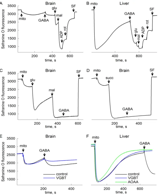
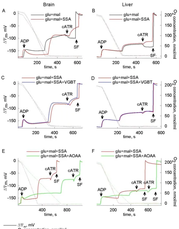
4.1. Catabolism of GABA, succinic semialdehyde or γ-hydroxybutyrate through the GABA shunt impairs mitochondrial substrate-level phosphorylation ... 35
4.1.1. GABA as a bioenergetic substrate ... 35
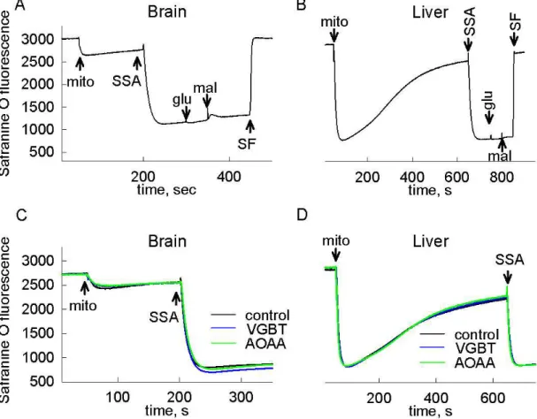
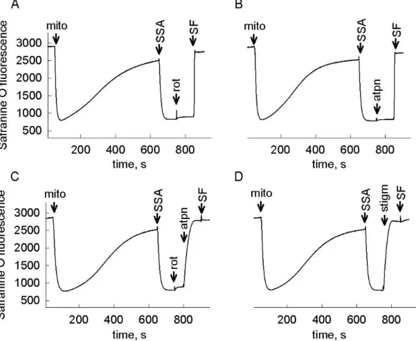
4.1.2. Succinic semialdehyde as a bioenergetic substrate ... 37
4.1.3. γ-Hydroxybutyrate as a bioenergetic substrate ... 41
4.1.4. Investigation of mitochondrial substrate-level phosphorylation ... 43
4.1.5. GABA abolishes mitochondrial SLP in anoxia ... 44
4.1.6. Succinic semialdehyde abolishes mitochondrial SLP in anoxia ... 46
4.1.7. γ-Hydroxybutyrate abolishes mitochondrial SLP in anoxia ... 48
4.2. Contribution of Nqo1 to NAD+ provision and mitochondrial SLP using endogenous or exogenous quinones ... 50
4.2.1. Determination of NAD(P)H oxidation and quinone reduction capacity in cytosolic extracts and permeabilized mitochondria from the livers of WT and Nqo1–/– mice ... 50
4.2.2. Effect of quinones on NADH oxidation capacity of permeabilized liver mitochondria from WT and Nqo1–/– mice ... 55
4.2.3. Effect of quinones on respiratory capacity of intact mitochondria from WT and Nqo1–/– mice ... 55
4.2.4. Effect of targeted ETC inhibition on quinone-assisted respiration in intact mitochondria of WT and Nqo1–/– mice ... 57
3
4.2.5. The contribution of mitochondrial Nqo1 to quinone-induced gain in ∆Ψm in
rotenone-treated mitochondria ... 60
4.2.6. Substrate-level phosphorylation supported by non-Nqo1 dicoumarol- sensitive mitochondrial diaphorases using endogenous quinones ... 61
4.2.7. MNQ supports mitochondrial SLP preferably through Nqo1 ... 63
4.2.8. Investigating the contribution of diaphorases including NQO1 to mitochondrial SLP in HepG2 cells ... 66
5. DISCUSSION ... 70
6. CONCLUSION ... 79
7. SUMMARY ... 80
8. ÖSSZEFOGLALÁS ... 81
9. BIBLIOGRAPHY ... 82
10. BIBLIOGRAPHY OF THE CANDIDATE’S PUBLICATIONS ... 106
10.1. Publications related to the PhD thesis ... 106
10.2. Publications not related to the PhD thesis ... 107
11. ACKNOWLEDGEMENTS ... 108
4 THE LIST OF ABBREVIATIONS
ADP adenosine 5'-diphosphate ANT adenine nucleotide translocase
AOAA aminooxyacetic acid (GABA-T inhibitor) A-SUCL ADP-forming succinate-CoA ligase (human) ATP adenosine 5'-triphosphate
ATPin/ADPin intramitochondrial ATP/ADP ratio
atpn atpenin A5 (succinate dehydrogenase inhibitor) cATR carboxyatractyloside (ANT inhibitor)
CHR chrysin (diaphorase inhibitor)
CN potassium cyanide
CoASH coenzyme A
CoQ1 coenzyme Q1 = ubiquinone 5
CoQ10 coenzyme Q10 = ubiquinone = ubiquinone 50 CoQ9 coenzyme Q9 = ubiquinone 45
D2HGDH D-2-hydroxyglutarate dehydrogenase DCPIP 2,6-dichlorophenol-indophenol DHODH dihydroorotate dehydrogenase DIC dicoumarol (diaphorase inhibitor) DLD dihydrolipoil dehydrogenase
dOH-F dihydroxyflavone (diaphorase inhibitor)
DQ duroquinone = 2,3,5,6-Tetramethyl-1,4-benzoquinone
e-D electron donor
e-D(red) or e-D(ox) electron donor in the reduced or oxidized state, respectively Erev_ANT reversal potential of the adenine nucleotide translocase Erev_ATPase reversal potential of the Fo-F1 ATP synthase
ETC electron transport chain
ETF electron-transferring flavoprotein
ETFDH electron-transferring flavoprotein dehydrogenase FAD flavin adenine dinucleotide (oxidized form) FADH2 flavin adenine dinucleotide (reduced form)
GABA γ-aminobutyrate
5
GABA-T γ-aminobutyrate aminotransferase = GABA transaminase GAD glutamate decarboxylase
GAD(65 or 67) glutamate decarboxylase, 65 or 67 kDa isoform GDP guanosine 5'-diphosphate
GHB γ-hydroxybutyrate
glu glutamate
GLUD glutamate dehydrogenase
GOT2 mitochondrial aspartate aminotransferase = glutamate- oxaloacetate transaminase 2
GPDH glycerol-3-phosphate dehydrogenase
G-SUCL GDP-forming succinate-CoA ligase (human) GTP guanosine 5'-triphosphate
HOT hydroxyacid-oxoacid transhydrogenase
IDB idebenone = 2-(10-hydroxydecyl)-5,6-dimethoxy-3- methylcyclohexa-2,5-diene-1,4-dione
IDH isocitrate dehydrogenase
KGDHC α-ketoglutarate dehydrogenase complex
mal malate
MCT monocarboxylate transporter
MCT(2 or 4) monocarboxylate transporter, isoform 2 or 4, respectively
mito mitochondria
mitoQ mitoquinone = [10-(4,5-Dimethoxy-2-methyl-3,6-dioxo-1,4- cyclohexadien-1-yl)decyl](triphenyl)phosphonium
mln malonate (succinate dehydrogenase inhibitor)
MND menadione = vitamin K3 = 2-methyl-1,4-naphtoquinone MNQ 2-methoxy-1,4-naphtoquinone
mtDNA mitochondrial DNA
NAD(P)+ nicotinamide adenine dinucleotide (phosphate), oxidized form NAD(P)H nicotinamide adenine dinucleotide (phosphate), reduced form NQO1 or Nqo1 NAD(P)H quinone oxidoreductase 1 (human or mouse) Nqo2 NAD(P)H quinone oxidoreductase 1 (mouse)
olgm oligomycin (Fo-F1 ATP synthase inhibitor)
6
PEPCK phosphoenolpyruvate carboxykinase PHND phenindione (diaphorase inhibitor)
Pi inorganic phosphate
Q lipophilic quinone
Q’ hydrophilic quinone
QH2 lipophilic quinol
QH2’ hydrophilic quinol RCR respiratory control ratio RET reverse electron transport ROS reactive oxygen species rot rotenone (complex I inhibitor) SDH succinate dehydrogenase
SF SF6847 = 3,5-di-tert-butyl-4-hydroxybenzylidenemalononitrile (uncoupler)
shRNA short hairpin RNA
siRNA small interfering RNA
SLP substrate-level phosphorylation
SSA succinic semialdehyde
SSADH succinic semialdehyde dehydrogenase SSAR succinic semialdehyde reductase stigm stigmatellin (complex III inhibitor)
succ succinate
SUCL succinate-CoA ligase = succinyl-CoA synthetase = succinate thiokinase (human)
SUCLA2 or Sucla2 ADP-forming succinate-CoA ligase β subunit (human or mouse) Suclg2 GDP-forming succinate-CoA ligase β subunit (mouse)
VGBT vigabatrin = γ-vinyl-GABA (GABA-T inhibitor)
WT wild type
α-KG α-ketoglutarate β-OH β-hydroxybutyrate
∆Ψm mitochondrial membrane potential
7 1. INTRODUCTION
Mitochondria, double-membrane-bound organelles found in the most eukaryotic cells, form a dynamic network with frequent changes in their number and morphology.
Their ubiquitous presence was first described by Altmann in 1890 and since then extensive literature has accumulated regarding their structure and operation [1].
Mitochondria are usually brought in context as the sites of cellular respiration since they consume oxygen for the aerobic metabolism of nutrients. Beside energy provision, mitochondria exhibit an important role in several other cell functions, such as heat production, regulation of Ca2+ homeostasis, or production of intracellular reactive oxygen species (ROS) as signaling molecules. They are also involved in the synthesis of steroid hormones, heme, Fe-S clusters, lipids and pyrimidine nucleotides. In addition to their role of maintaining these vital processes, it has turned out that they are key components in cell death. Another unique feature of these organelles is that they possess their own DNA (mtDNA). This encodes however 13 proteins only, all of which are components of the oxidative phosphorylation. The majority of mitochondrial proteins is thus nuclear-encoded and has to be actively imported into the mitochondrial compartments, followed by assembly into functional macromolecular complexes.
Mindful of their diverse roles, it is not surprising that the dysfunction of mitochondria has been described to participate in a number of pathological conditions.
Mutations in mtDNA or in genes of nuclear-encoded mitochondrial proteins lead to a heterogeneous group of – usually severe and incurable – diseases. The contribution of mitochondria to the pathomechanism of neurodegenerative diseases, cancer, ischaemia- reperfusion injury, obesity, metabolic syndrome, and aging rendered these organelles an important subject of research [2; 3].
ATP generation by mitochondria is a key component in determining the outcome of the above mentioned diseases. In case the condition is associated with an impaired respiration of these organelles, oxygen-independent energy-producing pathways such as mitochondrial substrate-level phosphorylation (SLP) become of particular importance.
The topic of the present thesis concerns the bioenergetic aspects of mitochondrial metabolism when the respiratory chain is dysfunctional, focusing on the metabolic pathways influencing SLP.
8
1.1. Oxidative phosphorylation and mitochondrial respiration
Catabolic pathways converge at mitochondria, where the final phase of fuel degradation, the oxidative phosphorylation takes place. Carbohydrates, amino acids and fatty acids are eventually oxidized in the citric acid cycle in the mitochondrial matrix.
Electrons arising from the action of dehydrogenases are transferred to the complexes of the inner mitochondrial membrane by universal electron carriers, NAD+ or FAD. The membrane-embedded four complexes of the respiratory chain allow the flow of electrons to their final electron acceptor, molecular oxygen. Electrons carried in the form of NADH enter the respiratory chain at the level of complex I, whereas those coming from succinate oxidation are transferred to the FAD prosthetic group of complex II (succinate dehydrogenase, SDH). Both complexes reduce a mobile electron carrier, ubiquinone to ubiquinol. Ubiquinol can also be produced by the electron- transferring flavoprotein (ETF) system, or by glycerol-3-phosphate dehydrogenase (GPDH) [4], or in the reaction catalyzed by dihydroorotate dehydrogenase (DHODH) [5]. The first one represents a pathway in fatty acid oxidation, in which electrons from fatty acyl-CoA are transferred to ETF and from there they are passed to ubiquinone via electron-transferring flavoprotein dehydrogenase (ETFDH). GPDH converts glycerol-3- phosphate to dihydroxyacetone phosphate, and DHODH is involved in de novo pyrimidine biosynthesis, oxidizing dihydroorotate into orotate – both reactions are coupled to the reduction of ubiquinone [4; 5]. Ubiquinol coming from either source donates electrons to complex III, which is then reoxidized by an intermembrane space protein, cytochrome c. This moves to complex IV, where electrons are ultimately passed to molecular oxygen. The utilization of substrates is connected to oxygen consumption this way, which is the basis of cellular respiration [4].
After years of searching for the mechanism by which mitochondria link nutrient oxidation to ATP production, the chemiosmotic theory was introduced by Mitchell in 1961 [6] and later confirmed experimentally by several laboratories [1]. The key aspect of this theory is that the respiratory chain complexes possess proton pump activity, and the free energy change arising from the electron transfer allows the translocation of protons from the matrix into the intermembrane space. Due to the proton impermeability of the inner membrane this creates an electrochemical gradient, termed the proton-motive force. This gradient, composed of a chemical (∆pH) and an electrical
9
potential energy (mitochondrial membrane potential, ∆Ψm), drives the synthesis of ATP as protons flow back into the matrix through the Fo-F1 ATP synthase complex [4].
The Fo-F1 ATP synthase consists of a membrane-embedded Fo part, through which protons can flow back into the matrix, and an extramembraneous F1 sector, which is responsible for ATP production. The enzyme establishes ATP synthesis through a rotational catalysis, with the c-ring of the Fo moiety rotating in a clockwise direction (as viewed from the membrane) at about 100 times per second. The structure of the mammalian c-ring has been determined recently and lead to the recognition, that the translocation of 2.7 protons is required to generate one ATP molecule [7].
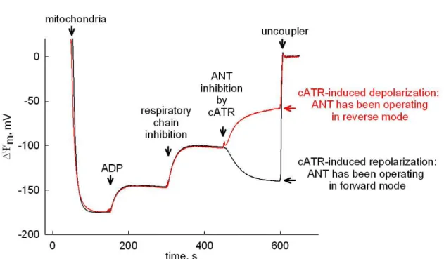
ATP synthesized in the matrix is transported to the cytosol by an integral protein of the inner membrane, the adenine nucleotide translocase (ANT). The enzyme exports ATP in exchange for ADP with a 1:1 stoichiometry. Since ATP has one more negative charge than ADP, the transport is electrogenic and is driven by the membrane potential [8; 9]. ANT is the most abundant protein in the inner mitochondrial membrane, representing approximately 10% of the total protein content. Until now four ANT isoforms have been identified in mammals, with a sequence homology between 80 and 90% and with a different tissue-specific expression pattern. Beside the exchange of ATP and ADP, the role of the different isoforms in apoptosis and cancer progression has been described as well [10]. ANT is known to be a modulatory element of the mitochondrial permeability transition pore [11] – a pore in the inner mitochondrial membrane the opening of which is triggered by Ca2+ and possibly leads to cell death –, and ANT1 has been recently identified as the voltage sensor of this phenomenon [12].
Unlike the Fo-F1 ATP synthase which utilizes Mg2+-complexes, the substrates of ANT are the free adenine nucleotides only [9; 13].
In intact mitochondria, ATP synthesis and oxygen consumption are obligatory coupled: in the absence of ADP respiration is impaired, and vice versa, the lack of oxygen prevents phosphorylation. Respiratory steady states in experiments using isolated mitochondria were defined by Chance and Williams in 1955 [14]. According to this terminology, state 1 refers to the presence of mitochondria in a phosphate- containing medium which lacks substrate and phosphate acceptor. State 2 is achieved by the addition of ADP, under these conditions the rate limiting component of respiration is the concentration of endogenous substrates. Provision of exogenous substrates results
10
in the acceleration of oxygen consumption, this is the state of oxidative phosphorylation termed state 3. After the exhaustion of ADP respiration slows down and state 4 is reached, and the consumption of all the dissolved oxygen (in a closed chamber) leads to state 5, the state of anoxia. This nomenclature has been modified later, based on a different sequence of additions in the experimental protocol [15; 16]. In the new system state 2 indicates the condition after the addition of respiratory substrates, followed by state 3 where ADP is present as well. The definition of state 1, 3, 4 and 5 remained basically unchanged. Oxygen consumption in state 2 - when exogenous substrates are provided but the phosphate acceptor is absent - reflects the proton conductance of the inner membrane (proton leak): some protons can return to the matrix through alternative pathways compared to the Fo-F1 ATP synthase. ANT was shown to be the main contributor to this basal proton leak [17].
These well-defined respiratory states cannot be applied directly for mitochondria under physiological conditions. Little is known about the rate of oxygen consumption of mitochondria in vivo, but it is thought that due to the physiological ATP turnover, mitochondria in the cell exist in an intermediate state between state 3 and 4 [18].
1.2. Reversibility of the Fo-F1 ATP synthase and the ANT
The Fo-F1 ATP synthase can not only synthesize ATP but is also capable of hydrolyzing it [19]. Since in the presence of high concentrations of inorganic phosphate (Pi) ∆pH across the mitochondrial membrane is small [20], the direction of the enzyme complex is mainly regulated by the ∆Ψm component of the proton-motive force. When this parameter moves towards more positive values, the Fo-F1 ATP synthase can reverse and pump protons out of the mitochondrial matrix in order to maintain a suboptimal membrane potential. The ∆Ψm value where the enzyme complex switches from ATP producing to ATP hydrolyzing mode is termed reversal potential (Erev_ATPase). The reversal potential of the Fo-F1 ATP synthase complex can be calculated using the following equation [21]:
11
E 316
n 2.3RT
F
n x log ATP free$% x K'()*+, ADP. free$% x K'()/+, x P$ $%
2.3 RT
F x (pH234 pH$%, and
P$ $% P$ 42456 $%
1 7 109:;<9=>
where in and out signify inside and outside of the mitochondrial matrix, n is the H+/ATP coupling ratio [7], R is the universal gas constant (8.31 J . mol–1. K–1), F is the Faraday constant (9.64 x 10⁴ C . mol–1), T is temperature (in degrees Kelvin), KM(ADP) and KM(ATP) are the true affinity constants of Mg2+ for ADP and ATP valued 10–3.198 and 10–4.06, respectively [22], and [Pi] is the free phosphate concentration (in molar) given by the second equation, where pKa2 is 7.2 for phosphoric acid.
During the reverse operation of the enzyme, the ATP that is hydrolyzed can originate either from intramitochondrial resources or from the cytosol. The latter requires the reversal of the ANT, so that the transporter will import extramitochondrial ATP and export ADP generated in the matrix. The reversal potential of the ANT (Erev_ANT) can be defined according to the following equation [21]:
E_)@/ 2.3 RT
F x logADP. free234 x ATP free$%
ADP. free$% x ATP free234
where the same symbols are used as in the case of Erev_ATPase.
In summary, when the oxidative phosphorylation is impaired, mitochondrial membrane potential can decrease below the reversal potential values, which leads to a reverse operating ANT and Fo-F1 ATP synthase. The reversal of the two enzymes serves the purpose of preventing further depolarization and a subsequent opening of the permeability transition pore. The glycolytic ATP being imported by the ANT will be hydrolyzed by the Fo-F1 ATP synthase while protons are translocated to the intermembrane space. In reverse mode, also the electrogenic nature of the ANT contributes to the maintenance of the potential difference across the inner membrane [23].
1.3. The B space of mitochondrial phosphorylation As the intramitochondrial ATP/ADP ratio in the two equations above,
function of ATPin/ADPin. computational estimation for
Figure 1. Computational estimation of
[ATP]in/[ADP]in ratios. A: ATPase forward, ANT forward; B: ATPase reverse, ANT forward; C: ATPase reverse, ANT reverse; D: ATPase forward, ANT reverse. Black triangles represent Erev_ATPase
for [ATP]out = 1.2 mM, [ADP]
pHout = 7.25. (Adapted from reference
The two curves define four areas in the graph:
an ATPin/ADPin which places them in the A space ANT working in forward mode,
Mitochondria with low membrane potential are found in F1 ATP synthase and ANT.
ATP. However, the directionality of the F
necessarily change simultaneously. In the B space, potential value more positive than the E
indicating that the Fo-F1 ATP synthase hydrolyzes ATP but the ANT still operates in ATP-exporting direction. Finally,
but the ANT also increases
12
of mitochondrial phosphorylation
As the intramitochondrial ATP/ADP ratio (ATPin/ADPin) is a common parameter , Erev_ATPase and Erev_ANT can be represented in one figure as a . Based on this, Fig. 1 from reference
for the reversal potential values of the two enzymes.
Computational estimation of Erev_ANT and Erev_ATPase at different free A: ATPase forward, ANT forward; B: ATPase reverse, ANT forward; C: ATPase reverse, ANT reverse; D: ATPase forward, ANT reverse. Black
rev_ATPase; white triangles represent Erev_ANT. Values were computed
= 1.2 mM, [ADP]out = 10 µM, [Pi]in = 0.02 M, n = 3.9, pH
= 7.25. (Adapted from reference [21])
curves define four areas in the graph: mitochondria with a
which places them in the A space have the Fo-F1 ATP synthase and the ANT working in forward mode, which implies that they produce and export ATP.
with low membrane potential are found in the C space, with ATP synthase and ANT. In this case the organelles import and hydrolyze
the directionality of the Fo-F1 ATP synthase and ANT do not simultaneously. In the B space, mitochondria have a membrane
positive than the Erev_ATPase but more negative than the E
ATP synthase hydrolyzes ATP but the ANT still operates in Finally, in the D space the Fo-F1 ATP synthase produces ATP, increases ATPin/ADPin by importing ATP from outside.
is a common parameter can be represented in one figure as a [21] shows a the reversal potential values of the two enzymes.
at different free A: ATPase forward, ANT forward; B: ATPase reverse, ANT forward; C: ATPase reverse, ANT reverse; D: ATPase forward, ANT reverse. Black . Values were computed 3.9, pHin = 7.38, and
mitochondria with a ∆Ψm value and ATP synthase and the they produce and export ATP.
with reversed Fo- and hydrolyze cytosolic ATP synthase and ANT do not mitochondria have a membrane than the Erev_ANT, ATP synthase hydrolyzes ATP but the ANT still operates in ATP synthase produces ATP, from outside. Nevertheless,
13
this region is characterized by a high membrane potential and a low matrical ATP/ADP ratio, which is very unlikely to occur under natural conditions, thus probably this area has no biological relevance. This also implies that in living cells, the Erev_ATPase is more negative than the Erev_ANT [23].
It is important to stress that from the reversal potential values only the directionality of the two enzymes can be concluded, the figure provides no information of the actual enzyme activities. Experiments on isolated [24; 25] and in situ [26]
mitochondria showed that during depolarization, the ADP-ATP exchange rate of the ANT decreases as the membrane potential is approaching Erev_ANT. At the reversal potential there is no net transport of adenine nucleotides across the inner mitochondrial membrane, whereas after passing this value, the transport of substrates in the opposite direction will start. The same scenario is assumed for the Fo-F1 ATP synthase [23].
The B space of the figure is of special interest, since in such case, despite a substantial depolarization, mitochondria do not consume cytosolic ATP for the maintenance of their membrane potential. This phenomenon could have significance under many pathological conditions, where generation of the proton-motive force is damaged. Neurodegenerative diseases are usually associated with different respiratory complex deficiencies, for example a defect in complex I activity is reported in patients with Parkinson’s disease [27]. In Alzheimer’s disease primarily complex IV was shown to have diminished activity [28]. In addition, many rare mitochondrial diseases are known where due to mtDNA or mitochondrial protein encoding nuclear gene abnormalities the assembly of the respiratory complexes is damaged [29], thus a loss of membrane potential and reversal of the Fo-F1 ATP synthase is expected. Another relevant pathological condition is ischaemic-reperfusion injury where mitochondrial membrane potential is diminished due to limited oxygen availability [30]. In either case, mitochondrial ∆Ψm generation is compromised, and if the organelles get into the C space, they will start consuming cytosolic ATP. A decrease in the cellular energy pool can lead to a damage of essential cell functions, like to the hampered operation of ATP- dependent pumps such as the Na+/K+-ATPase and Ca2+-ATPase. Therefore it can be critical for mitochondria to stay within the boundaries of the B space, so that cytosolic ATP pools will be preserved and the chances of the cell for survival will be improved [23; 31].
14
1.4. Mitochondrial substrate-level phosphorylation
Mitochondria with parameters located in the B space do not cease to export ATP despite a reverse working Fo-F1 ATP synthase. This implies that the ATP, which is hydrolyzed and exported simultaneously, comes from intramitochondrial sources other than the oxidative phosphorylation. Reactions capable of producing high energy phosphates independent of the respiratory chain are termed substrate-level phosphorylation. ATP originating from mitochondrial SLP can be crucial to keep mitochondria in the B space when their respiratory chain is dysfunctional [21].
Sufficient ATP production by SLP can prevent mitochondria from entering the C space in three ways: i) by providing ATP for the reverse operating Fo-F1 ATP synthase thereby maintaining a moderate ∆Ψm ii) an increase in ATPin/ADPin will translocate mitochondria upwards along the y axis in Fig. 1 making them more likely to stay in the B space, iii) it will affect the reversal potential values of both the Fo-F1 ATP synthase and the ANT. Based on equations 1 and 2, Erev_ATPase will be shifted towards more negative and Erev_ANT towards more positive values, leading to a broadening of the B space [23]. Thus a functional SLP can prevent mitochondria from ANT reversal and a subsequent consumption of cytosolic ATP.
In mitochondria, phosphoenolpyruvate carboxykinase (PEPCK) and the succinate- CoA ligase (SUCL, or succinate thiokinase or succinate-CoA ligase) are able to perform SLP. Mitochondrial PEPCK participates in the transfer of the phosphorylation potential from the matrix to the cytosol and vice versa [32]. SUCL catalyzes a reversible reaction in the citric acid cycle, it converts succinyl-CoA, ADP (or GDP) and Pi to succinate, ATP (or GTP) and CoASH [33]. The enzyme is a heterodimer, being composed of an invariant α subunit encoded by SUCLG1 and a substrate-specific β subunit encoded by either SUCLA2 or SUCLG2. This dimer combination results in either an ATP-forming succinate-CoA ligase (A-SUCL; EC 6.2.1.5) or a GTP-forming succinate-CoA ligase (G-SUCL; EC 6.2.1.4). The GTP-forming enzyme occurs mainly in anabolic tissues like liver and kidney, whereas A-SUCL can be found first of all in brain, heart and muscle [32; 33]. It was shown in human brain samples, that the ATP-forming β subunit is expressed exclusively in neurons of the cerebral cortex [34]. Moreover, in glial cells none of the two enzyme forms was found, suggesting that in these cells an alternative pathway bypasses this step of the citric acid cycle [35]. SUCL exists in the matrix in
15
association with a mitochondrial nucleoside diphosphate kinase known as NM23-H4 (NDPK-D, EC 2.7.4.6), an enzyme capable of the interconversion of nucleoside triphosphates, thus GTP produced by G-SUCL is also able to contribute to the matrix ATP pool [36; 37].
Growing evidence confirms that the adequate operation of SUCL is necessary to prevent ANT reversal when mitochondrial respiration is inhibited. It is known that the citric acid cycle is operational in anoxia, with succinate as an end product [38]. Also, the fact that anaerobic mitochondrial metabolism can generate ATP via SLP and maintain mitochondrial energization this way has been substantiated extensively [38].
The α-ketoglutarate dehydrogenase complex (KGDHC) provides succinyl-CoA for SUCL, and this is considered to be the rate-limiting step among the sequential reactions [39]. Experiments performed on mitochondria isolated from mice heterozygote for different subunits of KGDHC showed that the enzyme complex is essential for the operation of SUCL, and that impaired function of KGDHC results in ANT reversal during respiratory failure. This was supported by measurements performed on in situ neuronal somal mitochondria as well [40]. The role of SUCL itself was investigated in [41], in which Sucla2+/–, Suclg2+/–, or Sucla2+/–/Suclg2+/– mice were generated and characterized extensively from a bioenergetic point of view. Mitochondrial respiration and electron transport chain (ETC) activities showed no alterations in the transgenic animals (except for a significant elevation of complex II activity in the double heterozygote samples). Decreased mtDNA content and alterations in the concentration of blood carnitine esters were found in Sucla2+/– and in Sucla2+/–/Suclg2+/– mice.
Regarding mitochondrial SLP, no difference was observed in the heterozygote models compared to wild type (WT) littermates. This is either due to i) a rebound increase in Suclg2 expression and associated GTP-forming activity as it was found in Sucla2+/– and Sucla2+/–/Suclg2+/– mice; or ii) the small flux control coefficient of SUCL (compared to that of the KGDHC) which makes it possible that even a decreased enzyme activity is able to maintain sufficient ATP production for the ANT to work in forward mode.
Nevertheless, submaximal pharmacological inhibition of SUCL in the heterozygote samples lead to an abolition of SLP. The importance of SUCL in preventing ANT reversal has been strengthened in fibroblasts from patients suffering from SUCLA2 deficiency as well. These cells are unable to perform sufficient SLP, and start to import
16
cytosolic ATP when their respiratory chain is inhibited [41]. Another finding which confirms the significance of this reaction is that inorganic phosphate activates SUCL [42]. The Pi produced by a reverse working Fo-F1 ATP synthase in anoxia results in a greater activity of this rescue mechanism.
Mindful of these findings, enhancing SUCL function by succinyl-CoA provision could be helpful in diseases mentioned in the previous chapter. The practical relevance of this notion is confirmed by publications where substrates supporting mitochondrial SLP improved the outcome of anoxic or ischaemic conditions (as summarized in [38]).
In addition, recently methylene blue, a drug which shows beneficial effects in stroke and neurodegenerative disease models, was found to enhance mitochondrial SLP [43].
1.5. Metabolic pathways affecting mitochondrial substrate-level phosphorylation Regarding the role of SLP in preventing ANT reversal during respiratory arrest, it is worth considering the factors that could influence its operation. The succinyl-CoA – succinate interconversion mediated by SUCL is reversible, (∆G=0.07 kJ/mol) [44].
Therefore the availability of the substrates will largely determine the direction of the reaction, and any metabolic pathway influencing the concentration of the participants will predictably impact SLP.
First of all, provision of succinyl-CoA is crucial for keeping the reaction in ATP- producing direction [40]. A possible source of succinyl-CoA is the reaction mediated by KGDHC, which catalyzes the irreversible conversion of α-ketoglutarate, CoASH, and NAD+ to succinyl-CoA, NADH, and CO2 in the citric acid cycle. In experiments using isolated mitochondria, α-ketoglutarate and glutamate are the two substrates that support SLP to the greatest extent, especially when provided together with malate, which assists in their entry into mitochondria. It is obvious though, that for the operation of KGDHC oxidized NAD+ is needed, the availability of which is expected to be markedly decreased in anoxia or during complex I inhibition. Despite this, experiments showing that there is substantial SLP under these conditions indicate that sufficient NAD+ is generated for KGDHC [21; 40; 45]. The origin of this NAD+ in anoxia is assumed to be the mitochondrial diaphorases [45].
Other sources for succinyl-CoA can be the catabolism of certain biomolecules (methionine, threonine, isoleucine, valine, propionate, odd chain fatty acids and
17
cholesterol), the degradation of which converges to succinyl-CoA as an entry point into the citric acid cycle [46].
By the same token, any metabolic pathway which consumes succinyl-CoA can hamper SLP. When SUCL proceeds in the direction towards succinyl-CoA formation, this can take part in δ-aminolevulinate generation, a step of heme synthesis [47]. Ketone body catabolism requires succinyl-CoA as a CoASH donor for acetoacetyl-CoA formation, with succinate staying behind, bypassing the reaction by SUCL this way [48]. These reactions possibly steal succinyl-CoA away from the high-energy phosphate producing step.
In addition, succinate accumulation can shift the reaction mediated by SUCL into the ATP (GTP) hydrolyzing direction. Addition of succinate resulted in ANT reversal in isolated mitochondria when the electron transport chain was inhibited distal from complex II, reflecting an impairment of SLP [21]. It has been recently shown, that itaconate, an antimicrobial compound produced by macrophages upon lipopolysaccharide stimulation, abolishes mitochondrial SLP [49]. This was attributed to three different effects: i) itaconate is metabolized through thioesterification by SUCL, and this step requires ATP (or GTP); ii) the product of its metabolism, itaconyl-CoA traps CoASH from the KGDHC; iii) itaconate inhibits SDH, leading to a buildup of succinate. Another possible metabolic pathway which can lead to succinate accumulation in respiratory-inhibited mitochondria is the catabolism of the neurotransmitter γ-aminobutyrate (GABA) through the so-called GABA shunt [50; 51].
Inhibitory effect of GABA on SLP has been reported in a study [52], where incubation with GABA appreciably reduced ATP and GTP production in uncoupled rat brain mitochondria. However, the mechanism of inhibition was not established. γ- Hydroxybutyrate (GHB), a neurotransmitter and a psychoactive drug is also converted to succinate during its degradation [53], and possibly exerts similar effects on SLP.
The present thesis focuses on i) the effect of GABA and GHB metabolism on mitochondrial SLP; ii) the contribution of a diaphorase enzyme, NAD(P)H quinone oxidoreductase 1 (Nqo1) to SLP. Therefore, in the next chapters I will give a brief overview about mitochondrial diaphorases, GABA, the GABA shunt, and GHB.
18 1.6. γ-Aminobutyrate (GABA)
4-Aminobutyrate, also known as 4-aminobutanoate, γ-aminobutyrate or more frequently, GABA, is most widely known as the predominant inhibitory neurotransmitter in the adult brain [54]. Since its discovery in the central nervous system [55-57], GABA has been increasingly recognized to participate in processes other than neurotransmission as it is present in many organs other than the brain, such as pancreas, testes, gastrointestinal tract, ovaries, placenta, uterus and adrenal medulla [58; 59]. Most notably though, very high concentrations of GABA have been found in the livers of all animal species reported, particularly humans [60].
GABA exerts an inhibitory effect on synaptic transmission by interacting with ionotropic receptors on the postsynaptic membrane, resulting in an increased chloride conductance, thus an inward chloride current and a consequent hyperpolarization [61].
These channels are termed GABAA receptors. Based on different pharmacological properties previously another GABA-sensitive anion channel type was distinguished, designated as GABAC receptors, but later these were classified rather as a subfamily of GABAA receptors and the use of the term GABAC receptor is not recommended any more [62]. GABA can cause hyperpolarization of neurons and a diminished neurotransmitter release by acting on metabotropic GABAB receptors as well [63].
However, in neonatal hippocampal neurons the electrochemical gradient of chloride is outward directed, therefore, opening the receptor channels is associated with depolarization; hence, at this developmental stage, GABA is an excitatory neurotransmitter [64]. It is also worth mentioning that in the brain GABA has been further branded as a gliotransmitter [65; 66]. However, the concept of GABA as gliotransmitter has been met with skepticism from those asserting that many of the phenomena attributed to release of transmitters by the glia can be explained by changes in the activity or expression of astrocytic membrane transporters, reviewed in [67].
GABA can be found in numerous tissues outside the central nervous system.
Regarding the liver, it was hypothesized that this organ is responsible for clearing GABA from the systemic circulation. GABA is only catabolized but very little synthesized in the liver, and it originates from the intestinal flora [60] finding its way through the portal system; however, hepatic lobular GABA synthesis increases >300%
19
following partial hepatectomy [60]. Relevant to this, the high GABA concentration in liver has been implicated in the pathophysiology of hepatic encephalopathy [68].
In pancreas, GABA acts as an intra-islet transmitter regulating hormone release.
In α-cells, GABA induces membrane hyperpolarization and suppresses glucagon secretion, whereas in islet β-cells it induces membrane depolarization and increases insulin secretion [69]. This difference in the effect of GABA on membrane polarization is due to the different expression of cation-Cl−-cotransporters in islet α and β-cells [70], which leads to an opposite electrochemical driving force of chloride ion in the two cell types. In the gastrointestinal tract, GABA is involved in the regulation of gut motility [71], in the adrenal medulla it is thought to play a role in modulating the release of catecholamines [72], and it may have an important role in reproductive function [73; 74]
Furthermore, GABA is released by immune cells and has a number of immunomodulatory effect [75], acts as a developmental signal during brain organogenesis [76], and even inhibits mitophagy and pexophagy in mammalian cells of various tissues, in an mTOR-sensitive manner [77]. Finally, GABA's realm has been recently recognized to extent to plantae, playing a vital role as a plant-signaling molecule [78].
Altered GABA concentration and signaling plays a role in a number of pathological conditions such as epilepsy, Parkinson’s and Alzheimer’s disease, depression, anxiety, schizophrenia and panic disorders [79; 80]. It is not surprising therefore, that the GABAergic system is a center of interest as a pharmacological target [80].
1.7. The GABA shunt
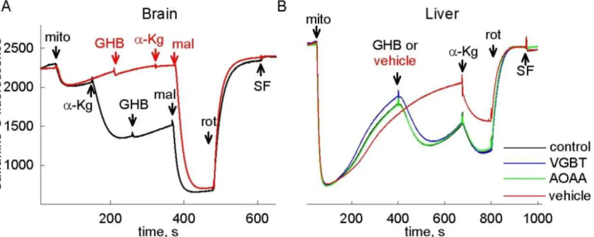
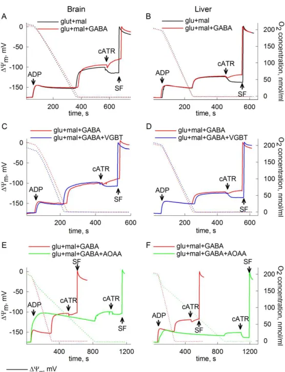
Despite that the participation of GABA in diverse biological processes implies different downstream effectors responsive to this molecule, its metabolism is rather uniform among all tissues: GABA is metabolized through the ‘GABA shunt’
(represented in Fig. 2), a pathway representing an alternative route for converting α- ketoglutarate to succinate in the citric acid cycle circumventing succinate-CoA ligase [50; 51]. Enzymes of the GABA shunt are expressed not only in the brain but also in a variety of nonneural tissues [81; 82].
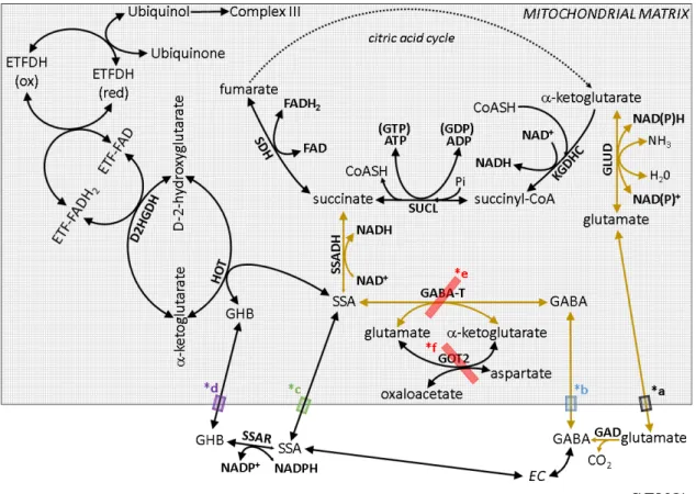
Figure 2. The GABA shunt (outlined by arrows in gold color) and pertinent reactions. SUCL: succinate
complex; GLUD: glutamate dehydrogenase; GAD: glutamate decarboxylase; GABA:
aminobutyrate; GABA-T: γ
aspartate aminotransferase; SSAR: s
hydroxybutyrate; SSA: succinic semialdehyde; SSADH: succinic semialdehyde dehydrogenase; HOT: hydroxyacid
hydroxyglutarate dehydrogenase; ETF: electron electron-transferring flavoprotein dehydrogenase
glutamate transporters; *b: putative mitochondrial GABA transporter; *c: putative mitochondrial SSA transporter; *d: putative mitochondrial GHB transporter; *e:
inhibitors for GABA-T used in this study: vigabatrin and this study: AOAA.
As shown in Fig. 2, GABA can be derived from glutamate by glutamate decarboxylase (GAD), encoded by either
different molecular weights (65 and 67 kDa) and
GAD65 found primarily in axon terminals and GAD67 more widely distributed in neurons [83]. Since GAD is a cytosolic enzyme, glutamate needs to be exported from mitochondria; this may occur through well
black semi-transparent box *a), as an electroneutral transport driven by
20
The GABA shunt (outlined by arrows in gold color) and pertinent SUCL: succinate-CoA ligase; KGDHC: α-ketoglutarate dehydrogenase complex; GLUD: glutamate dehydrogenase; GAD: glutamate decarboxylase; GABA:
T: γ-aminobutyrate aminotransferase; GOT2: mitochondrial aspartate aminotransferase; SSAR: succinic semialdehyde reductase; GHB:
hydroxybutyrate; SSA: succinic semialdehyde; SSADH: succinic semialdehyde dehydrogenase; HOT: hydroxyacid-oxoacid transhydrogenase; D2HGDH: D hydroxyglutarate dehydrogenase; ETF: electron-transferring flavoprotein;
flavoprotein dehydrogenase; SDH: succinate dehydrogenase. *a:
glutamate transporters; *b: putative mitochondrial GABA transporter; *c: putative mitochondrial SSA transporter; *d: putative mitochondrial GHB transporter; *e:
T used in this study: vigabatrin and f; *f: inhibitor for GOT2 in
As shown in Fig. 2, GABA can be derived from glutamate by glutamate decarboxylase (GAD), encoded by either GAD65 or GAD67. The two isoforms have molecular weights (65 and 67 kDa) and different subcellular localization, with GAD65 found primarily in axon terminals and GAD67 more widely distributed in . Since GAD is a cytosolic enzyme, glutamate needs to be exported from occur through well-characterized transporters (depicted as a transparent box *a), as an electroneutral transport driven by
The GABA shunt (outlined by arrows in gold color) and pertinent ketoglutarate dehydrogenase complex; GLUD: glutamate dehydrogenase; GAD: glutamate decarboxylase; GABA: γ-
aminobutyrate aminotransferase; GOT2: mitochondrial uccinic semialdehyde reductase; GHB: γ- hydroxybutyrate; SSA: succinic semialdehyde; SSADH: succinic semialdehyde
oxoacid transhydrogenase; D2HGDH: D-2- transferring flavoprotein; ETFDH:
; SDH: succinate dehydrogenase. *a:
glutamate transporters; *b: putative mitochondrial GABA transporter; *c: putative mitochondrial SSA transporter; *d: putative mitochondrial GHB transporter; *e:
; *f: inhibitor for GOT2 in
As shown in Fig. 2, GABA can be derived from glutamate by glutamate . The two isoforms have subcellular localization, with GAD65 found primarily in axon terminals and GAD67 more widely distributed in . Since GAD is a cytosolic enzyme, glutamate needs to be exported from characterized transporters (depicted as a transparent box *a), as an electroneutral transport driven by ∆pH [84; 85].
21
GABA may also arise by metabolism of putrescine [86] or homocarnosine [87] (not shown), or enter the cytoplasm from the extracellular space. In any case, in order for GABA to undergo further transamination it must first enter the mitochondrial matrix.
The transport of GABA across the inner mitochondrial membrane (depicted by a blue semi-transparent box *b) has been proposed to occur by “diffusion of a species with no net charge, at rates which are able to maintain maximum activity of the GABA shunt”
[88]. However, in plants, a mitochondrial GABA permease has been recently identified, termed AtGABP [89]. No such protein has been identified in animals, but a BLASTp homology search yielded a highly homologous (94%) predicted protein termed ‘amino acid permease BAT1 (partial)’ with a sequence ID: XP_019577258.1 expressed in Rhinolophus sinicus (Chinese rufous horseshoe bat), as well as some other proteins from other species but with low homology (below 35%). In mice and humans there is 23–31% homology of AtGABP to an ‘epithelial-stromal interaction protein 1’, and no homologous proteins were identified in tissues from rats and guinea pigs. Thus, although several isoforms of plasmalemmal GABA transporters have been identified [90], their reversibility documented [91-94], and GABA is known to permeate murine mitochondria [88] and become intramitochondrially metabolized [95], the means of GABA entry to mitochondria remains speculative.
Once in the matrix, GABA transaminates with α-ketoglutarate to form glutamate and succinic semialdehyde (SSA) by the mitochondrial GABA transaminase (GABA- T). Succinic semialdehyde will get dehydrogenated by succinic semialdehyde dehydrogenase (SSADH) yielding succinate and NADH, and thus enter the citric acid cycle. SSADH is also the enzyme responsible for further metabolism of aldehyde 4- hydroxy-2-nonenal, an intermediate known to induce oxidant stress [96]. Glutamate and α-ketoglutarate are in equilibrium with oxaloacetate and aspartate through a mitochondrial aspartate aminotransferase (GOT2). GABA-T is inhibited by vigabatrin and aminooxyacetic acid (AOAA) [88; 97]. The latter compound is also known to inhibit GOT2 as well as other pyridoxal phosphate-dependent enzymes [98; 99].
Regarding SSA, there are three possible scenarios for its appearance in the matrix:
i) from the cytosol, transported through the inner mitochondrial membrane by a protein (depicted by a green semi-transparent box *c) that is yet to be characterized [100]; ii) by the action of hydroxyacid-oxoacid transhydrogenase (HOT), encoded by ADHFE1,
22
transhydrogenating γ-hydroxybutyrate (GHB) and α-ketoglutarate to D-2- hydroxyglutarate and SSA; or iii) by succinic semialdehyde reductase (SSAR, encoded by AKR7A2), converting GHB to SSA in the cytosol [101-104] and the latter getting transported into the matrix; however, the equilibrium of the SSAR reaction is strongly favored towards GHB formation.
The first evidence regarding the existence of HOT came from experiments investigating GHB catabolism in rat brain and kidney samples [105]. The enzyme was isolated and further characterized from rat tissues [106] and later on, its gene was identified [107]. The existence of human HOT has been demonstrated in homogenates of human liver and fibroblasts as well [108; 109]. HOT is not the only enzyme interconverting D-2-hydroxyglutarate and α-ketoglutarate; D-2-hydroxyglutarate dehydrogenase (D2HGDH) localized in mitochondria [110; 111] also performs such an interconversion, but this is coupled to the ETF system, eventually donating electrons to complex III through ubiquinone. D-2-Hydroxyglutarate is formed as a degradation product of L-hydroxylysine [112] and possibly also from δ-aminolevulinate [113].
Interestingly, isocitrate dehydrogenase (IDH) 1 and 2 mutations confer a novel enzymatic activity that facilitates reduction of α-ketoglutarate to D-2-hydroxyglutarate impeding oxidative decarboxylation of isocitrate [114; 115]. The accumulation of D-2- hydroxyglutarate due to IDH mutations has been implicated in tumorigenesis [116];
however, accumulation of D-2-hydroxyglutarate in glutaric acidurias is associated with encephalopathy and cardiomyopathy, but not tumors [117]. This metabolite is known to permeate the cell membrane through a sodium-dicarboxylate cotransporter (NaDC3) and an organic anion transporter (OAT1) [118] but a mitochondrial transport mechanism is yet to be described.
The reaction catalyzed by HOT is reversible, therefore SSA produced by GABA- T in mitochondria could be converted to succinate or GHB as well, but the predominant pathway of SSA metabolism is probably oxidation by SSADH because of the significant lower Km of the enzyme for SSA, compared to HOT [81]. The rate limiting step of GABA catabolism is the reaction catalyzed by GABA-T, therefore SSA coming from GABA is rapidly oxidized by SSADH and its concentration is kept low [119; 120].
In SSADH deficiency though – a rare disease with nonprogressive encephalopathy,
23
hypotonia and delay in mental and motor development –, SSA accumulates, leading to elevated GABA and GHB levels [100].
1.8. γ-Hydroxybutyrate (GHB)
GHB is a neurotransmitter and neuromodulator in the human brain, but is also synthesized outside the central nervous system [121], however, its physiological role is not completely understood. As a therapeutical agent, GHB was originally developed for an anesthetic [122], today it is a drug for the treatment of narcolepsy with cataplexy and alcohol withdrawal [123; 124]. In the industry it is used in the production of polymers.
It is also used illegally as a recreational drug and a drug of abuse [125], and FDA placed it in Schedule I of the Controlled Substances Act, since 2000 [126].
The molecule acts with high affinity on GHB receptors [127], which probably belong to the G-protein family [128]. In higher concentrations, it binds to GABAB receptors as well, causing hyperpolarization [129; 130], and the majority of the reported pharmacological and behavioral effects of exogenous GHB are mediated via GABAB receptors [131]. GHB was shown to modulate neurotransmitter release [132;
133], alter the release of opioids [134], increase growth hormone and prolactin secretion [135], reduce blood cholesterol levels [122] and to induce slow-wave sleep [136].
The major precursor for the synthesis of GHB in neurons is GABA: it is converted to SSA by GABA-T, subsequently, SSA can be oxidized to succinate and enter the citric acid cycle, or it can be reduced to GHB by cytosolic SSAR. Nonetheless, studies have shown that only 0.05-0.16% of the metabolic flux coming from GABA takes the reductive pathway. Alternative routes for GHB synthesis are hydrolysis of γ- butyrolactone or reduction of 1,4-butanediol [53], these are administered illegally as GHB precursors.
GHB permeates the plasma membrane through monocarboxylate transporters (MCTs) [137; 138] Although it is still controversial whether mitochondrial and plasma membranes share at least some MCT isoforms [139; 140] – though MCT2 and MCT4 were recently reported to localize in mitochondria in addition to the plasma membrane [141] – it is very likely that GHB crosses the inner mitochondrial membrane through one or more mitochondrial MCT (purple semi-transparent box *d).
24
GHB is predominantly degraded through the conversion to SSA by a cytosolic NADP-dependent aldehyde reductase encoded by AKR1A1 [142]. Alternatively, in peripheral tissues GHB can be converted to SSA by HOT after being transported into mitochondria [105]. The reduction is followed by metabolism to GABA by GABA-T or conversion to succinate by SSADH, the latter providing energy through oxidation in the citric acid cycle [53].
1.9. Diaphorases
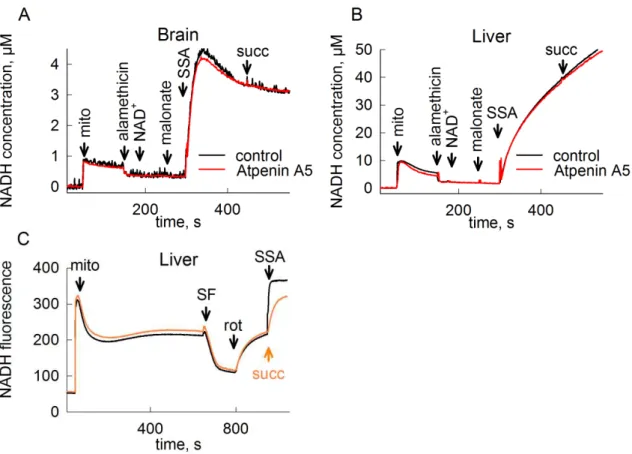
Diaphorases are flavoenzymes catalyzing the oxidation of reduced pyridine nucleotides by endogenous or artificial electron acceptors. The first diaphorase enzyme was purified in 1939 [143]), and was shown later to be identical to the DLD subunit of KGDHC [144]. Since then, several other mammalian proteins were found to exhibit diaphorase activity. NAD+ originating from diaphorases can be utilized by KGDHC to form succinyl-CoA, which is in turn converted to succinate by succinate-CoA ligase yielding ATP or GTP depending on the subunit composition of the enzyme. As recently shown in our laboratory, under respiratory chain inhibition when NADH cannot be oxidized by complex I, NAD+ supply by mitochondrial diaphorases is sufficient to maintain mitochondrial SLP [45]. In isolated mouse liver mitochondria supported by glutamate and malate, up to 81% of the NAD+ pool could be regenerated by intramitochondrial diaphorases. Applying different diaphorase inhibitors – dicoumarol, chrysin, dihydroxyflavone and phenindione – lead to the abolition of SLP under these conditions, pointing out the indispensable role of diaphorases for the adequate operation of KGHDC. In the reaction catalyzed by diaphorases, the electrons of NADH have to be passed on to a suitable electron acceptor. The effect of 14 quinone compounds as possible diaphorase substrates was tested, from which three – menadione (MND), mitoquinone (mitoQ) and duroquinone (DQ) – were shown to boost mitochondrial SLP when complex I was inhibited by rotenone. In anoxia, when the operation of the entire respiratory chain is hindered, only duroquinone was effective in improving SLP. From these experiments it was concluded that in freshly isolated, rotenone-treated mitochondria respiring on glutamate and malate, provision of exogenous quinones for NADH oxidation by diaphorases is not critical due to the presence of sufficient amounts of endogenous quinones. However, mitochondrial diaphorases are not saturated by
25
endogenous quinones, and the addition of exogenous reducible diaphorase substrates can boost SLP by providing NAD+ for KGDHC. A scheme for the pathway of electrons during respiratory arrest was proposed: diaphorases transfer electrons from NADH to suitable quinones, from where electrons are taken over by complex III, which is then oxidized by cytochrome c [45].
1.10. NAD(P)H quinone oxidoreductase 1 (NQO1)
The identity of the diaphorase enzymes participating in NAD+ regeneration for SLP is not known. NAD(P)H quinone oxidoreductase 1 (NQO1, EC 1.6.99.2.), a ubiquitously expressed flavoprotein was proposed as a potential candidate [45]. The enzyme was identified by Lars Ernster and colleagues and was originally named ‘DT- diaphorase’ because of its ability to react with DPNH (reduced diphosphopyridine nucleotide, i.e. NADH) and TPNH (reduced triphosphopyridine nucleotide, i.e.
NADPH) as well [145-147]. Although NQO1 is mostly considered a cytosolic enzyme, it – as well as DT-diaphorase activity with signatures similar to those of NQO1 – has been shown to localize also in the mitochondrial matrix [148-157] (except in [158]). All reports showed that mitochondrial diaphorase activity accounted for <15% of the total.
The reaction catalyzed by NQO1 is irreversible [152] and follows a ping-pong mechanism [159]. As electron acceptor, the enzyme can use a variety of quinones, from which naphtho- and benzoquinones without a long side-chain are the most active [149;
152].
Regarding its physiological role, the enzyme was found to exhibit vitamin K reductase activity [152], thereby it was suggested to play a role in the vitamin K cycle, but the in vivo significance of this has been questioned lately [160-162]. In a detailed study examining the in vivo role of Nqo1 it was demonstrated that Nqo1–/– mice exhibit lower levels of abdominal adipose tissue, and altered carbohydrate, lipid and nucleotide metabolism, due to an altered intracellular NAD(P)H/NAD(P) ratio [163].
NQO1 was also shown to be involved in carcinogenesis through multiple processes, but with opposing outcomes: the enzyme can protect against cancer development by: i) catalyzing a two-electron reduction of quinones and this way avoiding the production of highly reactive semiquinone intermediates [164] ii) preventing oxidative stress through superoxide scavenging [165; 166] and maintaining
26
endogenous antioxidants [167; 168] iii) inhibiting proteasomal degradation of p53 and p33ING1b [169; 170], proteins that are critical for tumor repression. Relevant to this, disruption of the Nqo1 gene in mice leads to increased susceptibility to menadione- and benzene-induced toxicity [171; 172], to increased risk of skin cancer induced by polycyclic aromatic hydrocarbons [173; 174], and to hyperplasia of bone marrow [175].
A polymorphism of the human NQO1 gene encodes a protein which has negligible enzyme activity. Individuals who are homozygous for the variant allele have greater risk for benzene-induced bone marrow toxicity and the resulting hematological malignancies [176], and the polymorphism has been associated with several types of cancer [177].
Even though these facts indicate that cancer development is associated with lower or absent NQO1 activity, a variety of solid tumors are known to overexpress the enzyme [177; 178], probably due to its ability to reduce oxidative stress. The contribution of NQO1 to carcinogenesis is supported by that NQO1 expression is induced by Nrf2 [179], a transcription factor that is being increasingly recognized to favor survival of malignant cells [180; 181]. Also, NQO1 may induce tumor formation through the bioactivation of environmental procarcinogens [182].
27 2. OBJECTIVES
The catabolism of GABA and GHB leads to SSA, an intermediate of the GABA shunt, which is finally converted to succinate, an entry point of the citric acid cycle.
Therefore it is assumable that in anoxia – when SDH is inhibited –, to a certain extent, succinate will accumulate in mitochondria. An elevation of succinate concentration shifts the reaction mediated by SUCL into succinyl-CoA producing direction, abolishing ATP (GTP) production this way. Mindful of the importance of SLP in preserving high energy phosphate levels in the matrix of respiration-impaired mitochondria [21; 23; 31; 40], it is hypothesized in the present thesis that the metabolism of GABA through the GABA shunt results in an inhibition of mitochondrial SLP and causes reverse operation of ANT in anoxia. The same conception is postulated for the metabolism of SSA and GHB. Therefore, the first aim of the present work is to test the effects of exogenous GABA, SSA and GHB addition on bioenergetic parameters and SLP of isolated mouse brain and liver mitochondria.
The second major issue of my thesis is to get closer to the identity of the diaphorase(s) which provide NAD+ for the KGHDC when the respiratory chain is inhibited, as this is critical for the uninterrupted operation of SLP and the prevention of ANT reversal. Because of its ubiquitous expression in numerous tissues, its localization in mitochondria, and its ability to reduce a great variety of quinone substrates, Nqo1 is a possible candidate for performing this. The second aim of my thesis is to address the contribution of Nqo1 to NAD+ provision and SLP under conditions of impaired mitochondrial respiration, by investigating bioenergetic parameters and ANT reversal in samples from wild type and Nqo1–/– mice.
Finally, in the previous work of our laboratory the effect of diaphorase substrates on SLP was tested [45], from which menadione, mitoquinone and duroquinone had a beneficial impact on ATP generation via SUCL. Thus, the question was raised whether these compounds exert their effect through being reduced by Nqo1. Two additional quinones, idebenone (IDB) and 2-methoxy-1,4-naphtoquinone (MNQ) are known to transfer electrons from cytosolic NADH to the mitochondrial respiratory chain, bypassing complex I [183; 184]. For idebenone this effect was proposed to be mediated by cytosolic NQO1 – in the reaction idebenol is generated, which is able to donate electrons to the respiratory chain at the level of complex III [185]. Hence it is
28
reasonable to hypothesize that these two substrates could assist in maintaining the mitochondrial NAD+ pool as well and consequently SLP when NADH oxidation through complex I is limited. My third aim in this thesis to examine the effect of the aforementioned five substrates on mitochondrial SLP and to scrutinize whether this effect is Nqo1-dependent or not.
29 3. METHODS
3.1. Animals
Mice were of mixed 129Sv and C57Bl/6 background. Nqo1–/– mice were a kind gift of Dr. Frank J. Gonzalez. The animals used in our study were of either sex and between 2 and 6 months of age. Mice were housed in a room maintained at 20-22 °C on a 12-h light-dark cycle with food and water available ad libitum. All experiments were approved by the Animal Care and Use Committee of the Semmelweis University and the EU Directive 2010/63/EU for animal experiments.
3.2. Isolation of mitochondria
Isolation of mitochondria from mouse liver and brain: liver mitochondria from all animals were isolated as described in [186], with minor modifications. Mice were killed by cervical dislocation. The liver was removed and immediately placed in ice-cold isolation buffer containing 225 mM mannitol, 75 mM sucrose, 5 mM HEPES (free acid), 1 mM EGTA and 1 mg/ml bovine serum albumin (fatty acid-free), with the pH adjusted to 7.4 with Trizma® (Sigma-Aldrich, St. Louis, MO, USA). The organs were chopped, washed, homogenized and the homogenate was centrifuged at 1,250 g for 10 min. The upper fatty layer of the centrifuged homogenate was aspirated and the pellet was discarded. The supernatant was transferred into clean centrifuge tubes and centrifuged at 10,000 g for 10 min. After this step the supernatant was discarded and the pellet was resuspended in isolation buffer and centrifuged again in clean tubes at 10,000 g for 10 min. At the end of the third centrifugation the pellet was resuspended in 0.2 ml of a buffer with the same composition as described above but containing only 0.1 mM EGTA.
Non-synaptic mouse brain mitochondria were isolated on a Percoll gradient as described previously [187; 188], with minor modifications. After cervical dislocation brains were removed, chopped and homogenized in ice-cold isolation buffer. For the preparation of brain mitochondria the same isolation buffer was used as for liver mitochondria but without BSA. The homogenate was centrifuged at 1,250 g for 10 min;
the pellet was discarded, and the supernatant was centrifuged at 10,000 g for 10 min.
The pellet was resuspended in 15% Percoll (Sigma) and layered on a preformed Percoll gradient (40 and 23%). The tubes were centrifuged at 40,000 g for 6 min, and non-