The contribution of the reaction catalyzed by succinyl- CoA ligase to substrate-level phosphorylation
PhD Thesis
Gergely Kacsó
Semmelweis University
János Szentágothai Doctoral School of Neurosciences Functional Neurosciences Program
Supervisor: Christos Chinopoulos, Ph.D
Official reviewers: Ferenc Gallyas Jr, D.Sc, Tamás Kardon, PhD
Chairman of the final examination board: József Mandl D.Sc
Members of the final examination board: Erzsébet Ligeti, D.Sc.
Olivér Ozohanics, Ph.D.,
Budapest
2018
1
TABLE OF CONTENTS
TABLE OF CONTENTS ... 1
1. THE LIST OF ABBREVIATIONS ... 4
2. INTRODUCTION ... 9
2.1 Mitochondrial ATP synthesis ... 10
2.2 Alternative mitochondrial ATP-producing biochemical pathways ... 12
2.3 Mitochondrial substrate-level phosphorylation and the SKDCC axis... 13
2.4 Succinyl-CoA ligase ... 15
2.5 Succinyl-CoA ligase and surrounding enzymatic pathways ... 17
2.6 Succinyl-CoA ligase mutations ... 22
2.6.1 Succinyl-CoA ligase deficiency ... 23
2.6.2 Metabolic alterations in succinyl-CoA ligase deficiencies... 24
2.6.3 Diagnostic aspects of succinyl-CoA ligase deficiencies ... 27
2.6.4 Potential treatment of patients with succinyl-CoA ligase deficiency ... 29
3. OBJECTIVES ... 31
4. METHODS ... 33
4.1 Animals ... 33
4.2 Isolation of mitochondria... 35
4.3 Determination of protein concentration ... 35
4.4 Mitochondrial substrates and substrate combinations ... 35
4.5 Determination of membrane potential (ΔΨm) in isolated liver mitochondria ... 36
4.6 Mitochondrial respiration ... 37
4.7 Cell cultures ... 37
4.8 Mitochondrial membrane potential (ΔΨm) determination in in situ mitochondria of permeabilized fibroblast cells... 37
4.9 Western blot analysis ... 38
4.10 mtDNA content ... 38
4.11 Protein purification ... 39
4.12 Electron transport chain complex and citrate synthase activity assays ... 39
4.13 Determination of succinyl-CoA ligase activity ... 41
4.14 Determination of acylcarnitines ... 41
4.15 Determination of Sucla2 mRNA by qRT-PCR ... 41
2
4.16 Statistics ... 42
4.17 Reagents ... 42
5. RESULTS ... 43
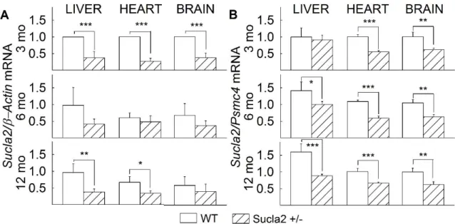
5.1 The effect of deleting one Sucla2 allele on Sucla2 mRNA level ... 43
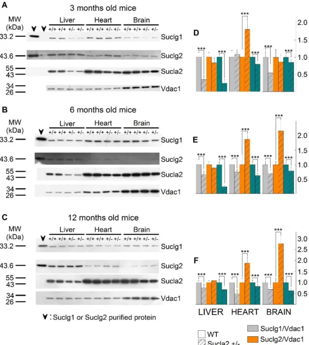
5.2 Characterization of succinyl-CoA ligase subunit expressions of WT and Sucla2+/‒ mice ... 44
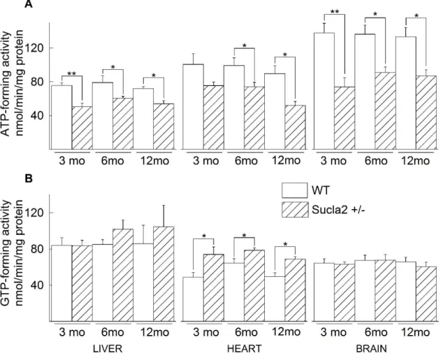
5.3 ATP- and GTP-forming succinyl-CoA ligase activities of WT and Sucla2+/‒ mice ... 46
5.4 The effect of deleting one Sucla2 allele on mtDNA content ... 47
5.5 The effect of deleting one Sucla2 allele on respiratory complex activities ... 48
5.6 The effect of deleting one Sucla2 allele on blood acylcarnitine ester levels ... 49
5.7 The effect of deleting one Sucla2 allele on substrate-level phosphorylation and bioenergetic parameters ... 51
5.7.1 The effect of deleting one Sucla2 allele on ΔΨm and substrate-level phosphorylation during inhibition of complex I by rotenone or true anoxia... 51
5.7.2 The effect of itaconate on Sucla2 heterozygous mice ... 56
5.7.3 The effect of KM4549SC on sucla2 heterozygous mice ... 58
5.7.4 The effect of deleting one Sucla2 allele on oxygen consumption ... 59
5.8 The lack of substrate-level phosphorylation in fibroblasts from patient suffering from complete SUCLA2 deletion ... 60
5.9 The effect of deleting one Suclg2 allele on Suclg2 mRNA level ... 62
5.10 Characterization of succinyl-CoA ligase subunit expressions of WT and Suclg2 +/‒ mice ... 63
5.11 The effect of deleting one Suclg2 allele on substrate-level phosphorylation and bioenergetic parameters ... 65
5.11.1 The effect of deleting one Suclg2 allele on ΔΨm and substrate-level phosphorylation during inhibition of complex I by rotenone or true anoxia... 65
5.11.2 The effect of deleting one Sucla2 allele on oxygen consumption ... 67
5.12 The effect of deleting both one Sucla2 and one Suclg2 allele on succinyl-CoA ligase subunit expressions ... 68
5.13 ATP- and GTP-forming succinyl-CoA ligase activities of WT and Sucla2+/‒ /Suclg2+/‒ mice ... 69
5.14 The effect of deleting both one Sucla2 and one Suclg2 allele on mtDNA content ... 70
3
5.15 The effect of deleting both one Sucla2 and one Suclg2 allele on respiratory
complex activities ... 71
5.16 The effect of deleting both one Sucla2 and one Suclg2 allele on blood acylcarnitine ester levels ... 71
5.17 The effect of deleting both one Sucla2 and one Suclg2 allele on ΔΨm and substrate-level phosphorylation during inhibition of complex I by rotenone ... 73
6. DISCUSSION ... 74
7. CONCLUSIONS ... 80
8. BIBLIOGRAPHY ... 82
9. BIBLIOGRAPHY OF THE CANDIDATE’S PUBLICATIONS ... 98
10. ACKNOWLEDGEMENTS ... 99
11. SUMMARY ... 100
12. ÖSSZEFOGLALÁS ... 101
4
1. THE LIST OF ABBREVIATIONS
AcAc: acetoacetate
AcAc-CoA: acetoacetyl-coenzyme A ADP: adenosine diphosphate ALA: aminolaevulinic acid
ALAS: aminolaevulinic acid synthase ANT: adenine nucleotide translocase ATP: adenosine triphosphate
BAEP: brain auditory evoked potential BCA: bicinchoninic acid
bp: base pair
BSA: bovine serum albumin
C: Coulomb
C10: decanoyl carnitine C10:1: decenoyl carnitine
C12: dodecanoyl carnitine (see also C6-1DS) C14: myristoyl carnitine
C14:1: tetradecenoyl carnitine C16: palmitoyl carnitine
C16-OH: 3-hydroxyhexadecenoyl carnitine C18: stearoyl carnitine
C18:1: oleyl carnitine
C18-OH: 3-hydroxystearoyl carnitine C2: acetyl carnitine
C3: propionyl carnitine C3-DC: malonyl carnitine
C4: butyryl/isobutyryl carnitine C4-DC: methylmalonyl/succinyl carnitine C4-OH: 3-hydroxybutyryl carnitine
C5: isovaleryl/2-methylbutyryl/pivaloyl carnitine C5-DC: glutaryl carnitine
C5-OH: 3-hydroxy isovaleryl/2-methyl 3-hydroxybutyryl carnitine
5 C6-1DS: dodecanoyl carnitine (see also C12) cATR: carboxyatractyloside
CoASH: coenzyme A
complex I: nicotinamide adenine dinucleotide-ubiquinone oxidoreductase complex II: succinate dehydrogenase
complex III: ubiquinone-cytochrome c oxidoreductase complex IV: cytochrome c oxidase (also known as COX)
CoQ: coenzyme Q, coenzyme Q10, ubiquinone, ubidecarenone, COX: cytochrome c oxidase (also known as complex IV)
Cox1: cytochrome c oxidase subunit 1 CS: citrate synthase
CSF: cerebrospinal fluid CT: computed tomography DCPIP: dichlorophenolindophenol dhz: double heterozygote
DLD: dihydrolipoyl dehydrogenase DLST: dihydrolipoyl succinyltrasferase DNA: deoxyribonucleic acid
DTNB: 5,5′-Dithiobis(2-nitrobenzoic acid) EEG: electroencephalogram
EGTA: ethylene glycol-bis(2-aminoethyleter)-N,N,N’,N’-tetraacetic acid EMG: electromyogram
Erev_ATPase: reversal potential of the adenine nucleotide translocase Erev_ATPase: reversal potential of the FoF1-ATP synthase
ES cells: embryonic stem cells ETC: electron transfer chain
F: Faraday constant = 9.64·104 C·mol-1
GABA T: 4-aminobutyrate-2-oxoglutarate transaminase GABA: 4-aminobutyrate-2-oxoglutarate
GAD: glutamate decarboxylase GDH: glutamate dehydrogenase GDP: guanosine diphosphate
6 glut: glutamate
GSC: glioma stem-like cells GTP: guanosine triphosphate
HEPES: 2-[4-(2-hydroxyethyl)piperazin-1-yl]ethanesulfonic acid HIF-1α: hypoxia-induced factor-1α
HVA: homovanillic acid Ile: isoleucine
IMM: inner mitochondrial membrane Irg1: immuneresponsive gene 1 K: Kelvin, absolute temperature KCN: potassium-cyanide
KM: affinity constant
L-DOPA: levodopa, L-3,4-dihydroxyphenylalanine LED: light-emitting diode
Lys: lysine
mal: malate
MCM: methylmalonyl-coenzyme A mutase MDC score: mitochondrial disease criteria score
MDS: mitochondrial deoxyribonucleic acid depletion syndrome Met: methionine
MMA: methylmalonyl acid
MRI: magnetic resonance imaging mRNA: messenger ribonucleic acid MS/MS: tandem mass spectrometry
mtDNA: mitochondrial deoxyribonucleic acid n: coupling ratio
NAD+: nicotinamide adenine dinucleotide (oxidized form) NADH: nicotinamide adenine dinucleotide (reduced form) OXCT1: 3-oxoacid CoA-transferase 1 (also known as SCOT) PCR: polymerase chain reaction
PDH: pyruvate dehydrogenase PEP: phosphoenolpyruvate
7 PEPCK: phosphoenolpyruvate-carboxykinase PHD: prolyl hydroxylase
Pi: inorganic phosphate PK: pyruvate kinase
pmf: protonmotive force/electrochemical gradient
pyr: pyruvate
qPCR: quantitative polymerase chain reaction R: universal gas constant = 8.31 J·mol-1·K-1 RC complex: respiratory chain complex
RNA: ribonucleic acid
ROS: reactive oxygen species
rot: rotenone
RPM: revolutions per minute
RT-PCR: reverse transcription polymerase chain reaction
SCOT: succinyl-CoA:3-oxoacid-CoA transferase (also known as OXCT1) SD: standard deviation
SDH: succinate dehydrogenase
SDS PAGE: sodium dodecyl sulfate polyacrylamide gel electrophoresis SEM: standard error of the mean
SIDS: sudden infant death syndrome SSA: succinate-semialdehyde
SSADH: succinate-semialdehyde dehydrogenase St2: state 2 respiration
St3: state 3 respiration
SUCL: succinyl-coenzyme A ligase enzyme
SUCLA2: human – gene, coding ATP-forming succinyl-CoA ligase β subunit SUCLA2: human – ATP-forming succinyl-CoA ligase β subunit
Sucla2: mouse – gene, coding ATP-forming succinyl-CoA ligase β subunit Sucla2: mouse – ATP-forming succinyl-CoA ligase β subunit
SUCLG1: human – gene, coding succinyl-CoA ligase invariant α subunit SUCLG1: human – succinyl-CoA ligase invariant α subunit
Suclg1: mouse – gene, coding succinyl-CoA ligase invariant α subunit
8
Suclg1: mouse – succinyl-CoA ligase invariant α subunit
SUCLG2: human – gene, coding GTP-forming succinyl-CoA ligase β subunit SUCLG2: human – GTP-forming succinyl-CoA ligase β subunit
Suclg2: mouse – gene, coding GTP-forming succinyl-CoA ligase β subunit Suclg2: mouse – GTP-forming succinyl-CoA ligase β subunit
T: absolute temperature in Kelvin ta: the timepoint of anoxia
Thr: threonine Val: valine
VDAC: voltage-dependent anion channels WT: wild type
α-Kg: α-ketoglutarate βOH: β-hydroxybutyrate
ΔG: change in Gibbs free energy (ΔG means equilibrium) ΔpH: transmembrane proton concentration difference ΔΨm: transmembrane potential
9
2. INTRODUCTION
Metabolism, a controlled cellular activity, consists of several processes that converts nutrients to cell-specific molecules, builds up or degrades macromolecules and biomolecules through thousands of enzymatic reactions. In living cells, the ability to generate, store and utilize chemical energy in the most efficient way is critical.
It is a fact found in all textbooks, that the primary power source (in other terms “the powerhouse”) of cells are mitochondria. But while their primary role is to provide ATP for cells, they also take part in several other processes, e.g., Ca2+-, ROS homeostasis, cellular death, and thermoregulation. Several other important biochemical pathways are also found here (oxidative phosphorylation, citric acid cycle, beta oxidation, heme synthesis, etc.) [1]. Additionally, in the last few decades it has been highlighted that impaired mitochondrial function is associated with several pathological conditions, such as excitotoxicity [2], ischemia/reperfusion-, neurodegenerative diseases [3], and oxidative stress [4] [5].
Energy limiting conditions lead to dysfunction of these organelles through losing inner mitochondrial membrane impermeability and/or losing mitochondrial membrane potential (ΔΨm). A serious dysfunction accompanied by complete depolarization can lead to cytosolic ATP pool depletion [5-8]. The consequence of decreased ATP level can be the partial or complete demolition of the primary and secondary active transports e.g., Na+/K+ ATPase. It is not surprising that there has to be a rescue mechanism that is able to support to maintain the cellular ATP pool in those cells, where ATP synthesis through the oxidative phosphorylation is not satisfactory. A possible rescue mechanism revealed by our laboratory is the mitochondrial substrate-level phosphorylation catalyzed by succinyl-CoA ligase and other succinyl-CoA ligase supporting enzymes (see Chapter 2.3).
Mitochondrial diseases are collectively considered to be a primary cause of encephalomyopathies and other multisystem maladies [9-11]. A sizeable fraction of this pool of diseases are associated with mtDNA depletion [12]. Many animal models have been generated to model mtDNA depletion by explicitly deleting genes essential for
10
mtDNA replication [13-20], although only one study has addressed the role of succinyl- CoA ligase [21].
2.1 Mitochondrial ATP synthesis
It is well known that mitochondria are responsible for ATP synthesis. In 1961, Peter Mitchell proposed his Chemiosmotic theory, in which the electrochemical potential difference generated by the flow of electrons through respiratory complexes and the synthesis of ATP are coupled [22]. This process, called oxidative phosphorylation, is fundamental to bioenergetics. Respiratory complex I (NADH-ubiquinone oxidoreductase, EC 1.6.5.3), complex III (ubiquinone-cytochrome c oxidoreductase, EC 1.10.2.2), and complex IV (cytochrome c oxidase, EC 1.9.3.1) act as proton pumps, translocating protons from the matrix side of the inner mitochondrial membrane (IMM) to the intermembrane space, and generating a proton motive force (pmf) across the inner membrane. The membrane potential can change in a wide scale and in the literature different values are found e.g., -180 ‒ -220 mV in [23] or -108 mV ‒ -158 mV in [24]
(see details below). The mitochondrial proton gradient drives the ATP synthase located in the IMM.
FoF1-ATP synthase [EC 3.6.3.14] is a reversible enzyme multicomplex which can both synthetize and hydrolyze ATP [25-31]. The directionality of the enzyme is governed by pmf (proton motive force) [32], which consists of membrane potential (ΔΨm) and proton concentration gradients (ΔpH) across the IMM. However, as it will be proved later, in case of sufficiently high matrix inorganic phosphate (Pi) concentration the ΔpH is subtle (<0.15) [33-35], so the directionality of the enzyme rather depends on ΔΨm. So, the ΔΨm value at which the enzyme shifts its operational direction is called the reversal potential (Erev_ATPase) and dictated by the concentration of participants [36]. Erev_ATPase can be calculated at ΔG=0 by the following equation:
𝐸𝑟𝑒𝑣_𝐴𝑇𝑃𝑎𝑠𝑒 = − (316
𝑛 ) − (2.3𝑅𝑇
𝐹 ∙ 𝑛 ) ∙ 𝑙𝑜𝑔 [( [𝐴𝑇𝑃4−]𝑓𝑟𝑒𝑒𝑖𝑛∙ 𝐾𝑀(𝐴𝐷𝑃) [𝐴𝐷𝑃3−]𝑓𝑟𝑒𝑒𝑖𝑛∙ 𝐾𝑀(𝐴𝑇𝑃)∙ [𝑃−]𝑖𝑛)]
− (2.3𝑅𝑇
𝐹 ) ∙ (𝑝𝐻𝑜𝑢𝑡− 𝑝𝐻𝑖𝑛) 𝑎𝑛𝑑 [𝑃−]𝑖𝑛 = [𝑃𝑡𝑜𝑡𝑎𝑙]𝑖𝑛 (1 + 10𝑝𝐻𝑖𝑛−𝑝𝐾𝑎2)
11
, where in and out means inside and outside of the mitochondrial matrix respectively; R is the universal gas constant 8.31 J·mol-1·K-1; T is the temperature (K); F is the Faraday constant 9.64 · 104 C·mol-1; KM(ADP) and KM(ATP) are the true affinity constants of Mg2+; [P-] is the free phosphate concentration in mol; pKa2=7.2 for phosphoric acid; and n is the coupling ratio (H+/ATP). The latest parameter depends on the number of the c subunits of ATP synthase [37, 38].
Adenosine phosphates can be exchanged only through adenine nucleotide translocase (ANT) and by a minor fraction through the ATP-Mg/Pi carrier [39]. ANT catalyzes a reversible reaction governed by mitochondrial membrane potential (ΔΨm). The membrane potential value at which ANT reverses its operation is Erev_ANT calculated as follows:
𝐸𝑟𝑒𝑣_𝐴𝑁𝑇 = 2.3𝑅𝑇
𝐹 ∙ 𝑙𝑜𝑔[𝐴𝐷𝑃3−]𝑓𝑟𝑒𝑒 𝑜𝑢𝑡 ∙ [𝐴𝑇𝑃4−]𝑓𝑟𝑒𝑒 𝑖𝑛 [𝐴𝐷𝑃3−]𝑓𝑟𝑒𝑒 𝑖𝑛 ∙ [𝐴𝑇𝑃4−]𝑓𝑟𝑒𝑒 𝑜𝑢𝑡
where in and out means inside and outside of the mitochondrial matrix, respectively; R is the universal gas constant 8.31 J·mol-1·K-1; T is the temperature (K); F is the Faraday constant 9.64·104 C·mol-1. In normal conditions when oxygen and nutrients are available, and ΔΨm is more negative than Erev_ATPase and Erev_ANT, both enzymes operate in a so called “forward mode”. FoF1-ATPase synthetizes ATP while ANT translocates ATP out of, and ADP into the matrix. The opposite status occurs when both operate in so-called
“reverse mode”, when FoF1-ATPase hydrolases ATP while ANT translocates ATP into, and ADP out of the matrix.
Having taken into account both equations (Erev_ATPase and Erev_ANT), it turns out to be evident that alterations in [ATP4-]free in and [ADP3-]free in affect the reversal potentials inversely; a decrease in intramitochondrial [ATP]/[ADP] ratio shifts Erev_ATPase towards more positive, while it shifts Erev_ANT towards more negative, and vice versa. In other words, the greater the matrix [ATP] and, the greater the force for a forward-operating ANT (ATP outward/ADP inward) is, the less will the ANT seek to reverse. Also, the higher the matrix [ATP] is -which is the product of the forward-operating ATP synthase- , the more it will seek to reverse. There are some other parameters which are also
12
participating in the equations and may exhibit wild fluctuations. As such, cytosolic [ATP]/[ADP] ratio [40] coupling ratio [41, 42], phosphate concentration [43] ΔΨm, and
“ΔΨm flickering”, which could be greater than 100 mV [44-46]. Based on computational estimations and following circumstantial experimental works we know that there is a transient state when the FoF1-ATPase is already acting as a hydrolase, while the ANT is still operating in forward mode, translocating ATP from the matrix towards cytoplasm.
Computer estimations were verified by experimental results in both isolated and in situ mitochondria. These results are showing that Erev_ATPase is always more negative than Erev_ANT,implying that progressively depolarizing mitochondria will first exhibit reversal of the FoF1-ATP synthase, followed by the reversal of ANT. The question arises at this point: what is the source of the matrix ATP? [6, 36].
2.2 Alternative mitochondrial ATP-producing biochemical pathways
As seen in the previous chapter, the main role in ATP synthesis is played by the mitochondrial FoF1 ATP synthase, nevertheless, it is controlled by membrane potential and oxygen availability.
In cells, other mechanisms can also act as ATP source independently from oxidative phosphorylation, e.g., the glycolysis in the cytoplasm. According to Otto Warburg’s lasting theory -the Warburg effect-, most cancer cells produce predominantly ATP by a high rate of aerobic glycolysis [47].
In the mitochondrial matrix, there are two reactions capable of producing high-energy phosphate at substrate-level:
i. The mitochondrial form of the phosphoenolpyruvate carboxykinase (PEPCK) [EC 4.1.1.32]. It catalyzes a reversible reaction:
𝑜𝑥𝑎𝑙𝑜𝑎𝑐𝑒𝑡𝑎𝑡𝑒 + 𝐺𝑇𝑃 ↔ 𝑝ℎ𝑜𝑠𝑝ℎ𝑜𝑒𝑛𝑜𝑙𝑝𝑦𝑟𝑢𝑣𝑎𝑡𝑒 + 𝐺𝐷𝑃 + 𝐶𝑂2
This enzyme participates in the pyruvate recycling pathway and is thought to take part in transferring phosphorylation potential from matrix to cytosol [48-50].
ii. GTP for the mitochondrial PEPCK may originate from the GTP-forming succinyl-CoA ligase [51]. It was considered earlier that the substrate-level phosphorylation catalyzed by
13
succinyl-CoA ligase might significantly contribute to the net ATP (or GTP) production [52].
As it was described in Chapter 2.1, while the FoF1-ATP synthase operates in reverse mode, ANT can operate in both reverse and forward modes. The reverse operation of the ANT would lead to the depletion of the cytosolic ATP pool and would further lead to cell death [5, 7, 53]. In case of the forward operation of the ANT (but a reverse operation of the FoF1-ATP synthase), ATP cannot originate from the cytosol. Thus, it should originate from the matrix and due to the lack of any other possible mechanism, the origin should be the succinyl-CoA ligase [6, 36, 48, 54].
2.3 Mitochondrial substrate-level phosphorylation and the SKDCC axis
As shown in Chapter 2.1 and 2.2, matrix substrate-level phosphorylation has a critical role in maintaining ANT operation in the forward mode, thereby preventing mitochondria from becoming cytosolic ATP consumers during respiratory arrest.
Furthermore, succinyl-CoA ligase does not require oxygen for ATP production and it is even activated during hypoxia [55]. Later, our research group showed that provision of succinyl-CoA by alfa-ketoglutarate dehydrogenase complex is essential for the maintenance of the substrate-level phosphorylation by supporting the succinyl-CoA ligase. Thus, substrate-level phosphorylation can prevent mitochondria -with a dysfunctional electron transport chain- to consume the extramitochondrial ATP pool. In order to prove this, experiments were performed in respiration-impaired isolated and in situ neuronal somal mitochondria from transgenic mice with a deficiency of either dihydrolipoyl succinyltransferase (E2 or DLST EC 2.3.1.61) or dihydrolipoyl dehydrogenase (E3 or DLD, EC 1.8.1.4) that exhibited 20-48% decrease in alpha- ketoglutarate dehydrogenase complex activity [56]. These results demonstrated the consumption of extramitochondrial ATP by mitochondria of DLD+/‒, DLST +/‒, or DLD+/‒
/DLST+/‒ double transgenic mice provided with substrates supporting substrate-level phosphorylation (See later in Chapter 4.4), by clamping ΔΨm in a depolarized range due to targeted respiratory inhibition [56].
At this point the question arose: What provides NAD+ for alpha-ketoglutarate dehydrogenase complex during anoxia, when complex I is not expected to oxidize NADH?
14
As it was shown, mitochondrial diaphorases and a finite pool of oxidizable quinones are the source of NAD+ generated within the mitochondrial matrix during respiratory arrest caused by anoxia or poisons of the electron transport chain [57]. Using pharmacological tools, specific substrates, and by examining tissue from pigeon liver exhibiting no diaphorase activity it was shown in this work that mitochondrial diaphorases in mouse liver contribute up to 81% to the NAD+ pool during respiratory inhibition. In situ, reoxidation of the reducible substrates for the diaphorases is mediated by complex III of the respiratory chain using cytochrome c and an unidentified cytosolic oxidant, i.e.
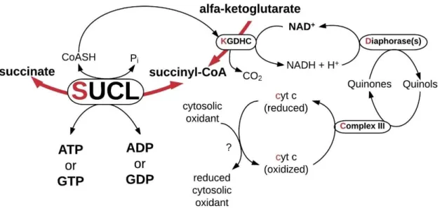
possibly p66Shc [57] [58]. An extensive body of work in our laboratory on animal models has shown that substrate-level phosphorylation substantiated by succinyl-CoA ligase is part of an axis consisting of enzyme complexes termed 'SKDCC' axis. The pathway produces high-energy phosphates in the mitochondrial matrix in the absence of oxidative phosphorylation. The SKDCC axis is summarized in Figure 1. This rescue mechanism protects against cytosolic/nuclear ATP depletion in conditions involving impaired respiration, as it may occur in cancer.
Figure 1 Illustration of the SKDCC axis
SKDCC stands for the initial letters (red) of the participant proteins: succinyl-CoA ligase, α-ketoglutarate dehydrogenase complex, diaphorases, complex III, cytochrome c.
(adapted from [57]) Abbreviations: ADP: adenosine diphosphate, ATP: adenosine triphosphate, cyt c: cytochrome c, GDP: guanosine diphosphate, GTP: guanosine triphosphate, KGDHC: α-ketoglutarate dehydrogenase complex, Pi: inorganic phosphate, SUCL: succinyl-CoA ligase
15 2.4 Succinyl-CoA ligase
Succinyl-CoA ligase -also known as succinyl coenzyme A synthetase or succinate thiokinase- is a heterodimer enzyme incorporating the same α-subunit, but different β- subunits. The combination of the invariant α-subunit encoded by SUCLG1 and the substrate-specific β-subunits encoded by SUCLA2 and SUCLG2 result in either ATP- forming (EC 6.2.1.5) or GTP-forming (EC 6.2.1.4) enzyme respectively (see Figure 2.) [59].
Figure 2 Different succinyl-CoA ligase enzyme compositions
Succinyl-CoA ligase is a heterodimer enzyme composed of invariant α-subunit encoded by SUCLG1, and substrate specific β-subunits encoded by SUCLA2 and SUCLG2 forming ATP and GTP, respectively.
ΔG is ~0.05 kJ/mol [60], therefore the reaction is reversible. Succinyl-CoA ligase is located in the mitochondrial matrix catalyzing the following reaction:
𝑠𝑢𝑐𝑐𝑖𝑛𝑦𝑙 − 𝐶𝑜𝐴 + 𝑃𝑖 + 𝐴𝐷𝑃(𝑜𝑟 𝐺𝐷𝑃) ↔ 𝑠𝑢𝑐𝑐𝑖𝑛𝑎𝑡𝑒 + 𝐶𝑜𝐴𝑆𝐻 + 𝐴𝑇𝑃(𝑜𝑟 𝐺𝑇𝑃) [61].
The catalytic mechanism of succinyl-CoA ligase has not been fully characterized.
Representative catalytic mechanism and associated kinetic parameters that can explain data on the enzyme-catalyzed reaction kinetics have not been established (reviewed by Xin LI et al. in ref. [62]).
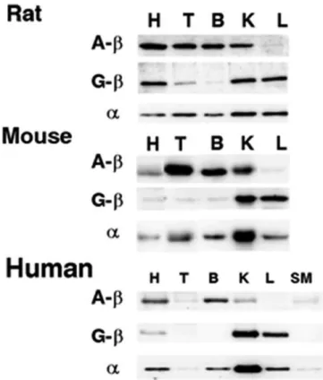
Generally, both GTP and ATP forming succinyl-CoA ligase are well represented in a variety of tissues, not only in humans but also in murine mitochondria. The GTP-forming subunit is rather expressed in anabolic tissues such as liver and kidney, the ATP-forming succinyl-CoA ligase is rather present in kidney, testis, and brain, whereas in cardiac tissue both ATP- and GTP-forming subunits are strongly expressed [61, 63-65]. In the human brain, SUCLA2 is exclusively expressed in neurons in the cerebral cortex and not in any other cell type [66], while SUCLG2 encoded GTP-forming succinyl-CoA ligase is not found either in neurons or in astroglial cells, it can be found only in cells forming the microvasculature [67] (See Figure 3).
16
Figure 3 Western blots of succinyl-CoA ligase subunits in rat, mouse, and human.
The figure was adapted from Lambeth et al. ‘Expression of two succinyl-CoA synthetases with different nucleotide specificities in mammalian tissues’.
Abbreviation on the figure: H – heart, T – testis, B – brain, K – kidney, L – liver, SM – skeletal muscle, A-β – ATP forming SUCL subunit, G-β – GTP-forming SUCL subunit, α – invariant SUCL α-subunit [63].
As it was described above, GTP also can form ATP due to a trans-phosphorylation by mitochondrial nucleotide diphosphate kinase [68]. While ATP is used by catabolic pathways, GTP is rather used for cellular anabolism [69], protein synthesis in mitochondria and also can support other GTP-binding regulatory proteins [70]. The GTP pool is outstanding in the mitochondrial matrix, since it is believed, that there is no mechanism for the export of GTP from the mitochondrial matrix to the cytosol in higher organisms [71]. Nevertheless, an atractyloside sensitive and insensitive guanine nucleotide transport mechanism was published by one group [72, 73], but it has not been confirmed yet.
Furthermore, there also exists another GTP using mechanism, i.e. the phosphoenolpyruvate (PEP) synthesis. PEP can be transported to the cytosol [74], where
17
it is used in gluconeogenesis for either GDP phosphorylation by cytosolic PEPCK or synthesis of ATP through the pyruvate kinase (PK, EC 2.7.1.40) reaction [63].
2.5 Succinyl-CoA ligase and surrounding enzymatic pathways
Figure 4 Succinyl-CoA ligase and surrounding enzymatic pathways
Succinate-CoA ligase is an intersection of several biochemical pathways, such as: citric acid cycle (1-red); Heme metabolism (2-yellow); in extrahepatic tissues, metabolism of ketone bodies (3-green); substrate-level phosphorylation (4-orange); succinyl-CoA ligase is associated with nucleoside-diphosphate kinase, therefore participate in maintaining mtDNA content (5-dark blue); through propionyl-CoA, succinyl-CoA is an entry point of several biomolecules (Met, Thr, Ile, Val, odd chain fatty acids, cholesterol) (6-brown);
increases in propionyl-CoA and methylmalonyl-CoA may cause secondary metabolic aberrations due to their ability to inhibit steps in urea cycle, gluconeogenesis, and the glycine cleavage system (7-purple); in some particular cells in brain, succinate is the entry point of the GABA shunt (8-light blue); in cells of macrophage linage, succinyl-CoA ligase metabolizes the endogenously produced itaconate to itaconyl-CoA (9-pink);
succinyl-CoA has been reported as a cofactor of lysine succinylation as a post- translational modification (10-grey); succinate has also been reported as an oncometabolite and a metabolic signal in inflammation (11-black); Abbreviations on figure: βOH: beta-hydroxybutyrate; AcAc: acetoacetate; AcAc-CoA: acetoacetyl-CoA;
ADP: adenosine diphosphate; ALA: aminolaevulinic acid; ALAS: aminolaevulinic acid synthase; ATP: adenosine triphosphate; CoASH: coenzyme A; GABA T: 4- aminobutyrate-2-oxoglutarate transaminase; GABA: 4-aminobutyrate-2-oxoglutarate;
GAD: glutamate decarboxylase; GDH: glutamate dehydrogenase; GDP: guanosine diphosphate; GHB: gamma hydroxybutyrate; GTP: guanosine triphosphate; Ile:
18
isoleucine; KGDHC: alpha-ketoglutarate dehydrogenase complex; Lys: lysine; MCM:
methylmalonyl-CoA mutase; Met: methionine; NDPK: nucleotide diphosphate kinase;
Pi: inorganic phosphate; SCOT: succinyl-CoA : 3-oxoacid-CoA transferase (also known as OXCT1); SDH: succinate dehydrogenase; SUCL: succinyl-CoA ligase; SSA:
succinate-semialdehyde; SSADH: succinate-semialdehyde dehydrogenase; Thr:
threonine; Val: valine
Succinyl-CoA ligase is at the intersection of several metabolic pathways (see Figure 4) [75]. The succinyl-CoA ligase enzyme itself has an important role in several functions, furthermore, there are several enzymatic reactions which may be affected by its performance or may affect the performance of the reaction through altering e.g., succinate, succinyl-CoA, inorganic phosphate, ADP, GDP, ATP, GTP, and/or CoA concentration in the matrix. In this chapter, most of these possible functions and reactions were summarized. These are the following:
a. Succinyl-CoA ligase is an enzyme of the citric acid cycle, one of the largest metabolic hub in mammalian cells [76]. As its name implies, it is a cycle, where every participating enzyme has a product which is a substrate of the following enzyme. This metabolic pathway has no end, but several entry points, where taken up fuels by mitochondria can fill up the cycle. When oxygen is available for cells, the cycle’s primary role is to reduce NAD+ and FAD+ by dehydrogenases for the respiratory chain. NADH and FADH2 oxidation lead to building up the electrochemical gradient of H+-s across the inner mitochondrial membrane that is utilized by the FoF1-ATP synthase to produce ATP [48]. (see Figure 4 - 1, red pathway);
b. Succinyl-CoA ligase catalyzes a reversible reaction as mentioned above, thus succinyl-CoA ligase can proceed towards succinyl-CoA production. Due to the irreversible reaction catalyzed by alpha-ketoglutarate dehydrogenase complex [48, 56] it cannot be processed further than succinyl-CoA. In this case, succinyl- CoA may follow heme metabolism through aminolaevulinic-acid synthesis [77].
(see Figure 4 - 2, yellow pathway);
19
c. In extrahepatic tissues, 3-hydroxy butyrate (βOH) is changed back into acetoacetate (AcAc). In the next step it is activated to acetoacetyl-CoA by succinyl-CoA:3-oxoacid CoA transferase (SCOT or OXCT1, EC:2.8.3.5) Therefore, succinyl-CoA ligase is responsible also for maintaining the normal ketone body homeostasis through the regulation of the succinyl-CoA level within mitochondria [78]. This process is one of those that can mediate the by-pass of succinyl-CoA ligase (see Figure 4 - 3, green pathway);
d. Substrate-level phosphorylation is a definition of an enzyme reaction that results in a formation of an ATP or a GTP by the direct transfer of a phosphoryl (PO3) group from a substrate to an ADP or GDP molecule respectively. In mitochondrial matrix, the major regulator of the ATP/ADP ratio is the FoF1-ATP synthase, however, this enzyme highly depends on the mitochondrial membrane potential (ΔΨm) [32].
There are two possible enzymatic transformations in mitochondria where GTP is utilized, and one of them is catalyzed by the reversible phosphoenolpyruvate carboxykinase (PEPCK, EC 4.1.1.32) (not shown in Figure 4). The reaction is the following [48]:
𝑜𝑥𝑎𝑙𝑜𝑎𝑐𝑒𝑡𝑎𝑡𝑒 + 𝐺𝑇𝑃 ↔ 𝑝ℎ𝑜𝑠𝑝ℎ𝑜𝑒𝑛𝑜𝑙𝑝𝑦𝑟𝑢𝑣𝑎𝑡𝑒 + 𝐺𝐷𝑃 + 𝐶𝑂2
The PEPCK’s mitochondrial isoform may operate rather as a GTPase linking hydrolysis of mitochondrial GTP made by succinyl-CoA ligase to an anaplerotic pathway resulting phosphoenolpyruvate (PEP) and also can contribute to glucose- stimulated insulin secretion in pancreas β cells [51].
The other reaction in the mitochondrial matrix, which is also capable for substrate- level phosphorylation, is succinyl-CoA ligase. As it was described above, the enzyme can form ATP and GTP as well. Nevertheless, GTP forming succinyl- CoA ligase also contributes to the ATP formation through the enzyme’s concerted action with mitochondrial nucleotide diphosphate kinase (NDPK, EC 2.7.4.6) [79- 81]. Thus, ATP can form GTP and vice versa.
20
Succinyl-CoA ligase as a mitochondrial substrate-level phosphorylation can yield high-energy phosphates in the absence of oxygen [36, 48, 56]. (see Figure 4 – 4, orange pathway)
e. Since succinyl-CoA ligase is associated with the nucleotide diphosphate kinase [80, 81], it has a major role in maintaining mtDNA content through the provision of phosphorylated deoxyribonucleotides. Mutation of succinyl-CoA ligase leads to mitochondrial DNA (mtDNA) depletion syndrome (MDS) [82], which is further explained in Chapter 2.6. (see Figure 4 – 5, dark-blue pathway)
f. The accumulation of succinyl-CoA due to the malfunction of succinyl-CoA ligase can be responsible for the alteration of different participants of the propionyl- methylmalonyl-CoA axis. Succinyl-CoA is the entry point to the citric acid cycle for endpoint products of several biomolecules, e.g., methionine, threonine, isoleucine, valine, propionate, odd-chain fatty acids and cholesterol.
These main reaction steps in the catabolic propionyl-methylmalonyl-CoA axis are mediated by the sequential actions of propionyl-CoA carboxylase (EC 6.4.1.3) and methylmalonyl-CoA mutase (EC 5.4.99.2) [83] (see Figure 4 – 6, brown pathway)
g. In addition, increases in propionyl-CoA and methylmalonyl-CoA (partially shown on Figure 4 – 7, purple pathway) may cause secondary metabolic aberrations due to their ability to inhibit steps in urea cycle [84-86], gluconeogenesis and the glycine cleavage system [87, 88];
h. In specialized cells of the brain, succinate is the entry point to the citric acid cycle of the ‘GABA shunt’ from succinate semialdehyde, a metabolite which is also in equilibrium with γ-hydroxybutyric acid [89-91]
GABA is found both in liver and brain, however, the hepatic tissue is not able to synthetize GABA, due to the relatively low glutamate decarboxylase (GAD, EC 4.1.1.15) activity, which is the key enzyme of the GABA synthesis. Unlike GABA
21
synthesis, GABA metabolism is possible in liver tissue, because of the presence of GABA transaminase (GABA-T, EC 2.6.1.19).
In tissues where succinyl semialdehyde dehydrogenase also can be found, GABA shunt also have an impact on succinyl-CoA ligase by leading to succinate production [92]. (See under publications related to the present thesis in Chapter 11.)
GABA shunt is also able to by-pass substrate-level phosphorylation (see Figure 4 – 8, light-blue pathway) and apparently produces less ATP from glutamate to succinate than the glutamate-α-ketoglutarate-succinyl-CoA-succinate pathway [75].
i. Itaconic acid (also known as 2-methylidenebutanedioic acid, methylenesuccinic acid) is a dicarboxylic acid. It has been identified in mammalian tissue specimens, such as Mycobacterium tuberculosis-infected lung tissue [93], glioblastomas [94], urine and serum samples [95]. It also has been shown that in human and mouse, macrophage lineage cells produce itaconic acid from cis-aconitate through an enzyme showing cis-aconitate decarboxylase activity. This enzyme is encoded by an immuneresponsive gene 1 (irg1). In cells, where irg1 can be activated, itaconate concentration is in millimolar range [96]. The role of itaconic acid in macrophages is to contribute to the antibacterial effect of these cells, since itaconate inhibits bacterial isocitrate lyase (EC 4.1.3.1), which is the key enzyme of bacterial glyoxylate shunt [97, 98] which renders bacteria able to grow on fatty acids and/or acetate [99]. More than a half decade ago it was described that itaconate is metabolized by succinyl-CoA ligase to itaconyl-CoA on a malonate sensitive manner, implying succinate dehydrogenase (EC 1.3.5.1) participation [100-103].
Considering substrate-level phosphorylation, itaconate:
- abolishes the process, resulting a “CoA trap” in the form of itaconyl-CoA;
- decreases ATP (or GTP) levels, since it diverts succinyl-CoA ligase towards thioesterification;
- inhibits SDH leading to a succinate build-up, which shifts succinyl-CoA ligase also towards thioesterification [104].
(See under publications related to the present thesis in Chapter 11.)
22 (see Figure 4 – 9, pink pathway)
j. According to a recent finding, succinyl-CoA might be the cofactor of enzyme- mediated lysine succinylation as an active form of succinate. This process is another way of protein post-translational modification, which can cause significant structural changes and is expected to have important cellular regulator functions [105] (see Figure 4 – 10, grey pathway).
k. In activated macrophages succinate can stimulate dendric cells through succinate receptor 1. Thus, succinate has an impact on cellular activation and immune response [106, 107].
l. Succinate can be named as ‘oncometabolite’ because succinate links the dysfunction of the citric acid cycle to oncogenesis through the activation of the hypoxia-inducible factor-1α (HIF-1α) [48, 108, 109]. Succinate can stabilize HIF- 1α in tumors through succinate accumulation, which is itself can inhibit prolyl hydroxylase enzyme (PHD, EC 1.14.11.29) [108]. PHD is responsible for hydroxylation, thus the regulation of HIF1α causes its proteasomal-mediated degradation [110] (partially see Figure 4 – 11, black pathway).
m. In mitochondria, inorganic phosphate can act as a signaling molecule by activating oxidative phosphorylation through several points. Succinyl-CoA ligase has been identified as a new target of phosphate-induced activation of the oxidative phosphorylation and substrate-level phosphorylation under energy- limited circumstances [55].
2.6 Succinyl-CoA ligase mutations
As it was described in Chapter 2.4, succinyl-CoA ligase is encoded by three genes with different roles. SUCLG1 encodes the invariant α subunit, SUCLA2 and SUCLG2 encode the β subunit forming ATP or GTP respectively.
To date, 51 patients have been reported with SUCLA2 deficiency [65, 111-121], 28 patients with SUCLG1 deficiency [112, 119, 122-132], and only 2 patients with SUCLG2
23
deficiency [122, 123]. Mitochondrial depletion syndrome (MDS), encephalomyopathy, lactic acidosis and methylmalonic aciduria are the most common features of succinyl- CoA ligase affecting diseases. In this chapter, the most typical symptoms, diagnostic aspects, metabolic alterations, and possible treatments are discussed highlighting the differences between subunit involvements.
Given the fact, that succinyl-CoA ligase plays a central role in the pathways mentioned beforehand, it is not surprising that this enzyme’s deficiency leads to pleiotropic pathology, which is also influenced by the tissue-specific expression of its subunits.
Significant clinical differences between mutations were hepatopathy found with SUCLG1 cases but not in SUCLA2 cases [112, 127, 132]. Hypertrophic cardiomyopathy was not reported in SUCLA2 patients (in one patient, a mild restrictive cardiomyopathy was found by echocardiography, but it resolved spontaneously [65]), but documented in several cases with SUCLG1 mutations [112]. Since SUCLG2 mutations have been described in the literature just twice recently, only few pieces of information are available about these patients. As it was reported, one of the patients had elevated methylmalonic acid and SIDS (sudden infant death syndrome), and the post-mortem blood spot analysis revealed a substantial elevation in C3 carnitine levels [122].
SUCLA2 mutant patients have significantly longer survival (median survival 20 years) than SUCLG1 deficient patients (median survival 20 months) significantly [112, 125], which is also true for missense mutations compared to loss-of-function (deletions, frameshift, nonsense) mutations [112].
2.6.1 Succinyl-CoA ligase deficiency
Succinyl-CoA ligase deficiency is a rare disease, however, it has a higher incidence of 1 in 1.700 in the Faroe Islands due to a founder effect. The carrier frequency of 1 in 33 [121] (frequency of the mutated allele is 2%, the estimated homozygote frequency is 1:2500 [111]). To date in Scandinavian population two founder mutations were identified as well [112]. Most of the mutation analyses have shown several novel mutations, including homozygous deletion of the entire SUCLA2 gene, but other types also were found, such as one bp duplication, one bp deletion, three bp insertion, nonsense mutations, and a missense mutation [112, 131, 133, 134].
24
The median onset of symptoms was two months for patients with SUCLA2 mutations and for SUCLG1 patients it occurred at birth [111] with intrauterine growth retardation [132].
Both ATP- and GTP-forming subunit involving mutation caused the elevation of blood and urine methylmalonic acid, mtDNA depletion syndrome (MDS), lactic acidosis, and dystonia.
Hypertrophic cardiomyopathy and liver involvement were exclusively found in patients with SUCLG1 mutations [132], whereas epilepsy was more frequent in patients with SUCLA2 mutations compared to SUCLG1 mutations [55].
According to MRI neuroimaging results, bilateral hyperdense lesions in basal ganglia (putamen, caudate nuclei) and a mild diffuse cerebral atrophy were found in both SUCLA2 and SUCLG1 cases as well [65, 91, 112, 131, 132]. Furthermore, EMG and nerve condition studies showed progressive axonal and demyelinating polyneuropathy especially of the lower limbs of SUCLA2 patients [65]. Other symptoms are also typical as in Leigh-like encephalomyopathy [125, 131], such as diarrhea, emesis, dehydration, metabolic alterations (see details below), myopathy revealed by muscle echogram [65], severe muscle hypotonia [83, 133] with progressive areflexia [82], later on progressive dystonia [60, 121, 132, 133], dysphagia leading a failure to thrive [122, 133], and developmental delay. BAEP (brain auditory evoked potential) studies proved severe progressive sensorineural hearing loss [83, 111, 123, 133] in SUCLA2 patients (Deafness in SUCLG1 mutations hasn’t been reported, due to the death during the first weeks, [18].) Interestingly liver failure [128, 131, 133] and hepatic encephalopathy [75] were also found in SUCLG1 patients, however, almost none of the patients with SUCLA2 mutation have been reported on so far suffered from cardiac, hepatic, gastrointestinal, or bone marrow involvement [131]. In SUCLA2 patients with longer survival there was no speech development, but the children were able to understand sign language [65].
2.6.2 Metabolic alterations in succinyl-CoA ligase deficiencies
Regarding metabolic patterns or changes (see Figure 5), the accumulation of succinyl- CoA leads to an elevation in carnitine ester concentrations, especially C4-dicarboxylic carnitine [65, 111] and other elements of the succinyl-, methylmalonyl-, propionyl-CoA axis [65, 122, 123, 135, 136]. Since the end product of odd chain fatty acid oxidation is propionyl-CoA [83] which is also ending in here, it is evident that the elevation of the
25
intramitochondrial succinyl-CoA concentration blocks this metabolic pathway as well.
Due to the fact that succinyl-CoA ligase deficiency causes an error in mtDNA maintenance, most succinyl-CoA ligase deficient patients had mtDNA depletion syndrome (MDS) resulting in a mitochondrial failure due to decreased respiratory complex activity [83, 125, 133, 136]. The most specific respiratory complex alterations are the combined complex I/IV; combined complex I/III/IV; isolated complex IV;
isolated complex I deficiency (and also in [133]) and decreased RC complex activities were also found [112]. The insufficient succinyl-CoA ligase function leads to the accumulation of the TCA cycle intermediers, for example succinyl-CoA and citrate [65].
Since both citrate and succinyl-CoA [137] inhibit citrate synthase activity, as a secondary inhibitory affect the accumulating acetyl-CoA may inhibit the pyruvate dehydrogenase complex (PDH) [138]. It will direct the glycolysis towards lactic acid production causing -in most of the cases severe- lactic acidosis [65, 83, 111, 122, 123, 125, 132, 136]. The acidosis further enhanced by the succinyl-CoA ligase deficiency caused the accumulation of the citric acid cycle intermediers [65, 122, 132]. In one case a SUCLA2 mutation accompanying PDH complex deficiency [136] was documented. The urinary excretion of L-leucine catabolism intermediers (3-hydroxyisovaleric acid and 3-methylglutaconic acid) were also found [65]. Isovaleric acid and isovaleryl-CoA are the direct precursors of 3-hydroxyisocvaleric acid. In case of the accumulation of isovaleric acid, the residual succinyl-CoA enzyme activity may be further decreased worsening the symptoms, since isovaleric acid has a succinyl-CoA ligase inhibitory effect [139]. (see also in Figure 5)
26
Figure 5 Schematic biochemical pathways demonstrating metabolic alterations in succinyl-CoA ligase deficiency caused by elevated succinyl-CoA (adapted from [65, 83]) Abbreviations: CS: citrate synthase; PDH: pyruvate dehydrogenase complex;
27
2.6.3 Diagnostic aspects of succinyl-CoA ligase deficiencies
Figure 6 Diagnostic flowchart for patients with common MMA-uria, developmental delay and muscle hypotonia (adapted from [65, 140]) Characteristic metabolic profile in SUCLA2 patients were increased e.g., lactic acid, methylmalonic acid, C4-DC carnitine, and 3-hydroxyisovaleric acid. Abbreviations: MDC: mitochondrial disease criteria; MMA: methylmalonic acid; CSF: cerebrospinal fluid; MRI: Magnetic resonance imaging;
As it has been described above there are only mild differences between SUCLA2/SUCLG1 and SUCLG2 mutations. As for diagnostic aspects, the common features are lactic acidaemia, elevated MMA concentration in blood and mildly in urine, and muscle dystonia should also be mentioned. As it can be seen in Figure 6 not only the succinyl-CoA ligase affected differential diagnoses are difficult, but also other
28
mitochondrial diseases. Morava et al. suggested that late-onset hearing loss and absence of muscular and ocular involvement can be differential diagnostic clues in SUCLA2 and SUCLG1 mutations, since those rather favor SUCLG1. Unlike to this, in SUCLG1 patients the mtDNA depletion in muscle is not a consistent finding, therefore cannot be used for diagnostic purposes [65].
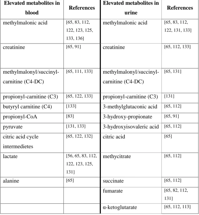
Table 1 Most frequented elevated metabolite levels in SUCLA2 and SUCLG1 patients in blood and urine samples
Hereafter the thesis will focus on succinyl-CoA ligase affecting mutations. In all succinyl-CoA ligase diseases Leigh-like syndrome and accompanying symptoms were
Elevated metabolites in
blood References Elevated metabolites in
urine References
methylmalonic acid [65, 83, 112, 122, 123, 125, 133, 136]
methylmalonic acid [65, 83, 112, 122, 131, 133]
creatinine [65, 91] creatinine [65, 112, 133]
methylmalonyl/succinyl- carnitine (C4-DC)
[65, 111, 133] methylmalonyl/succinyl- carnitine (C4-DC)
[65, 131]
propionyl-carnitine (C3) [65, 122, 133] propionyl-carnitine (C3) [131]
butyryl carnitine (C4) [133] 3-methylglutaconic acid [65, 112]
propionyl-CoA [83] 3-hydroxy-propionate [65, 91]
pyruvate [131, 133] 3-hydroxyisovaleric acid [65, 112]
citric acid cycle intermedietes
[65, 122, 132] citric acid [65]
lactate [56, 65, 83, 112,
122, 123, 125, 131]
methycitrate [65, 112]
alanine [65] succinate [65, 112]
fumarate [65, 82, 112,
131]
α-ketoglutarate [65, 112, 113]
29
typical, however muscular, cardiac, and liver involvement were described in SUCLG1 patients (Only limited data have been available about the SUCLG2 mutant patient). The summarization of metabolites which were elevated either in SUCLA2 and SUCLG1 patients can be found in Table 1.
Diagnostic results might come from metabolic investigations. There were several elevated metabolites identified such as lactic acid in all compartment, MMA in blood and mildly in urine, citric acid cycle intermediers, specified acyl-carnitines (C3/C4/C4DC) in all body fluids, etc. Furthermore 3-hydroxyisovaleric acid also should be listed as a biomarker of SUCLA2 and SUCLG1 deficiency. For final diagnosis, a direct sequence analysis of the SUCLA2 and or SUCLG1 gene is recommended in a suspected SUCLA2 or SUCLG1 deficiency.
2.6.4 Potential treatment of patients with succinyl-CoA ligase deficiency Given the fact that succinyl-CoA ligase plays a role in several metabolic pathways and the deficiency of the enzyme causes severe metabolic and physiologic alterations, it is not surprising that the prognosis is bad and the number of tools for the treatment is limited.
In children an individualized optimal dietary intake can increase the energy generating capacity of mitochondria, since it may be a possible way of therapy and could improve the quality of life [140]. Due to the fact that SUCLA2 mutation-carrying patients have higher than normal total- and free acyl-carnitine levels, a carnitine supplementation might be beneficial. For the treatment of mild to moderate dystonia and early sign of extrapyramidal involvement accompanying mildly decreased CSF (cerebrospinal fluid) HVA (homovanillic acid) concentration (major end-product of the catecholamine metabolism) a low dose L-DOPA treatment (3-5mg/kg/day) was used with transient success. It resulted in an improvement of facial expressions and some cases increased hand mobility for 6-10 months. Baclofen (a central nervous system depressant) or clonazepam (tranquilizer of the benzodiazepine class) treatment also can be considered for dystonia [112]. Additional antioxidant, riboflavin and thiamine treatment were also documented as an effective supplementary. There were trials of applying B12 vitamin intramuscular, however, these treatments didn’t show any improvement. It would be obvious to add succinate as anaplerosis, however, it would possibly worsen chronic encephalopathy due to an increase in methylmalonic acid concentration. Furthermore,
30
early cochlear implantation could help regarding the quality of life and increase the level of contact and vocalization dramatically [65]. The lactate acidosis was treated with bicarbonate and later citrate in one report [132].
31
3. OBJECTIVES
In the past several years our research group could demonstrate that mitochondrial substrate-level phosphorylation has a critical role in producing high energy phosphates in the absence of oxidative phosphorylation and in conditions where availability of oxygen and/or nutrients are limited e.g., in hypoxic tissue. It has also been shown that the provision of the succinyl-CoA by the alpha-ketoglutarate dehydrogenase complex is crucial for maintaining the function of the succinyl-CoA ligase, yielding ATP (or GTP) and preventing the ANT from reversing. The alpha-ketoglutarate dehydrogenase complex affecting mutations led to enzyme dysfunction, decreased amount of produced succinyl- CoA, and resulted in ATP consuming mitochondria with substrates supporting substrate- level phosphorylation (See Chapter 4.4). More recently it was shown that the NAD+ supply for alpha-ketoglutarate dehydrogenase complex under anoxic conditions is also critical to maintain substrate-level phosphorylation. The revealed diaphorase enzyme system in mouse liver mitochondria adds up to 81 % to the NAD+ pool during respiratory inhibition. As it was known from the literature, human succinyl-CoA ligase mutations confer a severe pathology.
Succinyl-CoA ligase plays a central role in several metabolic pathways as we have seen in Chapter 2.5. The enzyme deficiency resulted in mtDNA depletion in most of the cases and as a consequence of this respiratory complex failure. We proposed to investigate further the succinyl-CoA ligase deficiencies in order to contribute to the understanding of the succinyl-CoA ligase mutations related molecular pathology. See Publications related to the present thesis in Chapter 11. (Biochem J. 2016 Oct 15;473(20):3463- 3485. Epub 2016 Aug 5.)
To achieve this, we generated transgenic mice lacking either one Sucla2 or one Suclg2 allele. Homozygous knockout mice for either gene were never born, suggesting that the complete absence of either gene is incompatible with life in mice, as also reported in [21].
We also quantitated the expression of succinyl-CoA ligase subunits in mitochondria isolated from brains, hearts, and livers of 3-, 6- and 12-month-old wild-type (WT) and heterozygote mice. In the tissues of Sucla2 heterozygote mice we investigated respiration rates and membrane potential (ΔΨm) for an array of mitochondrial substrates and various metabolic states, complex I, II, II/III and IV activities, as well as substrate-level phosphorylation during respiratory inhibition or true anoxia. Substrate-level
32
phosphorylation was further investigated during submaximal inhibition of succinyl-CoA ligase by either itaconate or KM4549SC. We also compared mtDNA content, ATP- forming and GTP-forming succinyl-CoA ligase activities, and blood levels of 20 carnitine esters. Furthermore, we cross-bred Sucla2+/− with Suclg2+/− mice, yielding double heterozygote Sucla2+/−/Suclg2+/− mice, and investigated the expression of g1, g2 and a2 subunits, mtDNA content, blood carnitine esters and bioenergetic parameters.
Our aims for this thesis were:
1. Setting up animal models for succinyl-CoA ligase deficiencies and investigating the impact of the mitochondrial substrate-level phosphorylation on the bioenergetics parameters.
2. Advising the following questions:
a. How and to what extent do succinyl-CoA ligase mutations affect substrate-level phosphorylation?
b. What happens if the SKDCC axis, the primary, central enzyme, the succinyl-CoA ligase itself was damaged?
c. What would be the difference between ATP- or GTP-forming subunit affecting mutations?
3. Using this animal model revealing and elucidating the details of known symptoms, alterations in metabolites and mtDNA content in order to contribute to the better understanding of the succinyl-CoA ligase mutation and the function of the succinyl-CoA ligase enzyme.
4. Revealing a potential compensatory mechanism which is able to prevent phenotypic alterations in the heterozygous patients and animal model.
33
4. METHODS
4.1 Animals
Mice were of either 129/SvEv (Sucla2 heterozygote strain) or C57Bl/6N (Suclg2 heterozygote strain) background. The animals used in our study were of both sex and 3, 6 or 12 months of age. Mice were housed in a room maintained at 20–22 °C on a 12-hour light–dark cycle with food and water available ad libitum. All experiments were approved by the Animal Care and Use Committee of the Semmelweis University (Egyetemi Állatkísérleti Bizottság) and the EU Directive 2010/63/EU for animal experiments.
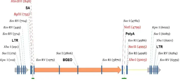
Sucla2+/‒ heterozygous mice were generated by Texas A&M Institute for Genomic Medicine (TIGM) using a gene-trapping technique [141]. Mice (strain C57BL/6N) were cloned from an ES cell line (IST10208H1; TIGM). The ES cell clone contained a retroviral insertion in the Sucla2 gene (intron 4) identified from the TIGM gene trap database and was microinjected into C57BL/6 albino host blastocysts to generate germline chimeras using standard procedures. The retroviral OmniBank Vector 76 contained a splice acceptor (see Figure 7) followed by a selectable neomycin resistance marker/LacZ reporter fusion (β-Geo) for identification of successful gene trap events further followed by a polyadenylation signal. Insertion of the retroviral vector into the Sucla2 gene led to the splicing of the endogenous upstream exons into this cassette to produce a fusion that leads to termination of further transcription of the endogenous Sucla2 exons downstream of the insertion. Chimeric males were bred to 129/SvEv females for germline transmission of the mutant Sucla2 allele.
Suclg2 heterozygote mice [B6-Suclg2Gt(pU-21KBW)131Card] were generated at CARD, Kumamoto University, Japan also using a gene-trapping technique [142]. Mice (strain Albino B6) were cloned from an ES cell line (Ayu21-KBW131; Exchangeable Gene Trap Clones: EGTC). The ES cell clone contained a trap vector insertion in the Suclg2 gene (first intron) identified from the EGTC database and was aggregated with morulae from ICR mice to generate germline chimeras using standard procedures. pU21- W (accession number: AB427140, 9333 bp) was a ‘promoter trap’ vector with three stop codons, which were arranged upstream of the ATG of the β-geo in all three frames (see Figure 8). Insertion of the trap vector into the Suclg2 gene led to the splicing of the
34
endogenous upstream exons into this cassette to produce a fusion transcript that leads to termination of further transcription of the endogenous Suclg2 exons downstream of the insertion. Chimeric males were bred to C57BL/6N females for germline transmission of the mutant Suclg2 allele. To investigate the expression level of Suclg2 mRNA of Suclg2 heterozygote, the original ES cell line (Ayu21-KBW131: +/−) was compared with the parental strain (KAB6: +/+). mRNA was purified from parental ES cells (+/+) and 21- KBW131 (+/−). Suclg2 expression levels of these cells were analyzed by real-time PCR using the TaqMan Gene Expression Assays, XS, Suclg2 (AB, 4448892, FAM/MGB- NFQ) kit. Heterozygous ES cells showed almost half the amount of Suclg2 mRNA compared with parent cells see results in Chapter 5.9.
Neither Sucla2 ‒/‒ nor Suclg2 ‒/‒ mice were ever born from mating heterozygous mice, suggesting that complete absence of either gene is incompatible with life in mice, and as also reported in [21]. By mating Sucla2 heterozygous mice with Suclg2 heterozygous mice, double transgenic (Sucla2+/‒/Suclg2+/‒) mice were born and viable.
Figure 7 Generation of Sucla2 mutant mice Gene trap vector for generating Sucla2 mutant mice.
35 Figure 8 Generation of Suclg2 mutant mice Gene trap vector for generating Suclg2 mutant mice
(adapted from http://egtc.jp/action/access/vector_detail?vector=pU-21W) 4.2 Isolation of mitochondria
Isolation of mitochondria from mouse liver, heart, and brain: liver and heart mitochondria from all animals were isolated as described in ref. [143], with the modifications described in refs [36] and [35]. Nonsynaptic brain mitochondria were isolated on a Percoll gradient as described previously [144], with minor modifications detailed in ref. [145]. Protein concentration was determined using the bicinchoninic acid assay as described below in Chapter 4.3.
Yields were typically 0.2 ml of ∼20 mg/ml per two brains; for liver, yields were typically 0.7 ml of ∼70 mg/ml per two livers, and for heart mitochondria, yields were typically 0.1 ml of ∼15 mg/ml per two hearts.
4.3 Determination of protein concentration
Protein concentration was determined using the bicinchoninic acid assay [146]
(ThermoFisher SCIENTIFIC ‒ Pierce™ BCA Protein Assay Kit) , and calibrated using bovine serum standards using a Tecan Infinite® 200 PRO series plate reader (Tecan Deutschland GmbH, Crailsheim, Germany).
4.4 Mitochondrial substrates and substrate combinations
In our experiments and protocols, it is critical to use adequate substrate combinations to maintain mitochondrial respiration. While some substrates would rather support, others
36
would rather not support substrate-level phosphorylation. Glutamate (glut) and α- ketoglutarate (α-KG) are two substrates that support substrate-level phosphorylation to the greatest extent, however, these should be combined with malate (mal) because it assists in the entry into mitochondria of other substrates including glutamate and α- ketoglutarate. Malate alone has no effect on mitochondrial substrate-level phosphorylation. β-hydroxybutyrate (βOH) is a ketone body, that can be converted to acetoacetate (AcAc) by β-hydroxybutyrate dehydrogenase in the following reaction [147]:
(R) − 3 − hydroxybutyrate + NAD+ ↔ acetoacetate + NADH + H+
It also has no direct effect on substrate-level phosphorylation; however, βOH decrease matrix NAD+/NADH ratio, and therefore reduce mitochondrial substrate-level phosphorylation through blocking succinyl-CoA production by alpha-ketoglutarate dehydrogenase complex, the enzyme that needs oxidized NAD+ [56, 57].
4.5 Determination of membrane potential (ΔΨm) in isolated liver mitochondria
m of isolated mitochondria (0.5-1 mg -depending on the tissue of origin- per two ml of medium containing, in mM: KCl 8, K-gluconate 110, NaCl 10, Hepes 10, KH2PO4 10, EGTA 0.005, mannitol 10, MgCl2 1, substrates as indicated in the figure legends, 0.5 mg/ml bovine serum albumin [fatty acid-free], pH 7.25, and 5 mM safranine O) was estimated fluorometrically with safranine O [148]. Traces obtained from mitochondria were calibrated to millivolts as described in [36]. Fluorescence was recorded in a Hitachi F-7000 spectrofluorometer (Hitachi High Technologies, Maidenhead, UK) at a 5-Hz acquisition rate, using 495- and 585-nm excitation and emission wavelengths, respectively, or at a 1-Hz rate using the O2k-Fluorescence LED2-Module of the Oxygraph-2k (Oroboros Instruments, Innsbruck, Austria) equipped with an LED exhibiting a wavelength maximum of 465 +/‒ 25 nm (current for light intensity adjusted to 2 mA, i.e., level '4') and an <505 nm shortpass excitation filter (dye-based, filter set
"Safranin"). Emitted light was detected by a photodiode (range of sensitivity: 350-700 nm), through an >560 nm longpass emission filter (dye-based). Experiments were performed at 37 oC. Safranine O is known to exert adverse effects on mitochondria if used at sufficiently high concentrations (i.e. above 5 μM, discussed in [57]). However, for
37
optimal conversion of the fluorescence signal to m, a concentration of 5 μM safranine O is required, even if it leads to a diminishment of the respiratory control ratio by approximately one unit (not shown). Furthermore, the non-specific binding component of safranine O to mitochondria (dictated by the mitochondria/safranine O ratio) was within 10% of the total safranine O fluorescence signal, estimated by the increase in fluorescence caused by the addition of a detergent to completely depolarized mitochondria (not shown). As such, it was accounted for, during the calibration of the fluorescence signal to m.
4.6 Mitochondrial respiration
Oxygen consumption was estimated polarographically using an Oxygraph-2k. 0.5-1 mg -depending on the tissue of origin- mitochondria was suspended in 2 ml incubation medium, the composition of which was identical to that for m determination.
Experiments were performed at 37 oC. Oxygen concentration and oxygen flux (pmol·s−1·mg−1; negative time derivative of oxygen concentration, divided by mitochondrial mass per volume and corrected for instrumental background oxygen flux arising from oxygen consumption of the oxygen sensor and back-diffusion into the chamber) were recorded using DatLab software (Oroboros Instruments).
4.7 Cell cultures
Fibroblast cultures from skin biopsies from the patient with no SUCLA2 expression and a control subject were prepared. Cells were grown on poly-L-ornithine coated flasks for 5-7 days in RPMI1640 medium (GIBCO, Life Technologies, Carlsbad, CA, USA) supplemented with 10% fetal bovine serum and 2 mM glutamine and kept at 37 oC in 5%
CO2. The medium was also supplemented with penicillin, streptomycin, and amphotericin (Sigma-Aldrich St. Louis, MO, USA).
4.8 Mitochondrial membrane potential (ΔΨm) determination in in situ mitochondria of permeabilized fibroblast cells
ΔΨm was estimated using fluorescence quenching of the cationic dye safranine O due to its accumulation inside energized mitochondria [148]. Fibroblasts were harvested by trypsinization, permeabilized as detailed in [61] and suspended in a medium identical to that as for ΔΨm measurements in isolated mitochondria. Substrates were 5 mM glutamate

![Figure 5 Schematic biochemical pathways demonstrating metabolic alterations in succinyl-CoA ligase deficiency caused by elevated succinyl-CoA (adapted from [65, 83]) Abbreviations: CS: citrate synthase; PDH: pyruvate dehydrogenase complex;](https://thumb-eu.123doks.com/thumbv2/9dokorg/1382308.114081/27.892.136.767.154.502/schematic-biochemical-demonstrating-metabolic-alterations-deficiency-abbreviations-dehydrogenase.webp)
![Figure 6 Diagnostic flowchart for patients with common MMA-uria, developmental delay and muscle hypotonia (adapted from [65, 140]) Characteristic metabolic profile in SUCLA2 patients were increased e.g., lactic acid, methylmalonic acid, C4-DC](https://thumb-eu.123doks.com/thumbv2/9dokorg/1382308.114081/28.892.145.752.181.810/diagnostic-flowchart-developmental-hypotonia-characteristic-metabolic-increased-methylmalonic.webp)