Novel aspects of the non-pharmacological management of tachyarrhythmias in chronic systolic heart failure in Adults
Ph.D. Dissertation
Dr. Pál Ábrahám MD
Doctoral School of Basic Medicine Semmelweis University
Consultants:
Dr. Béla Merkely MD, D.Sc.
Dr. Tamás Szili-Török MD, Ph.D.
Official reviewers:
Dr. András Vereckei MD, Ph.D., Med. Habil.
Dr. Gábor Duray MD, Ph.D.
Head of the final examination committee:
Dr. Klára Gyires MD, D.Sc., Med. Habil.
Members of the final examination committee:
Dr. Zoltán Csanádi MD, Ph.D., Med. Habil.
Dr. Lívia Jánoskuti MD, Ph.D., Med. Habil.
Dr. Zoltán Járai MD, Ph.D., Med. Habil.
Budapest 2015
Table of contents
1. Introduction ... 4
1.1. Supraventricular arrhythmias ... 4
1.2. Ventricular arrhythmias ... 7
1.3. Tachycardia-induced cardiomyopathy ... 12
1.4. Remote CIED monitoring and patient management ... 13
1.5. Instantaneous rate-control of rapid atrial fibrillation with coupled and paired ventricular pacing ... 14
1.6. Maximum voltage-guided technique for cavotricuspid isthmus ablation during ongoing atrial flutter ... 15
1.7. Defibrillation coil retention during orthotopic heart transplantation... 17
1.8. Cardiac resynchronisation in chronic renal failure ... 18
2. Objectives ... 19
2.1. Instantaneous rate-control of rapid atrial fibrillation with coupled and paired ventricular pacing ... 19
2.2. Maximum voltage-guided technique for cavotricuspid isthmus ablation during ongoing atrial flutter ... 19
2.3. Defibrillation coil retention during orthotopic heart transplantation... 19
2.4. Cardiac resynchronisation in chronic renal failure ... 20
3. Methods ... 21
3.1. Instantaneous rate-control of rapid atrial fibrillation with coupled and paired ventricular pacing ... 21
3.2. Maximum voltage-guided technique for cavotricuspid isthmus ablation during ongoing atrial flutter ... 23
3.3. Defibrillation coil retention during orthotopic heart transplantation... 25
3.4. Cardiac resynchronisation in chronic renal failure ... 25
3.5. Statistical methods ... 26
3.5.1. Instantaneous rate-control of rapid atrial fibrillation with coupled and paired ventricular pacing ... 26
3.5.2. Maximum voltage-guided technique for cavotricuspid isthmus ablation during ongoing atrial flutter ... 26
3.5.3. Defibrillation coil retention during orthotopic heart transplantation ... 26
3.5.4. Cardiac resynchronisation in chronic renal failure ... 27
4. Results ... 28
4.1. Instantaneous rate-control of rapid atrial fibrillation with coupled and paired ventricular pacing ... 28
4.2. Maximum voltage-guided technique for cavotricuspid isthmus ablation during ongoing atrial flutter ... 30
4.3. Defibrillation coil retention during orthotopic heart transplantation... 32
4.4. Cardiac resynchronisation in chronic renal failure ... 36
5. Discussion ... 42
5.1. Instantaneous rate-control of rapid atrial fibrillation with coupled and paired ventricular pacing ... 42
5.1.1. Rationale of coupled and paired pacing and energetic considerations ... 42
5.1.2. Alternative non-pharmacological methods to regulate or reduce the ventricular rate during atrial fibrillation ... 43
5.1.3. Future clinical implications ... 44
2
5.2. Maximum voltage-guided technique for cavotricuspid isthmus ablation during
ongoing atrial flutter ... 45
5.2.1. Former results with the MVGT and implications for clinical routine ... 45
5.2.2. Clinical impact of the results ... 46
5.3. Defibrillation coil retention during orthotopic heart transplantation... 47
5.4. Cardiac resynchronisation in chronic renal failure ... 50
5.4.1. Outcomes of CRT in renal failure ... 50
5.4.2. Mortality of ICD recipients suffering from CRF ... 51
5.4.3. Utilisation of CRT-D over CRT-P could not reduce mortality in our CRF patients ... 52
6. Conclusions ... 53
6.1. Instantaneous rate-control of rapid atrial fibrillation with coupled and paired ventricular pacing ... 53
6.2. Maximum voltage-guided technique for cavotricuspid isthmus ablation during ongoing atrial flutter ... 53
6.3. Defibrillation coil retention during orthotopic heart transplantation... 53
6.4. Cardiac resynchronisation in chronic renal failure ... 54
7. Summary ... 55
8. Összefoglalás ... 56
9. References... 57
10. List of publications ... 75
10.1. Publications related to my thesis ... 75
10.2. Publications not related to my thesis ... 75
11. Acknowledgements ... 76
List of abbreviations
AE: adverse event
AVNRT: atrio-ventricular node reentry tachycardia AVRT: atrio-ventricular reentry tachycardia
CI: coupling interval
CIED: cardiac implantable electronic device CL: cycle length
CP: coupled pacing CRF: chronic renal failure
CRT-D: cardiac resynchronization therapy defibrillator CRT-P: cardiac resynchronization therapy pacemaker CTI: cavo-tricuspid isthmus
DC-ICD: dual-chamber implantable cardioverter defibrillator ERP: effective refractory period
HF: heart failure HRA: high right atrium HTX: heart transplantation
ICD: implantable cardioverter defibrillator MPR: mechanical pulse rate
MVGT: maximum voltage guided ablation technique LVEF: left ventricular ejection fraction
NYHA: New York Heart Association
PJRT: paroxysmal junctional reciprocating tachycardia PP: paired pacing
PVC: premature ventricular complex RF: radiofrequency
RVA: right ventricular apex
SC-ICD: single-chamber implantable cardioverter defibrillator VNS: vagal nerve stimulation
4
1. Introduction
Both supraventricular and ventricular arrhythmias are prevalent in patients suffering from chronic systolic heart failure (HF), and their impact on mortality and quality of life is significant. Besides the well-established treatment with complex, optimised pharmacological therapy, promising non-pharmacological methods have been emerged in the past decades as frontline therapies: catheter ablation of cardiac arrhythmias and utilisation of cardiac implantable electronic devices (CIEDs). Many of them have already been clinically proved to be effective not only in treating and preventing tachyarrhythmias, but also in reducing morbidity and mortality.
The spectrum of tachyarrhythmias in patients suffering from chronic systolic heart failure is as broad as in the normal population: atrio-ventricular node reentry tachycardia (AVNRT), atrio-ventricular reentry tachycardia (AVRT), ectopic and reentrant atrial tachycardias, non-reentrant junctional tachycardias, atrial flutter, atrial fibrillation, premature ventricular complexes (PVCs), and different sorts of ventricular tachycardias are all can be found in heart failure patients. Nevertheless, the distribution of these arrhythmias shows some heart-failure and age-related variability. For example AVRT, paroxysmal junctional reciprocating tachycardia (PJRT), junctional ectopic tachycardia have a strong predilection in childhood (Ko et al. 1992, Coumel P et al.
1967), whereas atrial fibrillation is predominantly the arrhythmia of adulthood and its likelihood is growing with advanced age, as it became evident from the sub-analysis of the Framingham study (Kannel WB et al. 1982). Well-known fact, that malignant ventricular arrhythmias and sudden cardiac death are far more common in systolic heart failure, especially in patients suffering from post-infarction ischemic cardiomyopathy (Myerburg et al. 1992). The prevalence of atrial fibrillation is also considerably higher in congestive heart failure (Fabbri G et al. 2012).
1.1. Supraventricular arrhythmias
In the non-pharmacological treatment of supraventricular tachyarrhythmias catheter ablation techniques are impressively effective, therefore they have a primary role over CIEDs. Catheter ablation therapy of the common supraventricular arrhythmias in heart
ablation, cavo-tricuspid isthmus (CTI) ablation, and pulmonary vein isolation requires the same technical setup as in otherwise healthy patients. What can make these cases more challenging are the abnormal anatomy and the usually enlarged cardiac chambers and dilated tricuspid and mitral annuli. The atria in chronic systolic heart failure are dilated due to the elevated end-diastolic pressures and dilated cardiomyopathy usually causes annular dilatation which can promote significant functional valvular regurgitation. All of these pathologic variations can make manual catheter manipulation and effective energy delivery even more difficult (Ren JF et al. 1998), frequently requiring intracardiac ultrasound guidance and the use of long-sheaths to give sufficient support to the ablation-catheter.
Another important aspect of the ablation procedure is time. Patients suffering from heart failure can hardly tolerate long sessions and sometimes considerable amount of saline- overload without dyspnoea while lying supine on the table. Therefore every attempt to shorten procedure time without compromising success is welcomed. This can be achieved with single-shot devices like the cryoballon (Sarabanda AV et al. 2005) or the phased-array duty-cycled ablation device (Boersma LV et al. 2008) in pulmonary vein isolation of atrial fibrillation ablation, or with the maximum-voltage guided technique of CTI ablation performed in sinus rhythm (Redfearn DP et al. 2006). Ablation time, procedure time and success rate were compared using the maximum-voltage guided technique during ongoing isthmus-dependent atrial flutter in our work.
The impact of atrial fibrillation ablation on long term clinical endpoints and mortality is under investigation. The ongoing CASTLE-AF (Marrouche NF et al. 2009), and RAFT- AF trials are recruiting patients suffering from chronic heart failure. The results are due in 2016 and awaited with excitement, since no pharmacologic rhythm control strategy was proved to be superior over rate control both in preserved (AFFIRM: mean left ventricular ejection fraction (LVEF) 54.7% – Wyse DG et al. 2002), and in depressed left ventricular function (AF-CHF: mean LVEF 27.6% – Roy D et al. 2008). Results of some nonrandomized studies and a small randomized single-centre trial (ARC AF - Jones DG et al. 2013) that recruited heart failure patients who underwent successful
6
radiofrequency (RF) ablation for persistent atrial fibrillation are promising in terms of improved cardiopulmonary exercise capacity and quality of life when comparing them to the pharmacological rate control strategy. Results of atrial fibrillation ablation were compared between groups of congestive heart failure and normal systolic function in an early prospective trial conducted by the Bordeaux group (Hsu LF et al. 2004). They reported significant improvement not only in symptoms and exercise capacity, but also in ejection fraction. Assessing differences in mortality was beyond the scope of all these small studies. Nevertheless, these results may give rise to the speculation that rhythm control achieved by catheter ablation could have a priority over cumbersome antiarrhythmic drug therapy with its well-known and feared proarrhythmic effects.
Catheter ablation of paroxysmal or persistent atrial fibrillation (i.e. non-pharmacological rhythm control) in heart failure proved to be superior when it was compared to the
“ablate and pace” strategy of rate control. This was demonstrated in the randomized, multicenter PABA-CHF trial (Khan MN et al 2008). Not only the composite endpoint (LVEF, 6-minute walk test, and quality of life score), but also LVEF alone was significantly higher in the pulmonary vein isolation group in the end of the 6-month follow-up period (35 vs. 28 % p<0.001). Of note, there was no improvement in ejection fraction in the “ablate and pace” group. This is quite embarrassing given the fact that the pacing method used was biventricular regarding the baseline mean LVEF of 29%, and in the previously conducted PAVE trial biventricular pacing proved to be superior over conventional right ventricular apical pacing after complete AV-node ablation in a group of patients with a mean LVEF of 47% (Doshi RN et al. 2005).
Device therapy can also have a promising new role in atrial fibrillation rate control.
Rate control of rapid ventricular response can be achieved by AV-conduction slowing drugs or in drug-refractory cases, by AV-node catheter ablation with prior pacemaker implantation. The recommended pacemaker is preferably a biventricular system in systolic heart failure (Brignole et al. 2013) if we consider the almost 100% pacing ratio after AV-nodal ablation. Our group has tested two similar ventricular pacing methods to instantly control the mechanical pulse rate: coupled pacing and paired pacing.
1.2. Ventricular arrhythmias
In the non-pharmacological treatment of ventricular arrhythmias both CIEDs and catheter ablation techniques have a firm ground in heart failure. The landmark randomized trials on implantable cardioverter defibrillator (ICD) implantation for secondary prevention of sudden cardiac death due to malignant ventricular arrhythmias were done in the end of the nineties of the past century. The AVID trial (Zipes DP et al.
1997), the CASH trial (Kuck KH et al.2000) and the CIDS trial (Connolly S et al. 2000) proved first that ICDs decrease relative mortality by 20-33% in patients with impaired systolic function (mean LVEF were 32%, 46%, and 33%, respectively), as compared with antiarrhythmic therapy only. Both ischemic and non-ischemic cardiomyopathy patients benefited from ICD implantation, although sometimes in a statistically non- significant manner.
Not much later, larger trials were conducted in the field of primary prevention of sudden cardiac death of coronary patients with results of relative mortality reduction in a magnitude of 23-51% (SCD-HeFT – Bardy G et al. 2005; MADIT-II – Moss AJ et al.
2002 ; MUSTT – Buxton AE et al. 1999). In the DEFINITE trial (Kadish A et al. 2004) ICDs reduced the rate of sudden death from arrhythmia in non-ischemic cardiomyopathy significantly by 80%, while all-cause mortality reduction was non- significant. With the results of the DINAMIT (Hohnloser S et al. 2004) and IRIS (Steinbeck G et al. 2009) trials it became clear that primary prevention ICD implantation within 30-40 days of an acute coronary event complicated with depressed systolic function did not reduce long-term mortality. It is of note, however, that arrhythmic mortality decreased in both trials in a significant manner, but this gain was offset by the rate of non-arrhythmic deaths. To our current knowledge, only ICD implantations performed more than 40 days after myocardial infarction carry favourable clinical outcomes.
Since 1994, when Serge Cazeau and his colleagues performed the first biventricular device implantation (which was originally a four-chamber system, the left ventricular lead was implanted epicardially and the coronary sinus was used to place the left atrial lead: Cazeau S et al. 1994), cardiac resynchronisation therapy (CRT) has emerged as an effective tool in the treatment of advanced heart failure in patients with systolic dysfunction, wide QRS complex, and on optimal medical therapy. Besides improving
8
quality of life, ejection fraction, exercise tolerance and reducing heart failure-related symptoms, cardiac resynchronisation therapy pacemaker (CRT-P) added on top of optimal medical therapy could significantly reduce mortality as the landmark CARE-HF trial proved it (Cleland JG et al. 2005). Relative mortality reduction was 36% at 29 months, and this study also detected a decreased number of sudden death with CRT-P (9.4 % on optimal medical therapy vs. 7.1 % on CRT-P). This could also be attributed to the improvement in left ventricular systolic function. Because the target population for CRT inherently has poor left ventricular ejection fraction, combining CRT with an ICD for primary prevention became almost a must in the developed world, especially after the results of the COMPANION trial (Bristow MR et al. 2004). This trial showed some incremental benefit of cardiac resynchronisation therapy with defibrillator (CRT- D) over CRT-P in relative all-cause mortality reduction (a secondary end-point variable) against medical therapy-only (36% vs.24%). It is of note, however, that this trial was not primarily designed to directly compare CRT-P with CRT-D, so its conclusions refer strictly to the primary goal: comparison of CRT (with and without ICD) versus optimal medical therapy alone, and the authors correctly concluded at the end of their paper, that the choice between CRT-P or CRT-D should be made individually according to the particular clinical case (Bristow MR et al. 2004). In low symptomatic New York Heart Association (NYHA) I-II patients with an ejection fraction below 30%, CRT-D reduced the combined end-point of all-cause mortality or a heart-failure event by 34% versus ICD-alone according to the MADIT-CRT trial (Moss AJ et al. 2009). There was no significant mortality reduction in this trial. Similar results were reported in the RAFT trial conducted on NYHA II-III patients, with 25% relative reduction in the combined primary end-point of death or hospitalization for heart failure (Tang AS et al. 2010).
Moreover, this latter trial showed a significant risk reduction in all-cause mortality (25%), and in the subgroup analysis this remained true even for NYHA II patients.
Considerable hemodynamic improvement assessed by Doppler echocardiography after initiation of biatrial pacing in patients with interatrial conduction block, called “atrial resynchronization” was enthusiastically reported in a paper as a single-centre experience (Daubert JC et al. 2004). Here the right atrial lead was placed in the conventional way in the appendage, and an additional left atrial lead was placed far out in the coronary sinus.
resynchronization therapy has not gained wide acceptance in everyday clinical practice.
The above-mentioned strong results of ICD and CRT trials and the accumulating experience of the implanting centres published as single-centre studies have been embedded in the recommendations of the current international guidelines on treating ventricular arrhythmias (Zipes DP et al. 2006), device therapy (Epstein AE et al. 2013), and the 2013 cardiac pacing and resynchronization therapy guideline (Brignole M et al.
2013). Most ICD and CRT-P/D indications are backed with an A or B level of evidence.
Patient selection for CRT continuously refines as new data regarding the impact of comorbid conditions on survival come into the limelight. The most striking example is the unfavourable outcome with non-left bundle branch block in the subgroup analysis of the MADIT-CRT study. But other comorbidities as pulmonary hypertension, extreme left ventricular dilatation, and chronic renal failure are getting more attention even at the guideline level (Brignole et al. 2013). The role of chronic renal failure was addressed by our work in a series of 60 CRT patients.
On the downside of ICD therapy are the device-related complications and inappropriate ICD discharges. As the number of implantations exponentially rises, the absolute number of lead-related complications is also rising steeply, causing more need for complex extraction procedures. Insulation damage, conductor fracture, externalisation, dislodgement, and cardiac perforation are some of the mechanisms of ICD lead-related complications (Borleffs CJ et al. 2009). Implantation of an ICD is common for non- hospitalized patients awaiting heart transplantation (Sandner SE et al. 2001), and those ICD systems are usually totally explanted during the transplantation procedure.
Complication rates and possible causes of the failure of total surgical system explantation during heart transplantation were addressed by our work in Rotterdam.
CRT is only effective in patients with wide-QRS complexes, especially of left bundle branch block morphology. In fact, the greater proportion of heart failure patients has narrow QRS complexes therefore an effective and safe device-based therapy targeting this population would be of high clinical importance. Vagal nerve stimulation (VNS)
10
has been traditionally used in drug refractory epilepsy, but its use in congestive heart failure has been investigated in the last few years as it effectively reduces heart rate and increases heart rate variability. Although the concept of the human cervical vagal stimulation system is not new (US patent in 2001), the first in-man study was reported only in 2008 by Schwartz and colleagues. The system was well-tolerated and VNS improved NYHA class, quality of life and decreased left ventricular end-systolic volume during 6 months in 8 patients (Schwartz PJ et al. 2008). In 2011 the CardioFit Multicenter Trial showed similarly promising results in 32 patients after 6 months with an improvement in LVEF from 22+7 to 29+8%, although this came with the expense of numerous serious adverse events (40.6%) and non-negligible side-effects as death (3/32), pulmonary oedema, surgical complication, and stimulation pain (De Ferrari GM et al. 2011). Results of the NECTAR-HF study (NYHA class II-IV, LVEF <35%, on optimal medical therapy, right sided VNS, 96 patients) did not evidence any significant improvement in LVEF or a reduction in brain-natriuretic peptide levels at 6 months, although quality of life was improved in the actively treated group (presented at ESC Congress 2014, Barcelona by Zannad F). The ANTHEM-HF trial (NYHA class II-III, LVEF <40%, optimal medical therapy, right or left sided VNS, 60 patients) had somewhat better results as LVEF improved significantly in 6 months (presented at ESC Congress 2014, Barcelona by Anand IS). The average stimulation current applied in this study was somewhat higher than that in the NECTAR-HF (2.0 vs 1.4 mA, respectively).
Noteworthy, none of these trials were designed to assess hard clinical end-points.
Catheter ablation of outflow tract ventricular arrhythmias can be as effective in heart failure patients as in normal subjects. The technical setup, procedure, and endpoints are just the same. Left ventricular systolic function can improve considerably after outflow tract ablation in patients with frequent monomorphic PVCs. Navigation to the site of origin of PVCs can be challenging, and in the minority of cases the substrate can only be localised with epicardial mapping via the coronary sinus or via direct subxyphoid epicardial access (Abraham P, Abkenari LD et al. 2013). In difficult-to-reach areas remote magnetic navigation can help steer the catheter to the right spot and maintain reliable catheter contact: both are key factors of a successful and safe ablation procedure (Schwagten B et al. 2009).
A considerable proportion of heart failure patients suffer from post-infarction ischaemic cardiomyopathy. Infarct scar-related macroreentry ventricular tachycardias are characteristic arrhythmias of this population. Nevertheless, scar-related ventricular arrhythmias are also well known in other diseases as dilated cardiomyopathy, Chagastic disease, arrhythmogenic right-ventricular cardiomyopathy, and cardiac sarcoidosis.
Catheter ablation of a ventricular tachycardia in a patient suffering from heart failure is a rather challenging task both intellectually and manually and requires seamless team- work. The introduction of substrate mapping (Marchlinsky FE et al. 2000) and the use of open irrigated tip ablation catheters gave a boost to ventricular tachycardia ablation.
Multicenter studies like the Thermocool Ventricular Tachycardia Ablation trial (Stevenson WG et al. 2008) or the Euro-VT trial (Tanner H et al. 2010) conducted in experienced, large-volume sites presented promising results on acute success rates (81%), and on the reduction of ICD shock burden, but moderate success on the 6-month arrhythmia-free survival (53%, 51 %). These studies indicate that high recurrence rates (51%, 49 %) still remain an issue to deal with. Because frequent ICD discharges are not only painful, causing significant discomfort and psychological stress to patients, but are also independent predictors of adverse clinical outcomes, techniques to reduce avoidable appropriate and inappropriate ICD shocks are increasingly become part of the modern anti-tachycardia management (Israel CW 2008). Preventive substrate-based ablation of the left ventricular scar in post-infarction patients considerably reduced ICD discharges, as it has been straightforwardly demonstrated by the SMASH-VT trial investigators (Tung R, et al. 2010). These studies also imply that ICDs and antiarrhythmic drug therapy still remain in the mainstream of ventricular tachycardia management.
Bundle-branch ventricular tachycardias are common in dilated cardiomyopathy with 41% incidence, and sometimes after surgical aortic valve replacement (Caceres J et al.
1989). Fast rates, poor haemodynamic tolerance and bundle-branch block QRS pattern similar to sinus rhythm are quite characteristic of these tachycardias. Catheter ablation of the conduction system (mainly the right bundle) can cure the arrhythmia with nearly 100 % success even without the use of sophisticated three-dimensional electro-anatomic
12
mapping systems. Although curative bundle-branch ventricular tachycardia ablation could be a promising perspective, leaving the patient without an ICD carries a non- negligible risk, and future malignant arrhythmias are not uncommon if we consider the underlying cardiac disease. (Lopera G et al. 2004)
1.3. Tachycardia-induced cardiomyopathy
An evergreen aspect of the arrhythmia-heart failure relationship is the chicken-or-egg question of causality. Not only the malfunctioning heart can be more susceptible of even life-threating arrhythmias but also some forms of supraventricular and ventricular tachyarrhythmias can deteriorate left ventricular systolic function considerably, causing a special form of heart failure called tachycardia-induced cardiomyopathy. By definition, this is a cardiomyopathy with left ventricular dilation and systolic dysfunction in the absence of other causative structural heart disease, and myocardial function recovers after termination of the arrhythmia (Packer DL et al. 1986). The incidence of tachycardia-induced cardiomyopathy was 2.7 % in a group of 625 patients with congestive heart failure referred for catheter ablation. (Donghua Z et al. 2013).
Among atrial tachyarrhythmias incessant forms of focal atrial tachycardia (Medi C et al.
2009), PJRT (Dorotskar et al. 1999), atrial flutter (Pizzale et al. 2009), and atrial fibrillation with rapid ventricular conduction (Grogan M et al. 1992) are the most common forms causing tachycardia-induced cardiomyopathy. Males, young patients with incessant forms of atrial tachycardias are more susceptible of developing tachycardia-induced cardiomyopathy in focal atrial tachycardias (Medi C et al. 2009). It is interesting that not only ventricular rate matters but also rhythm irregularity and longer atrial cycle lengths can contribute to the development of this cardiomyopathy, and hence a reliably predictive threshold in heart rate does not exist. Even an arrhythmia other than a true tachycardia can cause systolic dysfunction. Frequent PVCs with a 24- hour burden of more than 20-24% (Takemoto M et al. 2005, Baman TS et al. 2010) or with a >10.000/day absolute number (Kanei Y et al. 2008) can considerably decrease left ventricular ejection fraction, and can lead to heart failure on the long run. These PVCs mostly arise from the outflow tracts of the ventricles or from the valve cusps, and can be ablated safely and effectively with success rates reaching 90%. Medical
rhythm but also left ventricular systolic function, usually within some weeks or months (Takemoto M et al. 2005, Vijgen J et al. 1997, Hasdemır C et al. 2013).
1.4. Remote CIED monitoring and patient management
What CIEDs can undoubtedly add to the improvement of standards in heart failure management are their extensive arrhythmia- and heart failure-related diagnostic functions combined with remote follow-up and remote monitoring capabilities. These features include -among many others- monitoring and trending lead and generator integrity, atrial and ventricular arrhythmia burden, patient activity, mean heart rate, heart rate variability, pulmonary artery and left atrial pressures, and intrathoracic impedance changes to monitor possible thoracic fluid accumulation. Similarly to the old clinical observation, the onset of a persistent atrial arrhythmia can lead to heart failure more frequently (relative risk 65%) which is heralded by a decrease in thoracic impedance indicating fluid accumulation, and vice versa: significant intrathoracic impedance drops are followed by new-onset atrial fibrillation with a relative risk of 32 percent over 30 days (Andriulli JA et al. 2014). The really innovative point in the CIED-based remote management is to forecast impending clinical deterioration and to response earlier than a routine in-office visit would take place, and before disease progression would lead to hospitalisation. The body of evidence of remote management is growing regarding its safety, utility and long-term positive impact on patient outcomes. The results of the impressively large North American ALTITUDE registry (remotely/standard monitored: 69.556/116.222) showed a striking 50 percent relative mortality risk reduction and long-term survival benefit in remotely monitored ICD patients (Saxon LA et al. 2010). When the information gained from remote monitoring of an ICD or CRT-D system was incorporated into a clinical response-system, earlier therapy decision and intervention could prevent further clinical deterioration, and the length of hospital stay due to a cardiovascular event was significantly shorter for patients in the remote monitoring arm. This was indicated by the results of the CONNECT trial (Crossley GH et al. 2011), and more recently by the IN-TIME study, where a significant difference in the modified Packer score (27.2% vs 18.9%; p<0.05)
14
and all-cause mortality (8.7% vs 3.4%; P<0.01 ; p< 0.012) was observed in favour of the telemonitored cohort after 12 months follow-up (Hindricks et al. 2014). The content of telemonitored data prompted clinician contact with 76% of the patients. Data from the COMPAS (Mabo P et al. 2012) and ECOST trials (Guedon-Moreau et al. 2013) showed that telemonitoring of both conventional pacemaker systems and ICDs is safe, reduces the annual number of in-office visits, and telemonitoring-based patient management could substantially decrease (70% relative reduction) the number of appropriate and inappropriate ICD shocks in the remotely monitored arm of the ECOST study. Yearly in-office follow-up of primary prevention ICD systems is as safe as quarterly checking, and it is mutually beneficial for the patient and for the caregiver as it improves quality of life and significantly decreases the in-office follow-up burden of the outpatient clinics according to the results of the REFORM trial (Hindricks et al. 2014).
As a consequence, the current European guideline on cardiac pacing endorses remote monitoring with an IIa recommendation (Brignole et al. 2013).
Another interesting remote monitoring target is implantable hemodynamic monitoring.
In the CHAMPION trial a wireless pulmonary artery pressure monitor delivered via right heart catheterisation was tested. Remote monitoring-guided timely patient management with diuretics or long-acting nitrates to reach targeted pulmonary arterial pressure values (mean pressure 10-25 Hgmm) yielded in a 37% relative risk reduction in heart failure hospitalizations (Abraham WT et al. 2011). A transvenously implanted pressure sensor lead capable of directly monitoring left atrial pressure changes on the interatrial septum is under clinical investigation. Both kinds of sensors are powered wirelessly from an external patient interface unit.
1.5. Instantaneous rate-control of rapid atrial fibrillation with coupled and paired ventricular pacing
Atrial fibrillation is the most common sustained cardiac arrhythmia and its prevalence keeps increasing worldwide. Because the arrhythmia is often asymptomatic the true prevalence is likely even higher (Camm AJ et al. 2010). When it is not possible or not reasonable to restore sinus rhythm, ventricular rate control is the option to alleviate
ventricular rates. The efficacy of the available pharmacological agents to control the ventricular rate during permanent atrial fibrillation is limited and in some cases non- pharmacological methods such as atrioventricular nodal ablation and implantation of a permanent pacemaker are necessary.
Premature activation of the heart results in impaired contraction and randomly occurring ventricular ectopic activations can prolong the compensatory pause during atrial fibrillation. Based on this observation the concept of coupled pacing (CP) in rapid atrial fibrillation has been developed (Lau CP et al. 1990) and was revisited recently to improve cardiac function by controlling ventricular rate (Yamada H et al. 2006). CP consists of premature electrical stimulation delivered shortly after the effective refractory period (ERP) of the sensed ventricular activation, resulting in electrical activation but no mechanical contraction. The concealed ventriculoatrial conduction caused by the extrastimulus can prolong the conducted RR interval in atrial fibrillation, and thus decrease the mechanical pulse rate (MPR). When pairs of stimuli are applied and the second stimulus is placed shortly after the ERP of the first one, minimal or no mechanical contraction occurs (Frommer PL et al. 1966). During this paired pacing (PP) the first stimulus of each pair results in an augmented contractile response by post- extrasystolic potentiation and can increase the cardiac output in heart failure patients (Braunwald E et al. 1964, Cooper MW et al. 1986). As rapid paroxysmal atrial fibrillation can lead to symptomatic heart failure, a non-pharmacological method to promptly control the pulse rate of such an episode would be of great clinical importance.
1.6. Maximum voltage-guided technique for cavotricuspid isthmus ablation during ongoing atrial flutter
Radiofrequency ablation of the CTI is a widely and effectively used procedure in patients with counterclockwise or clockwise typical atrial flutter (Cosio FG et al. 1993, Kirkorian G et al 1994, Shah DC et al. 1997). Ablation is superior to conventional
16
antiarrhythmic drug treatment (Natale A et al. 2000) because at the end of a 21±11 month-long follow-up only 36% of patients on antiarrhythmics were in sinus rhythm and 63% required rehospitalisation, compared to the 80% and 22% after CTI ablation.
Pathological studies revealed that the CTI is composed of distinct muscular bundles, formed to an isthmus by connective tissue binding them together (DaCosta A et al.
2004, Cabrera JA et al. 2005).
In most centres an empiric anatomical approach is performed by drawing a line of lesions from the tricuspid annulus to the inferior caval vein until bidirectional conduction block is achieved. Although, this approach is highly successful, it might require a high number of applications and long procedure times. Recently, an alternative approach was developed where large atrial signals were targeted as they represent bundles of conduction through the isthmus. This method was tested and was proven to be applicable (Redfearn DP et al. 2006, Subbiah RN et al. 2007, Posan E et al. 2007).
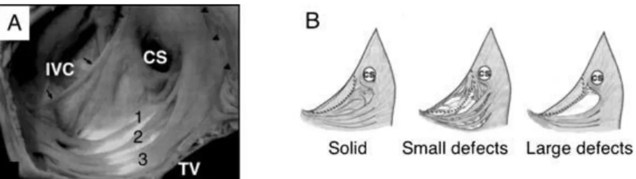
The results are promising in decreasing fluoroscopy and procedure times without compromising success rate. The success of this novel method is related to the anatomy of the CTI (Figure 1). It seems that the CTI does not appear to be a continuous homogenous atrial tissue. Indeed, careful histological examination of the isthmus
Figure 1 Cavo-tricuspid isthmus anatomy
Panel A shows the cavo-tricuspid region of the right atrium in a macroscopic anatomic specimen. Numbers show the discrete extensions of pectinate muscular bundles forming the isthmus. Black arrows highlight the Eustachian valve. Panel B depicts different isthmus anatomies.
IVC: inferior vena cava, CS: coronary sinus, TV: tricuspid valve (from Redfearn DP et al. 2006)
reveals very variable anatomy (Cabrera JA et al. 2005, DaCosta A et al. 2004, Redfearn DP et al. 2006). The length, the shape and some other parameters are very varying. On top of that it is clear that the isthmus is not composed of muscle bundles equally distributed throughout this region. These muscle fibers rather oriented as discrete bundles and pectinate extensions of the crista terminalis (DaCosta A et al. 2004). This certainly determines its conduction properties favouring longitudinal conduction over these bundles. Based on the hypothesis that these bundles are responsible for the conduction of electrical activation, the maximum-voltage guided technique (MVGT) was developed and introduced (Ozaydin M et al. 2003, Hall B et al. 2004, Okishige K et al. 2005, Redfearn DP et al. 2006). This technique was reported to be a feasible method to reach bidirectional isthmus block without the need for a complete anatomic ablation line, however all ablation procedures were performed in sinus rhythm or during pacing from the coronary sinus (Redfearn DP et al. 2006). Using this ablation technique shorter ablation times could be achieved among patients in sinus rhythm according to previous reports (Subbiah RN et al. 2007). This was associated with higher mean temperature, suggesting better catheter stability. However, this issue was not evaluated in patients undergoing CTI ablation during ongoing atrial flutter in a randomized prospective fashion.
1.7. Defibrillation coil retention during orthotopic heart transplantation
An implantable cardioverter-defibrillator has become the standard therapy for patients with chronic heart failure at high risk for sudden cardiac death. Therefore it is a reasonable choice for non-hospitalized patients awaiting heart transplantation (HTX) (Sandner SE et al. 2001). Numerous primary prevention trials conducted on sudden cardiac death in chronic heart failure patients have resulted in the expansion of ICD indications, thus the number of implanted ICD systems is growing continuously.
Defibrillation leads are the most vulnerable part of the ICD system, due to their complex design and to their need to withstand mechanical strain. Management of patients presenting with ICD lead-related adverse events can be very challenging. The estimated lead survival rates are 98% at 4 years, 85% at 5 years, and 62 to 60% at 8 years (Dorwarth U et al. 2003, Kleemann T et al. 2007). Better long-term lead survival
18
rates with 83 % at 10 years were reported when polyurethane and coaxial leads were excluded from analysis (Borleffs CJ et al. 2009). Malfunction or failure of ICD leads can occur by several mechanisms including dislodgement, cardiac perforation, conductor fracture, and insulation damage, some of them may be attributed to lead design (Porterfield JG et al. 2009). Malfunctioning, failed, or infected ICD leads require lead revision, extraction and/or replacement. The removal of a retained lead fragment after a transvenous lead extraction attempt is often possible only via thoracotomy (Wilkoff BL et al. 2009).
1.8. Cardiac resynchronisation in chronic renal failure
Cardiac resynchronisation therapy has emerged from the therapeutic options of chronic heart failure as an effective tool of treatment over the last two decades. By performing CRT in patients who do not adequately respond to optimal pharmacological therapy, clinicians can relieve HF symptoms, improve quality of life, and reduce long-term mortality (Cleland JG et al. 2005, Bristow MR et al. 2004). Previous guidelines have laid down general indications for CRT as ejection fraction below 35 %, New York Heart Association (NYHA) functional class of II-IV, and QRS duration longer than 120-130 ms (Vardas PE et al. 2007, Epstein AE et al. 2008). Chronic renal failure (CRF) is a condition that worsens the clinical course and mortality of HF (Smith GL et al. 2006), and data regarding its impact on the success of CRT are sparse in the literature.
2. Objectives
2.1. Instantaneous rate-control of rapid atrial fibrillation with coupled and paired ventricular pacing
The objective of this study was to compare the acute rate-controlling effects of CP and PP algorithms during atrial fibrillation with rapid ventricular rates in humans in an experimental setting. We also wanted to determine the drive train cycle length resulting in the lowest controlled rate during PP. Safety of the different ventricular pacing algorithms were analysed according to their ability to trigger life-threatening arrhythmias.
2.2. Maximum voltage-guided technique for cavotricuspid isthmus ablation during ongoing atrial flutter
In order to assess the feasibility of the MVGT during ongoing atrial flutter we compared procedural outcomes in patients undergoing standard anatomical ablation vs. patients with MVGT. The primary endpoints of this prospective randomized study were acute and chronic procedural success, fluoroscopy time, and procedure times. Secondary endpoints were the number of radiofrequency lesions and total radiofrequency application duration.
2.3. Defibrillation coil retention during orthotopic heart transplantation
Surgical removal of the whole ICD system is part of the orthotopic HTX procedure and it is usually carried out without complications (Smith MC et al. 2008). We wanted to explore the incidence and causes of defibrillation lead-related adverse events in our HTX patients who had an ICD implantation while awaiting heart transplantation. We collected data on lead retention, conductor fracture, insulation damage, dislodgement,
20
perforation, and lead migration. We compared the incidence of these adverse events according to the type of shock lead.
2.4. Cardiac resynchronisation in chronic renal failure
We aimed to assess the effect of CRF on the long-term clinical outcome and mortality of patients who underwent CRT implantation at our institute. Clinical outcome measures as CRT responder rate, NYHA functional class, LVEF, number of annual hospitalizations and mortality were compared between groups with normal and abnormal renal function. We also addressed the question whether the more frequent utilisation of CRT-D over CRT-P could be transformed to a survival advantage in CRF patients.
3. Methods
3.1. Instantaneous rate-control of rapid atrial fibrillation with coupled and paired ventricular pacing
Having their informed consent obtained sixteen patients (12 males), scheduled for elective pulmonary vein isolation because of paroxysmal or persistent atrial fibrillation, were enrolled into this prospective, single-centre study. The study was conducted in the Hungarian Institute of Cardiology. All patients had spontaneous atrial fibrillation during the procedure as we only selected those patients from the paroxysmal group who had an ongoing episode in order to avoid burst-induction of atrial fibrillation on the table. The enrolment into the study was not done in a consecutive manner, accordingly. All patients were free of angina, pulmonary, or peripheral oedema.
Electrophysiological studies were performed in the post-absorptive, non-sedated state during spontaneous atrial fibrillation. The procedure was done right before the pulmonary vein isolation. Standard 5 Fr quadripolar electrophysiological catheters (Bard Inc., Lowell, MA, USA) were introduced under fluoroscopic guidance into the right ventricular apex (RVA) and high right atrium (HRA) via the right femoral approach. An arterial line was inserted into the right radial artery for monitoring arterial blood pressure and MPR. Surface electrogram, intracardiac electrograms from the RVA and HRA, and arterial pressure were monitored and recorded simultaneously and continuously.
The distal pairs of the electrodes were used to stimulate the right ventricle and the proximal pairs to sense spontaneous ventricular signals. The pacing protocol was generated using a four-channel Radionics stimulator (Model PACE-1A, Radionics, Inc., Burlington, MA, USA). The pulse output was 5 V at 2 ms. Following the measurement of the ventricular ERP by ventricular extrastimulus testing, CP or PP was applied in random order in each patient. During CP a coupling interval (CI) of ERP plus 20 ms was used following a sensed ventricular event (Figure 2). To maintain the steady hemodynamic effects of pacing, stimulation protocols were delivered for 3 minutes.
Paired pacing was started with a basic cycle length of 500 ms followed by an extrastimulus with a CI of ERP plus 20 ms. The drive train was then changed at 50 ms
22
increments. Stimulation protocols were separated by 5-minute breaks. The MPR was recorded for 3 minutes at each increment until the lowest regular MPR frequency was reached. The MPR was analysed under steady-state conditions for all protocols during the last minute. Irregularity index of the pulse cycle length (CL) was defined as the ratio of (CLmax- CLmin)/CLmax > 0.10. Proarrhythmic effects were characterized by the number of PVCs and/or ventricular tachycardia.
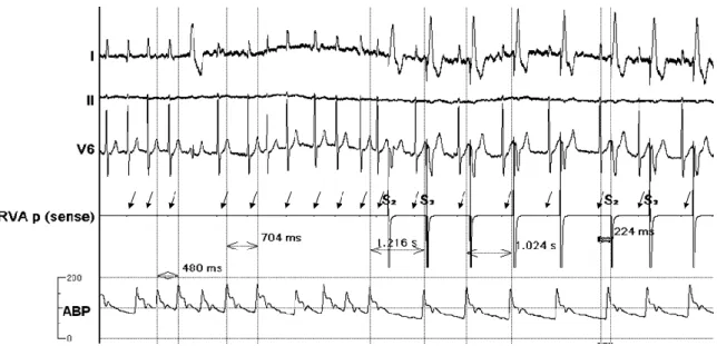
Figure 2 Initiation of coupled pacing
Invasive electrophysiological and hemodynamic recordings obtained from a patient during atrial fibrillation. When CP was initiated with a coupling interval of 224 ms, the previously fast and irregular mechanical pulse rate became significantly slower and less irregular. Time callipers showing arterial pulse cycle lengths before and during CP.
Arrows point to the diminished electrograms of spontaneous beats on the RVA p (sense) channel used for pacing. Registration speed: 100 mm/sec
Abbreviations:
I, II, V6: surface ECG recordings; RVA p (sense): pace/sense channel from the intracardiac electrode in the right ventricular apex; ABP: radial arterial pressure curve;
S2, S3: markers of pacing extrastimuli
3.2. Maximum voltage-guided technique for cavotricuspid isthmus ablation during ongoing atrial flutter
Twenty patients were recruited into this single-centre, prospective, randomized study conducted in the Hungarian Institute of Cardiology. Included patients were aged >18 years, all patients gave their informed consent to the study. The presence of cavotricuspid isthmus dependent atrial flutter was proven by the atrial activation sequence and concealed entrainment mapping from the CTI during the electrophysiological evaluation, with a difference in tachycardia cycle length and post- pacing interval of less than 30 milliseconds. Patients with prior CTI ablation were excluded from the study. The study patients were randomized into two groups. In group I (ten patients) CTI block was achieved with a complete, continuous anatomical ablation line using standard ablation technique. After completion of the line without termination of the flutter or without achievement of bidirectional block (Shah DC et al. 1999, Sasano T et al. 2002) another line was performed usually at a more anterior location.
Mapping for gaps were only performed if the second anatomical line was still unsuccessful. In group II (ten patients) the CTI was mapped, and peak-to-peak bipolar atrial electrogram amplitudes were measured continuously, and the application points were guided by the highest amplitude potentials on the CTI sequentially until bidirectional isthmus block was reached (Figure 3).
In both groups standard pacing manoeuvres were used to verify bidirectional isthmus block after the termination of the atrial flutter (Tada H et al. 2002). Cross over to another group was not allowed in this study. As part of our routine in 2006-2007, LocaLisa 3D navigation system was used in all patients. In group I, this allowed creating a smooth anatomical line without gaps, and in group II the system helped us to navigate the catheter back to any previous ablation spots.
24
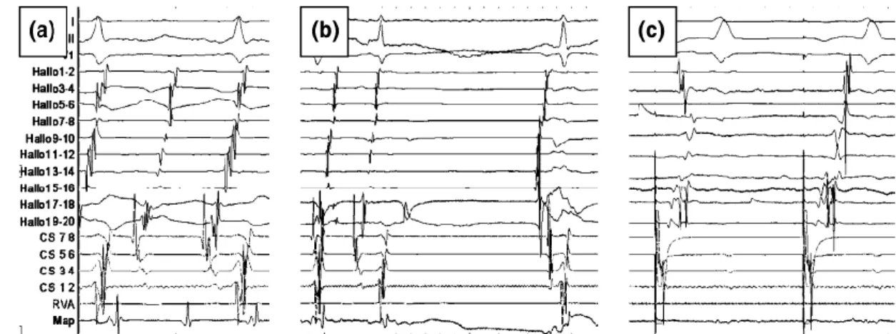
Figure 3 Typical counter-clockwise atrial flutter in a patient undergoing a three-
application MVGT isthmus ablation
Figure contains three surface electrograms, electrograms from a multipolar right atrial catheter and electrograms of an octopolar coronary sinus catheter. The mapping catheter is placed on the isthmus. (a) First ablation spot with rather large atrial bipolar signal. (b) Second RF application, results in immediate decrease of the local signal amplitude and fast termination of the flutter (c) As coronary sinus pacing showed no bidirectional block a third application was done resulting in isthmus block
The procedures were performed during a fasting state, using local anaesthesia. A 20- pole catheter was advanced to the right atrium, and positioned around the tricuspid annulus. An octopolar catheter was positioned in the coronary sinus and a quadripolar catheter in the right ventricular apex. All patients received full anticoagulation during the ablation procedure (heparine 100 IU/kg) following the local ablation protocol. An 8 mm tip ablation catheter was used in all cases (Blazer, Boston Scientific).
Radiofrequency energy was delivered for 60 s at each application point. Maximum power was set to 70 W, maximum temperature to 65°C in all patients regardless whether they were randomized to groups I or II. Procedural success was determined as the termination of the atrial flutter and achievement of a bidirectional CTI block.
The primary endpoints of the study were procedure time, fluoroscopy time, radiofrequency application numbers, and total radiofrequency time. For group II the maximum bipolar atrial signal was determined as target. For group I, after the ablation procedure atrial bipolar signal amplitudes were measured off line at all ablation spots.
Follow-up visits were scheduled at 3 and 6 months after the ablation procedure.
3.3. Defibrillation coil retention during orthotopic heart transplantation
We conducted a single-centre, retrospective study, and reviewed all the medical and surgical reports of 117 patients who underwent HTX in the Erasmus MC in Rotterdam, between March 2005 and May 2011. We further analyzed data of those 84 patients (72%) who had an ICD (n=53) or CRT-D implantation (n=31) prior to HTX. We determined the time from ICD implantation to HTX, the duration of the transplant procedure, the type and manufacturer of defibrillation leads, and the defibrillator lead- related adverse events (AEs). AEs were defined as lead fragment retention, lead perforation of cardiac chambers, evidence of conductor fracture on X-ray or as a sudden impedance rise, insulation failure, and macro-dislodgement of the lead. All patients were closely followed after HTX as a routine part of their clinical management. No patient was lost to follow-up.
3.4. Cardiac resynchronisation in chronic renal failure
Clinical and echocardiographic data of 60 consecutive patients who underwent CRT-P or CRT-D implantation for primary or secondary prophylaxis of sudden cardiac death were analysed in this retrospective study. Implantations were done between May 2005 and October 2006 in the Hungarian Institute of Cardiology.
We assumed the existence of chronic renal failure if serum creatinine concentration was >130 µmol/L on at least two separate days. Patients with both ischemic and non-ischemic dilated cardiomyopathy were included. The aetiology was considered ischemic when an old myocardial infarction was present, and/or any revascularisation procedure had been carried out before CRT. Patients ineligible of revascularisation were also included into the ischemic group.
Patient selection for CRT-P or CRT-D implantation had been performed according to the ACC/AHA/NASPE guidelines in operation at the time of implantation (Gregoratos G et al. 2002). Patients received optimal pharmacological therapy of chronic HF before selection to CRT.
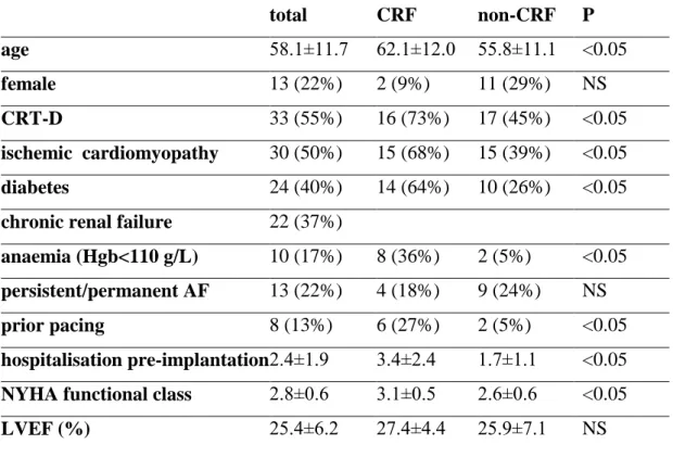
The following baseline characteristics were recorded: age, gender, NYHA functional class, history of ischemic cardiomyopathy, diabetes, CRF, anaemia, atrial
26
fibrillation, presence of a permanent pacemaker, proportion of CRT-D at implantation, annual hospitalisation rate prior to implantation, and left ventricular ejection fraction.
LVEF was calculated from the apical window of 2D-mode echocardiography using the modified Simpson’s rule. Available hospital records and outpatient follow-up documents were reviewed.
Data on change in NYHA functional class and change in LVEF were collected at a mean post-implantation period of 9.6 ±3.0 months. Improvement of one or more NYHA class was considered as a positive response to CRT. Data on all-cause mortality and the annual number of postoperative hospitalisations due to HF had been collected for 36 months. Seven patients were lost to follow-up, so 53 completed the whole course of the study.
3.5. Statistical methods
3.5.1. Instantaneous rate-control of rapid atrial fibrillation with coupled and paired ventricular pacing
Continuous data are shown as mean value±SD (or range); categorical data are expressed in absolute numbers and percentages. Continuous variables with normal distribution were compared using the paired t-test (changes in MPR due to CP or PP stimulation) and the unpaired t-test (comparison between MPR results of CP and PP). Analysis was performed using the statistical program R version 2.3.1 [R Development Core Team (2006), Foundation for Statistical Computing]. All tests were two-tailed and a P value of
<0.05 was considered statistically significant.
3.5.2. Maximum voltage-guided technique for cavotricuspid isthmus ablation during ongoing atrial flutter
Values were expressed as mean±SD. Independent samples t-test for parametric and Kologmorov–Smirnov test for nonparametric data was performed to compare data. A p value <0,05 was considered to be statistically significant.
3.5.3. Defibrillation coil retention during orthotopic heart transplantation
Continuous variables were expressed as mean ± SD, categorical variables as absolute numbers and their percentages. Variables were compared by using a Fisher’s exact test
lead age, HTX duration). All tests were two-tailed and a p value of <0.05 was reported as statistically significant. Analysis was performed using the statistical program SPSS PASW 17.0.2 (SPSS Inc., Chicago, 2009).
3.5.4. Cardiac resynchronisation in chronic renal failure
All continuous variables are expressed as mean±SD, non-continuous are as numbers and percentages. Differences in baseline characteristics and in follow-up data between separate patient groups were compared using the unpaired Student t-test for continuous and a chi-square test for dichotomous variables, as well as a Mann-Whitney U-test for NYHA classes. Comparison between identical groups to assess the effects of CRT was performed using the paired Student t-test (continuous), chi-square test (dichotomous), and a Wilcoxon signed-rank test (NYHA classification). After completion of the three- year long follow-up period, data on mortality were assessed using the Kaplan-Meier method. Comparison of cumulative survival rates was done using the log-rank test.
Univariate Cox regression analysis was performed to calculate crude hazard ratios and their respective 95% confidence intervals on mortality with the following baseline variables: age, sex, device type, diabetes, CRF, anaemia, atrial fibrillation, ischemic cardiomyopathy, NYHA class, LVEF, prior pacing, and number of annual hospitalisations pre-implantation. All p values were two-tailed, and a p value of < 0.05 was considered statistically significant for all tests. Analysis was performed with STATISTICA (version 8.0, StatSoft Inc, Tulsa, Oklahoma, USA).
28
4. Results
4.1. Instantaneous rate-control of rapid atrial fibrillation with coupled and paired ventricular pacing
Baseline patient characteristics
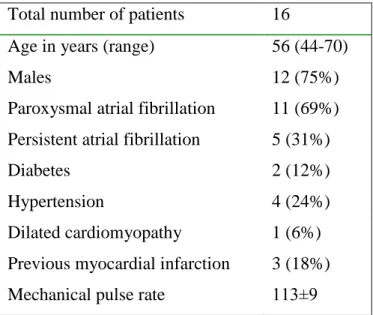
The study population consisted of 16 predominantly male (75%) patients with a mean age of 56 years (range 44–70). Demographic and clinical data are shown in Table 1. The mean right ventricular ERP was 222±10.5 ms before CP and 214±10.9 ms before PP.
Table 1 Baseline characteristics of patients
Total number of patients 16
Age in years (range) 56 (44-70)
Males 12 (75%)
Paroxysmal atrial fibrillation 11 (69%) Persistent atrial fibrillation 5 (31%)
Diabetes 2 (12%)
Hypertension 4 (24%)
Dilated cardiomyopathy Previous myocardial infarction
1 (6%) 3 (18%) Mechanical pulse rate 113±9
Results with the coupled pacing protocol
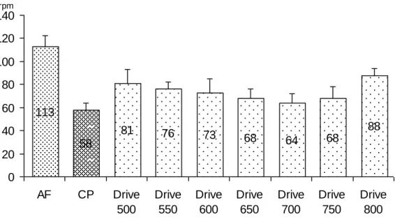
Successful MPR control was achieved in all patients using CP. The MPR significantly decreased from baseline (113±9 vs. 58±4/min; P < 0.001) (Figure 4). The controlled rhythm remained irregular, as the pulse CL ranged between 896±24 and 1452±67 ms, and the index of irregularity at baseline and during CP was 0.43 and 0.38, respectively.
Neither PVCs nor ventricular tachycardia occurred during CP.
113
58
81 76 73 68 64 68
88
0 20 40 60 80 100 120 140
AF CP Drive
500
Drive 550
Drive 600
Drive 650
Drive 700
Drive 750
Drive 800
rpm
Figure 4 The rate controlling effect of CP and PP at different drive trains
CP resulted in a significant reduction of the mean MPR. During PP all drive trains resulted in significantly lower pulse rates compared to baseline, and the lowest MPR was achieved at a ventricular drive train of 700 ms. Abbreviations: AF: baseline pulse rate during atrial fibrillation, rpm: rate per minute, Drive: drive trains with respective cycle lengths
Results with the paired pacing protocol
With different drive trains PP resulted in different regular MPRs (range 64±6 – 88±4/min). Increasing the drive train by 50 ms resulted in significantly lower MPR values as compared with the baseline (Figure 4). The lowest MPR achieved was found at the 700 ms drive train. With drive trains > 700 ms an increase in MPR was observed due to the occurrence of intercalated, spontaneously conducted atrial fibrillation beats.
Continuous MPR control was only achieved in seven patients, while PP caused premature beats in nine patients (56%) [mean 7 (range 1–72) PVCs per patient], resulting in failure of continuous MPR control (Figure 5). In these patients PP was regarded unsuccessful. No ventricular tachycardia was observed during PP.
There was no significant difference between the ERP of PP and CP protocols. Both CP and PP significantly reduced the MPR as compared to baseline (Figure 4). The lowest MPR achieved by PP was significantly higher than in the CP group
30
(64±6 vs. 58±4 /min; p<0.05). Neither death nor any life-threatening complication occurred during the study.
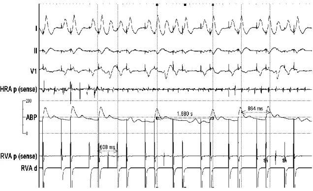
Figure 5 Unsuccessful PP due to frequent PVC’s
Frequent pacing-related PVC’s making the PP ineffective at a drive train of 600 ms.
Pulse cycle lengths vary considerably (864 ms and 1680 ms). HRA electrode shows ongoing atrial fibrillation. Registration speed: 100 mm/sec
Abbreviations:
I, II, V1: surface ECG recordings; HRA p (sense): intracardiac recordings from high right atrium; RVA p (sense): pace/sense channel from the intracardiac electrode in the right ventricular apex; RVA d: distal electrode channel from right ventricular apex;
ABP: radial arterial pressure curve; S1: marker of pacing extrastimuli
4.2. Maximum voltage-guided technique for cavotricuspid isthmus ablation during ongoing atrial flutter
Ablation data are shown in Table 2. In all primary and secondary endpoints the MVGT was superior to the anatomical approach. In all patients, atrial flutter was terminated
the patients. In this regard there was no difference between group I and group II (42 vs.
48%, respectively). At the conclusion of the ablation procedures bidirectional isthmus block was achieved in all cases. Procedure time was shorter in group II. Significantly less fluoroscopy was used in group II, less application needed for complete bidirectional isthmus block, and radiofrequency application duration was also shorter in group II.
There were no major complications related to the procedures. Minor complications such as groin haematoma were not different between the groups.
Mapping data
Atrial bipolar signal amplitudes of each ablation sites were assessed. There was no difference between the maximum atrial bipolar amplitudes in groups I and II. This also refers to the identical baseline conditions of the two groups.
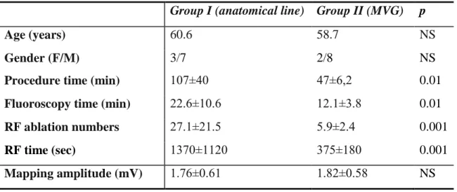
Table 2 Baseline characteristics and results of flutter patients treated with the MVG method or with conventional linear anatomical ablation
RF: radiofrequency
Group I (anatomical line) Group II (MVG) p
Age (years) 60.6 58.7 NS
Gender (F/M) 3/7 2/8 NS
Procedure time (min) 107±40 47±6,2 0.01
Fluoroscopy time (min) 22.6±10.6 12.1±3.8 0.01
RF ablation numbers 27.1±21.5 5.9±2.4 0.001
RF time (sec) 1370±1120 375±180 0.001
Mapping amplitude (mV) 1.76±0.61 1.82±0.58 NS
Follow-up data
During the median follow-up of 8.4±2.4 months, 1–1 recurrences of atrial flutter were detected in each group and redo procedures were performed successfully (not included
32
into the ablation results). The occurrence of atrial fibrillation was not statistically different between the groups (group I 25% vs. group II 34%).
4.3. Defibrillation coil retention during orthotopic heart transplantation
The study population consisted of 84 predominantly male (54 cases), end-stage heart failure patients aged 46.4 ± 9.9 years, who had a previously implanted primary prevention ICD system. Ischemic cardiomyopathy was the cause of heart failure in 43%
of the cases. The distribution of the implanted devices was: single-chamber ICD (SC- ICD) in 26 patients (31%), dual-chamber ICD (DC-ICD) in 26 (31%), and a CRT-D in 32 (38%). The implanted defibrillation lead configuration was single-coil in 33 patients (39%) and dual-coil in 51 (61%). The following defibrillation lead manufacturers were represented: Biotronik 10 leads (12%), Guidant (and former CPI) 19 leads (22%), Medtronic 25 leads (30%), and St Jude Medical 30 leads (36%). The mean time from defibrillation lead implantation to transplantation was 36 ± 30 months. Baseline characteristics of the ICD patients grouped by lead configuration are shown in Table 3.
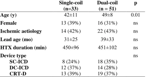
Patients in the single-coil group were younger, but other variables as gender, ischemic aetiology, duration of HTX procedure, and lead age did not differ significantly.
Table 3 Baseline characteristics of patients grouped by the configuration of defibrillation lead
Abbreviations: HTX heart transplantation, SC-ICD: single-chanber ICD DC-ICD: dual-chamber ICD, CRT-D: biventricular ICD
Single-coil (n=33)
Dual-coil (n = 51)
p
Age (y) 42±11 49±8 0.01
Female 13 (39%) 16 (31%) ns
Ischemic aetiology 14 (42%) 22 (43%) ns
Lead age (mo) 31±25 39±33 ns
HTX duration (min) 450±96 451±102 ns
Device type SC-ICD DC-ICD CRT-D
8 (24%) 12 (37%) 13 (39%)
18 (35%) 14 (28%) 19 (37%)
ns
ICD lead-related AEs were documented in the dual-coil group immediately after HTX.
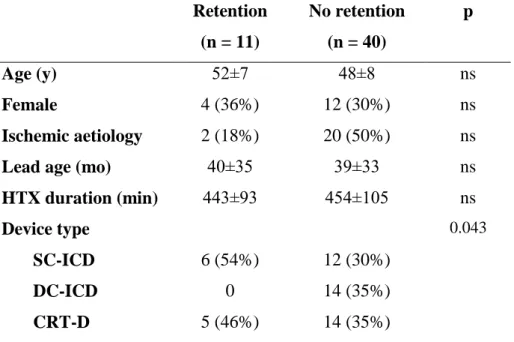
All of the AEs were retained proximal coil fragments of ICD leads. No other kind of defibrillator lead problems occurred post-transplantation. Additionally, a retained atrial lead fragment (CPI 4244 model) causing no further complications at 5 months follow- up is of note in the dual-coil group. Characteristics of the dual-coil cohort grouped by the presence or absence of fragment retention are displayed in Table 4. The type of implanted device has reached marginal significance (p=0.04) as only SC-ICDs and CRT-D devices, but no DC-ICDs were represented in the retained coil group.
Table 4 Characteristics of patients with dual-coil defibrillator leads
Abbreviations: HTX heart transplantation, SC-ICD: single-chanber ICD DC-ICD: dual-chamber ICD, CRT-D: biventricular ICD
Retention (n = 11)
No retention (n = 40)
p
Age (y) 52±7 48±8 ns
Female 4 (36%) 12 (30%) ns
Ischemic aetiology 2 (18%) 20 (50%) ns
Lead age (mo) 40±35 39±33 ns
HTX duration (min) 443±93 454±105 ns
Device type SC-ICD DC-ICD CRT-D
6 (54%) 0 5 (46%)
12 (30%) 14 (35%) 14 (35%)
0.043
Details and outcome of the patients who were affected by fragment retention are listed in Table 5. Nine different heart surgeons were involved in the HTX procedures, all with ample clinical experience in this field. There was no difference in the distribution of surgeons according to the presence or absence of lead retention. The proportion of manufacturers in the dual-coil group was: St Jude Medical 41%, Medtronic 26%,
34
Guidant 23%, and Biotronik 10%. There was no fragment retention among the Biotronik leads in our patients. The fragmented defibrillation leads came from the St.
Jude Medical Riata family in the majority of cases (7/11 = 64%, 43% of all St Jude Medical dual-coil leads); whereas the Medtronic Sprint (2/11 = 18%, 20% of all Medtronic dual-coil leads) and CPI-Guidant families (2/11 = 18%, 22% of all Guidant dual-coil leads) were represented in the other four cases (Table 5). Except three patients, the ICD extraction procedures of those eleven transplantations were considered complete removals at the time of the transplant procedure, and in eight cases only the postoperative chest X-ray or CT-scan revealed the retained proximal defibrillation coil fragments in the body (Figure 6). On follow-up, one coil fragment migrated into the pericardial space, another into the right atrium causing perforation of the interatrial septum. The latter patient died of multi-organ failure 3 months after HTX. One patient underwent rethoracotomy due to a S. aureus-related purulent mediastinitis and cardiac tamponade, and he also developed right jugular and right femoral venous thrombosis, but finally he has recovered.
Four patients died in the retention group, their cause of death is reported in Table 5. Of the 40 patients without retention in the dual-coil group, and of the 33 patients in the single-coil group also four-four patients died, but the small numbers and absence of any relation of the deaths to the retained fragments makes any comparison meaningless. In two patients an uncomplicated percutaneous transvenous lead fragment extraction was performed using the femoral route 19 and 56 days after transplantation. In the rest of the patients including the one with the coil in the pericardium, our clinical decision was to leave the fragments in place after individually weighing the inherently elevated risks against the potential benefits of a removal procedure.