RADIOLOGICAL PLANNING OF
LAPAROSCOPIC DONORNEPHRECTOMY
PhD thesis
Pathological Sciences Doctoral School Semmelweis University
Supervisor: Prof. MD, Ph.D.
Official reviewers:
Dr. MD, Ph.D
Dr. MD, Ph.D
Head of the Final Examination Committee:
Dr. MD, D.Sc
Members of the Final Examination Committee:
Dr. Kinga Karlinger MD, Ph.D
Dr. MD, Ph.D
Budapest, 2012
Summary
The most effective method of treating terminal kidney diseases is kidney transplantation. However, the number of cadaver donors does not grow with the number of patients suffering from end-stage kidney failure. Introducing living-donor kidney transplantation proved to be an effective way of increasing the number of donors. A preliminary examination of the anatomy of renal vessels is essential to reduce the potential complications of hand-assisted laparoscopic donor nephrectomy.
Being aware of the embryological development of the arterial and venous system may provide an in-depth understanding of the versatility of the variations. Our research seeks to answer the following questions: to what extent anatomical and other morphological features discovered via multidetector-row CT (MDCTA) examinations influence the choice between the donor kidneys, and whether a simplification of the anatomical description could make the process more effective.
We have analysed MDCTA examinations of 55 donors between 27 and 78 years of age.
We have pinpointed the arterial and venous variations we have found, we have surveyed them through radiologist-surgeon consultations, and validated the results after the surgery. With 67% (37/55) of the 55 donors we have found certain vascular variations in the transplanted organ. In 70% of the above cases (26/55) one such variation was discovered both on the preoperative CT images and during the surgery, while in 22% of the cases (26/55) two and in 8% (3/55) of the cases three variations have been identified. In the cases of the above 37 donors we have found 51 vascular variations altogether that were also validated during surgeries later on.
Based on the results of our research (1) the MDCTA examination have proved to be an excellent method to examine the renal vascular variations; (2) the changes we have implemented in the terminological system further clarified the anatomic situation which in turn facilitated the surgeon-radiologist consultations; (3) we have systematized the vascular variations; (4) and expressed our opinion concerning the anatomical variations and their significance with regard to the surgery.
Introduction
The most effective method of treating terminal kidney diseases is kidney transplantation. The longer a patient has to wait for the operation the worse their life- quality and life-expectancy will be and it also makes the treatment itself more expensive. Shortening this period of waiting is the interest of both the patient and the state or the insurance company that covers the expenses. The number of cadever donors, however, does not grow with the number of patients suffering from end-stage kidney failure. Owing to the recent developments of immunology, immune-suppressive therapy and histological standardization the transplanted organs live longer and function better. These achievements made it possible to transplant organs of monozygotic twins and related or non-related, living or cadaver donors.
Introducing living-donor kidney transplantation proved to be an effective way of increasing the number of donors.
The kidneys of living donors live longer than those of cadaver donors thus providing better life-expectancy for the recipients. Transplantation is an elective (i.e. not acute) operation which makes it possible to plan the entire procedure in advance, and the transplanted organs also come from a healthy donor. Shortening the waiting period also decreases the number of preoperative dialyses, thus the recipients undergo surgery in a considerably better condition. In the case of living-donor kidney transplantations early kidney functions are significantly better, which effectively decreases the number of postoperative dialyses. International results show that in the cases of kidneys transplanted from living donors graft-survival reaches 98 per cent, while this ratio is barely 90 per cent with kidneys of cadaver donors. This exceptionally high survival ratio is the result of modern immune-suppressive medication that significantly reduced the occurrence of acute rejection. The long-term (three-year, five-year and ten-year) graft-survival is also better in the cases of kidneys of living donors reaching up to 92 per cent while it is somewhere around 80 per cent with cadaver-kidneys. It is important to note, however, that long-term graft-survival is also affected by the number of preoperative dialyses, the results being better with patients receiving less dialyses before the operation. Pre-emptive transplantation (i.e. before dialyses is required) further improves the chance of survival. In terms of expenses transplantation also proves to be preferable to dialysis making it a more favourable solution not only for the
recipients but also considering the long-term aspects of national economy.
Transplanting an organ is always less expensive than treating the chronic disease that makes the operation inevitable, as research both in the USA (in the 1960s) and in Europe (in late 1970s) show. Based on the above we can conclude that living-donor transplantation is definitely a more cost-efficient solution than other methods of treatment.
The first living-donor kidney transplantation was performed on monozygotic twins by Joseph Murray in Peter Bent Brigham Hospital, Boston, in 1954. By choosing a genetically identical donor he managed to avoid the possibility of graft-rejection.
Laparoscopic living-donor nephrectomy was first performed on a pig by Inderbir Gill in 1994 and soon afterwards, in 1995, Lloyd Ratner performed the same surgery on human patients.
A great number of significant advantages for the donors also contributed to the fact that laparoscopic donor-nephrectomy has become a generally accepted and widely used surgical procedure. The lesser surgical exposure, the shorter hospitalization, the quicker recovery, the better cosmetic results and an essentially unchanged life-quality have all resulted in a greater willingness to donate organs. The first laparoscopic donor
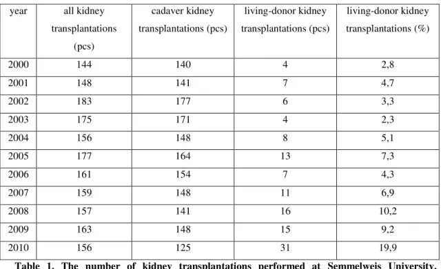
1. The Department of Transplantation and Surgery of Semmelweis University launched its laparoscopic donor-nephrectomy program led by -donor kidney transportations gathered new momentum, and by the end of 2010 31 such operations had been performed in our institute, almost twice as many as in the previous years.
year all kidney transplantations
(pcs)
cadaver kidney transplantations (pcs)
living-donor kidney transplantations (pcs)
living-donor kidney transplantations (%)
2000 144 140 4 2,8
2001 148 141 7 4,7
2002 183 177 6 3,3
2003 175 171 4 2,3
2004 156 148 8 5,1
2005 177 164 13 7,3
2006 161 154 7 4,3
2007 159 148 11 6,9
2008 157 141 16 10,2
2009 163 148 15 9,2
2010 156 125 31 19,9
Table 1. The number of kidney transplantations performed at Semmelweis University, Department of Transplantation and Surgery.
The improvement of diagnostic equipment has greatly contributed to the increasing number of surgical procedures in general, and laparoscopy in particular. With CT examinations the planning of the operations has become more accurate; in vivo anatomy could be pictured in never before seen detail.
Preliminary information on the anatomy of renal veins is essential for the surgeon to perform donor nephrectomy without complications. In order to be able to recognise and understand the anatomical variations radiologists have to be familiar with the embryology of the venous and arterial system. Multidetector CT (MDCT) is a widely accepted method of examining abdominal organs. Contrast-enhanced CT examinations have long been available for surgeons to decide whether certain abdominal conditions are operable or to determine the extent of damage the organs have suffered. The large amount of data gathered via MDCT examinations offer numerous opportunities of reconstruction that provide virtual 3D images, instead of the somewhat more indefinite abstract layers. These 3D images and other reconstructions help surgeons to interpret even more complex anatomical situations. With such an accurate picture of the anatomic circumstances
reduces the duration of the operation and helps avoid most dangerous complications that may result in bleeding.
Objectives
Laparoscopic donor nephrectomy is a special surgical method inasmuch as the surgeons have to operate in a limited field of vision and with restricted space for preparation.
Abdominal organs with a complex vascular system have their own terminology (e.g.
, these are intended to ensure the best possible graft-function in case of an ortotopic liver transplant by providing sufficient knowledge of the vascular system and by allowing the surgeons to create the most appropriate anastomoses. In the case of the kidney, however, the great number of possible variations make it too complicated to create an exhaustive list of variations like that of the liver. The logic and the ways of thinking the surgeons and the radiologists tend to follow may show certain differences which should under no circumstances result in misunderstandings that may eventually lead to critical situations, especially with respect to the survival and unchanged life-quality of the donor. Therefore the nomenclature and the terminology must be accurate and unambiguous at all times.
As a result of the close collaboration with our surgeons we have had the opportunity to compare the vascular anomalies shown by the MDCT images to the ones found during the surgery, which in turn allowed us to define the sensitivity and the specificity along with the positive and negative predictive value of MDCT examinations.
With laparoscopic donor nephrectomy the unchanged life-quality of the donor is of utmost importance even during the planning phase of the operation. With this in mind, no matter which of the kidneys is removed, there are other factors that need to be taken
principle. MDCT images also help the surgeons to determine which of the kidneys is
Based on the above in this paper I am trying to find an answer to the following questions:
Whether MDCT examination has been proved to be an appropriately sensitive and specific method for the preoperative planning of the surgeries in the newly launched Hungarian laparoscopic donor-nephrectomy program.
Whether it is possible to create a uniform terminology to describe the vascular variations of the kidneys, which would also promote surgeon-radiologists consultations.
The types, the number and the incidence rate of renal vascular variations in the Hungarian population we have examined.
Based on the Multidetector-row Computer Tomography (MDCT) examinations, which factors have proved to be important when deciding which kidney is to be removed.
Methods
Patients
This dissertation is intended to process the data of the kidney donors examined at Semmelweis University, Department of Surgery and Transplantation from January 2008 until September 2010 in a prospective way.
During the above period we examined 87 potential donors. Out of the 87 patients we analysed the data of the 55 donors (67% female, 33% male) who later underwent laparoscopic nephrectomy, as in their cases we could also compare the data to the ones we found during the surgery. The average age of the patients is 47 (ranging from 27 to 78) with the average BMI of 26 kg/m2. 91% of the donors were relatives, the remaining 9% were either friends or emotionally related to the recipients. The donor pool distribution was as follows: mother (n=22), spouse (n=9), sibling (n=8), father (n=6), friend/emotionally related (n=5), cousin (n=4), brother-in-law (n=1). When the donor was not related to the recipient an ethical board examined the case and decided whether it was possible to donate the organ. The surgeries were performed either transperitoneally or extraperitoneally. In 42 of the cases the left and in 13 cases the right kidney was removed. The choice between the right and the left kidney was based on the results of MDCT and renal sintigraphy examinations. All nephrectomies When there was no difference between the kidneys with respect to any of the above factors, surgeons chose the left kidney, which is easier to transplant due to the longer renal vein.
The Multidetector CT Angiography Protocol
Each donor underwent an MDCT examination. The examinations were performed cranio-caudally with a 16-slice Mutidetector-row CT equipment (Philips Brilliance 190 P; Philips Healthcare Systems). Each patient was examined by following the same protocol. To be able to determine the region to be examined first a native scout image was taken. Than we performed contrast-enhanced examinations which were followed by another native image in the secretion phase to provide a better view of the urinary tract. All examinations were performed on an empty stomach (after minimum 4 hours of fasting). The patients were only allowed to drink non-carbonated water. After having been informed about the CT examinations all donor candidates signed a statement to confirm their assent.
The images were all taken in the inspiratory phase in supine position. The native images were taken of the area ranging from the top of the diaphragm to the symphysis pubica in order to be able to determine the anatomical location of the kidneys, to discover possible arterioscleroses or calcifications in the kidneys or in the urinary tract, and to determine the base-density value of other potential lesions.
Before the contrast-enhanced examinations a 20 G cannula was inserted in the basilica vein of the patients. The contrast agent was injected with the help of an injector (Medrad Stellant Dualflow, Siemens AG). The required amount of contrast agent was determined based on the weight of the patient and the type of the contrast agent available. To determine the required amount of the contrast agent in millilitres the body weight of the patient in kilograms was multiplied by 1.3. We used Iomeron 400 (iomeprol, Bracco UK Ltd.), Ultravist 370 (iopromid, Bayer plc.) and Omnipaque 350 (iohexol, GE Healthcare) contrast agents with the injection rate of 5 ml/s.
To determine the timing of arterial-phase examinations bolus tracking method was applied. The density value was set at the section of the abdominal aorta above the renal artery. The series were started five seconds after reaching the 100 HU threshold value.
Arterial images were taken from the top of the diaphragm down to the spina iliaca anterior superior. The venous-phase examinations were started 70 seconds after the
arterial series, focusing on the area from the top of the diaphragm to the symphysis pubica. Instead of repeating the series in the excretion phase, we took a scout image on the abdominal and the pelvic (minor) regions ten minutes after the venous images were taken. Based on this latter examination, along with the data gained from the previous three phases, a clear picture of the urinary tract became available.
Visual reconstruction and the analysis of volumetric data
The data gained from the examinations were processed by using software and a workstation (Extended Brilliance Workspace, Philips Healthcare Systems) that is able to create both two- and three-dimensional reconstructions. The examinations were analysed in three phases. First a senior radiologist resident or a consultant radiologist evaluated the evidence and created the obligatory reconstructions along with the ones he or she deemed necessary. The evaluation was then validated by a specialist radiologist. The doctors discussed the differences (if there were any) and came to an agreement regarding doubtful cases. In the third phase the surgeon performing the laparoscopic donor-nephrectomy and the radiologist of the living-donor team reviewed the examination and the reconstructed images.
In the first phase, after reviewing the axial images, multiplanar reconstructions (MPR), maximum intensity projections (MIP) and volume rendering reconstructions were made. The radiologist primarily used MPR and MIP images to describe the vascular anatomy of the patient, while surgeon-radiologist consultations were best aided by the volume rendering reconstructions.
The radiologists described the number, the length, the branching pattern and the diameter of the renal arteries along with the potential extra arteries and the capsular arteries that may be relevant from surgical point of view. In addition to all the above the gonadal and adrenal veins we also described.
The description also included all genetic or acquired abnormalities of the arterial or venous system, the parenchyma of the kidneys, or the urinary system as well as any anomalies concerning other abdominal organs.
Terminological changes
The description of the above examinations often made surgeon-radiologist consultations complicated, which eventually resulted in changing some features of the ordinary terminology. All such changes were made with respect to the characteristics of laparoscopic surgery. During the course of laparoscopic preparation surgeons experience the anatomical spaces differently from radiologists therefore the description of the length of the artery had to be adapted to their special field of vision.
The radiomorphological features of anatomical structures are usually described from
difference in the viewpoints. Renal arteries are measured from their stemming from the aorta to their entering the renal hilum, whereas veins are measured from the point they leave the renal hilum to the one they reach the vena cava inferior. The data registered by using this method is not always perfectly comprehensible for the surgeon, and the radiologist needs to provide further explanations so that the surgeon would have all the relevant data. In some cases the terminology that might solve the problem simply does not exist, therefore, based on the specific questions of the surgeons, we have created new terminology.
With respect to the right kidney it means that while during the course of preparation the entire vena can be seen, the artery remain partly hidden. Only the part of the right retrocaval renal artery can be seen that reaches beyond the right margin of the vena cava inferior, so its length is of utmost importance for the surgeon. Therefore on this side the reference point was where the right margin of the vena cava inferior meets the artery. This point was termed the technical end of the artery. Consequently by technical length we mean the distance between technical end and the primary branching.
In the case of the left kidney the entire length of the artery can be seen, the vena, however, can only be safely recovered to the left margin of the aorta, so for surgeons the distance between this point and the last branching was important. Consequently in the case of the left renal vein the technical end is the point where the vena renalis meets
the left margin of the aorta, whereas technical length is measured from the last branching to this point of reference. These terminological changes have also been applied in radiological reports. In addition to the above the anatomical length is also included in the report.
Figure 1: Terminology used to describe the anatomy of the donor kidney
Analysis of the renal arterial system
During the course of our examinations the arteries of both kidneys were analysed in great detail, paying special attention to the conditions that are especially important from a surgical point of view.
Multiple arteries: more renal veins that stem from the aorta separately.
Polar arteries: veins stemming from arteria renalis and running towards the superiror or the inferior pole of the kidneys.
Early-branching arteries, late-junction veins: In the case of the left kidney, arteries that branched off 10 millimetres or closer from the left lateral wall of the aorta were described as early-branching. On the right side, the medially bifurcating veins off the right margin of vena cava inferior also fall under the same category. Given that there is no internationally accepted consensus regarding this issue, when determining early- branching, surgical aspects were considered. Our description was based on the length
required for inserting the clips (in the case of arteries), and the length required for using the stapler (in the case of veins). We had to distinguish between the reference points on the right and the left side, because in the case of a right nephrectomy the surgical preparation is limited by the right margin of vena cava inferior.
Capsular arteries: the branches of arteries supplying the capsulae that also penetrate into the parenchyma.
The scleroses, the stenoses, the asymmetric thickenings of the arterial wall along with other indications of vascular anomalies were also included in the radiological report.
The luminal diameter, i.e. the diameter of the contrast-agent column was indicated in the results.
In the case of the arteries we always performed volume rendering and MIP reconstructions. Our objective was to map the arteries found on the axial images or MIP reconstructions, and to provide the surgeons with a more comprehensible three- dimensional picture. The software automatically creates a virtual 3D image based on the data gained from the axial series. The resulting picture, however, does not always correspond with the reality. The radiologist completing post-processing had the opportunity to change the required parameters in order to achieve the most authentic image available.
Analysis of the renal venous system
Knowledge of the angle, the length and the calibre of venous system is as important as that of the arterial. The anatomical reference points used to determine the length, however, are different from those of the renal arteries. Similarly to the analysis of the arterial system we kept surgical aspects in view. In the case of the right renal vein we measured the distance between the last junction of the veins and the point it penetrates into the vena cava inferior, whereas with the left vein we based our description on the left margin of the aorta that limits the surgical preparation. Therefore the surgically relevant length of a renal vein is measured from the last junction of the veins to the point where the left renal vein crosses the left margin of the aorta.
Late-junction veins: based on the above distinction veins with a 10-millimetre or shorter distance between the last junction and the point where the left renal vein crosses the left margin of the aorta are described as late-junction. The description of gonadal and adrenal veins was also based on this point of reference.
Retroaortic veins: left renal veins coming from the renal hilum and passing behind the aorta before penetrating into the vena cava inferior.
Circumaortic veins: one of the two branches of the renal veins pass in front of, and the other behind the aorta before reaching the vena cava inferior.
Multiple veins: renal veins that penetrate into the vena cava inferior separately, and do not fall under either of the above categories.
The anomalies concerning the renal venous system were subdivided by major and minor ones.
Major anomalies of the left kidney are the ones that affected the laparoscopic approach or the technique of creating venous anastomoses during implantation.
Retroaortic and circumaortic veins fall under this category.
Major anomalies of the right kidney include multiple veins.
Minor anomalies on both sides are the ones that are relevant in terms of donor nephrectomy but do not affect venous anastomoses in any way. Lumbar veins with a diameter of 5mm or more belong to this category. The reports include all lumbar veins with their diametric values.
The method of comparing Multidetector CT Angiography examinations to the intra-operative circumstances
The 55 donor-nephrectomies provided an excellent opportunity to determine the accuracy of the preoperative MDCTA examinations, i.e. to compare the results with the circumstances found during surgery. The sensitivity, the specificity, the accuracy, and the positive and negative predictive value were determined as follows:
MDCTA evidence
Intraoperative evidence
Normal Variant
Normal TN FN
Variant FP TP
Table 2. Determining sensitivity, specificity, accuracy, and positive and negative predictive value
TN (true negative) all de facto negative cases: no vascular variations can be seen in MDCTA images, and none found during surgery either.
FN (false negativ) all falsely negative cases: no vascular variations can be seen in MDCTA images, but some were found during the surgery.
TP (true positive) all de facto positive cases: vascular variations can be seen in MDCTA images, and they were found during surgery
FP (false positive) all falsely positive cases: vascular variations can be seen in MDCTA images, but were not found during surgery.
Sensitivity = TP/(TP+FN) Specificity = TN/(TN+FP)
Accuracy = (TP+TN)/(TP+TN+FP+FN) Positive predictive value = TP/(TP+FP) Negative predictive value = TN/(TN+FN)
Results
We performed MDCTA examinations on 55 healthy kidney donors between January 2008 and September 2010. We set up a laparoscopic surgical plan for all 55 candidates.
In 54 out of the 55 cases the kidneys were removed by laparoscopy. In one case the surgical plan had to be changed intra-operatively, and in another conversion for open surgery was indicated. All kidneys removed were implanted subsequently.
The examination of the arterial and venous system of the kidneys was performed on 110 kidneys of 55 patients.
With 14.5% of the 55 patients (8/55) neither arterial no venous abnormalities were found. All of the above mentioned kidneys had one vein and one artery. In 18% of the cases (10/55) some vascular variation was found but they concerned the kidney that remained with the patient. Out of the 10 cases in 2 the left and in 8 the right kidney was removed due to the anomalies of the opposite ones. Via the MDCTA examinations we discovered 108 vascular variations altogether in the 110 kidneys of the 55 donors.
The 55 removed organs also provided opportunity to compare the results of the examinations to the circumstances found during surgery. 67% of the 55 donors (37/55) had some kind of vascular variation in the kidney that was subsequently removed and transplanted. In 70% of these cases (26/55) one, in 22% (8/55) two and in 8% (3/55)
three such abnormalities were found both in the preoperative CT images and during the surgery. Of the 37 above donors 51 vascular variations were discovered, that were also confirmed by the surgery. Based on these data we were able to measure the sensitivity, the specificity, the accuracy, and the positive and negative predictive value of the examinations.
The role of MDCTA examinations in choosing the kidney to be removed
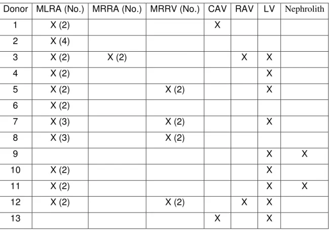
During the given period we examined, 55 donor-nephrectomies were performed; in 42 cases the left and in 13 cases the right kidney was removed. In addition to renal scintigraphy, MDCTA examinations played an important role when choosing the kidney to be removed. As scinitgraphy did not show considerable differences between the right and left kidneys of the healthy donors, decisions were mostly based on the results of MDCTA examinations, as follows:
1) If nephrolites were discovered in any of the kidneys, but there were no other metabolic anomalies contraindicating donor-nephrectomy, always the kidney with the stone was chosen
2) The left kidney was preferred, being easier to implant. In the 13 cases when the right kidney was removed the decision was always based on the anatomical circumstances.
3) In most cases when the right kidney was removed it was because the left one had multiple arteries.
4) The right kidney was removed in all cases when the number of arteries supplying the left kidney was higher than those supplying the right one.
5) In the rest of the cases the choice of the right kidney was indicated by the vascular variations of left renal vein, mostly due to the presence of retroaortic or circumaortic veins. In two cases out of the three circumaortic veins found, the presence of the vascular variation itself contraindicated choosing the left kidney, in the remaining one case, however, the left kidney with the circumaortic vein was chosen due to the short vein found in the right one.
6) Large lumbar branches and retroaortic veins did not play any role in choosing the kidney to be removed, but they were still significant from a surgical aspect.
Donor MLRA (No.) MRRA (No.) MRRV (No.) CAV RAV LV Nephrolith
1 X (2) X
2 X (4)
3 X (2) X (2) X X
4 X (2) X
5 X (2) X (2) X
6 X (2)
7 X (3) X (2) X
8 X (3) X (2)
9 X X
10 X (2) X
11 X (2) X X
12 X (2) X (2) X X
13 X X
Table 3. Anatomical variations of right donor-nephrectomies
MLRA: multiple left renal artery; MRRA: multiple right renal artery; MRRV: multiple right renal vein; CAV: circumaortic vein; RAV: retroaortic vein; LV: lumbar vein
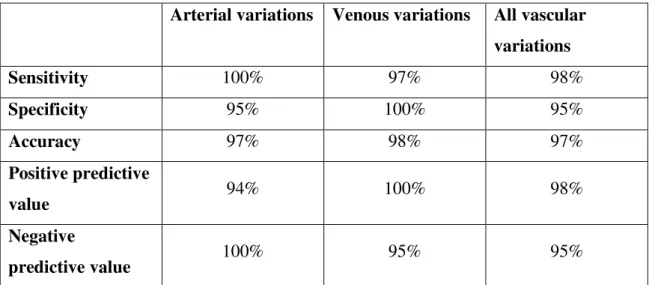
Sensitivity, specificity, accuracy, and positive and negative predictive value od MDCTA examinations
To measure the above indicated values we used the data in Table 4 on the preoperative CT images of the 55 kidneys with postoperative references. The results are summarised in Table 5:
Arterial vascular variations (pcs)
Venous vascular variations (pcs)
All vascular
variations (pcs)
TN 18 18 18
FN 0 1 1
TP 17 34 51
FP 1 0 1
Table 4. Vascular variations of the kidney with postoperative references
Arterial variations Venous variations All vascular variations
Sensitivity 100% 97% 98%
Specificity 95% 100% 95%
Accuracy 97% 98% 97%
Positive predictive
value 94% 100% 98%
Negative
predictive value 100% 95% 95%
Table 5. Sensitivity, specificity, accuracy, and positive and negative predictive value
Conclusions
To summarise the results of our examination, we can conclude that
1) the MDCTA examination proved to be an extremely sensitive method of mapping the vascular variations of the kidneys in the newly launched Hungarian laporascopic donornephrectomy program;
2) the uniform terminology makes the anatomical situation more comprehensible thus promoting surgeon-radiologist consultations;
3) systematising the vascular anomalies provides a clearer picture of the anatomical variations within the Hungarian population;
4) the arterial and venous variations are indeed significant from a surgical aspect, a detailed description reduces the chance of intraoperative and postoperative complications, and it also plays an important role when choosing the kidney to be removed.
List of own publications (thesis-related publications)
Deak PA, Doros A, Lovro Z, Juhasz E, Branstetter G, Kovacs JB, Piros L, Jaray J Significance and Imaging of Lumbar Veins and Early-Branching Arteries in Planning Living-Donor Laparoscopic Nephrectomy: Two Case Reports From 21 Months' Experience. TRANSPLANTATION PROCEEDINGS 42:(6) pp. 2347-2349. (2010) IF:
0.993
Deak PA, Doros A, Lovro Z, Toronyi E, Kovacs JB, Vegso G, Piros L, Toth S, Langer RM. The Significance of the Circumaortic Left Renal Vein and Other Venous Variations in Laparoscopic Living Donor Nephrectomies. TRANSPLANTATION PROCEEDINGS 43:(4) pp. 1230-1232. (2011) IF: 0.993
Vegso G, Toronyi E, Hajdu M, Piros L, Gorog D, Deak PA, Doros A, Peter A, Langer RM. Renal Cell Carcinoma of the Native Kidney: A Frequent Tumor After Kidney Transplantation With Favorable Prognosis in Case of Early Diagnosis.
TRANSPLANTATION PROCEEDINGS 43:(4) pp. 1261-1263. (2011) IF: 0.993
List of own publications (other publications)
Doros A, Nemeth A, Deak PA, Hartmann E, Gerlei Z, Fazakas J, Kobori L. (2010) Successful Treatment with a Covered Stent and 6-Year Follow-Up of Biliary Complication After Liver Transplantation. Cardiovasc Intervent Radiol, 33: 425-429.
Zadori G, Gelley F, Torzsok P, Sarvary E, Doros A, Deak PA, Nagy P, Schaff Z, Kiss A, Nemes B. (2011) Examination of Claudin-1 Expression in Patients Undergoing Liver Transplantation Owing to Hepatitis C Virus Cirrhosis. Transplant. Proc., 43:
1267-1271.
(2009)
83: 80-85.
(2009)
83: 8-13.
(2009)
- Orvosi Hetilap 150: 1231-1234.
(2009) Downstaging of hepatocellular carcinoma with radiofrequency ablation on the Hungarian liver transplantation waiting list.: Early results and learned lessons. Interventional Medicine
& Applied Science 1: 41-45.
(2010) Treatment of early hepatic artery complications after adult liver transplantation : A single center experience. Interventional Medicine & Applied Science 2: 159-164.
(2010) Radiofrequency ablation of an intercalyceal neoplasm in a transplanted kidney using percutaneous nephrostomy for cooling. Safety and early result. Interventional Medicine & Applied Science 2: 37-41.
(2010) Successful urinary tract reconstruction following ureteral necrosis in kidney transplant patient. Interventional Medicine & Applied Science 2: 134-138.
Doros A, Nemes
(2007) Are metallic biliary stents feasible in treatment of post- transplant biliary stenosis: Long term follow up. Transplant International 20: 301.
(2007) Interventional radiological treatment of venous complications after liver transplantation. Transplant International 20: 302.