Horseshoe kidney transplantation
BALÁZS NEMES1,*, ZSOLT KANYÁRI1, GERGELY ZÁDORI1, LAJOS ZSOM1, MARIANN BERHÉS2, MÁTYÁS HAMAR3, KRISZTINA KÓBOR4, ANTAL PÉTER3
1Institute of Surgery, Department of Organ Transplantation, Faculty of Medicine, University of Debrecen, Debrecen, Hungary
2Department of Anesthesiology and Intensive Care, Faculty of Medicine, University of Debrecen, Debrecen, Hungary
3Clinic of Transplantation and Surgery, Semmelweis University, Budapest, Hungary
4FMC Miskolc Nephrology Centre, Miskolc, Hungary
*Corresponding author: Balázs Nemes MD, PhD; Institute of Surgery, Department of Organ Transplantation, Faculty of Medicine, University of Debrecen, Móricz Zsigmond krt. 22, H-4032 Debrecen, Hungary; E-mail: abnemes@hotmail.com
(Received: March 5, 2015; Revised manuscript received: March 30, 2015; Accepted: April 5, 2015)
Abstract: Horseshoe kidney is a fusion anomaly found in approximately one in 400–600 people. Due to vascular and ureteral variations, transplantation with a horseshoe kidney presents a technical challenge. In our case, the isthmus connected the upper poles and contained parenchyma. It consisted of three renal arteries, fi ve veins collected to the inferior vena cava, and two ureters and pyelons. It was implanted en bloc to the left side retroperitoneally. During the early period, cellular and humoral rejection was confi rmed and treated. For a urine leak, double J catheters were implanted into both ureters. Later, the fi rst catheter was removed. Subsequently, urinary sepsis developed, necessitating graftectomy. The uncommon anatomy of ureters and antibody-mediated rejection (AMR) may both be factors for a ureter tip necrosis led to an infected urinoma. After other Hungarian authors, we also report a horseshoe kidney transplantation that was technically successful. However, after an adequately treated but severe acute humoral rejection, the patient developed sepsis, and the kidney had to be removed. We conclude that transplantation with horseshoe kidney is technically feasible but may increase the risk for urinary complications and resultant infections. Careful consideration of risk and benefi t is advised when a transplant professional is faced with this option.
Keywords: horseshoe kidney, kidney transplant, duplicated ureter, antibody-mediated rejection, urinary leakage, graftectomy
Introduction
Horseshoe kidney (HK) is a fusion anomaly of the kid- ney found in approximately 1 in 400–600 people [1, 2].
It is more common in men than in women. Da Carpi was the fi rst to document a case with horseshoe kidney in 1522 [3]. This is an anatomic variation where the kidneys are connected by an isthmus consisting of either fi brous tissue or parenchyma. The connection may be located either at the lower or upper poles [1]. Fusion anomalies are commonly asymptomatic with a normal renal function [4]. Horseshoe kidney usually presents together with other congenital anomalies [2, 5]. The vascular anatomy of a horseshoe kidney is usually com- plex [1, 6, 7]. Ureteral and collecting system abnor- malities are also common. Due to common vascular and ureteral variations, transplantation of a horseshoe kid- ney presents a technical challenge. V. A. Politano was the fi rst to transplant a horseshoe kidney from a living- donor in 1963 (not published). The kidney functioned well, but the recipient died 8 months later of hepatitis
[8]. Horseshoe kidneys can be transplanted en bloc or separated fi rst and then transplanted separately [9]. Ma- rofka et al. performed horseshoe kidney transplantation in 2000 fi rst, and in 2003, the second time. Their cases were reported at the biannual congress of the Hun- garian Surgical Society in 2008 [10]. Also, there are unpublished results from Pécs, Hungary group, who transplanted horseshoe kidney into 2 patients in 2012.
Both patients are well today (personal communication).
Here, we report a further case of horseshoe-kidney transplantation in Hungary.
Case Report
The kidney of a 34-year-old male donor, died of trau- ma-related cerebral oedema, was off ered to our institute for transplantation. The donor received 0.03 mg/kg/
min norepinephrine and 250 mL hydroxyethyl starch solution to maintain organ perfusion. On routine im- aging procedures, a horseshoe kidney was identifi ed.
Serum creatinine was 0.8 mg/dL, and urinary output was 6300 mL/day.
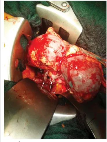
During harvesting a horseshoe kidney was removed en bloc. The isthmus connected the upper poles and contained parenchyma, so it could not be split. Vascu- lar anatomy consisted of three renal arteries with two patches and fi ve veins connecting to the inferior vena cava. There were two ureters with separate collecting systems (Fig. 1).
The recipient was a 49-year-old female with a history of hypertension and idiopathic nephrotic syndrome. Re- nal biopsy performed in 2007 showed advanced scar- ring, so no primary etiology could be identifi ed. Focal segmental glomerulosclerosis was suspected by the treat- ing physician on clinical grounds. The patient has re- ceived peritoneal dialysis since 2008. She was switched to hemodialysis due to ultrafi ltration failure after 3 years.
Donor and recipient were both cytomegalovirus (CMV) IgG positive. The transplantation was performed with 2 DR human leukocyte antigen (HLA) matches, recipi- ent panel reactive antibody being 0%. The horseshoe kidney was implanted en bloc to the left side retroperi- toneally. An end-to side cavovenostomy (between do- nor inferior vena cava [IVC] and recipient common iliac
vein), arterio-arteriostomies (one anastomosis to the common iliac artery and one to the external iliac artery), and two separate ureteroureterostomies were performed (Fig. 2). Cold ischemic time was 12 h, and warm isch- emia time was 75 min (counted from the beginning of venal anastomosis to the time of declampage).
The recipient received combined immunosuppressive therapy with tacrolimus, MMF, and tapering dosages of steroids with valgancyclovir given as CMV prophylaxis.
Patient’s anemia was corrected with two units of irradi- ated selected blood cell transfusions. Graft function was immediate. On the 4th postoperative day, graft function started to decline, proteinuria was detected, and oliguria.
A three-day course of steroid pulse with 500 mg/day iv.
methylprednisolone boluses was begun, and renal biopsy performed. Histology revealed signs for acute antibody- mediated rejection (peritubular capillaritis, glomeruli- tis, and vascular microthrombosis) as well as borderline T-cell mediated acute cellular rejection. The diagnosis of antibody-mediated rejection was also confi rmed by el- evated donor specifi c antibody (DSA) titers. The patient received four sessions of plasmapheresis with intrave- nous immunoglobulin (IVIG) replacement three times a week resulting in a complete elimination of DSA and
Fig. 1. Back-table preparation of the horseshoe kidney. The + sign represents the two graft ureters, VCI = segment of vena cava inferior, iliaca patch shows the iliac artery prepared for anastomosis
Fig. 2. Intraoperative picture of the graft and the vascular anas- tomoses. The yellow arrow represents the venal, the two white arrows show the arterial anastomoses
improvement of graft function and proteinuria. Mean- while, lower abdominal pain developed. On abdominal ultrasonography, a fl uid collection was identifi ed next to the graft. A computed tomography (CT)-guided drain- age of this fl uid was performed. Subsequent cystoscopy showed leakage of urine without precise localization, so double J catheters were implanted into both donor ureters resolving urine leakage. Patient was discharged home with stable graft function.
During follow-up, urinary infection recurred but was successfully treated with antibiotics. Patient was subsequently hospitalized in order to remove the dou- ble J catheters. After the fi rst catheter was removed, urinary sepsis ensued with massive thrombocytopenia.
CT scan identifi ed a fl uid collection next to the graft.
The patient was taken to the operation room for explo- ration. There was a needle-sized leak at the proximal ureteroureteral anastomosis. Due to persistent signs of sepsis despite the appropriate antibiotics, a graftectomy was performed.
Discussion
The experience with horseshoe kidney transplantation remains limited. A review, analyzing cases between 1983 and 2000, found a total of 47 cases of horseshoe kid- ney explantations within the Eurotransplant region [11].
From these grafts, 13 were discarded because of severe atherosclerosis or complex vascular anatomy. In fi ve cas- es, there were no data for the reason of horseshoe kidney refusal. Eight horseshoe kidneys were transplanted en bloc, and 26 grafts were divided and then transplanted separately into 47 recipients. In fi ve cases, one half of the separated kidneys were discarded due to vessel damage or because of complex vascular anatomy rendering the transplantation unfeasible. From the 26 split horseshoe kidneys, 23 had parenchymatous isthmus. Average cold ischemic time was 24 h in the en bloc group and 25 h in the split group. The rate of primary nonfunction (PNF) did not alter signifi cantly after transplanting horseshoe kidney compared with transplanting normal kidneys.
Surgical complications (bleeding from the surface of di- vided isthmus) were only seen in the split group. One- year graft survival did not diff er between the normal and horseshoe kidney groups. Another review published in 2010 analyzed 28 case reports on horseshoe kid- ney transplants [12]. In 15 cases, kidneys were trans- planted en bloc, and in the remaining cases, horseshoe kidneys were separated. Three horseshoe kidneys were transplanted into 3 recipients after splitting because of anomalies of vascular anatomy. From these cases, 9 grafts (18%) experienced primary nonfunction, and there were 2 recipient deaths. In the rest of the cases, the graft func- tion was good at 6 month follow-up. The average cold ischemic time was 24.4 h.
In the majority of cases, kidneys are fused at the lower poles [1, 2]. On the other hand, fusion at the upper pole as it was seen in our case, is very rare. Transplanting a horseshoe kidney demands technical expertise because an average, normal vascular anatomy occurs only in the 33% of these cases [12]. Caution is needed during organ harvesting. In cadaveric donors, arterial perfusion can- nula should be placed in the common or the external iliac artery to avoid injury of accessorial arteries. H.P.
Tan et al. recommend harvesting horseshoe kidney en bloc with long segments of aorta, vena cava, and iliac vessels [13]. In our case, the kidney was harvested en bloc, and because of a broad parenchymal isthmus and complex vascular anatomy, the kidney was implanted as a single graft. During the transplantation, we closed the proximal end of donor vena cava with a running suture and anastomosed the other end in an end-to-side way to the common iliac vein. This technique is recommended when broad, multiple veins are present branching from the vena cava [14]. Because of limited information on preparing horseshoe kidneys for transplantation on the
“back table” and also for the technical challenges of transplanting them, Uzzo et al. published an algorythm for the evaluation and utilization of horseshoe kidneys for transplantation [15]. Kidneys with a thick isthmus and a wide parenchymal bridge are recommended for transplantation en bloc [13]. However, Stroosma et al.
analyzed 26 cases of split horseshoe kidneys and found that 23 horseshoe kidneys had parenchymatous isthmus [11]. They recommended using a stapler in order to safely seal the surface of the divided isthmus. The col- lecting system rarely crosses the isthmus. It is more com- mon to fi nd the isthmus to contain only fi brous tissue.
In cases of a thick isthmus, one study recommends eval- uating the collecting system with contrast to ascertain anatomy and implanting it en bloc in case the collecting system crosses the isthmus [16]. After the transplanta- tion, we observed urine leakage, which later led to sepsis.
Therefore, graftectomy could not be avoided. Ureteric complications after renal transplantation are often dif- fi cult to manage. Occasionally, challenging surgery pro- cedures are necessary to solve the problem. Piros et al.
published a case with a totally necrotized graft ureter after kidney transplantation managed by nephrectomy with pyelon transection and pyelo-pyelar anastomosis [17]. Another group observed urine leakage presenting on 7th postoperative day after the transplantation of a divided horseshoe kidney [18]. The kidney had an addi- tional lower pole artery, which had been transsected dur- ing organ harvesting. After an unsuccessful attempt with conservative treatment (decompression of the urinary tract with insertion of Foley catheter into the bladder), the patient needed surgical exploration. The lower pole of the kidney and the retained isthmus was found to be ischemic, and a urinary leak was identifi ed in the collect- ing system. The defect was closed in 3 layers with suture.
After the reoperation, no complication occurred and the patient had excellent graft function thereafter [18]. In addition to receiving a horseshoe kidney, our patient also suff ered from acute antibody-mediated rejection (AMR) combined with T-cell mediated acute cellular rejection.
Appropriate treatment for combined humoral and cel- lular rejection was promptly started as recommended by others [19]. Lefaucheur et al. analyzed the impact of pre-existing donor-specifi c HLA-antibodies and AMR on graft survival and found that when acute antibody- mediated rejection occurred, graft survival was signifi - cantly worse [20]. Despite eliminating DSA from our patient serum and successful treatment of acute rejec- tion, the graft ended up failing in our case. The sepsis leading to eventual graft failure could have developed due to increased immunosuppression with the anatomi- cal anomaly being an additional contributing factor.
Hau et al. reported a case with a 19-year-old donor suff ering brain trauma. The horseshoe kidney was dis- covered with a wide isthmus, two arteries. The organ harvesting had been performed en bloc. The curiosity of this case is that the kidney was transplanted intraab- dominally, with an aortic segment (containing the 2 ar- teries) to the iliac artery, and the veins separately to the caval vein. They extensively discussed the pros and cons of separating a horseshoe kidney. The conclusion is that, in case of a complex vasculature, and a wide (more than 2 cm) isthmus, the en bloc implantation is suggested.
In case of separation, a ureterography is recommended:
if the two pyelons are separated, the surgical division is to be chosen. They also recommend the intraabdominal implantation due to the extreme importance of position- ing the graft properly [21].
Urography was also used and reported by Sieńko et al. in 2014, as a useful toolkit for decision making [22].
Vernadakis et al. also reported horseshoe kidney trans- plantation in 2013 with similar milestone in the diagnos- tics and implantation [23].
Verbelen et al. also mentioned horseshoe kidneys in relation to pretransplant nephrectomies. According to the authors, it is recommended to remove polycystic kid- ney before transplantation. In their reported series, the recipient had a polycystic horseshoe kidney. Their main argument for nephrectomy was to organize and clear up the vascular accesses before transplantation [24]. One of the most exciting reports is about a living related kid- ney transplant, when the donor was his 43-year-old sister who had an uncomplicated horseshoe kidney with nega- tive results on a urinalysis. An aortogram showed that the arterial supply to the kidney consisted of 2 superior arteries (1 on each side) and 1 inferior accessory artery that was divided to feed the lower fused parenchyma of the kidney. Surgery was performed via a retroperitoneal lumbotomy incision; the left half of the kidney was mo- bilized. The left kidney was procured by clamping the in- ferior accessory renal artery, transecting the parenchyma
within the demarcation boundary. The transplant kidney was placed in the recipient’s contra lateral iliac fossa. Au- thors report on excellent long-term results [25].
Conclusions
Horseshoe kidney is an infrequent choice as a donor organ. If one decides to accept a horseshoe kidney for kidney transplantation, the option exists to divide and transplant separately or transplant en bloc. As our case demonstrates, dividing the horseshoe kidney is not al- ways feasible when a large parenchyma bridge is present.
As to the postoperative course, the uncommon anatomy of the ureter together with the development of AMR may have contributed to the development of a ureter tip necrosis giving rise to urinoma infection. When faced with the option whether or not to accept horseshoe kid- ney for transplantation, the increased risk for urinary complications should be kept in mind.
* * *
Funding sources: No funding was obtained.
Authors’ contribution: BN – staff surgeon, in the operation of the case, writing of the manuscript; ZSK – assistant surgeon to the case, ad- visor in surgical part; GZ – literature review, taking photos and editing them; LZS – chief transplant nephrologist, advisor, review the nephro- logical part; MB – intensivist, advisor for the intensive management part, review; MH – assistant surgeon at the harvesting of the kidney from the donor; KK – chief nephrologist for the pretransplant period, advisor for the nephrological part; AP – staff surgeon for the donor procurement, photos, and advisor for the organ harvesting part. All authors had full access to all data in the study and take responsibility for the integrity of the data and the accuracy of the data analysis.
Confl ict of interest: No confl ict of interest was present.
Abbreviations: AMR: antibody-mediated rejection; CMV: cytomega- lovirus; DSA: donor specifi c antibody; HK: horseshoe kidney; HLA:
human leukocyte antigen; IVAG: intravenous immunoglobulin; IVC:
inferior vena cava
References
1. Eisendrath DN, Phifer FM, Culver HB: Horseshoe kidney. Ann Surg 82, 735–764 (1925)
2. Glodny B, Petersen J, Hofmann KJ, Schenk C, Herwig R, Trieb T, Koppelstaetter C, Steingruber I, Rehder P: Kidney fusion anomalies revisited: clinical and radiological analysis of 209 cases of crossed fused ectopia and horseshoe kidney. BJU Int 103, 224–235 (2009)
3. Da Carpi B (1535): Isagogae breves perlucide ac uberrime in anatomiam humani corporis. National Library of Medicine.
Woodcut, Bologna
4. Glenn JF: Analysis of 51 patients with horseshoe kidney. N Engl J Med 261, 684–687 (1959)
5. Boatman DL, Kolln CP, Flocks RH: Congenital anomalies as- sociated with horseshoe kidney. J Urol 107, 205–207 (1972) 6. Boatman DL, Cornell SH, Kolln CP: The arterial supply of
horseshoe kidneys. Am J Roentgenol Radium Ther Nucl Med 113, 447–451 (1971)
7. Graves FT: The arterial anatomy of the congenitally abnormal kidney. Br J Surg 56, 533–541 (1969)
8. Nelson RP, Palmer JM: Use of horseshoe kidney in renal trans- plantation: technical aspects. Urology 6, 357–359 (1975) 9. Stroosma OB, Scheltinga MR, Stubenitsky BM, Kootstra G:
Horseshoe kidney transplantation: an overview. Clin Transplant 14, 515–519 (2000)
10. Marofka F, Szederkényi E, Mihalovits G, Lázár Gy, Morvay Z, Szenohradszky P: Patkóvesével történő veseátültetés hosszú távú eredményei [Long-term results of transplantation with horseshoe kidney]. Magy Seb. 61(3), Suppl. 174 (2008), in Hungarian 11. Stroosma OB, Smits JM, Schurink GW, de Boer J, Persijn GG,
Kootstra G: Horseshoe kidney transplantation within the eu- rotransplant region: a case control study. Transplantation 72, 1930–1933 (2001)
12. Pontinen T, Khanmoradi K, Kumar A, Kudsi H, Cheng Kung S, Chewaproug D, Zaki R, Ortiz J: Horseshoe kidneys: an un- derutilized resource in kidney transplant. Exp Clin Transplant 8, 74–78 (2010)
13. Tan HP, Samaniego MD, Montgomery RA, Burdick JF, Maley WR, Kraus ES, Ratner LE: Donor horseshoe kidneys for trans- plantation. Transplantation 72, 869–873 (2001)
14. Ratner LE, Zibari G: Strategies for the successful transplantation of the horseshoe kidney. J Urol 150, 958–960 (1993)
15. Uzzo RG, Hsu TH, Goldfarb DA, Taylor RJ, Novick AC, Gill IS:
Strategies for transplantation of cadaveric kidneys with congeni- tal fusion anomalies. J Urol 165, 761–765 (2001)
16. Stroosma OB, Schurink GW, Smits JM, Kootstra G: Transplant- ing horseshoe kidneys: a worldwide survey. J Urol 166, 2039–
2042 (2001)
17. Piros L, Deak PA, Dallos G, Mathe Zs, Doros A: Successful uri- nary tract reconstruction following ureteral necrosis in kidney transplant patient. Interv Med Appl Sci 2(3), 134–138 (2010)
18. Foster JT, Morrissey PE: Segmental renal ischemia following transplantation of horseshoe kidney as separate allografts. Case Rep Transplant 2013, 852127 (2013)
19. Slatinska J, Honsova E, Burgelova M, Slavcev A, Viklicky O:
Plasmapheresis and intravenous immunoglobulin in early anti- body-mediated rejection of the renal allograft: a single-center experience. Ther Apheresis Dial 13, 108 (2009)
20. Lefaucheur C, Loupy A, Hill GS, Andrade J, Nochy D, Antoine C, Gautreau C, Charron D, Glotz D, Suberbielle-Boissel C: Pre- existing donor-specifi c HLA antibodies predic outcome in kidney transplantation. J Am Soc Nephrol 21(8), 1398–1406 (2010) 21. Hau HM, Morgul HM, Uhlmann D, Thelen A, Fellmer
P, Benckert C, Tautenhahn HM, Bartels M, Jonas S: Horseshoe kidney for transplantation: technical considerations. Scand J Urol 47(1), 76–79 (2013)
22. Sieńko J, Kotowski MJ, Nowacki A, Romanowski M, Sulikows- ki T, Ostrowski M: Methylene blue usage in horseshoe kidney graft separation: case report. Transplant Proc 46(8), 2923–2926 (2014)
23. Vernadakis S, Moris D, Kaiser G, Kykalos S, Sotiropoulos GC:
Horseshoe kidney transplantation. Am Surg Sep 79(9) 298–299 (2013)
24. Verbelen T, Darius T, Pirenne J, Monbaliu D: Decision making in pretransplant nephrectomy for polycystic kidneys, is it valid for horseshoe kidneys? Transplant Int 25(8), 96–97 (2012)
25. Sezer TO, Solak I, Sozbilen M, Firat O, Yilmaz M, Toz H, Sarsik B, Isayev C, Harman M, Hoscoskun C: A horseshoe kidney from a live donor as a renal transplant case report. Exp Clin Transplant 11(5), 454–457 (2013)