Downloadedfromhttps://journals.lww.com/co-pediatricsbyBhDMf5ePHKav1zEoum1tQfN4a+kJLhEZgbsIHo4XMi0hCywCX1AWnYQp/IlQrHD3tIQ5gQCIeyzeik+O9MyT0/My2IHHQVKVNr3hUK93/gteovjGwLvt1A==on05/08/2020
Downloadedfrom https://journals.lww.com/co-pediatricsby BhDMf5ePHKav1zEoum1tQfN4a+kJLhEZgbsIHo4XMi0hCywCX1AWnYQp/IlQrHD3tIQ5gQCIeyzeik+O9MyT0/My2IHHQVKVNr3hUK93/gteovjGwLvt1A==on
05/08/2020
C O
URRENTPINIONEvaluation of a child with suspected nephrolithiasis
George S. Reusza, Adam Hosszub, and Eva Kisc
Purpose of review
As the incidence of nephrolithiasis in children doubles every 10 years it is becoming a common disease associated with significant morbidity along with considerable economic burden worldwide. The aim of this review is to summarize current data on the epidemiology and causes of renal stones in children and to provide a frame for the first clinical evaluation of a child with suspected nephrolithiasis.
Recent findings
Dietary and environmental factors are the driving force of changing epidemiology. Diagnosis should be based on medical history, presenting signs, examination, first laboratory and radiological workup.
Ultrasound should be the initial diagnostic imaging performed in pediatric patients while low-dose computed tomography is rarely necessary for management. Metabolic factors including hypercalciuria, hypocitraturia, low fluid intake as well as specific genetic diseases should be explored after the resolution of initial signs and symptoms.
Summary
Appropriate initial evaluation, imaging technique, identification of risk factors and other abnormalities are essential for early diagnosis and prevention of stone-related morbidity in children with suspected
nephrolithiasis.
Keywords
epidemiology, kidney stones, low-dose computed tomography, metabolic evaluation, nephrolithiasis, ultrasound, urolithiasis
INTRODUCTION
Nephrolithiasis is the process in which a solid for- eign body (kidney stone) is formed by precipitation and aggregation of urine constituents within the kidney or the urinary tract. Stone formation occurs if the concentration of a solute surpasses its limit of solubility and is further facilitated by sites of aggre- gation such as Randall plaques [1,2&&,3&&] at the urinary surface of the renal papillae, bacterial or epithelial debris in the urine. Conditions leading to increased concentrations of stone forming solutes or the decrease of the inhibitory activity of urine promote stone formation [1,4].
Stones breaking loose from the kidney exit the body in the urine stream passing through the ureters, the bladder and the urethra. During the passage they can cause ureter blockage result- ing in severe pain in the lower back or abdomen
[5&,6&&].
Herein, we will review some new aspects of the epidemiology of pediatric nephrolithiasis followed by analysis of the clinical features of acute stone disease. Signs and symptoms as well as first-line lab work and radiological approach to the disease will be discussed.
Detailed metabolic evaluation, medical and uro- logical management and the aspects of prevention are beyond the scope of this survey and will be discussed in the following articles of this issue.
EPIDEMIOLOGY
The occurrence of kidney stone disease shows high geographical variability due to – among others – environmental, metabolic, dietary and genetic fac- tors [7,8].
Nephrolithiasis is rather common in adults.
Over the past decade incidence increased from 4%
to about 9%, with a slight male preponderance (11%
aFirst Department of Pediatrics, Semmelweis University, bMTA-SE
‘Lend€ulet’ Research Group, Hungarian Academy of Sciences andcPedi- atric Heart Center, Gottsegen Gyo¨rgy Hungarian Institute of Cardiology, Budapest, Hungary
Correspondence to George S. Reusz, PhD, ScD, First Department of Pediatrics, Semmelweis University, Bo´kay J u 53, H-1083 Budapest, Hungary. Tel: +36 1 3247795;
e-mail: reusz.gyorgy@med.semmelweis-univ.hu Curr Opin Pediatr2020, 32:265–272 DOI:10.1097/MOP.0000000000000880
REVIEW
among men compared with 7% among women).
The increase in the frequency of renal stones has been linked to the ‘epidemic’ of obesity [7,9,10]. A study including around 5000 adult stone patients showed that obesity and weight gain are indepen- dent risk factors for the development of nephroli- thiasis in both sexes [11]. In children the occurrence of pediatric nephrolithiasis is one magnitude lower than in adults, nevertheless, incidence has been steadily growing by 6–10% annually in the past 25 years, with an estimated incidence of 36–57 per 100 000 in the United States [12–15]. Signifi- cantly, the highest increase has been reported in teenage girls [14]. Although the epidemic of obesity has reached the pediatric population as well and parallels the increase in nephrolithiasis [16,17], its direct relation to nephrolithiasis is less evident than in adults. Recent pediatric studies do not agree on this issue [15,18,19&&], presumably due to geograph- ical and ethnical factors among others, moreover because of the different methodology of individual surveys. Results from a study in a network of 30 primary care pediatric practices including 110 cases and 396 matched controls from the United States [18] and a tertiary center from the United Kingdom [15] suggested that BMI should not be considered as a separate risk factor in the development of kidney stones in children. In contrast, a large epidemiologic survey from Israel found that the odds ratio for nephrolithiasis in candidates with a BMI of more than 30 kg/m2 was 1.7 compared with candidates with a BMI of 18.5–24.9 kg/m2 [19&&]. Thus, the relationship between increasing prevalence of neph- rolithiasis and higher body mass should be explored further.
With increasing occurrence, the number of hos- pital admissions, emergency department (ED) visits, surgical interventions and as a consequence, the economic burden increases as well [20–22,23&].
The great geographical variability may be explained inter aliaby environmental factors, such as arid climate, as well as dietary habits, such as the extent of salt and fluid intake, diversity of processed food and animal protein load and of vegetable and fruit consumption [24,25]. As an example, in the so- called stone belt in the southeast of the United States the prevalence of renal stones is 50% higher than in the northwest [26]. In the Afro-Asian ‘stone belt’ zone stretching from Morocco over Egypt to India, Indonesia and the Philippines there is also a positive correlation between nephrolithiasis preva- lence and temperature. However, the high fre- quency of bladder stones in these regions suggests the role of possible additional factors (e.g. diet, malnutrition and infections) [27,28].
WHEN SHOULD NEPHROLITHIASIS BE SUSPECTED?
The typical stone patient’s symptoms are flank pain, hematuria, nausea and vomiting [25,26,29]. How- ever, the signs and symptoms are largely dependent on the localization of the stone, its dimensions, the degree of consequent obstruction [30] and the age of the patient [25,26,29]. In particular, young children do not present with the usual acute onset of flank pain seen in adults, therefore children are very often evaluated for other conditions before the diagnosis of nephrolithiasis is made. A diagnostic pathway for the identification of nephrolithiasis is shown in Fig. 1.
Pain and accompanying vegetative symptoms A kidney stone formed at and fixed to the papilla is usually asymptomatic [29,31&&]. In a study from the United Kingdom, 13% of stones were diagnosed as accidental findings in asymptomatic patients [15].
In a Canadian report of the 244 unique children identified by radiological methods [ultrasound or computed tomography (CT)] to have renal stones, 140 (57%) were symptomatic while the other 104 patients (43%) were asymptomatic [29,31&&].
As the stone breaks away from the papilla and moves down the ureter it may get stuck at one of the three preformed areas of decreased luminal diame- ter: the pyelo-ureteral junction, the crossing site of the iliac vessels or the uretero-vesical junction, caus- ing obstruction, consequent distension of the uri- nary tract and abrasion of the mucosa. In the same study, 32% of patients presented with pain, 13% had painful gross hematuria and 36% presented with urinary tract infection [31&&].
Due to the conjoined innervation of the gastroin- testinal, genitourinary and somatic systems, patients may feel pain in the intestines, groin, bladder or
KEY POINTS
Dietary and environmental factors are the driving force of changing epidemiology of nephrolithiasis.
Family history and medical history should be explored, focusing on fluid intake as well as on specific
genetic diseases.
Ultrasound is the cornerstone of radiological imaging while low-dose computed tomography is rarely necessary in children.
Detailed metabolic evaluation and analysis of stones is necessary after the resolution of initial signs
and symptoms.
genitalia, further vegetative signs such as nausea and vomiting frequently accompany the painful colic [32].
Dilatation of the urinary tract due to obstruction is not an obligatory diagnostic mark of the passage of a stone. According to a recent study including 248 consecutive patients presenting with ureteral colic, nearly 11% did not demonstrate any dilatation and the majority (nearly 71%) had only mild hydro- nephrosis. Stone diameter was related to the degree of hydronephrosis, whereas age, sex and stone loca- tion were not [30]. Smaller stone size and lower incidence of hydronephrosis could explain the lower diagnostic accuracy of ultrasound compared with CT for detecting ureteral stones [30].
Lower incidence of hydronephrosis in the case of small stones is a matter of debate and may raise the question whether the symptoms conceivably result from transient hydronephrosis which cannot be detected with imaging modalities [33].
Clinical signs are also largely dependent on the age of the child. The younger a child is the less characteristic the symptoms are. In infants, irrita- bility, inconsolable crying, poor feeding, vomiting and in the case of urinary tract infection, signs and symptoms of sepsis may occur [25,26,29].
In young children poorly localized (often perium- bilical or umbilical) abdominal pain, vomiting, diar- rhea or constipation can be major challenges for the differential diagnosis. Only older children present symptoms similar to those seen in adults. The pain typically begins as waxing and waning flank pain in the acute phase [5&,34]. Ipsilateral genital pain is a common symptom of distal ureteral stones. As renal stones not causing obstruction are usually asymptom- atic, the detection of a nonobstructive stone with imaging techniques requires consideration of another etiology of the patient’s symptoms [29,35].
Hematuria
Hematuria can be macroscopic or more commonly microscopic. In the case of incidentally discovered asymptomatic stones hematuria may be absent.
Accordingly, using hematuria to predict the pres- ence of urolithiasis has an accuracy of only 60% and the absence of hematuria does not rule out urolith- iasis [7,36].
Microscopic hematuria may precede the appear- ance of kidney stones by years and is often associ- ated with hypercalciuria [37&,38]. Hypercalciuria is Suspected urolithiasis based on paent’s history and presentaon:
Pain, nausea, voming, hematuria
Perform focused family history, medical history and examinaon, Basic laboratory assessment with creanine and urinalysis
Consider complicaons or mimic:
Young age, no prior stone, fever, chills, solitary kidney, suspicion of urolithiasis mimic
No
History of prior stone and low risk for alternave diagnosis or complicated urolithiasis
1. Provide symptom relief 2. Bedside ultrasound
No history of prior stone, but low risk for alternave diagnosis or complicated urolithiasis
1. Provide symptom relief 2. Imaging with ultrasound, 3. Consult with radiologist CT or MRI
Suspicion of complicated nephrolithiasis or other dangerous eology 1. Ultrasound
2. Consult with radiologist CT or MRI
Infected renal stone 1. Treat symptoms, fluids, anbiocs,
2. Ultrasound 3. Consult urology CTI or MRI
Other condion:
manage accordingly
Detailed metabolic evaluaon and analysis of stones or fragments
FIGURE 1.The urolithiasis diagnostic pathway (based on [5&,23&,25,29]).
considered to be an important contributor to stone formation; however, individual studies provide very different prevalence data, ranging from 10 to 50%
[15,39,40,41&]. Further, isolated hypercalciuria without renal stone disease is associated with an increased frequency of urinary tract infections [42].
Thus, hypercalciuria and stone disease should be considered among the risk factors for UTI and should be investigated particularly in patients with a family history of urinary stones [43&].
Infection
Urinary tract infections may be present in up to 30%
of patients with urolithiasis. Age below 2 years at diagnosis, the presence of a metabolic risk factors and size of stone above 5.3 mm are significant risk factors for infection [43&].
Fevers and chills are not common in uncompli- cated urolithiasis, but if present, should raise con- cern for an infected stone [25,44].
DIAGNOSTICS
Initial evaluation depends on the situation in which the stone is discovered and includes laboratory test- ing and radiological imaging.
Laboratory testing
In the ED laboratory testing and imaging should focus on the detection of the suspected renal stone and its eventual acute consequences, such as obstruction, infection, and occasionally pre and/
or post renal failure [5&,25].
A more detailed metabolic evaluation [40,41&] including urine osmolarity, excretion of solutes such as calcium, sodium, oxalate, urate, citrate, magnesium, cysteine, urine pH, etc., should take place after the resolution of the initial violent signs and symptoms, or in the case of an accidentally discovered ‘silent’ stone, because in the acute phase dehydration (due to vomiting) and therapeutic mea- sures (intravenous fluids with NaCl load) can signif- icantly influence urinary concentration and excretion of solutes.
Blood tests
Dehydration due to vomiting can lead to a tran- sient rise in serum creatinine of prerenal origin
[5&,36]. More severe creatinine elevation may be
detected in patients with solitary kidney or cases with bilateral obstruction (postrenal mechanism) [45], advanced chronic kidney disease at baseline (acute on chronic mechanism). Eventually drug-
induced crystal nephropathy may induce serious acute kidney injury [46&&,47&,48]. Medication may further be associated with renal stone disease and increase incidence by influencing the enteral microbiome [49&&].
Parameters indicative of inflammation, such as increment of neutrophil count or C reactive protein, may help to differentiate complicated urolithiasis with infection from an uncomplicated stone
[5&,29,36]. However, as an increase in neutrophil
count can also be due to a stress reaction, it should be considered rather as a supplement to clinical decision-making [36].
Urinalysis
Urinalysis reveals the presence of hematuria. How- ever, hematuria may be absent in the case of a silent stone or in a patient with complete obstruction.
Urinary crystals are common in healthy patients.
As the voided urine specimen cools down during storage on room temperature, the solubility of urine components decreases leading to in-vitro crystal formation. Thus, the presence of crystals per se is insufficient for the diagnosis of urolithiasis. As exception to the general rule, specific crystal forms (e.g. cystine) are informative of the metabolic con- dition leading to stone disease [1]. The presence of urinary white blood cells under light microscopy, a positive leukocyte esterase reaction and nitrites on the test strip are suggestive for urinary tract infec- tion. However, urolithiasis can cause sterile ureteral inflammation, thus urinalysis should always be eval- uated together with clinical symptoms and the result of urine culture [43&].
Diagnostic imaging
While native abdominal radiography and intrave- nous urography were the standard methods to detect urolithiasis and consequent urinary obstruc- tion in adults as well as in children for decades, with the arrival of ultrasound and CT diagnostic algo- rithms have undergone fundamental changes. In adult urology CT has become the standard for stone imaging. Low-dose CT scans can now be performed with a similar or less amount of radiation as plain radiographs, without the need for intravenous con- trast [50]. In pediatrics however, both the American Urological Association and the European Society for Pediatric Radiology recommend ultrasound as the initial imaging modality [51,52]. Accordingly, abdominal radiography is not used routinely in children anymore. If clinical suspicion of urolithia- sis is high enough ideally ultrasound is usually chosen for imaging [53&] (Figs. 2–4).
Indeed, ultrasound evaluation has its limits. In a pediatric comparative study from 68 renal stones detected by CT 30 were not recognized by ultra- sound. Altogether, ultrasound was 66.7% (48.8–
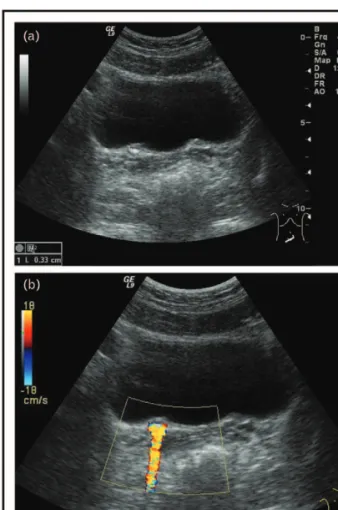
80.8%) sensitive and 97.4% (86.8–99.9%) specific for detecting stones. However, of the 30 stones not detected by ultrasound, only three were more than 3 mm according to CT. Thus, in the clinical practice, ultrasound has high specificity for detecting neph- rolithiasis in children but only moderate sensitivity for stones more than 3 mm, where false negatives are common [54&]. The sensitivity of ultrasound can be enhanced by the color Doppler technique using the stone-triggered artifact called twinkling artifact. The twinkling artifact is a mixture of rapidly alternating red and blue pixels behind a strongly reflective object (e.g. calculus) resembling turbulent blood flow. This phenomenon is thought to be secondary to intrinsic machine ‘noise’ within the color Dopp- ler circuitry of the ultrasound device [55,56&&,57&] (Fig. 5a and b).
Meanwhile, thanks to continuous technical progress the radiation burden of CT has also decreased substantially; however, it is indeed still not negligible [50]. Thus, there is a constant effort to
FIGURE 3. Staghorn stone filling the pyelum seen in a cystinuria patient. Please note the marked acoustic shadow.
The pyelum and some of the chalices are markedly dilated.
FIGURE 4.An ureterolith with an acoustic shadow is seen in the ureter near the ureterovesical junction. As part of the consequent obstruction, a dilated ureter is visible behind the bladder.
FIGURE 5. A nonobstructive small stone is shown at the ureterovesical junction (a). A stone-triggered artifact called twinkling artifact is seen (in the same patient) with color Doppler imaging (b).
FIGURE 2. A nonobstructive hyperechogenic renal stone with acoustic shadow is seen in the pyelum.
assist clinical diagnostics with appropriate imaging algorithms [52,53&,58] to reduce rates of initial and overall CT utilization without adversely impacting downstream care.
Evaluating risk factors of stone formation The risk factors should be assessed in two steps.
The first examination is intended to support general orientation. History of underlying structural abnormalities of the kidney and urinary tract should be explored together with the history of nephroli- thiasis in parents and siblings, as the offspring of renal stone formers may carry several metabolic risk factors similar to their parents and which are pre- disposing to stone formation [59]. Further, cystin- uria and hyperoxaluria may show increased occurrence in consanguineous families. Perinatal medical history with focus on prematurity, vitamin D supplementation, conditions leading to immobi- lization, dietary habits with special focus on salt and animal protein intake should be explored, daily fluid intake should be evaluated [60&,61&].
Further questions concerning medical history should refer to medications associated with stone formation [46&&,47&,48] and history of recurrent uri- nary tract infection, especially when caused by a urease-producing organism, such as Proteus or Kleb- siella [28,62].
The patient should be examined for the presence of manifest or latent malabsorptive intestinal dis- eases and conditions, such as Crohn’s disease, ulcer- ative colitis and short gut syndrome as they increase the risk of stone formation [63,64,65&].
In a second step, following the resolution of the acute symptoms a more detailed metabolic evalua- tion should take place.
While maintaining habitual fluid intake and dietary habits, a quantitative analysis of urinary solutes, both promoters and inhibitors of stone formation, and the assessment of daily fluid and food intake urine analysis should be performed [66].
The most appropriate method of urine collec- tion is still a matter of debate. 24-h urine collection [67] can be achieved in toilet-trained children, whereas in children who are not toilet-trained the solute/creatinine ratio in a single spot urine can be utilized to assess solute excretion. Alternatively, the use of 12 h urine collection and second morning sampling or afternoon single spot urine has also been proposed. Pediatric normal values for the con- stituents, such as calcium, oxalate and citrate are available and should be used as reference [67–70].
Using the concentration of urine constituents, different equations have been constructed to predict the risk of crystal precipitation [67,71,72&]. Further,
laboratory methods to explore the point of super- saturation and crystal formation have been designed
[73&]. However, these are not part of the everyday
clinical practice.
The analysis of stones or fragments of stones obtained after spontaneous passage or surgical inter- vention should always be performed. As stone com- position can change over time, recurrent stones should always be analyzed as well [66]. The methods of choice for analysis are radiograph diffraction or infrared spectroscopy [74]. This can lead directly to the diagnosis of rare diseases, such as cystinuria or adenine phosphoribosyl transferase defect.
CONCLUSION
The diagnosis of suspected nephrolithiasis is based on medical and family history, presenting signs, physical examination, first laboratory and radiolog- ical workup. Ultrasound should be the initial diag- nostic imaging performed in pediatric patients while low-dose CT is rarely necessary for manage- ment. Metabolic factors including hypercalciuria, hypocitraturia, low fluid intake as well as specific genetic diseases should be explored after the resolu- tion of initial signs and symptoms.
Acknowledgements
Figs. 2–5 were kindly provided by the Radiologic Division of the First Department of Pediatrics, Semmelweis Uni- versity Budapest.
Financial support and sponsorship
The study was supported by NKFI grant 124549 and 131637.
Conflicts of interest
There are no conflicts of interest.
REFERENCES AND RECOMMENDED READING
Papers of particular interest, published within the annual period of review, have been highlighted as:
& of special interest
&& of outstanding interest
1. Baumann JM, Affolter B. From crystalluria to kidney stones, some physicochem- ical aspects of calcium nephrolithiasis. World J Nephrol 2014; 3:256–267.
2.
&&
Evan AP, Coe FL, Lingeman J,et al.Randall’s plaque in stone formers originates in ascending thin limbs. Am J Physiol Renal Physiol 2018;
315:F1236–F1242.
Based on serial sections of papillary biopsies the authors conclude that plaques form preferentially in the basement membranes of ascending limbs of Henle, confirming the prediction of the washdown theory of plaque pathogenesis.
3.
&&
Wiener SV, Ho SP, Stoller ML. Beginnings of nephrolithiasis: insights into the past, present and future of Randall’s plaque formation research. Curr Opin Nephrol Hypertens 2018; 27:236–242.
A comprehensive review of the history of Randall’s plaque research.
4. Sa´ez-Torres C, Grases F, Rodrigo D,et al.Risk factors for urinary stones in healthy schoolchildren with and without a family history of nephrolithiasis.
Pediatr Nephrol 2013; 28:639–645.
5.
&
Gottlieb M, Long B, Koyfman A. The evaluation and management of urolithia- sis in the ED: a review of the literature. Am J Emerg Med 2018; 36:699–706.
A review focusing on the initial evaluation and management of stone patients including risk assessment at the emergency department.
6.
&&
Wiener SV, Chen L, Shimotake AR,et al.Novel insights into renal miner- alization and stone formation through advanced imaging modalities. Connect Tissue Res 2018; 59:102–110.
A study using high-resolution radiograph micro-computed tomography (CT), light and electron microscopy techniques to characterize the stepwise progression of events from crystallization to nephrolithiasis in human renal pyramids and kidney stones.
7. Shoag J, Tasian GE, Goldfarb DS, Eisner BH. The new epidemiology of nephrolithiasis. Adv Chronic Kidney Dis 2015; 22:273–278.
8. Liu Y, Yasheng A, Chen K,et al.Difference in urinary stone composition between Uyghur and Han children with urolithiasis. Urolithiasis 2017;
45:435–440.
9. Kovesdy CP, Furth SL, Zoccali C; World Kidney Day Steering Committee.
Obesity and kidney disease: hidden consequences of the epidemic. J Nephrol 2017; 30:1–10.
10. Sorokin I, Mamoulakis C, Miyazawa K,et al.Epidemiology of stone disease across the world. World J Urol 2017; 35:1301–1320.
11. Taylor EN, Stampfer MJ, Curhan GC. Obesity, weight gain, and the risk of kidney stones. JAMA 2005; 293:455–462.
12. Tasian GE, Ross ME, Song L,et al.Annual incidence of nephrolithiasis among children and adults in South Carolina from 1997 to 2012. Clin J Am Soc Nephrol 2016; 11:488–496.
13. Dwyer ME, Krambeck AE, Bergstralh EJ,et al.Temporal trends in incidence of kidney stones among children: a 25-year population based study. J Urol 2012;
188:247–252.
14. Kusumi K, Becknell B, Schwaderer A. Trends in pediatric urolithiasis: patient characteristics, associated diagnoses, and financial burden. Pediatr Nephrol 2015; 30:805–810.
15. Issler N, Dufek S, Kleta R, et al.Epidemiology of paediatric renal stone disease: a 22-year single centre experience in the UK. BMC Nephrol 2017;
18:136.
16. C¸ altık Yılmaz A, B€uy€ukkarago¨z B, Oguz U, C¸ elik B. Influence of body mass index on pediatric urolithiasis. J Pediatr Urol 2015; 11:350e1–350e6.
17. Kuroczycka-Saniutycz E, Porowski T, Protas PT, et al. Does obesity or hyperuricemia influence lithogenic risk profile in children with urolithiasis?
Pediatr Nephrol 2015; 30:797–803.
18. Kim SS, Luan X, Canning DA,et al.Association between body mass index and urolithiasis in children. J Urol 2011; 186:1734–1739.
19.
&&
Alfandary H, Haskin O, Davidovits M,et al.Increasing prevalence of nephro- lithiasis in association with increased body mass index in children: a popula- tion based study. J Urol 2018; 199:1044 –1049.
A large, population based study from Israel documenting an increasing prevalence of nephrolithiasis in children with a possible association with the increase in BMI during the same period.
20. Sas DJ, Hulsey TC, Shatat IF, Orak JK. Increasing incidence of kidney stones in children evaluated in the emergency department. J Pediatr 2010;
157:132–137.
21. Gonza´lez R, Ludwikowski BM. Editorial: Progress in pediatric urology in the early 21st century. Front Pediatr 2019; 7:349.
22. Hyams ES, Matlaga BR. Economic impact of urinary stones. Transl Androl Urol 2014; 3:278–283.
23.
&
Bowen DK, Tasian GE. Pediatric stone disease. Urol Clin North Am 2018;
45:539–550.
The review analyzes – among others – the overall economic burden of nephro- lithiasis.
24. Fakheri RJ, Goldfarb DS. Ambient temperature as a contributor to kidney stone formation: implications of global warming. Kidney Int 2011;
79:1178–1185.
25. Rodriguez Cuellar CI, Wang PZT, Freundlich M, Filler G. Educational review:
role of the pediatric nephrologists in the work-up and management of kidney stones. Pediatr Nephrol 2020; 35:383–397.
26. Marra G, Taroni F, Berrettini A,et al.Pediatric nephrolithiasis: a systematic approach from diagnosis to treatment. J Nephrol 2019; 32:199–210.
27. Jazayeri SM, Mamaghani ME, Pourmoghaddam A,et al.Need for emergent studies on dietary factors among infancy nephrolithiasis. Arch Ital Urol Androl 2011; 83:133–135.
28. Soliman NA, Rizvi SAH. Endemic bladder calculi in children. Pediatr Nephrol 2017; 32:1489–1499.
29. Marzuillo P, Guarino S, Apicella A,et al.Why we need a higher suspicion index of urolithiasis in children. J Pediatr Urol 2017; 13:164–171.
30. Song Y, Hernandez N, Gee MS,et al.Can ureteral stones cause pain without causing hydronephrosis? World J Urol 2016; 34:1285–1288.
31.
&&
Cassim R, Van Walraven C, Lavalle´e LT,et al.Systematic radiologic detection of kidney stones in Canadian children: a new era of asymptomatic stones? J Pediatr Urol 2019; 15:467e1–467e7.
A study on the incidence of nephrolithiasis in pediatric patients undergoing abdominal ultrasound or computerized tomography for all indications, showing that 43% of the stones were asymptomatic, following a benign course with minimal need for intervention.
32. Shokeir AA. Renal colic: new concepts related to pathophysiology, diagnosis and treatment. Curr Opin Urol 2002; 12:263–269.
33. So¨ylemez H, Yıldırım K. How can a ureteral stone cause pain without hydronephrosis? World J Urol 2016; 34:1289.
34. Pfau A, Knauf F. Update on nephrolithiasis: core curriculum. Am J Kidney Dis 2016; 68:973–985.
35. Carter MR, Green BR. Renal calculi: emergency department diagnosis and treatment. Emerg Med Pract 2011; 13:1–17.
36. Ingimarsson JP, Krambeck AE, Pais VM Jr. Diagnosis and management of nephrolithiasis. Surg Clin North Am 2016; 96:517–532.
37.
&
Valavi E, Nickavar A, Aeene A. Urinary metabolic abnormalities in children with idiopathic hematuria. J Pediatr Urol 2019; 15:165e1–165e4.
The study confirms that metabolic alterations commonly seen in nephrolithiasis may be at the origin of microscopic isolated hematuria without renal stone diseases.
38. Srivastava T, Schwaderer A. Diagnosis and management of hypercalciuria in children. Curr Opin Pediatr 2009; 21:214–219.
39. Lee ST, Cho H. Metabolic features and renal outcomes of urolithiasis in children. Ren Fail 2016; 38:927–932.
40. Bevill M, Kattula A, Cooper CS, Storm DW. The modern metabolic stone evaluation in children. Urology 2017; 101:15–20.
41.
&
Chan KH, Moser EA, Whittam BM,et al.The ability of a limited metabolic assessment to identify pediatric stone formers with metabolic abnormalities. J Pediatr Urol 2018; 14:331e1–331e6.
The value of a limited metabolic evaluation including measurement of 24-h calcium, citrate, oxalate and low urinary volume were compared with a complete urinary metabolic profile. Restricted evaluation detected most of the clinically significant metabolic abnormalities suggesting a measure to reduce healthcare costs.
42. Nacaroglu HT, Demircin G, B€ulb€ul M,et al.The association between urinary tract infection and idiopathic hypercalciuria in children. Ren Fail 2013;
35:327–332.
43.
&
Cetin N, Gencler A, Kavaz Tufan A. Risk factors for development of urinary tract infection in children with nephrolithiasis. J Paediatr Child Health 2020;
56:76–80.
In this retrospective follow-up study, the patients were divided in groups with and without urinary tract infection. The age at diagnosis, the presence of a metabolic risk factor and size of stone were significant risk factors for infection.
44. Borghi L, Nouvenne A, Meschi T. Nephrolithiasis and urinary tract infections: ’the chicken or the egg’ dilemma? Nephrol Dial Transplant 2012; 27:3982–3984.
45. Wen JG, Li ZZ, Zhang H,et al.Melamine related bilateral renal calculi in 50 children: single center experience in clinical diagnosis and treatment. J Urol 2010; 183:1533–1537.
46.
&&
Chatchen S, Pongsakul N, Srisomsap C,et al.Unravelling pathophysiology of crystalline nephropathy in ceftriaxone-associated acute kidney injury: a cel- lular proteomic approach. Nephron 2018; 139:70–82.
The study presents a cellular model of crystalline nephropathy in ceftriaxone- associated acute kidney injury and the related pathophysiology by using a proteomic approach.
47.
&
Sighinolfi MC, Eissa A, Bevilacqua L,et al.Drug-induced urolithiasis in pediatric patients. Paediatr Drugs 2019; 21:323–344.
This is a comprehensive review of drugs involved in pediatric nephrolithiasis.
Ceftriaxone, furosemide and topiramate are the most common drugs concerned.
48. Tang X, Lieske JC. Acute and chronic kidney injury in nephrolithiasis. Curr Opin Nephrol Hypertens 2014; 23:385–390.
49.
&&
Tasian GE, Jemielita T, Goldfarb DS,et al.Oral antibiotic exposure and kidney stone disease. J Am Soc Nephrol 2018; 29:1731–1740.
The population-based study analyzed data from 13 million adults and children with nephrolithiasis. Antibiotics associated with the highest risk of nephrolithiasis were sulfas, cephalosporins, fluoroquinolones, nitrofurantoin/methenamine and broad- spectrum penicillins. Recent exposure and exposure at younger age were factors for increased risk.
50. Rob S, Bryant T, Wilson I, Somani BK. Ultra-low-dose, low-dose, and standard- dose CT of the kidney, ureters, and bladder: is there a difference? Results from a systematic review of the literature. Clin Radiol 2017; 72:11–15.
51. Fulgham PF, Assimos DG, Pearle MS, Preminger GM. Clinical effectiveness protocols for imaging in the management of ureteral calculous disease: AUA technology assessment. J Urol 2013; 189:1203–1213.
52. Riccabona M, Avni FE, Blickman JG,et al.Imaging recommendations in paediatric uroradiology. Minutes of the ESPR uroradiology task force session on childhood obstructive uropathy, high-grade fetal hydronephrosis, child- hood haematuria, and urolithiasis in childhood ESPR Annual Congress, Edinburgh, UK, June. Pediatr Radiol 2009; 39:891–898.
53.
&
Ellison JS, Crowell CS, Clifton H, et al.A clinical pathway to minimize computed tomography for suspected nephrolithiasis in children. J Pediatr Urol 2019; 15:518e1–518e7.
A study showing that children with suspected nephrolithiasis can be safely managed with an ultrasound-first approach.
54.
&
Roberson NP, Dillman JR, O’Hara SM,et al.Comparison of ultrasound versus computed tomography for the detection of kidney stones in the pediatric population: a clinical effectiveness study. Pediatr Radiol 2018; 48:962–972.
The diagnostic performance of renal ultrasound compared to CT for pediatric nephrolithiasis was assessed. Ultrasound has high specificity for detecting nephro- lithiasis in children but false negative results are common in stones below 3 mm.
55. Gao J, Ng A, Dang MN, Min R. Flow turbulence or twinkling artifact? A primary observation on the intrarenal color Doppler sonography. Clin Imaging 2010;
34:355–360.
56.
&&
Dai JC, Bailey MR, Sorensen MD, Harper JD. Innovations in ultrasound technology in the management of kidney stones. Urol Clin North Am 2019; 46:273–285.
The article reviews new methodical advances in ultrasound technology for urinary stone disease, such as the twinkling signal and posterior acoustic shadow.
57.
&
Laher AE, McDowall J, Gerber L,et al.The ultrasound ’twinkling artefact’ in the diagnosis of urolithiasis: hocus or valuable point-of-care-ultrasound? A sys- tematic review and meta-analysis. Eur J Emerg Med 2020; 27:13–20.
The systematic review evaluates the validity of the color Doppler sonographic twinkling artefact sign as a diagnostic tool for the presence of urolithiasis.
58. Colleran GC, Callahan MJ, Paltiel HJ,et al.Imaging in the diagnosis of pediatric urolithiasis. Pediatr Radiol 2017; 47:5–16.
59. Dissayabutra T, Kalpongkul N, Rattanaphan J,et al.Urinary stone risk factors in the descendants of patients with kidney stone disease. Pediatr Nephrol 2018; 33:1173–1181.
60.
&
Veser J, O¨ zsoy M, Seitz C. Congenital and acquired diseases related to stone formation. Curr Opin Urol 2018; 28:414–419.
The review summarizes the latest findings of congenital and acquired diseases related to stone formation.
61.
&
Gonza´lez-Castro TB, Blachman-Braun R, Herna´ndez-Dı´az Y,et al.Associa- tion of vitamin D receptor polymorphisms and nephrolithiasis: a meta-analysis.
Gene 2019; 711:143936.
The meta-analysis provides comprehensive evidence that vitamin D receptor polymorphisms are associated with upper urinary tract stones.
62. Schwaderer AL, Wolfe AJ. The association between bacteria and urinary stones. Ann Transl Med 2017; 5:32.
63. Stark CM, Gorman GH, Nylund CM. Association of inflammatory bowel disease and urolithiasis in hospitalized pediatric patients. Inflamm Bowel Dis 2017; 23:1777–1782.
64. Bianchi L, Gaiani F, Bizzarri B,et al.Renal lithiasis and inflammatory bowel diseases, an update on pediatric population. Acta Biomed 2018;
89:76 – 80.
65.
&
Berman CM, Merritt RJ. Stoned-a syndrome ofD-lactic acidosis and urolithia- sis. Nutr Clin Pract 2018; 33:897–901.
A case report on two children with short bowel syndrome with bothD-lactic acidosis and urolithiasis with review of the literature.
66. Pearle MS, Goldfarb DS, Assimos DG,et al.Medical management of kidney stones: AUA guideline. J Urol 2014; 192:316–324.
67. Saitz TR, Mongoue-Tchokote S, Sharadin C,et al.24 Hour urine metabolic differences between solitary and multiple stone formers: results of the collaboration on urolithiasis in pediatrics (CUP) working group. J Pediatr Urol 2017; 13:506e1–506e5.
68. Sa´ez-Torres C, Rodrigo D, Grases F,et al. Urinary excretion of calcium, magnesium, phosphate, citrate, oxalate, and uric acid by healthy schoolchildren using a 12-h collection protocol. Pediatr Nephrol 2014; 29:1201–1208.
69. Hoppe B. Urinary excretion of calcium, magnesium, phosphate, citrate, oxalate, and uric acid by healthy schoolchildren using a 12-h collection protocol. Pediatr Nephrol 2014; 29:2065–2067.
70. Kirejczyk JK, Porowski T, Konstantynowicz J,et al.Urinary citrate excretion in healthy children depends on age and gender. Pediatr Nephrol 2014;
29:1575–1582.
71. Pak CY, Moe OW, Maalouf NM,et al.Comparison of semi-empirical and computer derived methods for estimating urinary saturation of brushite. J Urol 2009; 181:1423–1428.
72.
&
Ferraro PM, Ticinesi A, Meschi T,et al.Short-term changes in urinary relative supersaturation predict recurrence of kidney stones: a tool to guide preventive measures in urolithiasis. J Urol 2018; 200:1082–1087.
This is a post-hoc analysis of data from a randomized controlled trial comparing the effect of two diets in 120 men with recurrent calcium oxalate stones and hypercalciuria.
73.
&
Porowski T, Kirejczyk JK, Mrozek P,et al.Upper metastable limit osmolality of urine as a predictor of kidney stone formation in children. Urolithiasis 2019;
47:155–163.
A sophisticated method is presented to assess metastable osmolality limit after water evaporation from urine. Its usefulness as a new risk index to evaluate the individual lithogenic potential is assessed.
74. Selvaraju R, Raja A, Thiruppathi G. FT-IR spectroscopic, thermal analysis of human urinary stones and their characterization. Spectrochim Acta A Mol Biomol Spectrosc 2015; 137:1397–1402.
![FIGURE 1. The urolithiasis diagnostic pathway (based on [5 & ,23 & ,25,29]).](https://thumb-eu.123doks.com/thumbv2/9dokorg/1365311.111460/3.982.127.831.115.631/figure-urolithiasis-diagnostic-pathway-based-amp-amp.webp)