Evaluation of Two New Antibody Detection Techniques in Kidney Transplantation
Doctoral Dissertation
Dr. Petra Gombos
Semmelweis University Doctoral School of Pathology
Supervisor: Dr. Róbert Langer, Ph.D.
Consultant: Dr. Caner Süsal
Official reviewers: Dr. Péter Sótonyi, Ph.D.
Dr. Zsolt Káposztás, Ph.D.
Ph.D. Final Examination Board Chair: Dr. Zoltán Szabó, Ph.D.
Ph.D. Final Examination Board: Dr. László Szőnyi, Ph.D.
Dr. Krisztina Czebe, Ph.D.
Budapest
2014
1
TABLE OF CONTENTS
1. ABBREVIATIONS ... 3
2. INTRODUCTION ... 5
2.1 Kidney transplantation ... 5
2.1.1 History of kidney transplantation ... 5
2.1.2 Diseases leading to end-stage kidney disease ... 6
2.2 Transplantation immunology ... 8
2.2.1 History of transplantation immunology ... 8
2.2.2 Antigen systems that are responsible for rejection of organ transplants: HLA, AB0 and non-HLA ... 10
2.2.3 Structure and role of HLA antigens in kidney transplantation... 11
2.2.4 Recognition of alloantigens on the transplanted organ ... 14
2.2.5 Forms of allograft rejection ... 16
2.2.6 Different pretransplant approaches to prevent antibody-mediated rejection . 22 2.2.7 Diagnostic approaches before kidney transplantation to prevent antibody- mediated rejection ... 25
2.2.7.1 Crossmatching ... 25
2.2.7.2 Antibody screening ... 27
2.2.7.3 Determination of unacceptable HLA antigen mismatches ... 29
3. THE AIMS OF THE THESIS ... 31
3.1 Comparison of the clinical relevance of ELISA and B-cell CDC crossmatch before kidney transplantation ... 31
3.2 Evaluation of the influence of the recently introduced Luminex SAB on the sensitization status of patients on the kidney transplant waiting list ... 32
4. PATIENTS AND METHODS ... 33
4.1 Patients ... 33
4.2 Methods ... 33
4.2.1 Study 1: Comparison of the clinical relevance of ELISA and B-cell CDC crossmatch before kidney transplantation ... 33
4.2.2 Study 2: Evaluation of the influence of the recently introduced Luminex SAB on the sensitization status of patients on the kidney transplant waiting list ... 37
4.3 Statistics ... 40
5. RESULTS ... 42
2
5.1 Comparison of the clinical relevance of ELISA and B-cell CDC
crossmatch before kidney transplantation ... 42
5.2 Evaluation of the influence of the recently introduced Luminex SAB on the sensitization status of patients on the kidney transplant waiting list ... 45
5.2.1 HLA antibodies in waiting list patients detected by CDC, ELISA or SAB ... 45
5.2.2 Subanalysis of waiting list patients without a history of immunization ... 47
5.2.3 Analysis of waiting list patients with a history of immunization ... 49
5.2.4 Specificity of SAB-detected reactivities in patients without a history of immunization ... 49
5.2.5 HLA antibody reactivities in patients without a history of immunization. Control testing using the SAB assay of a second vendor ... 52
5.2.6 LabScreen® PRA results of patients without a history of immunization ... 52
6. DISCUSSION ... 54
6.1 Evaluation of two new antibody detection techniques in kidney transplantation ... 54
6.1.1 Comparison of the clinical relevance of ELISA and B-cell CDC crossmatch before kidney transplantation ... 54
6.1.2 Evaluation of the influence of the recently introduced Luminex SAB on the sensitization status of patients on the kidney transplant waiting list ... 56
6.2 Future directions: optimizing the clinical utility of SAB and ELISA crossmatch ... 60
7. CONCLUSIONS ... 62
7.1 Comparison of the clinical relevance of ELISA and B-cell CDC crossmatch before kidney transplantation ... 62
7.2 Evaluation of the influence of the recently introduced Luminex SAB on the sensitization status of patients on the kidney transplant waiting list .... 62
8. SUMMARY ... 63
9. ÖSSZEFOGLALÁS ... 64
10. REFERENCES ... 65
11. LIST OF PUBLICATIONS ... 77
11.1 Publications related to the thesis ... 77
11.2 Additional publications ... 77
12. ACKNOWLEDGMENT ... 78
13. ATTACHMENT ... 79
3 1. ABBREVIATIONS
AMR Antibody-mediated rejection AMS Antibody Monitoring System APC Antigen presenting cells ATN Acute tubular necrosis
BXM B-cell complement-dependent cytotoxicity crossmatch C1q Complement component 1q
C3 Complement component 3
C4d Complement component 4d
C5 Complement component 5
CD Cluster designation
CDC Complement-dependent cytotoxicity CTS Collaborative Transplant Study DC Dendritic cells
DGF Delayed graft function
DSA Donor-specific HLA antibodies DTT Dithiothreitol
ELISA Enzyme-linked immunosorbent assay ESKD End-stage kidney disease
FXM Flow-cytometry crossmatch HLA Human leukocyte antigen
IA Immunoadsorption
Ig Immunoglobulin
IgG Immunoglobulin G
IgM Immunoglobulin M
Mb Mega base pairs
MFI Mean fluorescence intensity MHC Major histocompatibility complex MICA MHC-class I-related chain A MICB MHC-class I-related chain B
µl Microliter
NC Negative control beads
4 NK Natural killer cells
OD Optical density PC Positive control beads
PE Phycoerythrin
PoD Peroxidase
PPh Plasmapheresis
PCR Polymerase chain reaction PRA Panel reactive antibody SAB Single antigen bead TBI Total-body irradiation Tc Cytotoxic T-lymphocytes Th Helper T-lymphocytes TMB Tetramethylbenzidine U Unseparated lymphocytes
UAM Unacceptable HLA antigen mismatches
V Vasculitis
XM Crossmatch
5
2. INTRODUCTION
2.1 Kidney transplantation
2.1.1 History of kidney transplantation
Organ transplantation is the ideal method to treat end-stage kidney disease (ESKD) and to ensure normal quality of life in patients who lost their kidney function (1). The first successful experimental kidney transplantation was performed in 1902 in Vienna by a Hungary-born surgeon Emerich (Imre) Ullmann who transplanted the kidney of a dog into the neck of the animal (2-5).
The next milestone was the achievement of an extraordinary, visionary surgeon Alexis Carrel, whose work led to the elucidation of fundamental surgical techniques for clinical transplantation (6-8). Carrel described his famous triangulation method for anastomosis of small vessels. This method profoundly contributed to the improvement of the surgical techniques available at that time (2). In 1912 he received for his findings the Nobel Prize in Physiology and Medicine (6).
After several attempts, the first successful kidney transplantation in humans was performed in Paris on Christmas Eve in 1952 at the clinic of Jean Hamburger. The surgeons removed a kidney of a mother who was desperately willing to save her son’s life. This first allotransplantation of an organ given by a living healthy person, which failed after three weeks of good function, despite a new attempt of using cortisone, was the source of important knowledge, including the first biopsy of a rejected kidney (9).
Two years later, on December 23, 1954 Joseph E. Murray performed in Boston the first long-term successful kidney transplantation. Based on Hamburger’s experiences, he decided to implant a kidney from a genetically identical twin to the other. The transplanted kidney did not have any immunological problems and functioned for eight years, until the recipient died (2, 9, 10). Murray received the Nobel Prize in Physiology and Medicine in 1990 (10).
6
2.1.2 Diseases leading to end-stage kidney disease
The indication for kidney transplantation is the ESKD, caused by primary kidney diseases, trauma or developmental abnormalities. ESKD can also be treated with hemo- or peritoneal dialysis, however, the quality of life is strikingly increased after organ transplantation. Several primary kidney diseases, such as chronic glomerulonephritis (55%), diabetic nephropathy (20%), chronic pyelonephritis (8%), malignant nephrosclerosis (6%), polycystic kidney disease (5%) and other diseases (2%) can lead to ESKD (Table 1) (11, 12).
7 Table 1. Diseases that can lead to kidney failure Glomerular diseases:
IgA-glomerulonephritis
Membranous glomerulonephritis
Focal segmental glomerulosclerosis
Diabetic nephropathy
Amyloidosis Vasculitis:
Wegener's granulomatosis
Microscopic polyangiitis
Cryoglobulinemic vasculitis
Goodpasture syndrome Interstitial diseases:
Interstitial nephritis
Analgesic nephropathy
Multiple myeloma Collagenosis:
Systemic lupus erythematosus
Scleroderma
Hereditary kidney diseases:
Familial cystic kidney disease
Alport syndrome
Steroid-resistant nephrotic syndrome Urologic diseases:
Tumors
Congenital diseases of the kidney or the efferent urinary passage Based on reference 12.
8 2.2 Transplantation immunology
2.2.1 History of transplantation immunology
Kidney transplantation in 1954 by Murray was a huge breakthrough and the surgical techniques developed rapidly, but the problem of immunological rejection of foreign tissue was not resolved, not every patient had a monozygotic twin. The mechanism and genetic differences which cause graft rejection were unknown.
Already before Murray’s success, Nobel Prize Winner Peter Medawar documented in 1940 in rabbits that transplantation induces systemic, specific “active immunization”
and demonstrated the central role of lymphocytes in the rejection process (13). His study design was kindled by Tom Gibson’s observations which showed that repeated donor skin grafts in man were rejected more quickly than the initial ones (13).
Porter and Edelman described in 1959 the heavy- and light-chain structure of antibody molecules, and the major role of antibodies in hyperacute rejection was recognized in 1960th by Kissemeyer-Nielsen who described the destructive effects of preformed cytotoxic antibodies on allografts (13).
In 1954, Mitchinson reported on the passive transfer of immunity. He showed that cells carry immunologic memory toward foreign tumor grafts (13). Based on mice experiments, in 1948 George D. Snell established the nomenclature of
“histocompatibility genes” (14) and began defining and naming the H-2 region, from which these genes are encoded in mice (15, 16). Four years later in 1952, Jean Dausset made the interesting observation that sera of patients who received multiple blood transfusions agglutinated white blood cells of not all but certain individuals (17, 18).
Dausset attributed this observation to an antibody in the recipient serum directed against the white blood cells of the donor. He showed that the human version of the major histocompatibility complex (MHC), namely human leukocyte antigen (HLA) system was directly comparable to the H-2 gene system discovered by Snell in mice. In 1958 he described the first HLA, which was first called MAC (the initials of the Dausset’s first three donors with whom he achieved the identification of the leukocyte antigens) and then renamed first to Hu-1 and thereafter to HLA-A2 (17, 18). In the 1960s, Dausset
9
together with the surgeon Felix Rapaport performed hundreds of skin-graft experiments on volunteers, correlating graft survival with the extent of HLA incompatibility and thereby demonstrating the role of HLA in human transplantation. In 1980 Dausset shared the Nobel Prize for Physiology and Medicine with the immunologist Baruj Benacerraf and George Snell “for their discoveries concerning genetically determined structures on the cell surface that regulate immunological reactions”. Today many years after these discoveries, tissue typing is an essential tool for selecting donors for organ transplantation (17-20).
Not only the discovery of the HLA molecules but also the development of immunosuppressive therapies was necessary to overcome the problem of graft rejection.
Immunomodulation proceeded in stages of increasing improvement. The first step to prolong stable kidney function included total-body irradiation (TBI) and chemical agents that rapidly destroyed dividing cells in a non-selective manner. Mannick et al. as well as Rapaport and his research group reported that TBI elongated canine renal allograft survival (13). During this period, TBI was the only option to prevent immunological rejection. TBI allowed in the Hamburger’s series successful human kidney transplantations in 9 of 25 patients with survival times beyond two years (13).
Although the adverse effects of irradiation were known (21), since patients with ESKD would die very soon without a transplant, the risk associated with irradiation and transplantation was considered acceptable. The pharmacologic era of immunosuppression had actually already begun in 1914, when Murphy and later Hektoen in 1916 documented the effects of the simple organic compounds benzene and toluene, but the modern era of pharmacological immunosuppression started in 1959 when Schwartz and Dameshek initiated the antiproliferative drug 6-mercaptopurine which inhibited antibody production and prolonged rabbit skin allograft survival (13).
Since Zukoski et al. not only confirmed the benefit of the azathioprine but also showed the advantages of the corticosteroid in 1966, the azathioprine-prednisone combination became the conventional therapeutic method until 1978 (13). The second stage in the development of immunosuppression focused on the attack on T-cells. Compared to the wide spectrum polyclonal antibody technology, only the selective monoclonal reagents gave the opportunity not only to dissect but also neutralize cells bearing specific surface markers. However, the cornerstone of immunosuppression was the introduction of
10
cyclosporine by Jean-Francois Borel in 1976 (13, 22). Cyclosporin was isolated from Tolypocladium inflantum Gams, a member of the Fungi imperfecti. It inhibits calcineurin and blocks lymphokine synthesis and the generation of cytotoxic T- lymphocytes (Tc) (13, 22).
Introduction of cyclosporine together with the molecular typing of HLA and T-cell- targeted immunosuppression greatly improved graft as well as patient survival after kidney transplantation. Currently, thanks to additional advances, such as introduction of organ allocation institutions, blood level measurements of immunosuppressive agents and antivirals, and with everyday improving patient management and rejection treatment, the patients enjoy excellent graft survival rates, especially, in living donor kidney transplantation where we have a high quality organ. Despite these improvements, however, it cannot be claimed that all problems have been solved in kidney transplantation; e.g. prevention of antibody-mediated rejection (AMR) still remains a major challenge.
2.2.2 Antigen systems that are responsible for rejection of organ transplants:
HLA, AB0 and non-HLA
Mainly two different antigen systems are responsible for rejection episodes of foreign organ transplants: AB0 and HLA. However, there is evidence from the literature that also non-HLA antigen systems, such as MHC-class I-related chain A (MICA) or MHC- class I-related chain B (MICB) antigens, angiotensin II type 1 receptor and other endothelial cell antigens, can also cause immunological rejection of allografts (23).
The AB0 blood group was discovered in 1900 by an Austrian scientist, Karl Landsteiner. He described three different blood types, namely A, B and 0, from serological differences in blood (24). Later, in 1902 Decastello and Sturli discovered the fourth type, AB. Since then, many other blood groups were found, however, the AB0 blood group is the most allogeneic one and consists of two antigens and their four combinations A0, B0, 00 and AB. The genes of the AB0 blood group are located in humans on chromosome nine (24). The antigens are expressed on red blood cells, lymphocytes, and platelets, as well as epithelial and endothelial cells. In the 1970’s
11
blood group antigens were recognized as targets of renal allograft rejection. At that time, the first reports of AB0 incompatible kidney transplantation were released.
Currently, desensitization protocols enable transplantation across AB0 antibody barriers with similar outcomes; nevertheless AB0-compatible transplantations are the most practiced ones (25-27).
Beside the AB0 blood group, the HLA antigen system, which is part of the MHC complex and mentioned in detail below, plays a critical role in rejection of kidney allografts. The injurious role of circulating and graft deposited antibodies is well recognized in the context of acute humoral rejection and graft vasculopathy caused by donor-specific HLA antibodies (DSA) (28-31).
While HLA antibodies have already been widely associated with poor graft survival, the recognition of the role of non-HLA antibodies, particularly those directed against endothelial cells, has just recently been realized. Thus, organ transplant injury in the form of both acute and chronic rejection can also occur in the absence of demonstrable DSA (32). Previous studies demonstrated a significant correlation between the development of anti-endothelial cell antibodies and hyperacute or acute rejection after kidney transplantation, even in HLA-identical sibling transplants. As mentioned above, other potential targets of non-HLA antibodies are MICA or MICB antigens, angiotensin II type 1 receptor and other endothelial cell antigens (23, 33-37).
2.2.3 Structure and role of HLA antigens in kidney transplantation
Since its discovery in the mouse, the MHC has become one of the most intensively studied regions in vertebrate genomes. Discovered on the surface of white blood cells, namely leukocytes, the first MHC gene products became known as leukocyte antigens, which is why the human MHC is also referred to as the HLA complex (38).
These genes encompass 7.6 Mb (mega base pairs) on the short arm of the chromosome six (6p21) and encode a variety of cell surface markers, antigen-presenting molecules and other proteins involved in different immune functions, and play an important role in
12
antigen presentation in tissue and organ transplantation (Figure 1) (39). One of the major functions of HLA molecules is to distinguish self from non-self (38, 40, 41).
Figure 1. Location and organization of the HLA complex on chromosome six (39)
The HLA region can be divided into three different classes. The HLA class I and class II loci contain the most polymorphic genes in the human genome. The HLA class I region includes classical class I genes (HLA-A, -B and -C), non-classical class I genes (HLA-E, -F, -G) and class I-like genes (MICA, MICB). The class I antigens are expressed nearly on all nucleated cells of the body at varying density. The human class I antigens are made up of a genetically polymorphic heavy chain (α-chain) encoded within the HLA region, which combines non-covalently with the non-polymorphic light chain (β2-microglobulin) (Figure 2), which is encoded on chromosome fifteen, outside of the HLA region and fix the final dimerized molecule. Intracellular antigens, cut into peptides in the cytosol of the antigen presenting cells (APC), bind to HLA class I molecules and are recognized by cluster designation (CD) 8+ Tc-lymphocytes, which, once activated, can directly kill the target cell (38-47).
13
Figure 2. Structure of the MHC class I molecule (46)
The HLA class II region contains classical class II genes (HLA-DP, -DQ, -DR) and non-classical class II genes (HLA-DM and -DO). Class II antigens are constitutively expressed on B-cells, dendritic cells (DC), macrophages and can be induced during inflammation on many other cell types that normally have little or no expression. The class II proteins consist of a heavy (α-chain) and a light (β-chain) chain and both of them are encoded within the HLA region (Figure 3). Extracellular antigens that have entered the endocytic pathway of the APC are processed there and presented by HLA class II molecules to CD4+ helper T-lymphocytes (Th), which, when turned on, have profound immunoregulatory effects (38-46).
Figure 3. Structure of the MHC class II molecule (46)
The region between class I and class II is termed as class III region. This region includes several complement components and cytokines (38-41, 43-46). Recently the role of the complement cascade in organ transplantation is intensively studied, and suggested that the activation of the complement components, such as complement
14
component 1q (C1q), complement component 3 (C3), complement component 4d (C4d) or complement component 5 (C5) by DSA might be the primary mechanism of acute AMR in sensitized recipients. However, the role of complement activation in chronic AMR is not clear (48, 49).
Thus, HLA antibodies are not only a relevant factor in the early phase after transplantation but are also capable of causing allograft dysfunction in later phases. Full prevention of incompatibilities for HLA class I and II together with intelligent introduction of incompatibilities could be seen as a first step in a series of possibilities to diminish allosensitization without the need of additional immunosuppressive treatments (50, 51). Therefore, matching for all identifiable HLA antibody epitopes could be a useful approach in the prevention of AMR (52).
2.2.4 Recognition of alloantigens on the transplanted organ
In immune protection of the human organism, the coordinated balance of the innate and adaptive immune system against foreign antigens plays an important role. The innate immune system consists of anatomical barriers, phagocytic cells and soluble molecules, and delivers a non-specific protection against foreign antigens, such as foreign tissue antigens in solid organ transplantation (53).
In contrast, the adaptive immune system is specific. Upon antigenic challenge, it can create a large diversity of antigen-specific responses, with the development of immunological memory. The memory response includes predominantly lymphocytes, such as T- and B-cells, and antibodies, and is more intense. Using the immunological memory, the adaptive immune system can rapidly eliminate foreign antigens that already had contact with the immune system (53).
Professional APC such DC, macrophages and mature B-cells play an important role in the regulation of both innate and adaptive immune systems. The processed antigen- peptide couple bound to class I or class II MHC molecules. The endogenous antigens in the cytoplasma are presented by class I MHC to CD8+ Tc-lymphocytes, while the exogenous proteins are presented on class II MHC to CD4+ Th-cells. In the classical
15
immune pathway, function of the CD4+ T-cells is to help and support the CD8+ effector T-cells on the one hand and B-cells on the other (23, 42, 53).
The recognition of donor antigens are mediated either by donor-derived or by recipient’s APC. There are two main pathways of allorecognition, namely the direct and the indirect pathway (Figure 4). Allorecognition is defined as T-cell recognition of MHC molecules between genetically non-identical individuals of the same species (23, 54).
Figure 4. Development of alloreactivity against the transplanted organ. During the direct allorecognition pathway, T-cells recognize determinants on the intact donor MHC molecules on donor APC that are present on the surface of transplant tissue. During the indirect allorecognition pathway donor MHC molecules are presented as peptides by recipient APCs and self-MHC molecules. Based on reference 54.
During the direct allorecognition, donor APC present donor peptides mounted on donor MHC molecules to recipient’s T-cells following migration of donor APC to the T-cell areas of the secondary lymphoid tissues in response to surgery. This type of presentation is predominant in early acute rejection resulting from a powerful alloantigen-specific T-cell response directed against alloantigens (23, 53, 55, 56).
During the indirect allorecognition, recipients’ APC present donor MHC-derived peptides loaded to self-MHC molecule to recipient’s T-lymphocytes. This type of
16
presentation seems to be more important in the process of chronic rejection (23, 53, 55, 56).
Besides T-lymphocytes, there is increasing evidence that B-lymphocytes, which through the production of antibodies can cause AMR, also play an important role in the immune response after organ transplantation. Furthermore, B-cells can also serve as APC and support T-cells, leading to the development of acute cellular rejection (53, 57).
Peripheral B-cells are produced in the bone marrow and continuously circulate as immature cells through secondary lymphoid organs until they meet antigens. After activation, B-cells become professional efficient APCs and capture antigen through the B-cell antigen receptor and can interact with naive T-cells. Therefore, B-cells can also activate T-cells and play an important role in the development of T-cell memory.
Activated B-cells may also differentiate into memory B- and plasma cells, and a small proportion of the latter cell type may persist as long-lived plasma cells in the bone marrow and allografts indefinitely, continuously producing immunoglobulin (Ig) G antibodies. Antibodies produced by terminally differentiated B-cells directed against donor antigens are critical mediators of AMR and graft damage (53, 58, 59).
2.2.5 Forms of allograft rejection
According to the time of its appearance, allograft rejection can be categorized into hyperacute, acute and chronic rejection. For the histological characterization, the Banff criteria are used (Table 2) (46, 60).
Since the publication of Patel and Terasaki in 1969 (61) on immediate graft failures in patients with pretransplant positive crossmatch (XM), cell XMs became mandatory in kidney transplantation and hyperacute rejection, which appear within minutes or hours after preparation of the anastomosis, became a rare event. During the hyperacute rejection, which can result in thrombotic occlusion of graft vessels, preexisting antibodies in the recipient’s serum bind to donor antigens of the endothelial cells and activate the complement cascade. Preformed alloantibodies can recognize AB0 blood type antigens and vascular endothelial cells, but are directed mainly against HLA
17
antigens. The histology includes thrombotic and necrotic areas, resulting, macroscopically, in a purple, clotted graft that can also rupture (Figure 5) (33, 46, 61).
Figure 5. Hyperacute allograft rejection. Preformed donor-specific antibodies react with alloantigens, such as blood group or HLA on the vascular endothelium of the graft, and activate complement, cause inflammation and trigger rapid intravascular thrombosis and necrosis of the vessel wall. Based on reference 46.
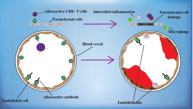
Depending on the involved immunological components, acute allograft rejection can be categorized into acute cellular (interstitial and vascular) (Figure 6) and acute humoral rejection, and their mixed forms. While in the new immunosuppression era, graft damage caused by acute cellular rejection has almost disappeared, due to the increasing number of HLA-mismatched transplantations and sensitization of the recipients at B- cell level, acute humoral rejection still remains a problem (46, 62).
18
Figure 6. Acute allograft rejection. In cellular forms of acute rejection, CD8+ T- lymphocytes recognize alloantigens on the endothelial and parenchymal cells of the graft and cause their damage (upper part), whereas in humoral acute rejection, alloreactive antibodies induce vascular injury (lower part). Based on reference 46.
Acute humoral rejection, which appears hours to weeks after transplantation, is also known as AMR and its pathology shows overlaps with the hyperacute allograft rejection. Acute AMR is characterized by graft dysfunction manifesting over days and is a result of an immune attack mediated by DSA that may either be preformed and persistent or develop de novo after transplantation. Clinically it presents as acute accelerated oliguria or delayed graft function (DGF) without symptoms, such as fever or allograft tenderness. It is more common in patients with preformed anti-donor antibodies. Besides the declining urine output, decreased renal blood flow, increased resistive index and a missing diastolic flow is detected in ultrasound. In the pathogenesis of AMR complement activation caused by preformed donor-specific IgG antibodies play a critical role. Histological fibrinoid necrosis of vascular wall, acute glomerulitis, infiltration of the glomerulus by mononuclear cells, peritubular capillary with polymorphonuclear cell infiltrate, peritubular capillary with C4d staining, perivascular T-cells, natural killer cells (NK), and other mononuclear cells can be seen in the biopsy. C4d deposition is strongly associated with the development of class I and class II DSA (46).
19
Acute AMR occur in about 5-7% of all kidney transplant recipients, and is responsible for 20-48% of acute rejection episodes among presensitized positive XM patients.
These early damages later play an important role in the development of chronic rejection. The prevalence of chronic AMR 1 year after transplantation is about 5% and can occur rapidly during an ongoing acute AMR (23, 33, 46, 63).
Chronic allograft rejection can occur one month to years after kidney transplantation.
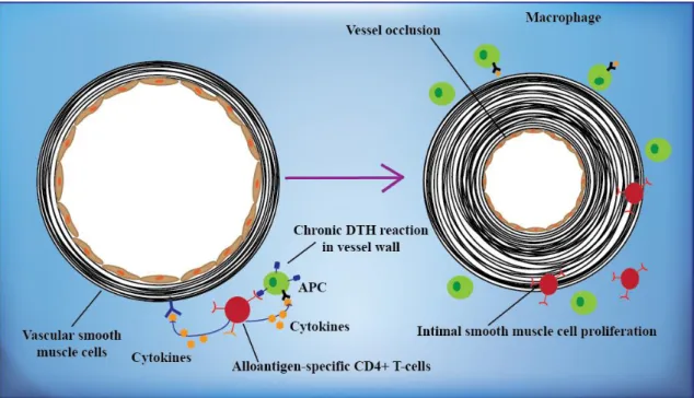
Especially, in the early phase there are no symptoms, but later on proteinuria and edema can develop. Furthermore, progressive loss of renal function, proteinuria, hypertension and hyperlipidemia can be monitored as most common causes of posttransplant nephritic syndrome. In the pathogenesis of chronic rejection many processes play a role in combination, such as severe acute rejection episodes, early posttransplant tubular injury, subclinical rejection, chronic ischemia, calcineurin inhibitor toxicity or increased transforming growth factor β. Furthermore, hypertension, hyperlipidemia, infections, smoking, oxygen radicals or proteinuria can also lead to chronic rejection. Patients who especially had multiple acute rejection episodes, deceased donor graft, acute tubular necrosis (ATN) on implantation biopsy, dialysis requirement DGF, HLA mismatches, previous sensitization, panel reactive antibody (PRA) >50%, increased donor age, suboptimal immunosuppression or non-compliance have a higher risk for the development of chronic rejection. Glomerulosclerosis, splitting of glomerular basement membranes, tubular atrophy, interstitial fibrosis, casts, simplification of the tubular epithelium and vascular fibrointimal proliferation can be detected in the biopsy (Figure 7) (46).
20
Figure 7. Chronic allograft rejection. T-lymphocytes reactive with graft alloantigens may produce cytokines that induce proliferation of endothelial cells and intimal smooth muscle cells, resulting in arteriosclerosis of the graft. Based on reference 46.
21
Table 2. Banff 97 diagnostic categories for renal allograft biopsies
1. Normal
2. Antibody-mediated rejection
Rejection due, at least in part, to documented antidonor antibody (“suspicious for” if antibody not demonstrated). May coincide with categories 3, 4, and 5.
Type (grade):
I. ATN–like-C4d+, minimal inflammation
II. Capillary-C4d+,capillary margination and/or thrombosis III. Arterial-C4d+, transmural arteritis (v3)
3. Borderline changes
Suspicious for acute cellular rejection. No intimal arteritis is present, but there are foci of mild tubulitis (1-4 mononuclear cells/tubular cross-section). May coincide with categories 2 and 5.
4. Acute/active cellular rejection
T-cell–mediated rejection. May coincide with categories 2 and 5.
Type (grade) of histopathological findings:
IA Cases with significant interstitial infiltration (>25% of parenchyma affected) and foci of moderate tubulitis (>4 mononuclear cells/tubular cross-section or group of 10 tubular cells)
IB Cases with significant interstitial infiltration (>25% of parenchyma affected) and foci of severe tubulitis (>10 mononuclear cells/tubular cross-section or group of 10 tubular cells)
IIA Cases with mild to moderate intimal arteritis (v1)
IIB Cases with severe intimal arteritis comprising >25% of the luminal area (v2)
III Cases with “transmural” arteritis and/or arterial fibrinoid change and necrosis of medial smooth muscle cells with accompanying lymphocytic inflammation (v3)
5. Chronic/sclerosing allograft nephropathy
Fibrosing changes in the allograft, with or without features of true alloimmune injury to the graft. May coincide with categories 2, 3, and 4
Grade histopathological findings:
Grade I: Mild interstitial fibrosis and tubular atrophy without (a) or with (b) specific changes suggesting (mild) chronic rejection.
Grade II: Moderate interstitial fibrosis and tubular atrophy (a) or (b) (moderate)
Grade III: Severe interstitial fibrosis and tubular atrophy and tubular loss (a) or (b) (severe) 6. Other
Changes not considered being due to rejection. May coincide with categories 2, 3, 4, and 5.
Based on reference 46. v: vasculitis, ATN: acute tubular necrosis
22
2.2.6 Different pretransplant approaches to prevent antibody-mediated rejection Thus, further developments of immunosuppression medications in the 1980’s and 1990’s were centered also on the control of T-cell alloimmunity, leading to a significant decrease in the incidence of acute cellular rejection and graft loss. However, after this success, the problem of AMR emerged which currently is recognized as one of the major causes of late kidney graft failure. Therefore, prevention of AMR became an important issue in the field of kidney transplantation (31, 62, 64, 65).
To protect graft function after transplantation, a combination of different measures is needed. Besides diagnostic measures, such as HLA-typing, XM and antibody screening, therapeutic measures, such as plasmapheresis (PPh) or immunoadsorption (IA) and immunosuppressive therapies are also mandatory to prevent AMR. Prevention of antibody-mediated allograft damage must actually start already before transplantation, as the patient is on the waiting list and thereafter be continued after organ transplantation.
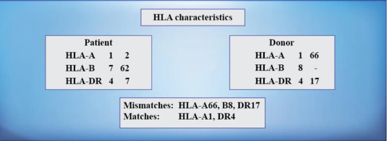
The selection of the appropriate donor for the recipient on the waiting list includes the matching for the AB0 blood group system and HLA compatibility. First of all patients have to be tested, which AB0 blood group they have, to avoid an AB0-incompatible transplantation. After that, all patients awaiting a kidney must be typed, either with serologic or molecular techniques, for HLA, and during the allocation process the kidney organs should be offered to recipients with the lowest number of HLA mismatches. Specific HLA antigen characteristics of the donor that the recipient does not possess are called mismatches. Good matching of donor and recipient is the easiest way to prevent AMR, especially in highly-immunized patients (66) (Figure 8).
23
Figure 8. Example for HLA matching between a donor-recipient pair. HLA antigens that are present in the donor but not in the recipient are called mismatches.
Conversely, HLA antigens that both donor and recipient possess are the matches.
Organ allocation of deceased donor kidneys is commonly based on matching for HLA- A, -B, and -DR. Several registry reports still report a significant influence of the number of mismatches at the HLA-A, -B, and -DR loci on kidney graft survival. Ten years after transplantation, the graft survival rate of first cadaver kidney transplants with a complete six mismatches is lower than that of grafts with no mismatch. During the first posttransplant year, the class II HLA-DR locus has a stronger impact than the class I HLA-A and HLA-B loci. Therefore for optimal graft outcome, compatibility at all three HLA loci is preferable (28-30, 67). Furthermore in the last years several studies have shown a possible impact on graft outcome of the HLA-C and -DP loci on kidney transplantation in certain subgroups of patients, particularly in presensitized recipients (52, 68-70). HLA-C and-DP molecules have usually been considered to be less immunogenic than other HLA markers. Because previously techniques to identify anti- HLA-C or -DP antibodies were not available, their contribution to graft rejection and graft loss have been poorly understood. However, recent developments in XM and screening technologies that have proven useful to detect and identify antibodies in kidney recipients made the exploration of the role of anti-C and -DP antibodies in renal transplantation possible (52, 68-70).
Directly before kidney transplantation, prospective XM has to be performed, to detect DSA in the recipients’ serum, which can appear for example after different sensitizing events. Therefore, the avoidance of sensitizing events is an important part of the measures in the pretransplant phase. Patients could be sensitized through previous blood
24
transfusions, pregnancies or transplantations. Especially in sensitized patients, precise characterization of HLA alloantibodies with different screening techniques, including the accurate determination of “unacceptable HLA antigen mismatches” (UAM) is mandatory.
HLA antibody screening is usually practiced in different steps. Initially, a complement- dependent cytotoxicity (CDC) test with and without dithiothreitol (DTT), is applied, identifying antibodies and calculate PRA (%) values, which refers to the percentage of an antibody screening panel with which the patient’s serum reacts. PRA ≤5% patients count as non-immunized, 6-85% as sensitized and ≥85% as highly sensitized. With the presence of DTT, IgM antibodies could be recognized, and made sure the absence of IgG antibodies. Detected IgG antibodies are generally considered to reflect the true sensitization against HLA antigens, while IgM antibodies which have a lower binding capacity are classified as less dangerous. Transplantation should not proceed if there is evidence of a positive XM caused by a cytotoxic IgG anti-HLA antibody. In parallel to the CDC screening, HLA antibody enzyme-linked immunosorbent assay (ELISA) screening and PRA test should be implemented for the detection of HLA class I and II antibodies for an exact determination of the sensitization status. Finally, the precise definition of antibody specificities in complex sera of sensitized patients is possible only with the Luminex single antigen bead (SAB) assay.
The risk assessment of kidney transplant waiting list patients and the exact definition of highly sensitized patients is a very important step of the diagnostics. To ensure timely and successful transplantation of highly sensitized patients, desensitization using PPh or IA, and inclusion in special programs such as the Eurotransplant Acceptable Mismatch Program should be considered (62, 71-73).
25
2.2.7 Diagnostic approaches before kidney transplantation to prevent antibody- mediated rejection
To choose the most appropriate donor for the recipient on the waiting list and prevent allograft rejection, prospective crossmatching and antibody screening at regular intervals are required before kidney transplantation. The diagnostic procedure is mandatory for the determination of UAM.
2.2.7.1 Crossmatching
XM techniques are assays to identify the presence of preformed DSA against donor HLA class I and II antigens in the serum of recipients before transplantation on the kidney transplant waiting list. For crossmatching, the recipient’s serum and donor lymphocytes have to be available. Lymphocytes are obtained from peripheral blood, lymph nodes or spleen of the donor. Different assays for identification of HLA antibodies vary in the type of target, format, sensitivity and specificity. Assay targets can either be cells tested for example in cytotoxicity assay or soluble antigens tested in solid-phase immunoassays.
Since the publication of Patel and Terasaki in 1969 on the clinical value of the cell based CDC XM assay (61), it has been routinely implemented in the practice for determination of HLA antibodies. While it has a low sensitivity, it is capable of identifying antibodies that can cause hyperacute rejection (31). The CDC XM technique utilizes separated T-, B- or unseparated (U) lymphocytes of the donor and detects complement-dependent lymphocytotoxic HLA antibodies in the recipients’ serum.
A positive XM result with donor T-cells is interpreted as the presence of donor-specific HLA class I antibodies and is a definitive contraindication of transplantation. In contrast, the value of a positive B-cell CDC XM (BXM) is unclear because B-cells do not express uniform HLA antigens. B-lymphocytes express HLA class II molecules and constitutively express HLA class I molecules at a higher density than T-lymphocytes.
Therefore, CDC BXM positivity may reflect a donor-specific anti-HLA class II, anti- HLA class I activity or a combination of both. In addition, non-HLA antibodies and clinically not harmful autoantibodies may also cause positivities in CDC BXM. This
26
heterogeneity of CDC BXM contributes to the debates surrounding the effects of CDC BXM positivity on renal transplantation with a concomitant negative CDC T-cell XM.
Hyperacute vascular rejection in CDC B-cell positive recipients has been reported, but other investigators claimed that CDC BXM has no influence on transplant outcome (74- 78).
The detection of DSA by CDC assay is limited by its low sensitivity and the quality of the donor lymphocytes. Therefore, more sensitive assays were required in order to detect antibodies which are not detectable by classical CDC assays and are relevant for the clinical outcome. Therefore despite of its benefits, CDC technique has been criticized, especially because AMR occurs often in sensitized patients in the presence of a negative CDC result.
The more modern version of a cell-based test is the flow-cytometry XM (FXM) test, which was first described in 1983. In the FXM assay, the recipient’s serum is incubated in the first step with donor T- or B-lymphocytes and in the next step with fluorescein–
labeled antibodies against human IgG. If DSA of the IgG class is present, antibodies in the recipient serum will be detected and the strength of the fluorescence can be measured and expressed as channel shifts above the control sample (79). Thus this XM technique is more sensitive and enables the detection of both complement fixing and non-complement fixing antibodies of all Ig isotypes that went unrecognized in the CDC and can therefore, bridge over limitations of the cytotoxic technique. However, a negative CDC in parallel with a positive FXM result should not necessarily mean a contraindication to kidney transplantation. Furthermore, because of a possibility of false positive reactions, especially in patients without any immunization events, not all transplant centers use flow crossmatching in their pretransplant diagnostic algorithm.
This technique is most informative in immunized patients who are already sensitized to HLA, including those recipients who had a previously failed transplant (71, 80).
Beside cell based assays mentioned above, solid-phase techniques are also available for crossmatching before transplantation. As mentioned below in detail, solid-phase techniques are obtained as commercially manufactured kits that use solubilized or
27
recombinant HLA molecules bound to a solid matrix that is either a microtiter plate using the ELISA or polystyrene beads using the Luminex assay.
2.2.7.2 Antibody screening
To avoid a positive XM in the transplant centers and thus prevent AMR, kidney transplant recipients are screened periodically for the presence of HLA antibodies before transplantation with the above mentioned cell-based or solid phase assays to define the UAM. For the detection of DSA, serum of the recipient has to be available.
The test principles are the same as by XM techniques, but instead of the lymphocytes of potential donor, a panel of HLA-typed lymphocytes from healthy blood donors, their solubilized HLA antigens or artificially produced recombinant HLA molecule panels are utilized.
In the case of CDC-PRA testing, recipient serum is added to a CDC plate which includes a panel of different HLA-typed donor cells. For detection of HLA class I antibodies U-lymphocytes and for the detection of HLA class II antibodies cells from B- lymphoid lines are used as targets. PRA against a panel of U-, mainly T- or separated B- lymphocytes can be calculated in presence or absence of DTT to detect not only IgG but also IgM antibodies. CDC-reactive antibodies against particular HLA antigens are defined as UAM. Due to its low sensitivity and resolution grade, however, CDC method often misses clinically relevant antibody specificities. Therefore, the guidelines recommend the usage of more sensitive solid-phase assays additionally (31) that utilize solubilized or recombinant HLA molecules instead of cells.
According to previous studies the solid-phase ELISA screening method, which utilize purified class I or class II antigens bound to a microtiter plate, is quite successful to predict AMR (81). Purified HLA class I glycoproteins obtained from platelets of healthy blood donors or HLA class II from EBV transformed B-lymphocyte cell lines are used. HLA antibodies of the recipient can be determined against these pooled class I or class II HLA molecules. Patients positive for both HLA class I and II antibodies of the IgG isotype in ELISA screening, respectively, had significantly poorer graft outcome than patients who were negative in this assay (81).
28
Not only the ELISA screening but also the ELISA-PRA technique is available as part of the screening diagnostic and is helpful in the determination of the UAM. The solid phase ELISA-PRA utilizes cell lysates from single individuals instead of pooled lysates in the microtiter plate and therefore contains in each well of the plate well characterized purified HLA class I or class II specificities.
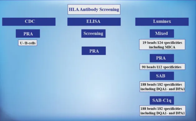
The most sensitive technique that revolutionized the HLA antibody diagnostics is the Luminex methodology. Presently, three types of panels are offered which vary in the composition of their target antigens. Luminex Mixed utilizes pooled HLA antigen panels, Luminex PRA uses phenotype panels and the last one, offered by two vendors, is the SAB, in which each bead population is coated with a recombinant molecule representing a single cloned allelic HLA class I or II antigen. A modification of the SAB technique which detects C1q binding HLA antibodies is also available (Figure 9).
Figure 9. Different HLA antibody screening methodologies. For antibody screening, cell-based assays, such as CDC and solid-phase assays, such as ELISA and Luminex, are available. Different Luminex methods utilize different amount of beads and specificities.
SAB technique enables precise antibody specificity analysis in complex sera. It detects many additional HLA antibody reactivities that go undetected in CDC or ELISA
29
methods (Figure 10) (31). However, there is an ongoing debate whether kidney transplant recipients with HLA antibodies detectable only in the highly sensitive SAB assay and not reactive in the less sensitive CDC or ELISA assays are at increased risk of graft failure (82-85). Previous studies suggested that SAB testing alone is problematic because, with this technique, HLA antibodies can be found in sera of healthy blood donors without a history of a sensitizing event (86).
Figure 10. With increasing sensitivity of the diagnostic assays, weak DSA can be detected appropriately in serum of recipients on the kidney transplant waiting list.
While CDC or flow assays can detect from very high to high or moderate DSA levels, ELISA assay is more sensitive to determine moderate or low antibody reactivity in the recipient’s serum. Currently, Luminex is the only methodology with its high sensitivity, which can detect low titer DSA with high accuracy. HLA antibody specificities detected negative in the Luminex SAB assay can be categorized as acceptable antigen mismatches.
2.2.7.3 Determination of unacceptable HLA antigen mismatches
UAM are HLA specificities against which the recipient developed antibodies and based on which potential kidney donors are excluded during the organ allocation procedure. It is an obligatory step before transplantation, because even in the absence of a positive CDC or FXM, pretransplant DSA are believed to have a negative impact on kidney graft survival.
The determination of UAM is a critical decision. Inappropriate determination of the UAM can cause prolonged waiting times or death on the kidney transplant waiting list, futile organ shipments and decreased graft survival. Thus, definition of too many unacceptable antigens can lead in case of an organ offer to exclusion of probably
30
appropriate donors; conversely, unrecognized antibodies can cause positive XM in the transplant centers, and cause inappropriate organ shipments and inferior graft survival.
Because there are still no standardizations for the definition of UAM, currently the transplant centers work using their own algorithms. While it is generally accepted that CDC-detected DSA are a contraindication for transplantation, the relevance of additional antibodies detected by more sensitive assays are questionable. Since the highly sensitive Luminex SAB assay has become available, many transplant laboratories do not use hereby the CDC assay alone anymore, but support it with SAB.
In contrast, some other laboratories use only the Luminex SAB for the UAM determination at varying cutoffs (87).
31 3. THE AIMS OF THE THESIS
Because the ultimate aim of the transplantation diagnostics is to offer the most appropriate donor organ for the recipient in the shortest time on the waiting list, inappropriate exclusion of donors because of an imperfect XM or antibody screening technique should be avoided. We analyzed the advantages and the problems associated with two recently introduced HLA antibody detection methods. False positive results as well as low sensitivity can create difficulties in the correct assessment of the patient’s HLA antibody status on the kidney transplant waiting list.
In the first study we investigated the potential superiority of the commercially available AbCross® ELISA XM over the CDC BXM in predicting graft loss. Because there is debate about the sensitivity and clinical relevance of the CDC BXM in renal transplantation (74-78), we analyzed, whether with the new AbCross® technique the disadvantages of the CDC BXM, such as the detection of unspecific reactions or autoantibodies, can be eliminated.
In the second study, to estimate the impact of the problem of potentially “false positive”
results detected with the highly sensitive Luminex SAB technique on the sensitization status of patients on the kidney transplant waiting list, we investigated the prevalence of HLA antibodies in waiting list patients of the Heidelberg transplant center using three different assays, namely CDC T-cell screening, AbScreen® ELISA screening, and SAB in parallel. A high prevalence of HLA antibody reactivity with a given assay in patients without any history of immunization would indicate that this particular assay generates
“false positive” results. We also examined in detail the HLA specificity and strength of the “false positive” reactions. Such information could be useful in the daily routine, when SAB results are evaluated in the individual patients.
3.1 Comparison of the clinical relevance of ELISA and B-cell CDC crossmatch before kidney transplantation
The following questions were addressed:
a. What is the rate of 2-year graft loss after kidney transplantation in AbCross® ELISA XM-positive and AbCross® ELISA XM-negative patients?
32
b. What is the rate of 2-year graft loss after kidney transplantation in CDC BXM- positive patients compared to CDC BXM-negative patients?
c. Is the impact of positivity in AbCross® ELISA XM on graft survival supported by AbScreen® ELISA screening results?
d. Is there a relationship between kidney graft survival and CDC BXM and AbCross® ELISA XM or AbScreen® ELISA screening results?
3.2 Evaluation of the influence of the recently introduced Luminex SAB on the sensitization status of patients on the kidney transplant waiting list
In this context, the following questions were addressed:
a. What is the prevalence of the positive patients on the kidney transplant waiting list in the SAB technique compared to the less sensitive ELISA and CDC methods?
b. What is the prevalence of HLA antibody-positive patients without any immunization history?
c. Which mean fluorescence intensity (MFI) values have patients without any history of immunization?
d. Whether the problem of “false positive” results could be solved by increasing the cutoff values?
e. What is the prevalence of SAB-positive patients according to reaction with the percentage of beads?
f. Which HLA allele specificities react positive in SAB in patients without history of immunization?
g. Whether test offered by another vendor or another test principle can solve the problem of “false positive” results?
33 4. PATIENTS AND METHODS 4.1 Patients
In the first study in which the potential superiority of the ELISA XM over the CDC BXM before kidney transplantation was evaluated, pretransplant sera of 271 living or deceased donor kidney transplant recipients who were transplanted at the Heidelberg transplant center between 1998 and 2010 and on whom frozen donor cell material was available were tested in the AbScreen® ELISA screening assay for the presence of HLA antibodies and in CDC BXM and AbCross® ELISA XM assays for antibody reactivity against donor B-cells or donor HLA class I and II antigens, respectively.
In the second study in which the influence of the Luminex SAB test on the sensitization status of patients on the waiting list was evaluated in parallel with the ELISA and CDC screening methods, pretransplant sera of 534 patients on the Heidelberg kidney transplant waiting list were additionally analyzed using the SAB assay.
Patient consent and ethics committee approval was obtained, and the investigations were performed in accordance with the Declaration of Helsinki.
4.2 Methods
4.2.1 Study 1: Comparison of the clinical relevance of ELISA and B-cell CDC crossmatch before kidney transplantation
In the first study, for the CDC XM, the donor's separated B-lymphocytes were used. B- cells were isolated using monoclonal antibodies against the β-monomorphic antigen of the HLA class II, attached to magnetic beads. One microliter (µl) of donor cell suspension (1 × 10⁶ cells/ml) was incubated with 1 µl of recipient serum for 30 minutes at room temperature. After washing the cells, 5 µl rabbit complement (BioRad, Munich, Germany) was added and incubated for 30 minutes at room temperature. The cells were stained with 3 µl fluorescent dye, containing acridine orange and ethidium bromide, and incubated for 10-15 minutes. If DSA were present and bound to donor cells, the complement cascade have been activated via the classical pathway resulting in lysis of the lymphocytes. The assessment of the test occurred through the percentage of dead
34
cells relative to live cells and the cytotoxicity effect was examined using a fluorescent microscope (Leica, Wetzlar, Germany) (Figure 11). Cell death >20% was considered positive.
Figure 11. The CDC B-cell crossmatch assay. Recipient serum potentially containing DSA is added to donor B-lymphocytes in the presence of complement (A). If DSA are not present in the recipient serum, no lysis occurs and the result is considered negative (B). If DSA bind to the lymphocytes and activate complement, cell lysis occur and the XM result becomes positive (C). The percentage of death cells is assessed by fluorescent dye using a fluorescence microscope, and the XM is graded. Live cells are green (B) and dead cells are red (C) in the fluorescence microscope. Based on reference 79.
In addition, sera were tested using the recently introduced AbCross® ELISA XM (BioRad, Munich, Germany) assay, in which solubilized donor HLA molecules are used to detect DSA.
AbCross® ELISA XM is a commercially available solid phase XM technique with advantages over the standard CDC BXM, such as higher reproducibility, objectivity, sensitivity and specificity for HLA antigens. Furthermore, in contrast with the CDC BXM, which has been claimed to detect in >60% of patients clinically irrelevant non- HLA antibodies (77), AbCross® utilizes solubilized donor HLA class I and II antigens,
35
which allows the specific detection of anti-donor HLA antibodies with high sensitivity.
Interference of autoantibodies is excluded. Because no viable cells are required, the test provides an objective assessment with low variability. Compared with the currently available ELISA XM technique Antibody Monitoring System (AMS) (88), which is carried out in 96-well ELISA microtiter plates, AbCross® utilizes the 60-well CDC microtiter plate format and can therefore be easily performed with little amounts of sera and donor cells parallel to CDC testing. Moreover, owing to the lysis of donor cell- antibody immune complexes outside of the detection plate, it creates fewer background signals than AMS. This enables specific capture of HLA–HLA antibody complexes instead of cell–HLA antibody complexes, thereby minimizing the interference of donor cell proteins other than HLA by nonspecific binding (Table 3).
Table 3. Advantages of AbCross® XM over the AMS assay
Advantages of AbCross® ELISA XM over the AMS
AbCross® can easily be performed parallel to CDC testing using 60-well CDC Terasaki plate format
Extremely low background due to lysis of donor cell-antibody immune complexes outside of the detection plate
Small amount of serum and cells
The AbCross® ELISA XM assay detects antibodies on the microtiter plate coated with monoclonal antibodies. Donor lymphocytes were incubated with the recipient’s serum and, if present, anti-HLA antibodies bound to the HLA molecules on the cell surface.
The antigen-antibody complexes were incubated with peroxidase (PoD)-conjugated goat anti-human IgG antibodies. Specifically bound antibodies were detected in a final enzymatic reaction with the substrate tetramethylbenzidine (TMB). The results were detected with photometric measurement in an ELISA reader and expressed as optical density (OD) ratios compared with a negative control, giving a semiquantitative assessment of antibody binding (Figure 12). Results of OD greater than or equal to the double of the negative control were considered positive.
36
Figure 12. ELISA crossmatch. Recipient serum potentially containing DSA is added to donor lymphocytes, in presence of anti-human IgG conjugat with fluorescence reporter- (A). Donor lymphocytes with recipient serum are incubated in a polymerase chain reaction (PCR) plate. If present, DSA bind to HLA proteins on the cell surface (B). Cells with DSA are lysed in the PCR plate (C). Lysates are transferred to anti-HLA class I- or II-coated wells of the microtiter plate and DSA-donor HLA immune complexes are captured to the plates. Anti-human IgG-conjugate and substrate make positive reactions visible (D). The strength of optical density (OD) is measured in an ELISA reader.
The sera were also tested for the presence of IgG-anti-HLA class I and II alloantibodies using AbScreen® ELISA (BioRad) kits, which use pooled HLA molecules on 96-well microtiter plates for the detection of HLA antibodies. Affinity purified HLA class I glycoproteins obtained from platelets of healthy blood donors or HLA class II from EBV transformed B-lymphocyte cell lines are used. HLA antibodies of the recipient are determined on separate plates against pooled class I or class II HLA molecules. Based on previous clinical findings, an OD of ≥0.300 was used as cutoff for anti-HLA positivity (81).
Two-year clinical follow-up data were collected and documented for 223 of 271 patients.
37
4.2.2 Study 2: Evaluation of the influence of the recently introduced Luminex SAB on the sensitization status of patients on the kidney transplant waiting list
In the second study, the different antibody screening techniques were analyzed. At the Heidelberg transplant center, waiting list patients are routinely screened every three months for HLA antibodies employing ELISA and CDC. In addition, the 534 sera from the third quarter of 2010 were examined using the SAB method.
PRA against total lymphocytes (mainly T-cells) of a panel of 56 cell donors on frozen/thawed cell trays were determined using the CDC method in the absence of DTT (http://www.ctstransplant.org/public/reagents/serolCell.shtml). Following standard procedure, the patient’s serum was incubated with lymphocytes, complement was added and the trays were read using a fluorescent microscope (Leica, Wetzlar, Germany). PRA of >5% was considered positive.
Furthermore, all 534 sera were tested for the presence of HLA class I and II alloantibodies using AbScreen® ELISA kits of BioRad (Munich, Germany), which as mentioned already above, utilize pooled HLA molecules attached to microtiter plates and enable the detection of HLA-A, -B, -C, -DR, and -DQ antibodies of the IgG isotype. Based on previous clinical findings, an OD of ≥0.300 was used as cutoff for anti-HLA positivity in ELISA (81). In one patient who was negative with SAB but positive by ELISA AbScreen®, the ELISA-PRA assay (AbIdent®, BioRad, Munich, Germany) which utilizes cell lysates from single individuals instead of pooled lysates was used to confirm the absence of HLA antibodies.
In addition, all sera were tested using the LABScreen® Luminex kits of One Lambda (Canoga Park CA, USA, LS1A04 Lot006 and LS2A01 Lot008), using SAB-coated beads that enable the identification of IgG antibody specificities against HLA-A, -B, -C, -DRB1/3/4/5, -DQA1, -DQB1, -DPA1 and -DPB1.
The bead-based Luminex technology use polystyrene beads impregnated with different ratios of two fluorescent dyes (classifier signals), and is capable of differentiating up to
38
100 different beads in one test cavity for HLA class I and 100 different beads in one test cavity for class II. The antiglobulin reagent in the bead assays is labeled with a third fluorescent dye (the reporter signal) so that, using a dual-laser instrument, the fluorescence signature of each bead can be interrogated to identify the bead population by one laser, whereas the reporter fluorescence simultaneously assesses HLA-specific antibody binding (Figure 13).
Figure 13. The bead-based Luminex technology. Recipient serum potentially containing DSA is added to a mixture of synthetic microbeads. Each bead is coated with a set of antigens (screening beads) or with recombinant single HLA antigen molecules (single antigen beads). A unique dye signature (up to 100) specifies the identity of each bead (A). If DSA are present, these will bind to the appropriate bead (B) and be visualized in the next step by an additional phycoerythrin (PE)-conjugated goat anti- human IgG antibody which serves as reporter dye (C). Each unique bead can then be interrogated for the presence of the reporter dye on its surface using a dual beam laser (D). A profile of antibodies can thus be identified in the recipient. Based on reference 79.
While the SAB assay of One Lambda® utilizes 97 beads for HLA class I and 91 beads for HLA class II, Lifecodes® SAB assay uses 94 beads for HLA class I and 72 beads for HLA class II. Each single bead coated with either one recombinant HLA class I (HLA- A, -B, -C) or one (HLA-DRB1/3/4/5) or two (HLA-DQA1/DQB1 or HLA- DPA1/DPB1) HLA class II proteins.
39
The bead-based array assay is analyzed on the Luminex platform and is semiquantitative. The level of HLA-specific antibody binding is expressed as the MFI of the reporter signal (Figure 14).
A
B
Figure 14. HLA antibody specificities detected by the highly sensitive Luminex SAB assay. SAB results for typical serum of a highly sensitized (A) and a non- sensitized patient (B). Mean fluorescence intensity (MFI) of the reacted HLA specificities on the single antigen beads was determined using a cutoff of ≥1000 MFI.
Furthermore, when the Negative Control Beads (NC) value was 500 MFI or above, Adsorb Out (One Lambda) treatment was used to reduce high background caused by non-specific binding of sera to latex beads. When the NC value was still high after Adsorb Out treatment, the test was judged as invalid. Therefore, in all analyzed cases the NC value was below 500 MFI. Seven patients were excluded from the analysis because of the high NC value (≥500 MFI). According to manufacturer’s instructions,
MFI values
Cutoff ≥1000 MFI
MFI values
Cutoff ≥1000 MFI
HLA specificities HLA specificities