Nephrol Dial Transplant (2011) 26: 727–732 doi: 10.1093/ndt/gfq401
Advance Access publication 4 July 2010
High frequency of ulcers, not associated with Helicobacter pylori , in the stomach in the first year after kidney transplantation
Gabor Telkes
1, Antal Peter
1, Zsolt Tulassay
2and Argiris Asderakis
31Transplantation and Surgical Clinic,2II. Internal Medicine Clinic, Research Group of Hungarian Academy of Sciences, Semmelweis University, Budapest, Hungary and3University Hospital of Wales, Cardiff, UK
Correspondence and offprint requests to: Gabor Telkes; E-mail: telkesdr@gmail.com
Abstract
Background.Although gastrointestinal (GI) symptoms are very frequent in organ transplant patients, there is a paucity of data about the endoscopic findings of kidney recipients.
Methods.Two thousand one hundred and thirty-five kid- ney transplants were performed between 1994 and 2007.
During that period, 672 gastroscopies were performed in 543 of those patients. Their mean age was 49.5 years and 56.9% were male. Immunosuppressive combinations in- cluded cyclosporine–mycophenolate–steroids, cyclosporine– steroids and tacrolimus–mycophenolate mofetil–steroids.
Ninety-eight percent of the patients received acid suppres- sion therapy.
Results.The rate of clinically significant endoscopic find- ings was 84%. Macroscopic findings included inflamma- tion in 46.7%, oesophagitis in 24.7%, ulcer in 16.9%
and erosions in 14.8% of cases. Twenty-nine percent of en- doscopies showed ulcer disease more frequently in the first 3 months (P = 0.0014) after transplantation than later, and 45.7% of all ulcers developed in the first year. The pres- ence of Helicobacter pylori was verified in 20.9% of cases, less than in the general, and also in the uraemic population (P < 0.0001). There was no association between the presence ofH. pyloriand ulcers (P = 0.28). Steroid pulse treatment for rejection was not associated with more ulcers (P = 0.11); the use of mycophenolate mofetil increased the risk of erosions by 1.8-fold.
Conclusion.More than 25% of all kidney recipients re- quired upper endoscopy in their‘post-transplant life’; the prevalence of ‘positive findings’ and ulcer disease was higher than in the general population (P < 0.0001). The most vulnerable period is the first 3 months. Mycopheno- late mofetil had an impact on GI complications, whilst the presence ofH. pylori in the transplant population is not associated with the presence of ulcers.
Keywords:endoscopy;Helicobacter pylori; kidney transplantation; ulcer disease; upper gastrointestinal tract
Introduction
Upper gastrointestinal (GI) symptoms are frequent in organ transplant recipients. Peptic ulcers and related pathologies such as gastritis and duodenitis are known to occur with in- creased frequency (20–60%) and severity in renal transplant recipients. The frequency of severe complications is about 10% among transplant recipients and 10% of those might prove fatal [1–5]. Compared to the general population, renal transplant recipients have an age-adjusted ratio for GI bleeding of 10.69 at 1 year of follow-up [6]. Frequency of ulcer disease was proven to be 8.3–10.7% of endoscopically examined general gastroenterology patients [7,8]. Similar data among transplant patients are not available. GI compli- cations might play a role in the outcome of kidney trans- plantation, as they are associated with an increased risk of graft loss, since dose reduction or even withdrawal of immunosuppressive (IS) agents might be necessary [3, 9–11].Different factors can cause GI symptoms: surgi- cal stress, the operation itself, local irritant and pro-inflam- matory effects of IS and/or other drugs, the numerous pills some patients have to take every day and bacterial, viral and mycotic infections [12,13]. The GI tract accounts for a large component of non-allograft-related complications seen after all types of solid organ transplantation [14]. Whilst endo- scopic alterations of liver transplant recipients are the focus of numerous studies [15–18], only few studies have been done on the endoscopic findings of renal patients and in- clude only small numbers [19,20].
By summarizing and analysing the largest endoscopic database in the literature, our aim was to ascertain the fre- quency and the time of occurrence of ulcer disease after kidney transplantation.
Materials and methods Patients
From January 1994 to December 2007, 2143 kidney transplants were per- formed in a large transplant centre. Six hundred and seventy-two upper GI
© The Author 2010. Published by Oxford University Press on behalf of ERA-EDTA. All rights reserved.
For Permissions, please e-mail: journals.permissions@oxfordjournals.org
at Semmelweis Ote on December 22, 2014http://ndt.oxfordjournals.org/Downloaded from
endoscopies were done in 543 kidney transplant patients (25.3% of reci- pients). Patients with GI complaints following informed written consent, underwent an upper endoscopy examination; 56.9% of them were male, with a mean age of 49.5 ± 12.8. All patients received an acid-blocker as ulcer prophylaxis. Data of kidney transplant patients were collected for this cross-sectional (date of endoscopy), descriptive study.
Endoscopy
Two senior surgeon-gastroenterologists performed all the endoscopic ex- aminations esophago-gastro-duodenoscopy = gastroscopy (EGD). The upper GI tract was examined. The endoscopic abnormalities were evalu- ated according to international standards [21–25]. Forceps mucosal biop- sies were taken from a specific lesion (e.g. ulcer) if present and from every patient from the first part of the duodenum, the gastric antrum and the gastric corpus (2-2 biopsy samples from each localization). Bi- opsy samples were investigated by conventional histology for mucosal changes (haematoxylin–eosin and Giemsa). Patients were considered as Helicobacter pylori(H. pylori) positive, if histology proved its presence.
No biopsy was taken in cases of bleeding, in patients with known coagu- lation disorders or those on anticoagulant therapy (except aspirin).
Immunosuppression and acid suppressive therapy
The IS medication used at the time of endoscopy was collected and ana- lysed. Cyclosporine (CSA), tacrolimus (TAC), mycophenolate mofetil (MMF) and steroids (ST) were used in various combinations for mainten- ance therapy. ST were administered in a tapering dose once a day. CSA, TAC and MMF were administered twice daily. Target trough levels of CSA were 150–200 ng/mL and TAC were 7–10 ng/mL at the long term.
The initial dose of MMF was 2 × 1 g, according to an established protocol.
Prednisolone or methylprednisolone was used as ST. At the time of EGD, the most frequently used (75.1%) IS combinations were CSA–MMF–ST triple treatment, CSA–ST double treatment and TAC–MMF–ST triple therapy. All other combinations occurred in <5% in that material, and these were excluded from the IS-related analysis. A total of 34.51% of all transplant recipients and 34.56% of endoscopically examined patients received intravenous ST pulse therapy. Out of the latter ones, 15.53%
received it in the 3 months period prior to the endoscopy.
Data of acid suppressive therapy (AST)‘before’endoscopy were also collected and analysed. Nearly all (97.8%) patients received AST. Out of them, 56.1% received a histamine receptor antagonist (H2RA) and 43.8%
a proton pump inhibitor (PPI). There was a variable usage of PPIs and H2
receptor antagonists; however, the general use did not change during the study period. There was no difference between the IS groups according to AST treatment received, P = 0.33.
Statistical analysis
Qualitative data were analysed by Fisher’s exact test or with chi-square test with a two-sided probability value. Continuous variables were com- pared using the Mann–WhitneyUor Kruskal–Wallis test. Frequencies of specific pathologic phenomena were calculated by direct counting. The influence of risk factors was quantified using odds ratio (OR). P-values of below 0.05 were considered significant. Statistics were performed
using STATISTICA (data analysis software system; StatSoft, Inc., 2008, version 8.0, www.statsoft.com).
Results
Macroscopic findings
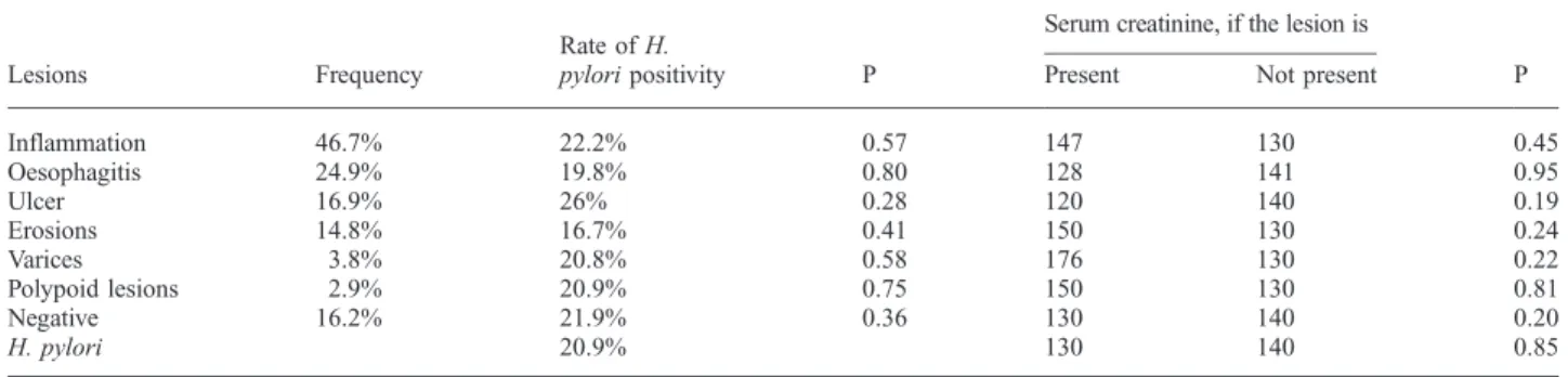
Only 16.2% of the patients examined had the absence of any abnormal endoscopic findings. The most frequent ab- normality found were inflammatory changes, with almost one-half of the patients showing signs of gastritis and/or duodenitis. Oesophagitis, varices, duodenal and/or gastric ulcer disease, erosions, oesophageal varices and polypoid lesions were frequently observed findings. Their frequency was independent from renal function assessed by serum- creatinine level (Table 1). Fifty-two out of 92 ulcer patients (56.5%) had gastric, 33 (35.8%) duodenal and 7 (7.6%) both kinds of ulcer. In five cases, malignant tumour was suspected during the endoscopy with three of them histologi- cally proven: one of those was mucosa associated lymphoid tissue (MALT) lymphoma, the others were adenocarcinomas.
Altogether six malignant tumours were found, in the other three cases, previously a diagnosis of polyp or ulcer was made. Other rare abnormalities seen were diverticula, vas- cular angiectasia (suspicious for Kaposi-sarcoma) and Mallory–Weiss syndrome.
H. pylori
In 62 (11.4%) cases, the H. pylori status was not deter- mined. These examinations were mainly repeated or acute endoscopies for bleeding. Out of the 481 histologically examined, the presence of H. pylori was verified in 101 cases, representing 20.9%. For a two-year period during the study we performed histology and rapid urease test (RUT) in parallel. Whilst the specificity of RUT was 98.5%, its sensitivity was only 38.5%, so we stopped per- forming it in 2004. There was no difference according to gender or age (Table 2).
The presence of H. pyloriwas not associated with the presence of peptic ulcer: 20 of 101 H. pylori-positive pa- tients as compared to 57 of 380H. pylori-negative patients had ulcers (P = 0.28). Vice versa: 26% of ulcer patients were positive forH. pylori. There were no significant associa- tions between positivity forH. pyloriand erosions, oeso-
Table 1.Most frequent macroscopic lesions, creatinine medians (µmol/L), rate ofH. pyloriand their relationship
Lesions Frequency
Rate ofH.
pyloripositivity P
Serum creatinine, if the lesion is
P
Present Not present
Inflammation 46.7% 22.2% 0.57 147 130 0.45
Oesophagitis 24.9% 19.8% 0.80 128 141 0.95
Ulcer 16.9% 26% 0.28 120 140 0.19
Erosions 14.8% 16.7% 0.41 150 130 0.24
Varices 3.8% 20.8% 0.58 176 130 0.22
Polypoid lesions 2.9% 20.9% 0.75 150 130 0.81
Negative 16.2% 21.9% 0.36 130 140 0.20
H. pylori 20.9% 130 140 0.85
at Semmelweis Ote on December 22, 2014http://ndt.oxfordjournals.org/Downloaded from
phagitis, macroscopic signs of inflammation, metaplasia or atrophy (Table 1).
Impact of IS agents
We compared the main groups according to the observed diagnosis and infection rate. There were no differences in the incidence of negative findings, inflammation, ulcer and oesophagitis and the presence ofH. pylori. However, there were differences in the incidence of erosive lesions;
both the differences between the groups and the trend (P = 0.01) are significant (Table 3). This table displays the rate of the particular lesions of those receiving ST pulse every three months prior to endoscopy as well. Out of them, 18.75% had an ulcer (P = 0.106). MMF was proven as an independent risk factor for erosive lesions, OR: 1.83 (1.02–
3.29, P = 0.043) when correcting for the presence of other risk factors.
Impact of acid suppression therapy
We compared the H2RA and PPI groups according to the indications, observed diagnoses and infections. These groups did not differ in their nature of presenting com- plaints: pain, dyspepsia, anaemia and bleeding occurred at the same rate. Respective P-values were 0.9, 0.55, 0.16 and 0.86. There was no difference in the incidence of in- flammation (P = 1.0), oesophagitis (P = 0.68), erosions (P = 0.60), ulcers (P = 0.20) or negative findings (P = 0.87) depending on the AST given, and there was no differ- ence in mycosis (P = 0.16). The only significant difference was in the presence ofH. pylori; it was present in 22.6%
of patients who received H2RA and in 12.4% of those who received PPI (P = 0.044).
Timing of endoscopy
The time between transplantation and endoscopy varies from 3 days up to almost 19 years, median was 3.39 years.
However, 29% (157) of the patients were examined in the first post-transplant year, and 58.5% (92) of them, which is 16.9% of all, in the first 3 months, as shown in Figure 1.
Ulcers were more commonly found in patients requiring earlier endoscopy: 1.65 vs 3.66 years for patients where no
ulcer was found (P = 0.009). The frequency of ulcer disease was 29.3% of the examinations in the first 3 months, 26.3%
in the first year and only 12.9% later on, P = 0.0014.
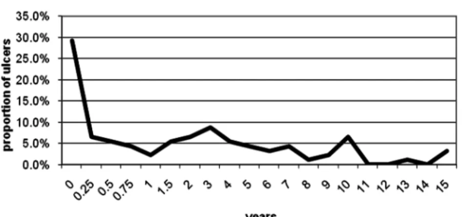
Twenty-seven (29.3%) out of 92 ulcers developed in the first 3 months, forty-two (45.7%) in the first year and all the others at a constant rate later on (Figure 2). TheH. pyl- oripositivity rate on examination decreased with time after transplantation, but this was statistically non-significant.
Discussion
This is the largest series, to our knowledge, of endoscopic findings in consecutive kidney transplant patients scoped for a specific indication. Endoscopy is not part of the rou- tine pre- or postoperative schedule in this unit. By nature, this examination is relatively unpleasant, invasive and ex- pensive, with a small but existing hazard of perforation of about 0.06% [26].
We included only endoscopies performed in our centre on those patients to guarantee homogeneity. The way of follow-up in our centre, though, means that only few en- doscopies (perhaps <5%) were performed elsewhere. Ac- cording to our study, at least 25% of all patients require upper endoscopy in their ‘post-transplant life’. We did not find data to compare this rate.
Endoscopy was completely negative in about 16% of our cases, which reflects other reports [20]. This rate (84%) of clinically significant endoscopic findings is much higher than expected from data of general gastro- enterology patients, where this figure is around 58%
[7,27].
The most important period for upper GI symptoms re- quiring endoscopy was the first year and particularly the first 3 months. Almost one-third of the patients were inves- tigated in the first year. The importance of the first year was observed in other reports as well, where 51% of the verified GI complications occurred during that time [5].
In the first 3 months 29%, and in the first year 26% of the endoscopic examinations revealed an ulcer disease.
Out of 92 ulcers, 29% developed in the first 3 months and 45.7% in the first post-transplant year. The nearly 17% incidence of well-defined ulcer disease is signifi- cantly (P < 0.0001) higher comparing it with gastroenter- ology patients undergoing endoscopy [7,8]. The risk of ulcer disease is 1.69-fold (1.32–2.15 confidence interval (CI) 95%) for a kidney recipient. There are no similar data for the prevalence of ulcers in transplant patients in the literature. The true prevalence of ulcer disease might be even higher, as a large proportion of patients do not experience symptoms [28].
Table 2.Demographic data accordingH. pylori
H. pyloripos. H. pylorineg. P
Male (56.55%) 20.96% 79.04% P = 1.0
Female (43.45%) 21.05% 78.95%
Age (year) 50.54 49.24 0.53
Table 3.GI lesions and their frequency according to the IS regimen and steroid pulse therapy
CSA–ST CSA–MMF–ST TAC–MMF–ST P ST pulse P
Negative 12.0% 13.9% 17.9% 0.53 25.0% 1.00
Inflammation 51.0% 50.5% 38.5% 0.15 31.23% 0.18
Ulcer 19.0% 16.1% 15.4% 0.77 18.8% 0.11
H. pylori 24.7% 23.9% 17.3% 0.45 7.1% 0.18
Erosions 15.9% 18.4% 24.4% 0.03 31.2% 0.0547
at Semmelweis Ote on December 22, 2014http://ndt.oxfordjournals.org/Downloaded from
The individual IS drugs and combinations received seemed to have a significant impact on the patient’s GI status. MMF was proven to increase the risk of erosions of the gastro-duodenal mucosa 1.83-fold (1.02–3.29 CI 95%). Patients receiving the TAC–MMF–ST regimen had the highest frequency of erosive lesions. The effect of corticosteroids is still controversial; high doses are sus- pected to be ulcerogenic, but real evidence is not available [12,29–31]. ST treatment can mask the symptoms and delay treatment [32]. In our series, even high-dose intra- venous ST pulse therapy for rejection was not associated with GI lesions observed endoscopically.
Administration of AST for ulcer prophylaxis is com- mon at most transplant centres [1,12,33,34]. The specific
drug used in our centre was dependent on availability, price etc. and not only on medical grounds. The rate of observed endoscopic alterations did not differ in the H2RA and in the PPI group, indicating the equivalence of these two groups of AST in this setting. The only dif- ference was in the presence of H. pylori; PPIs facilitated the eradication ‘by chance’more efficiently than they do in the general population.
Over the last 20 years there has been considerable inte- rest in the role ofH. pyloriin the pathogenesis of gastritis and peptic ulceration in the general population [35]. Data reported on the prevalence of Helicobacter pyloriamong transplant recipients are contradictory. There are only a few studies reporting endoscopic results of transplant pa- No and rate of endoscopies in the posttransplant years
1 2 3 4 5 6 7 8 9
Posttransplant years 0
20 40 60 80 100 120 140 160 180
No. of endoscopies
157; 29%
55; 10%
40; 7%
47; 9% 45; 8%
37; 7%
32; 6% 33; 6%
15; 3%
26; 5%
Fig. 1. No. of endoscopies in the post-transplant years.
Fig. 2. Rate of observed ulcers according to post-transplant years.
at Semmelweis Ote on December 22, 2014http://ndt.oxfordjournals.org/Downloaded from
tients [2,36–38], and there are some more reporting ofH.
pyloriserologic examinations [39–41]. Published data vary from 29 to 70%. Seroprevalence ofH. pyloriis 49% in a large Hungarian uraemic cohort [42]. In our endoscopic unit experience, 47%H. pyloripositivity rate was detected with biopsy in general gastroenterology patients during the same period (data not published). The observed 20.9% fre- quency of biopsy-provenH. pyloriinfections in our study represented a highly significant difference (P < 0.0001).
Our results demonstrated a high,‘spontaneous’eradication rate in transplanted patients. The rate ofH. pyloridid not change in time in our present material, and the age of posi- tivity and negativity was the same. These results suggest that eradication happens at the very early peri-operative period, when prophylactic antibiotics and AST are given together. The presence ofH. pyloridid not result in signifi- cant postoperative gastric complications. Due to the con- stant use of AST, the sensitivity of RUT was only 38.5%.
As a result, we do not recommend it in this particular group of patients. As bothHelicobacter pylori[43] and immuno- suppression have been linked to an increased risk of devel- oping malignancy, we considered our patients as‘high-risk patients’, and a‘test and treat’strategy was followed. The Maastricht Consensus Reports were used as basis for the eradication protocol [44].
In the general population, only 15% ofH. pylori-infected persons develop peptic ulcer disease, suggesting that spe- cific factors are required for ulceration to occur [45]. Less than one-third (26%) of gastric ulcer patients wereH. pylori positive and even less (17%) were positive among patients found to have a duodenal ulcer. Neither the type of IS drugs nor the presence ofH. pylorihad a statistically significant impact on ulceration. This finding suggests that ulceration in transplant recipients is a multifactorial process that may involve the interaction of acid secretion,H. pylori, IS agents and other medications rather than a single factor.
Specific IS drugs, opportunistic infections and other clinical circumstances that affect transplant patients are not seen frequently in the general practice of gastroenter- ology. Thus, the endoscopist at a transplant centre has to be able to recognize, identify and treat the unique pro- blems seen in a transplant population. Giving prophylac- tic acid secretion blockers, minimizing the number of pills a patient has to take a day and adopting a low threshold for endoscopy are among the most important measures that could be used to avoid GI complications after trans- plantation [46].
Conflict of interest statement.None declared.
References
1. Kleinman L, Faull R, Walker R et al. Gastrointestinal-specific patient-reported outcome instruments differentiate between renal transplant patients with or without GI complications. Transplant Proc 2005; 37: 846–849
2. Abu Farsakh NA, Rababaa M, Abu Farsakh H. Symptomatic, endo- scopic and histological assessment of upper gastrointestinal tract in renal transplant recipients.Indian J Gastroenterol2001; 20: 9–12 3. Hardinger KL, Brennan DC, Lowell Jet al. Long-term outcome of
gastrointestinal complications in renal transplant patients treated with mycophenolate mofetil.Transpl Int2004; 17: 609–616
4. de Francisco AL. Gastrointestinal disease and the kidney.Eur J Gas- troenterol Hepatol2002; 14: S11–S15
5. Sarkio S, Halme L, Kyllonen Let al. Severe gastrointestinal compli- cations after 1,515 adult kidney transplantations.Transpl Int2004;
17: 505–510
6. Matsumoto C, Swanson SJ, Agodoa LYet al. Hospitalized gastro- intestinal bleeding and procedures after renal transplantation in the United States.J Nephrol2003; 16: 49–56
7. Sonnenberg A, Amorosi SL, Lacey MJet al. Patterns of endoscopy in the United States: analysis of data from the Centers for Medicare and Medicaid Services and the National Endoscopic Database.Gastrointest Endosc2008; 67: 489–496
8. Montes Teves P, Salazar Ventura S, Monge Salgado E. Epidemio- logical changes in peptic ulcer and their relation with Helicobacter pylori. Hospital Daniel A Carrion 2000-2005.Rev Gastroenterol Perú 2007; 27: 382–388
9. Knoll GA, MacDonald I, Khan A et al. Mycophenolate mofetil dose reduction and the risk of acute rejection after renal transplantation.
J Am Soc Nephrol2003; 14: 2381–2386
10. Pelletier RP, Akin B, Henry MLet al. The impact of mycophenolate mofetil dosing patterns on clinical outcome after renal transplantation.
Clin Transplant2003; 17: 200–205
11. Akioka K, Okamotoa M, Nakamuraa Ket al. Abdominal pain is a critical complication of mycophenolate mofetil in renal transplant re- cipients.Transplant Proc2003; 35: 300–301
12. Ponticelli C, Passerini P. Gastrointestinal complications in renal trans- plant recipients.Transpl Int2005; 18: 643–650
13. Péter A, Telkes G, Varga Met al. Endoscopic diagnosis of cyto- megalovirus infection of upper gastrointestinal tract in solid organ transplant recipients: Hungarian single-center experience.Clin Trans- plant2004; 18: 580–584
14. Gautam A. Gastrointestinal complications following transplantation.
Surg Clin N Am2006; 86: 1195–1206
15. Van Thiel DH, Wright HI, Fagiuoli Set al. Upper gastrointestinal endoscopy: its clinical use and safety at a transplant center.J Okla State Med Assoc1994; 87: 116–121
16. Giraldez A, Sousa JM, Bozada JMet al. Results of an upper endos- copy protocol in liver transplant candidates. Rev Esp Enferm Dig 2003; 95: 555–560, 549–554
17. Zaman A, Hapke R, Flora Ket al. Prevalence of upper and lower gastrointestinal tract findings in liver transplant candidates undergo- ing screening endoscopic evaluation.Am J Gastroenterol1999; 94:
895–899
18. Donovan JP. Endoscopic management of the liver transplant patient.
Clin Liver Dis2000; 4: 607–618
19. Troppmann C, Papalois BE, Chiou Aet al. Incidence, complications, treatment, and outcome of ulcers of the upper gastrointestinal tract after renal transplantation during the cyclosporine era.J Am Coll Surg1995; 18: 433–443
20. Graham SM, Flowers JL, Schweitzer Eet al. Opportunistic upper gastrointestinal infection in transplant recipients.Surg Endosc 1995; 9: 146–150
21. Misiewicz JJ. The Sydney System: a new classification of gastritis.
Introduction.J Gastroenterol Hepatol1991; 6: 207–208
22. Tytgat GN. The Sydney System: endoscopic division. Endoscopic ap- pearances in gastritis/duodenitis.J Gastroenterol Hepatol1991; 6:
223–234
23. Stolte M, Meining A. The updated Sydney System: classification and grading of gastritis as the basis of diagnosis and treatment.Can J Gastroenterol2001; 15: 591–598
24. Miller G, Savary M. Is gastro-esophageal prolapse responsible for le- sions of the gastric mucosa?Minerva Gastroenterol1977; 23: 42 25. Levy N, Stermer E, Boss JM. Accuracy of endoscopy in the diagnosis
of inflamed gastric and duodenal mucosa.Isr J Med Sci1985; 21:
564–568
26. Misra T, Lalor E, Fedorak RN. Endoscopic perforation rates at a Canad- ian university teaching hospital.Can J Gastroenterol2004; 18: 221–226 27. Thomson ABR, Barkun AN, Armstorng Det al. The prevalence of clinically significant endoscopic findings in primary care patients with uninvestigated dyspepsia: the Canadian Adult Dyspepsia Em-
at Semmelweis Ote on December 22, 2014http://ndt.oxfordjournals.org/Downloaded from
piric Treatment –Prompt Endoscopy (CADET–PE) study.Aliment Pharmacol Ther2003; 17: 1481–1491
28. Steger AC, Timoney AS, Griffen Set al. The influence of immuno- suppression on peptic ulceration following renal transplantation and the role of endoscopy.Nephrol Dial Transplant1990; 5: 289–292 29. Conn HO, Poynard T. Corticosteroids and peptic ulcer: meta-analysis of adverse events during steroid therapy.J Intern Med1994; 236: 619–632 30. Pecora PG, Kaplan B. Corticosteroids and ulcers: is there an associ-
ation?Ann Pharmacother1996; 30: 870–872
31. Black HE. The effects of steroids upon the gastrointestinal tract.
Toxicol Pathol1988; 16: 213–222
32. Sarkio S. Gastrointestinal complications after kidney transplantation.
Doctoral dissertation, April 2006. University of Helsinki, Faculty of Medicine, Institute of Clinical Medicine, Department of Transplant- ation and Liver Surgery. http://ethesis.helsinki.fi/julkaisut/laa/kliin/
vk/sarkio/ ISBN 952-10-3005-4
33. Logan AJ, Morris-Stiff GJ, Bowrey DJet al. Upper gastrointestinal complications after renal transplantation: a 3-yr sequential study.Clin Transplant2002; 16: 163–167
34. Reese J, Burton F, Lingle Det al. Peptic ulcer disease following renal transplantation in the cyclosporine era.Am J Surg1991; 162: 558–562 35. Marshall B. Helicobacter pylori: 20 years on.Clin Med2002; 2:
147–152
36. Ozgur O, Boyacioglu S, Ozdogan Met al. Helicobacter pylori infec- tion in haemodialysis patients and renal transplant recipients.Nephrol Dial Transplant1997; 12: 289–291
37. Teenan RP, Burgoyne M, Brown ILet al. Helicobacter pylori in renal transplant recipients.Transplantation1993; 56: 100–103
38. Khedmat H, Ahmadzad-Asl M, Amini Met al. Gastro-duodenal le- sions and Helicobacter pylori infection in uremic patients and renal transplant recipients.Transplant Proc2007; 39: 1003–1007 39. Davenport A, Shallcross TM, Crabtree JEet al. Prevalence of Heli-
cobacter pylori in patients with end-stage renal failure and renal transplant recipients.Nephron1991; 59: 597–601
40. Yildiz A, Besisik F, Akkaya Vet al. Helicobacter pylori antibodies in hemodialysis patients and renal transplant recipients.Clin Transplant 1999; 13: 13–16
41. Sarkio S, Rautelin H, Halme L. The course of Helicobacter pylori infection in kidney transplantation patients.Scand J Gastroenterol 2003; 38: 20–26
42. Telkes G, Rajczy K, Varga Met al. Seroprevalence of Helicobacter pylori in Central-European uraemic patients and its possible associ- ation with presence of HLA-DR12 allele.Eur J Gastroenterol Hepa- tol2008; 20: 906–911
43. Sepulveda AR, Graham DY. Role of Helicobacter pylori in gastric carcinogenesis.Gastroenterol Clin N Am2002; 31: 517–535 44. Malfertheiner P, Megraud F, O’Morain Cet al. Current concepts in
the management of Helicobacter pylori infection: the Maastricht III Consensus Report.Gut2007; 56: 772–781
45. Tadataka Y. Handbook of Gastroenterology. Philadelphia: Lippincott- Raven Publishers, 1998, 266.
46. Helderman JH. Prophylaxis and treatment of gastrointestinal compli- cations following transplantation.Clin Transplant2001; 15: 29–35
Received for publication: 24.5.09; Accepted in revised form: 15.6.10
Nephrol Dial Transplant (2011) 26: 732–738 doi: 10.1093/ndt/gfq388
Advance Access publication 5 July 2010
Barriers to living kidney donation identified by eligible candidates with end-stage renal disease
Lianne Barnieh
1, Kevin McLaughlin
2, Braden J. Manns
1,2, Scott Klarenbach
3, Serdar Yilmaz
4, Brenda R. Hemmelgarn
1,2For the Alberta Kidney Disease Network
1Department of Community Health Sciences,2Department of Medicine, University of Calgary, Calgary, Alberta, Canada,3Department of Medicine, University of Alberta, Edmonton, Alberta, Canada and4Department of Surgery, University of Calgary, Calgary, Alberta Canada
Correspondence and offprint requests to: Brenda R. Hemmelgarn; E-mail: Brenda.Hemmelgarn@albertahealthservices.ca
Abstract
Background. Among eligible transplant candidates with end-stage renal disease, only a minority receive a living donor kidney transplant (LDKT), suggesting that there are barriers to receipt of this optimal therapy.
Methods. A validated questionnaire was administered to adults active on the deceased donor transplant waiting list, identified from the Southern Alberta Renal Program data- base. The questionnaire included both quantitative and
qualitative items addressing issues related to LDKT in the categories of knowledge, opportunity, fear and guilt.
Results.Of the 196 subjects invited to complete the ques- tionnaire, 145 (74%) responded. Not knowing how to ask someone for their kidney was the most frequently reported barrier, identified by 71% of respondents. Those that sta- ted that living donation did not pose significant long-term health risks to the donor [odds ratio (OR) = 3.40, 95% CI 1.17–9.46, P = 0.01] and those who understood how and
© The Author 2010. Published by Oxford University Press on behalf of ERA-EDTA. All rights reserved.
For Permissions, please e-mail: journals.permissions@oxfordjournals.org
at Semmelweis Ote on December 22, 2014http://ndt.oxfordjournals.org/Downloaded from