Krisztina Hagymási, Zsolt Tulassay, 2nd Department of Internal Medicine, Semmelweis University, H-1088 Budapest, Hungary Author contributions: Hagymási K and Tulassay Z drafted and wrote the manuscript; all authors read and approved the final manuscript.
Correspondence to: Krisztina Hagymási, MD, PhD, 2nd Depart- ment of Internal Medicine, Semmelweis University, H-1088 Buda- pest, Hungary. hagymasi.krisztina@med.semmelweis-univ.hu Telephone: +36-1-2660926 Fax: +36-1-2664616 Received: September 28, 2013 Revised: January 5, 2014 Accepted: February 26, 2014
Published online: June 7, 2014
Abstract
Helicobacter pylori (H. pylori) infects more than half of the world’s human population, but only 1% to 3% of infected people consequently develop gastric adenocarcinomas. The clinical outcome of the infection is determined by host genetic predisposition, bacterial virulence factors, and environmental factors. The as- sociation between H. pylori infection and chronic active gastritis, peptic ulcer disease, gastric cell carcinoma, and B cell mucosa-associated lymphoid tissue lym- phoma has been well established. With the exception of unexplained iron deficiency anemia and idiopathic thrombocytopenic purpura, H. pylori infection has no proven role in extraintestinal diseases. On the other hand, there is data showing that H. pylori infection could be beneficial for some human diseases. The un- predictability of the long-term consequences of H. py- lori infection and the economic challenge in eradicating
it is why identification of high-risk individuals is crucial.
© 2014 Baishideng Publishing Group Inc. All rights reserved.
Key words: Helicobacter pylori; virulence factor; Host fac- tors; Gastroduodenal diseases; extraintestinal disorders Core tip: Helicobacter pylori (H. pylori ) infects more
than half of the world’s human population. The as- sociation between H. pylori infection and chronic active gastritis, peptic ulcer disease, gastric cell carcinoma, and B cell mucosa-associated lymphoid tissue lympho- ma, unexplained iron deficiency anemia and idiopathic thrombocytopenic purpura has been well established.
H. pylori screening and treatment is a recommended gastric cancer risk reduction strategy in high-risk popu- lations. The unpredictability of the long-term conse- quences of H. pylori infection and the economic chal- lenge in eradicating it is why identification of high-risk individuals is crucial.
Hagymási K, Tulassay Z. Helicobacter pylori infection: new pathogenetic and clinical aspects. World J Gastroenterol 2014;
20(21): 6386-6399 Available from: URL: http://www.wjgnet.
com/1007-9327/full/v20/i21/6386.htm DOI: http://dx.doi.
org/10.3748/wjg.v20.i21.6386
INTRODUCTION
Helicobacter pylori (H. pylori) is a micro-aerophilic, Gram- negative, slow-growing, spiral-shaped, and flagellated organism which infects more than half of the world’s human population[1]. H. pylori colonization itself does not cause any symptoms, and fewer than 20% of all infected patients will develop symptoms from their infection[2]. Approximately 10% of infected individuals develop pep- tic ulcer disease, 1% to 3% develop gastric adenocarci- noma, and less 0.1% [mucosa-associated lymphoid tissue (MALT)] develop lymphoma[3].
The outcome of H. pylori infection may involve a combination of bacterial, host, and environmental fac- tors. The association between H. pylori infection and chronic active gastritis, peptic ulcer disease, gastric cell carcinoma, and B cell MALT lymphoma has been well established. On the other hand H. pylori infection could TOPIC HIGHLIGHT
Help Desk: http://www.wjgnet.com/esps/helpdesk.aspx
DOI: 10.3748/wjg.v20.i21.6386 ISSN 1007-9327 (print) ISSN 2219-2840 (online)
© 2014 Baishideng Publishing Group Inc. All rights reserved.
Helicobacter pylori infection: New pathogenetic and clinical aspects
WJG 20th Anniversary Special Issues (6): Helicobacter pylori
Krisztina Hagymási, Zsolt Tulassay
be beneficial for humans[2] (Table 1).
PATHOGENETIC ASPECTS
Virulence factors of H. pylori
Bacterial virulence factors play a significant role in the outcome and progression of H. pylori infection[4]. The linkages of virulence factors may show how they interact with each other[5].
The cag pathogenicity island (cag PAI) contains 27-31 genes flanked by a 31-p direct repeats. H. pylori exhibits a high degree of genetic heterogeneity due to genomic rearrangements, gene insertions, and/or deletion[6].
At least 18 cag genes encode components of the bacterial type Ⅳ secretion system, which functions to export bacterial protein across the bacterial membrane and into host gastric epithelial cells. The presence of cag PAI (cag+) amplifies the risk for severe gastritis, atrophic gastritis, and distal gastric cancer in comparison with cag- deficient (cag-) bacteria[6].
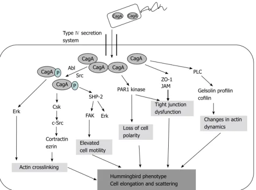
CagA: Cytotoxin-associated gene A product (CagA) is translocated into the host cell by the type Ⅳ secre- tion system. Phosphorylation of CagA at the glutamate- proline-isoleucine-tyrosine-alanine (EPIYA) motifs by the host Abl and Src kinases results in morphological chang- es to the cell (the so-called “hummingbird phenotype”).
Four EPIYA motifs (-A, -B, -C, and -D) are distinguished with different degrees of phosphorylation and geograph- ical distribution[6]. EPIYA-A and EPIYA -B sites are less phosphorylated in comparison with EPIYA-C. EPIYA-C is typically found only in strains from Western countries (Europe, North America, and Australia), and is an indi- cator of gastric cancer risk. EPIYA-D is found in East Asian strains. EPIYA-D containing strains induce more relief of interleukin-8 (IL-8) from gastric epithelial cells[6]
(Figure 1).
Phospho-CagA interacts with numerous intracellu- lar effectors, including eukaryotic tyrosine phosphatase with sustained activation of extracellular signal-regulated kinases 1 and 2 (ERK ½), Crk adaptor, and C-terminal
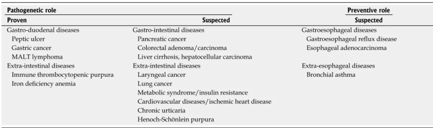
Table 1 Summary of the pathogenetic and preventive role of Helicobacter pylori
Pathogenetic role Preventive role
Proven Suspected Suspected
Gastro-duodenal diseases Gastro-intestinal diseases Gastroesophageal diseases
Peptic ulcer Pancreatic cancer Gastroesophageal reflux disease
Gastric cancer Colorectal adenoma/carcinoma Esophageal adenocarcinoma
MALT lymphoma Liver cirrhosis, hepatocellular carcinoma
Extra-intestinal diseases Extra-intestinal diseases Extra-esophageal diseases
Immune thrombocytopenic purpura Laryngeal cancer Bronchial asthma
Iron deficiency anemia Lung cancer
Metabolic syndrome/insulin resistance Cardiovascular diseases/ischemic heart disease Chronic urticaria
Henoch-Schönlein purpura MALT: Mucosa-associated lymphoid tissue.
Glutamate racemase gene
Cag Ⅱ IS 605 Cag Ⅰ
CagA
ePIYA-containing region
ePIYA site
ePIYA-A segment
ePIYA-B segment
ePIYA-C or D segment 1112 13 1415161718T S RQPO M N L I H G F e D C B
Figure 1 Cytotoxin-associated gene pathogenicity island. CagA: Cytotoxin-associated gene A product; EPIYA: Glutamate-proline-isoleucine-tyrosine-alanine.
10
Src kinase[6]. The activation of ERK and focal adhesion kinase with the tyrosine dephosphorylation of the actin binding proteins cortactin, ezrin, and vinculin leads to cell elongation[1,6] (Figure 2).
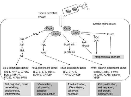
The targets of non-phosphorylated CagA comprise E-cadherin, β-catenin, hepatocyte growth factor receptor c-Met, phospholipase C gamma, adaptor protein Grb2, kinase partitioning-defective 1b/microtubule affinity- regulating kinase 2, epithelial tight junction scaffolding protein zonula occludens 1, and the transmembrane protein junctional adhesion molecule A. The main effects are pro-inflammatory and mitogenic cell-cell junction dis- ruption and loss of cell polarity that may be important in gastric carcinoma development[1,6] (Figures 3 and 4).
Activity of CagA on tumor-suppressor pathways has also been investigated. CagA is able to modulate the H.
pylori induced apoptotic signal, but the exact mechanism remains to be elucidated. The initial host response up- regulates p53 expression followed by the proteasomal degradation of p53[8].
Almost all cagA+ strains are classified as vacA s1 gen- otypes (either m1 or m2), whereas almost all cagA- strains are classified as the vacA s2/m2 strain (see below)[5]. Spe- cific vacA genotypes of H. pylori strains are associated with a level of in vitro cytotoxin activity with clinical con- sequences[9].
Peptidoglycans: Peptidoglycans translocated by the cag secretion system interact with the nucleotide-binding oligomerization domain 1 (Nod1) molecule which leads to the activation of nuclear factor κB (NF-κB), pro- inflammatory secretion of interleukin-8 (IL-8), and
β-defensin-2[6,10]. H. pylori enhances the phosphoinosit- ide 3-kinase Akt signaling pathway, leading to decreased apoptosis and increased cell migration. NOD1 ligand binding can activate the interferon (IFN)-stimulated gene factor 3 signaling cascade, resulting in type Ⅰ IFN pro- duction usually associated with protection against viral infection and possibly other mucosal infections[11]. VacA toxin: The cytotoxin gene vacA is present in all strains. The VacA cytotoxin induces the vacuolation, gastric epithelial barrier function disruption, disturbance of late endosomal compartments, and modulation of the inflammatory response. VacA reduces the mitochondrial transmembrane potential, releases cytochrome c from mitochondria, activates caspase 8 and 9, and induces apoptosis[6,12].
Binding of VacA to receptor-type protein tyrosine phosphatase (RPTPβ) regulates cell proliferation, dif- ferentiation, and adhesion, which all play a role in ulcero- genesis[13].
Variations in vacA gene structure (in the signal s: s1, s2, or in the middle regions m: m1, m2) make differences in vacuolating activity and specificity. The intermediate (i) region also plays role in the vacuolating activity of H.
pylori. All s1, m1 strains were classified as i1 (vacuolat- ing) type, and all s2, m2 strains were classified as i2 (non- vacuolating) type, while s1, m2 alleles could be i1 or i2.
A novel intermediate variant (i3) has been identified. The fourth pathogenic region is d, a 69-81 bp-region between the m and i regions[1,5].
The variants in s and m regions seem to be a good indicator of clinical outcomes. However the roles of i
Ras Raf Mek Erk CagA
CagA CagA
CagA CagA
CagA
CagA CagA Type Ⅳ secretion
system
Gastric epithelial cell
Morphological changes
Inflammation, cell growth, adhesion, immunregulation Cell migration, tissue
remodelling, angiogenesis, inflammation Elk-1 dependent genes Cortractin
Actin polymerisation Arp 2/3
Wave Rac1 Dock Crk
SHP-2
NFκB dependent genes P
P Src Abl P
FAK Csk
Src
Figure 2 Targets of phosphorylated cytotoxin-associated gene A. Based on the article from Current Opinion in Microbiology, Hatakeyama M, SagA of CagA in Helicobacter pylori pathogenesis, 11, 30-37, Copyright (2008), with permission from Elsevier[7]. CagA: Cytotoxin-associated gene A product; NFκB: Nuclear factor κB;
FAK: Focal adhesion kinase; Csk: C-terminal Src kinase.
bohydrate structure sialyl-Lewis antigen expressed on the gastric epithelium. SabA can mediate the binding of H. pylori to neutrophils and erythrocytes, but the patho- physiological importance of these findings is uncertain[1]. SabA positive status was associated with increased gastric cancer risk and a negative status associated with duodenal ulceration[12].
The outer inflammatory protein (OipA) has a role in the increased expression of mucosal IL-1, -8, -17, tu- mor necrosis factor-α (TNF-α), and in gastric mucosal inflammation. Upregulation of matrix metalloproteinase 1, inhibition of glycogen synthase kinase 3β, and nuclear accumulation of β-catenin can influence carcinogenesis[6]. OipA positive status was significantly associated with duodenal ulcer and gastric cancer[12].
Others: Duodenal ulcer promoting gene (dupA) product induces the production of IL-8 and -12[5]. DupA may enhance duodenal ulceration and /or decrease gastric cancer development in some populations[1,5,6].
Variants of gene encoding flagellar proteins (flaA) of H.
pylori may affect motility and colonization, and, therefore, the carcinogenic effect[6].
Annexin family members (AnxA1 and AnxA4) are involved in epithelial cell membrane repair response induced by H. pylori-generated VacA and CagA-indepen- dent plasma membrane disruption. Plasma membrane disruption and AnxA4 can promote cell proliferation[19].
TNF-α-inducing protein (Tipα) binds to cell-surface nucleolin and then enters the gastric cancer cells where and d regions should be further investigated[5]. The s1,
m1 strains can induce greater vacuolation, and are as- sociated with peptic ulcer disease and gastric cancer in Western countries, but have no pathogenic role in East Asian countries[1,6]. vacA i1 strains were associated with gastric cancer in Iranian patients[14], but not in the East Asian or Southeast Asian populations[14]. i1 genotype ap- peared to be a better predictor of carcinoma-associated H. pylori strains than the s or m genotype[15]. In Western countries, d1 strains without the deletion of the d region are predictors of histological inflammation, atrophy, and an increased risk of peptic ulceration and gastric cancer, compared with the presence of the vacA s-, m-, and i-region strains[16].
Adhesins and outer membrane proteins: 4% of the H. pylori genome encodes for outer membrane proteins (BabA, BabB, SabA, and OipA) which function as adhes- ins and porins, and are implicated in complement resis- tance and immune regulation[17].
The blood group antigen binding adhesin BabA is thought to mediate host-bacterial interactions and maintain colonization of the H. pylori targeting human Lewis-b surface epitopes[18,19]. The babA2 gene is associ- ated with duodenal ulcer and gastric cancer. When in conjunction with cagA and vacA s1 alleles (“triple-pos- itive strains”), it is associated with a greater risk of the more severe duodenal ulcer and gastric adenocarcinoma in Western populations[1,6,19].
Sialic acid-binding adhesin (SabA) binds to the car-
CagA CagA
CagA CagA CagA
Grb2 CagA
PLC Ras
Raf Mek Erk
NFAT
e-cadherin β-catenin
ZO-1 JAM
C-Met
PAR1 kinase
Elk-1 dependent genes NFκB dependent genes NFAT dependent genes Wnt/β-catenin dependent genes Morphological changes
PAI-1, MMP-2, 9, FOS, eGR-1, NUR77, PTGS2, HIF1A, PFN1
IL-2, 3, 6, 8, TNF-α,
ICAM-1, GM-CSF IL-2, 3, 4, 8,
TNF-α, GM-CSF cyclinD1, cdx1, c-myc, Nr-CAM, FGF18, gastrin, veGF
Cell migration, tissue remodelling, angiogenesis, Inflammation
Inflammation, cell growth, adhesion, immunregulation
T cell activation, differentiation, cell cycle, apoptosis
Cell proliferation, cell migration, cell growth, angiogenesis Gastric epithelial cell Type Ⅳ secretion
system
Figure 3 Targets of non-phosphorylated cytotoxin-associated gene a product. Based on the article from Current Opinion in Microbiology, Hatakeyama M, SagA of CagA in Helicobacter pylori pathogenesis, 11, 30-37, Copyright (2008), with permission from Elsevier[7]. CagA: Cytotoxin-associated gene A product; PLC: Phospho- lipase C gamma; PAR1: Kinase partitioning-defective 1b; ZO-1: Zonula occludens 1; JAM: Junctional adhesion molecule A; NFκB: Nuclear factor κB; TNF-α: Tumor necrosis factor-α; IL: Interleukin.
TNF-α and chemokine gene expressions are induced by NF-κB activation in a cag PAI independent manner[20].
Bacterial factors like urease, AmiE, AmiF, hydrog- enase, and arginase are essential for H. pylori survival in the acidic gastric environment[4].
Immune response to H. pylori: The host’s innate and adaptive immune system plays a crucial role in the initia- tion and progression of H. pylori infection[21].
Innate immunity effectors and a complex mixture of T helper (Th) 1, Th17, and regulatory T cells (Treg) adaptive immunity effectors are involved in H. pylori infection[22].
H. pylori initially targets gastric epithelial cells which form part of the innate immune response via signaling through pattern recognition receptors, such as Toll-like receptors (mainly TLR2)[21].
The neutrophil-activating protein of H. pylori polar- izes Th1 cells, stimulating IL-12 and IL-23 secretion from neutrophils and macrophages. Th1 cytokines, such as gamma interferon (IFN-γ) and TNF-α, can increase the release of pro-inflammatory cytokines and augment apoptosis induced by H. pylori[22,23].
IL-17 expressing Th17 cells are important in the pro- inflammatory immune response to H. pylori. Th17 cells produce Il-17, IL-21, and IL-22 cytokines[6]. H. pylori in- fected macrophages produce IL-6, IL-23, and transform- ing growth factor (TGF)-β, which are required for Th17 cell development and maintenance[6,21]. The literature on Th1 and Th17 H. pylori-associated gastric pathology is confusing and requires intensive investigation[6].
Tregs (formerly suppressor T cells) are also implicated in the pathogenesis of H. pylori infection. TGF-β and IL-18 are responsible for Treg development[21]. H. pylori-
specific Tregs suppress memory T cell responses that prolong the infection[6]. Tregs suppress the inflammatory reaction driven by IL-17, thereby also favoring bacterial persistence[24].
Antimicrobial defense of macrophages is nitric oxide (NO) dependent. H. pylori’s arginase enzyme can compete with macrophages for the inducible nitric oxide synthase (iNOS) substrate L-arginine so that host NO production is impaired; this leads to enhanced bacterial survival. H.
pylori can evade macrophage phagocytosis. VacA protein prevents the fusion of phagosomes with lysosomes need- ed for phagocytosis. Fused phagosomes contain large numbers of live bacteria[6].
The role of B cells in the host response to H. pylori has been suggested[21]. Immunoglobulin (Ig) G and IgA antibody release from B cells in response to H. pylori may be involved in protective immunity, however it was suggested this antibody-mediated response may be counterproductive. B cells can also produce autoreactive antibodies that may be pathogenic[6]. B cell activation and survival may have implications for MALT lymphoma de- velopment[6].
CLINICAL ASPECTS
Gastroduodenal diseases
Peptic ulcer: Some H. pylori colonized individuals may develop corpus gastritis associated with gastric hypo- chlorhydria, gastric atrophy, gastric ulcer, and an increased risk of gastric cancer. Conversely, others may develop antral-predominant gastritis, which is associated with gas- tric hyperchlorhydria and an increased risk of duodenal ulcer[8,25].
CagA CagA
CagA
CagA CagA CagA CagA
CagA
Type Ⅳ secretion system
P P
SHP-2
ZO-1 JAM
PLC
PAR1 kinase
Erk Csk Erk
c-Src
FAK Src Abl
Actin crosslinking
elevated cell motility
Loss of cell polarity
Tight junction dysfunction
Changes in actin dynamics Gelsolin profilin cofilin
Hummingbird phenotype Cell elongation and scattering Cortractin
ezrin
Figure 4 Development of “hummingbird phenotype”. CagA: Cytotoxin-associated gene A product; PLC: Phospholipase C gamma; PAR1: Kinase partitioning- defective 1b; ZO-1: Zonula occludens 1.
Since the discovery of H. pylori in the 1980s, the avail- ability of effective eradication therapy has led to a decline in recurrent peptic ulcer disease and its complications.
The pathogenetic role of H. pylori in 90% of duodenal ulcers and 80% of gastric ulcers is proven[26,27]. Effective eradication decreased the yearly recurrence rate of duo- denal and gastric ulcers from 80% and 60%, respectively, to less than 5%[28].
Gastric cancer: H. pylori is a class I carcinogen in hu- mans[1]. It is considered to be the most common aetio- logical factor of infection-related cancers (followed by human papilloma, hepatitis B and C, Epstein-Barr, hu- man immunodeficiency, and human herpesvirus-8)[1,29]. H. pylori infection-related cancer represents 5.5% of the global cancer burden[6].
Gastric cancer develops in 2.9% of H. pylori infected patients[30]. H. pylori infection is responsible for about 75% of all non-cardia gastric cancers and 63.4% of all stomach cancers worldwide[1]. H. pylori infection also plays a fundamental role in non-cardia gastric carcinogenesis, but its association with cardia cancer is still uncertain[31].
The prevalence of infection is statistically significantly much higher in patients with intestinal-type gastric cancer (89.2%) compared to the diffuse-type (31.8%)[32]. H. pylori infection is regarded as the trigger of intestinal-type gastric adenocarcinoma[33]. According to Correa and Piazuelo, intestinal-type gastric carcinogenesis progresses as follows:
normal gastric mucosa - no atrophic gastritis - multifocal atrophic gastritis without intestinal metaplasia - intestinal metaplasia of complete (small intestine) type - low-grade dysplasia - high-grade dysplasia - invasive adenocarcino- ma[34]. Altered cell proliferation, apoptosis, epigenetic modifications to the tumor suppressor genes, onco- gene activation, and dysregulation of DNA repair may occur and eventually lead to inflammation-associated carcinogenesis[35].
Eradication of H. pylori infection decreases the risk of premalignant lesions and gastric cancer in infected individuals[36-38]. Follow-up endoscopy and histology is crucial, even in patients with apparently non-malignant gastric ulcers, in improving the malignancy detection rate in populations with a high prevalence of gastric cancer[39].
H. pylori plays a role in the development and pro- gression of gastric (MALT) lymphoma[40]. The average prevalence of H. pylori infection in MALT lymphoma was 79%; it was higher in low-grade (79%) than in high-grade (60%) cases[41]. Treatment for localized stage Ⅰ gastric MALT lymphoma with H. pylori infection is eradication[40]. Eradication of H. pylori resulted in a complete remission in 60%-80% of patients with MALT lymphoma[42,43], and a 10-year sustained remission in up to 64% of cases[44].
The carcinogenic effect of H. pylori can be modified by dietary and environmental factors. H. pylori infection is more frequent in less developed Asian countries (e.g., India, Bangladesh, Pakistan, and Thailand) in comparison with the more developed Asian countries (e.g., Japan and China). However, the frequency of gastric cancer is para-
doxically very low in these less developed regions than in Japan and China (the so-called “Asian enigma”)[33,45]. Several other large populations with high infection preva- lence show a very low rate of gastric cancer. The so- called “African enigma” remains unexplained as well, but it does verify that not all H. pylori infected patients have an increased risk of gastric cancer[32,33,46].
Host and environmental factors also affect the devel- opment of gastroduodenal diseases in H. pylori infected individuals[6,47]. Individuals with a high-expression of IL- 1β polymorphisms (C-T or T-C transitions, at positions -511, -31, and +3954 base pairs from the transcriptional start site) have an increased risk for hypochlorhydria, gas- tric atrophy, and distal gastric adenocarcinoma in com- parison with low-expression polymorphisms; they have no effect on cancers associated with high acid exposure such as esophageal adenocarcinomas and some cardia cancers[47,48]. The combined effects of pro-inflammatory IL-1 genotypes and H. pylori bacterial virulence factors have been reported[48].
Gene polymorphisms (-308 G > A) of the pro-in- flammatory cytokine TNF-α that increase the expression of the cytokine and polymorphisms (promoter polymor- phisms at positions -592, -819, and -1082) that reduce the production of the anti-inflammatory cytokine (IL-10) have been associated with an increased risk of distal gas- tric cancer[48-50].
The effects of pro-inflammatory genotypes (IL-1β, TNF-α and IL-10) are additive[6,50].
High dietary salt intake increases the risk of gastric cancer by directly damaging gastric mucus and mucosa, improving temporary epithelial proliferation, increasing the incidence of endogenous mutations, upregulating cy- tokine production, and H. pylori gene expression modula- tion, especially that of virulence factors[51-53].
Co-infection with helminths (Ascaris lumbricoides) and Toxoplasma gondii reduces the severity of H. pylori-induced gastritis via a reduced Th1 response with higher levels of Th2 cytokines[54].
Fruit and vegetables are rich sources of carotenoids, vitamin C, folate, and phytochemicals, which may modu- late xenobiotic-metabolizing enzymes and have antioxi- dant activity, thereby playing a preventive role in carcino- genesis[6,55-57].
Smoking is an established risk factor for gastric can- cer. Swallowed carcinogenic substances (nitrosamine and other nitroso compounds), greater concentrations of smoking-related DNA adducts in the gastric mucosa, lower levels of free radical scavengers (ascorbic acid and β-carotene), and increased mRNA expression of chemo- kines in the gastric mucosa are in the background[58]. Pancreatic cancer
Epidemiological studies have suggested that H. pylori might be involved in the pathogenesis of pancreatic can- cer (OR = 1.87, 2.1), however results are inconsistent[59,60]. A meta-analysis showed significant association between H. pylori seropositivity and development of pancreatic
cancer (pooled adjusted OR = 1.38), but further research is needed to confirm this result[61,62].
Despite good scientific reasoning for the involvement of H. pylori in pancreatic diseases, direct pancreatic infec- tion seems unlikely[63] (Table 2).
Colorectal adenoma/carcinoma
On the basis of the epidemiological results showing
high mortality rates from gastric and colorectal cancer in similar areas, it can be speculated that gastric cancer and colorectal cancer have common risk factors like H. pylori infection[68]. Although the role of H. pylori in colorectal carcinogenesis has been widely examined, the association has remained inconclusive[69]. Several studies demon- strated conflicting positive and negative associations[68,69]. A meta-analysis showed that H. pylori infection was asso- ciated with an increased risk of colorectal adenoma (OR
= 1.66) and colorectal cancer (OR = 1.39), however there was significant heterogeneity among the studies[70]. The inconsistent results might be due to sample bias, small sample size, varying frequencies of cagA+ strains in the study population, incomplete colonoscopies, and evalua- tion of H. pylori infection with the IgG serum test[69].
H. pylori was detected in colorectal carcinoma tissue in a pilot study[71]. Higher prevalence was proven in adeno- ma and colorectal cancer compared with control[72,73]. H.
pylori was more prevalent in moderate/severe dysplastic adenomas compared with mild dysplasia, and in tubular and tubulovillous adenoma compared with villous type[72].
The pathogenetic mechanisms of H. pylori induced colorectal carcinogenesis are not fully understood[69]
(Table 2). However, not every study confirms the cor- relation between atrophic gastritis, hypergastrinemia, and colorectal cancer[68]. Conversely, atrophic gastritis and hypergastrinemia demonstrated a significant eleva- tion in the odds ratio (3.15) for rectal cancer[68]. Overall, chronic atrophic gastritis did not seem to contribute to an increase in colorectal adenoma risk. Chronic atrophic gastritis and its progression appear to further increase the risk for proximal colorectal adenoma formation[77]. The inconsistent results correlating hypergastrinemia and colorectal carcinogenesis may be explained by the fact that gastrin precursors (progastrin and glycine-ex- tended gastrin) act as important promoters of colorectal carcinogenesis, but cannot be measured by most com- mercially available assays[77,78].
Concomitant H. pylori infection with metabolic syn- drome further increases the possibility of colorectal adenoma formation; however the pathomechanism for this possible association is still unclear[69]. Insulin might exert proliferative effects on colonic tumor cells directly or indirectly via the insulin-like growth factor pathway[79]. Chronic inflammation, increased pro-inflammatory cytokine production, and decreased anti-inflammatory adiponectin production might be associated with carci- nogenesis[69,80]. Triglycerides are energy sources for cancer cell growth and are linked with increased synthesis of bile acids, which have a carcinogenesis promoting effect[81]. Extra-intestinal diseases
It has been shown that H. pylori may play a potential pathogenic role in extra-intestinal diseases via multiple mechanisms[82]. Atrophic gastritis caused by infection, an increase in gastric vascular permeability and therefore increased exposure to alimentary antigens, release of in- flammatory mediators, and systemic immune responses (auto-immunity, pro-inflammatory substances, and im-
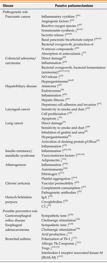
Table 2 Putative pathomechanisms of Helicobacter pylori
Disease Putative pathomechanisms
Pathogenetic role
Pancreatic cancer Inflammatory cytokine ↑[61]
Angiogenic factors ↑[61]
Reactive oxygen species ↑[61]
Somatostatin synthesis ↓[64,65]
Secretin release ↑[64,65]
Basal pancreatic bicarbonate output ↑[64,65]
Bacterial overgrowth, production of N-nitroso compounds ↑[66]
Absorption of antioxidants ↓[67]
Colorectal adenoma/
carcinoma
Direct damage[69]
Inflammation ↑[69]
Bacterial overgrowth, bacterial fermentation (ammonia)↑[69-71,74,75]
NO release ↑[76]
Hypergastrinemia[68,69]
Hepatobiliary disease Ammonia ↑[90]
Endotoxemia[90]
Inflammation ↑[90]
Hepatic fibrosis ↑[87]
Hepatoma cell adhesion and invasion ↑[91]
Laryngeal cancer Sensitivity to smoke and dust ↑[92]
Cell proliferation ↑[92]
Apoptosis ↓[92]
Lung cancer Direct damage[97]
Sensitivity to smoke and dust ↑[98]
Inhalation of gastrin and urea[95]
Hypergastrinemia[94]
Activation of docking protein p130cas[95]
Inflammation ↑[94]
Insulin resistance/
metabolic syndrome
Inflammation ↑[103,105]
Vasoconstrictor factors ↑[103,105]
Adiponectin ↓[104]
Atherogenesis Inflammation ↑[108]
Autoimmunity[108]
Fibrinogen ↑[112]
Platelet aggregation ↑[114]
Chronic urticaria Vascular permeability ↑[83]
Complement consumption ↑[83]
Pathogenetic antibodies ↑[83]
Henoch-Schönlein purpura
IgA ↑[82]
Cryoglobulins ↑[82]
C3 ↓[82]
Possible preventive role Gastroesophageal reflux disease
Sympathetic tone ↑[128]
Cholinergic stimulation[128]
Esophageal adenocarcinoma
Sympathetic tone ↑[128]
Cholinergic stimulation[128]
Acid production ↓[129]
Bronchial asthma Polarization of Th-1 ↓[131]
Allergic Th-2 response ↓[131]
Tregs ↓[132,133]
Interleukin-1 receptor associated kinase M (IRAK-M) ↑[133]
↑: Increase; ↓: Decrease.
mune complex formation induced by molecular mimicry and cross-reactive antibodies) have been suspected in the background[82,83].
With the exception of unexplained iron deficiency anemia (evidence level 1a) and idiopathic thrombocyto- penic purpura (evidence level 1b), H. pylori infection has no proven role in other extra-intestinal diseases[82,84,85]. Hepatobiliary diseases
Helicobacter DNA has been detected in hepatic tissues from patients with various hepatobiliary diseases, hepati- tis C virus-related chronic hepatitis, cirrhosis, and hepa- tocellular carcinoma (HCC)[86,87]. The association between H. pylori and Child-Pugh classification is inconsistent[87]. It can be proposed that H. pylori infection may play a role in hepatic carcinogenesis as well[88]. The odds ratio for the association between H. pylori infection and the risk of HCC was 13.63[89] (Table 2).
Respiratory tract disorders
Laryngeal cancer: Colonization of bacteria in the upper aerodigestive tract was confirmed, however the relation- ship between H. pylori infection and laryngeal cancer risk have produced conflicting results. Meta-analysis showed a 2.03-fold increased risk[92].
H. pylori was detected in larynx cancerous tissue. The presence of the cagA gene in larynx cancer tissues signifi- cantly decreased survival rate and increased the possibility of disease recurrence[93] (Table 2).
Lung cancer: The results of previous studies of H. py- lori seropositivity and lung cancer are inconclusive[94], with an odds ratio between 1.24 and 17.78 on the basis of the epidemiological studies[95]. The NHANES study observed an inverse association between H. pylori and lung cancer in older participants, with a significant inverse association for cagA+ strains; this was without histological examina- tion[96]. A case-control study found no evidence of an association between H. pylori and lung cancer in Finish male smokers. Neither overall H. pylori seropositivity nor CagA-specific H. pylori seropositivity were associated with lung cancer[94]. Causal relationships must be confirmed with exact determination of smoking status[95] (Table 2).
Insulin resistance and metabolic syndrome
Epidemiological studies showed significant associations with metabolic syndrome (OR = 1.39)[99,100]. Furthermore, multiple linear regression analysis showed that H. pylori seropositivity was significantly associated with higher systolic blood pressure, lower high-density lipoprotein (HDL)-cholesterol level, and higher low-density lipo- protein (LDL)-cholesterol level[99]. It has been suggested that H. pylori eradication could lead to an improvement of atherogenic blood lipid profile, insulin resistance, and low-grade inflammation, which were deduced from a de- creased C-reactive protein level[101]. Other studies did not find an association between H. pylori infection and insulin resistance[102,103].
The relationship between H. pylori infection and meta- bolic syndrome is both poorly understood[104] (Table 2) and controversial[106-108].
Cardiovascular diseases
Studies investigating the pathogenetic role of H. pylori in cardiovascular diseases have produced conflicting re- sults[108-111]. A meta-analysis of 18 epidemiological studies involving 10,000 patients did not find any positive associ- ation between H. pylori and cardiovascular risk factors and coronary heart diseases[112]. A higher prevalence of more virulent cagA+ H. pylori was reported in patients with ischemic heart disease, unstable angina, acute myocardial infarction, and restenosis after percutaneous transluminal coronary angioplasty and essential hypertension[108,111,113].
Evidence on the relationship between H. pylori infec- tion and ischemic heart disease is weak, with some incon- clusive, albeit plausible, mechanisms (Table 2). There are also no adequate interventional studies done to demon- strate that H. pylori eradication is associated with a lower incidence of ischemic heart disease[108].
Dermatological disorders
Chronic urticaria: A correlation between H. pylori infec- tion and chronic urticaria has been suggested (Table 2). H.
pylori eradication in patients with chronic urticaria leads to symptomatic improvement in some patients, while others showed no improvement[83].
Hematological disorders
Immune thrombocytopenic purpura: The prevalence of H. pylori infection in patients with immune thrombo- cytopenic purpura (ITP) is significantly higher than that in age- and gender-matched controls[108,115,116]. The most plausible mechanism is cross-mimicry involving H. py- lori, platelet antigens, and infected host factors (antibody production cross-reacts with platelet glycoprotein anti- gens)[108,117].
Eradication of H. pylori results in an increasing plate- let count in nearly half of infected ITP patients, although geographical differences in the efficacy of eradication were also presumed[83,115,116]. The European Helicobacter Study Group consensus in 2012 and the Second Asia-Pa- cific Consensus Guidelines have recommended H. pylori infection eradication in patients with chronic idiopathic thrombocytopenic purpura[84,85]. However, larger random- ized controlled trials with long-term follow-up are still required before a firm conclusion can be drawn[108]. Henoch-Schönlein purpura: A study in China found increasing evidence suggesting that Henoch-Schönlein purpura (HSP), especially abdominal HSP, might be asso- ciated with H. pylori infection (OR = 4.62); this underlines the necessity of screening H. pylori infection in children with HSP with gastrointestinal manifestations[82].
It was found that eradication of H. pylori infection resulted in prompt resolution of the HSP, or at least pre- vented its recurrence[118].
More investigations are needed to confirm the patho- genetic role of H. pylori in HSP (Table 2). HSP children with serious gastrointestinal symptoms must be screened and treated for H. pylori infection[82].
Iron deficiency anemia: Several epidemiological studies have shown lower ferritin levels among patients with H.
pylori infection, although there were studies that produced a negative association[108]. Meta-analyses showed an as- sociation between H. pylori infection and iron deficiency anemia (IDA)[119,120]. H. pylori eradication improves iron absorption[121].
Possible pathomechanisms are: increased iron loss due to active hemorrhage secondary to gastritis, peptic ulcer, gastric cancer, reduced iron absorption caused by achlorhydria induced by chronic pangastritis, reduced secretion of ascorbic acid to the gastric mucosa, and iron utilization for protein synthesis by the bacterium for colonization in the host environment[122]. Elevated serum prohepcidin might also indicate the role of inflammation in its aetiology[123].
Testing and eradication of H. pylori for unexplained IDA are supported by the current evidence and approved by the Maastricht Ⅳ Consensus and the Second Asia-Pa- cific Consensus Guidelines[84,85]. However, larger sample randomized controlled trials are necessary to clarify the reason why only a small proportion of H. pylori-positive patients develop IDA[108].
Possible beneficial clinical consequences of H. pylori infection
Gastroesophageal reflux disease/esophageal adeno- carcinoma: A meta-analysis showed that H. pylori infec- tion displays a negative association with the development of endoscopic gastroesophageal reflux disease (GORD).
Eradication of the infection may be a risk factor for de- velopment of de novo GORD[124].
H. pylori infection protects against gastroesophageal reflux[2]. H. pylori-induced corpus gastritis and profound suppression of gastric acid secretion have also been shown to prevent patients from developing GORD[125]. cagA+ H. pylori strains have a more protective effect against GORD[126], and it was found that H. pylori infec- tion was inversely associated with Barrett’s esophagus[127].
The Maastricht consensus Ⅳ confirmed a negative association between the prevalence of H. pylori and the severity of GORD. The consensus stated that H. pylori status exerts no effect on symptom severity, recurrence, or treatment efficacy in GORD. H. pylori eradication does not exacerbate pre-existing GORD or affect treatment efficacy[85].
Esophageal adenocarcinoma risk due to H. pylori in- fection was 0.58-fold, and squamous cell carcinoma risk was 0.80-fold compared with that of controls. Compared with cagA- H. pylori, cagA+ H. pylori markedly decreased esophageal cancer risk[129].
The underlying mechanism in the background of the protective effect of H. pylori against GORD is not fully
understood (Table 2).
H. pylori infection acts as neither a preventive factor nor a risk factor for squamous cell carcinoma. This dis- crepancy might be due to the relatively small number and heterogeneity of the included studies[129].
There is further need to assess the benefits of H. py- lori in connection with GORD and its complications.
Bronchial asthma: An infection in the early phase of life is essential for the normal maturation of the immune system, achieving a balance between T-helper type 1 (pro- tective immunity) and T-helper type 2 (allergic diseases) cytokine responses, which can reduce the risk of atopy later[129].
H. pylori infection might play a role in the develop- ment of chronic bronchitis, bronchiectasis, tuberculosis, and lung cancer[130]. Moreover, H. pylori might have an in- fluence on the developing immune system, which might reduce the risk of asthma in later life[131].
The associations between H. pylori and asthma were contradictory. Inverse associations were reported, but other studies demonstrated different results[131]. A meta- analysis found weak evidence (OR = 0.81, 0.84) for an inverse association between H. pylori infection and asthma in children and adults, respectively[131,132]. Another meta- analysis failed to prove a significant association between H. pylori infection and asthma risk[130].
The mechanism of the preventive effect of H. pylori on asthma has been unambiguous (Table 2).
It seemed that H. pylori infection (especially cagA+
strains) may prevent children from developing asthma, but must be studied in the future[131] due to the inconsis- tent result[134].
CONCLUSION
The clinical outcome of H. pylori infection is determined by host genetic predisposition, bacterial strain factors, and environmental factors[1]. Bacterial virulence factors (VacA, CagA) can modulate the immune response involved in the initiation of the carcinogenesis in the stomach. Host genetic factors including IL-1β, IL-10, and TNF-α influ- ence the inflammatory response and the exasperation of mucosal damage. Environmental factors, including salt intake and smoking tobacco, are well-known harmful aetiological factors. The ingestion of fruit and vegetables has some protective effect[135].
The mechanisms of H. pylori-associated gastric car- cinogenesis are still poorly defined; further recognition may provide possibilities to develop effective strategies for gastric cancer prevention and treatment[1].
Indications for H. pylori therapy have been extended and now include idiopathic thrombocytopenic purpura, iron deficiency anemia, and vitamin B12 deficiency. New data are presented on the role of H. pylori in neurodegen- erative disorders and in metabolic syndrome. H. pylori is associated with a small increase in the risk for colorectal adenoma and colon cancer[80] (Table 3).
H. pylori screening and treatment is a recommended gastric cancer risk reduction strategy in high-risk popula- tions. In low-risk populations for gastric cancer, H. pylori screening is not recommended[84]. The removal of H.
pylori from a large section of the population may be eco- nomically difficult, and the long-term consequences are still unpredictable. Identification of high-risk individuals is thus very important[40].
REFERENCES
1 Wen S, Moss SF. Helicobacter pylori virulence factors in gastric carcinogenesis. Cancer Lett 2009; 282: 1-8 [PMID:
19111390 DOI: 10.1016/j.canlet.2008.11.016]
2 Mishra S. Is Helicobacter pylori good or bad? Eur J Clin Microbiol Infect Dis 2013; 32: 301-304 [PMID: 23132690 DOI:
10.1007/s10096-012-1773-9]
3 Peek RM, Crabtree JE. Helicobacter infection and gastric neoplasia. J Pathol 2006; 208: 233-248 [PMID: 16362989 DOI:
10.1002/path.1868]
4 Molnar B, Galamb O, Sipos F, Leiszter K, Tulassay Z. Mo- lecular pathogenesis of Helicobacter pylori infection: the role of bacterial virulence factors. Dig Dis 2010; 28: 604-608 [PMID:
21088410 DOI: 10.1159/000320060]
5 Yamaoka Y. Pathogenesis of Helicobacter pylori-Related Gastroduodenal Diseases from Molecular Epidemiological Studies. Gastroenterol Res Pract 2012; 2012: 371503 [PMID:
22829807 DOI: 10.1155/2012/371503]
6 Wroblewski LE, Peek RM, Wilson KT. Helicobacter py- lori and gastric cancer: factors that modulate disease risk.
Clin Microbiol Rev 2010; 23: 713-739 [PMID: 20930071 DOI:
10.1128/CMR.00011-10]
7 Hatakeyama M. SagA of CagA in Helicobacter pylori patho- genesis. Curr Opin Microbiol 2008; 11: 30-37 [PMID: 18243773 DOI: 10.1016/j.mib.2007.12.003]
8 Ruggiero P. Helicobacter pylori infection: what’s new. Curr Opin Infect Dis 2012; 25: 337-344 [PMID: 22555448 DOI:
10.1097/QCO.0b013e3283531f7c]
9 Atherton JC, Cao P, Peek RM, Tummuru MK, Blaser MJ, Cover TL. Mosaicism in vacuolating cytotoxin alleles of Heli- cobacter pylori. Association of specific vacA types with cyto- toxin production and peptic ulceration. J Biol Chem 1995; 270:
17771-17777 [PMID: 7629077 DOI: 10.1074/jbc.270.30.17771]
10 Backert S, Naumann M. What a disorder: proinflammatory signaling pathways induced by Helicobacter pylori. Trends Microbiol 2010; 18: 479-486 [PMID: 20863705 DOI: 10.1016/
j.tim.2010.08.003]
11 Watanabe T, Asano N, Fichtner-Feigl S, Gorelick PL, Tsuji Y, Matsumoto Y, Chiba T, Fuss IJ, Kitani A, Strober W. NOD1 contributes to mouse host defense against Helicobacter py- lori via induction of type I IFN and activation of the ISGF3 signaling pathway. J Clin Invest 2010; 120: 1645-1662 [PMID:
20389019 DOI: 10.1172/JCI39481]
12 Yamaoka Y, Ojo O, Fujimoto S, Odenbreit S, Haas R, Gutier- rez O, El-Zimaity HM, Reddy R, Arnqvist A, Graham DY.
Helicobacter pylori outer membrane proteins and gastrodu- odenal disease. Gut 2006; 55: 775-781 [PMID: 16322107 DOI:
10.1136/gut.2005.083014]
13 Fujikawa A, Shirasaka D, Yamamoto S, Ota H, Yahiro K, Fukada M, Shintani T, Wada A, Aoyama N, Hirayama T, Fukamachi H, Noda M. Mice deficient in protein tyrosine phosphatase receptor type Z are resistant to gastric ulcer induction by VacA of Helicobacter pylori. Nat Genet 2003; 33:
375-381 [PMID: 12598897 DOI: 10.1038/ng1112]
14 Rhead JL, Letley DP, Mohammadi M, Hussein N, Mo- hagheghi MA, Eshagh Hosseini M, Atherton JC. A new Helicobacter pylori vacuolating cytotoxin determinant, the intermediate region, is associated with gastric cancer. Gastro- enterology 2007; 133: 926-936 [PMID: 17854597 DOI: 10.1053/
j.gastro.2007.06.056]
15 Ogiwara H, Graham DY, Yamaoka Y. vacA i-region subtyp- ing. Gastroenterology 2008; 134: 1267; author reply 1268 [PMID:
18395110 DOI: 10.1053/j.gastro.2007.11.062]
16 Ogiwara H, Sugimoto M, Ohno T, Vilaichone RK, Mahachai V, Graham DY, Yamaoka Y. Role of deletion located between the intermediate and middle regions of the Helicobacter pylori vacA gene in cases of gastroduodenal diseases. J Clin Microbiol 2009; 47: 3493-3500 [PMID: 19726606 DOI: 10.1128/
JCM.00887-09]
17 Dossumbekova A, Prinz C, Gerhard M, Brenner L, Backert S, Kusters JG, Schmid RM, Rad R. Helicobacter pylori outer membrane proteins and gastric inflammation. Gut 2006; 55:
1360-1361; author reply 1361 [PMID: 16905702]
18 Gerhard M, Lehn N, Neumayer N, Borén T, Rad R, Schepp W, Miehlke S, Classen M, Prinz C. Clinical relevance of the Helicobacter pylori gene for blood-group antigen-binding adhesin. Proc Natl Acad Sci USA 1999; 96: 12778-12783 [PMID:
10535999 DOI: 10.1073/pnas.96.22.12778]
19 Lin LL, Huang HC, Ogihara S, Wang JT, Wu MC, McNeil PL, Chen CN, Juan HF. Helicobacter pylori Disrupts Host Cell Membranes, Initiating a Repair Response and Cell Pro- liferation. Int J Mol Sci 2012; 13: 10176-10192 [PMID: 22949854 DOI: 10.3390/ijms130810176]
20 Suganuma M, Watanabe T, Yamaguchi K, Takahashi A, Fujiki H. Human gastric cancer development with TNF-α- inducing protein secreted from Helicobacter pylori. Cancer Lett 2012; 322: 133-138 [PMID: 22459353 DOI: 10.1016/
j.canlet.2012.03.027]
21 Ihan A, Pinchuk IV, Beswick EJ. Inflammation, immunity, and vaccines for Helicobacter pylori infection. Helicobacter 2012; 17 Suppl 1: 16-21 [PMID: 22958150 DOI: 10.1111/
j.1523-5378.2012.00977.x]
22 Müller A, Solnick JV. Inflammation, immunity, and vac- cine development for Helicobacter pylori. Helicobacter 2011; 16 Suppl 1: 26-32 [PMID: 21896082 DOI: 10.1111/
j.1523-5378.2011.00877.x]
23 Tsai HF, Hsu PN. Interplay between Helicobacter pylori and immune cells in immune pathogenesis of gastric inflam- Table 3 Other possible pathogenetic roles of Helicobacter
pylori[83,94,110,117,136,137]
Renal diseases
Renal resistive index, proteinuria Hepatobiliary diseases
Alcoholic damages of the liver, cholestatic autoimmune liver diseases (primary biliary diseases, primary sclerosing cholangitis),
cholelithiasis, cholangiocellular carcinoma Pancreatic disorders
Autoimmune pancreatitis Intestinal diseases
Enteric diseases, inflammatory bowel diseases Neurological diseases
Alzheimer-disease, idiopathic parkinsonism Dermatological diseases
Alopecia areata, atopic dermatitis, lichen planus, chronic prurigo multiformis, nodular prurigo, pruritus, psoriasis, recurrent aphthous stomatitis, rosacea, Sweet’s syndrome
Ophthalmological diseases
Glaucoma, central serous chorioretinopathy, uveitis, blepharitis Autoimmune disorders
Autoimmune thyroiditis, Behçet’s disease, Sjögren’s syndrome, progressive systemic sclerosis
Others
Impaired bioavailability of medication such as thyroxin and l-dopa, pre-eclampsia, chronic prostatitis, growth retardation
mation and mucosal pathology. Cell Mol Immunol 2010; 7:
255-259 [PMID: 20190789 DOI: 10.1038/cmi.2010.2]
24 Kabir S. The role of interleukin-17 in the Helicobacter pylori induced infection and immunity. Helicobacter 2011; 16: 1-8 [PMID: 21241406 DOI: 10.1111/j.1523-5378.2010.00812.x]
25 Malfertheiner P. The intriguing relationship of Helicobacter pylori infection and acid secretion in peptic ulcer disease and gastric cancer. Dig Dis 2011; 29: 459-464 [PMID: 22095010 DOI: 10.1159/000332213]
26 Sonnenberg A. Time trends of ulcer mortality in Europe.
Gastroenterology 2007; 132: 2320-2327 [PMID: 17570207 DOI:
10.1053/j.gastro.2007.03.108]
27 Zapata-Colindres JC, Zepeda-Gómez S, Montaño-Loza A, Vázquez-Ballesteros E, de Jesús Villalobos J, Valdovinos-An- draca F. The association of Helicobacter pylori infection and nonsteroidal anti-inflammatory drugs in peptic ulcer dis- ease. Can J Gastroenterol 2006; 20: 277-280 [PMID: 16609757]
28 Hagymási K, Tulassay Z. [Peptic ulcer: facts and questions -- 2010]. Orv Hetil 2010; 151: 1054-1061 [PMID: 20558352 DOI:
10.1556/OH.2010.28892]
29 de Martel C, Ferlay J, Franceschi S, Vignat J, Bray F, For- man D, Plummer M. Global burden of cancers attributable to infections in 2008: a review and synthetic analysis. Lancet Oncol 2012; 13: 607-615 [PMID: 22575588 DOI: 10.1016/
S1470-2045(12)70137-7]
30 Uemura N, Okamoto S, Yamamoto S, Matsumura N, Yama- guchi S, Yamakido M, Taniyama K, Sasaki N, Schlemper RJ.
Helicobacter pylori infection and the development of gastric cancer. N Engl J Med 2001; 345: 784-789 [PMID: 11556297 DOI: 10.1056/NEJMoa001999]
31 Venkateshwari A, Krishnaveni D, Venugopal S, Shashiku- mar P, Vidyasagar A, Jyothy A. Helicobacter pylori infection in relation to gastric cancer progression. Indian J Cancer 2011;
48: 94-98 [PMID: 21248438 DOI: 10.4103/0019-509X.75840]
32 Pandey R, Misra V, Misra SP, Dwivedi M, Kumar A, Tiwari BK. Helicobacter pylori and gastric cancer. Asian Pac J Cancer Prev 2010; 11: 583-588 [PMID: 21039020]
33 Hu Y, Fang JY, Xiao SD. Can the incidence of gastric cancer be reduced in the new century? J Dig Dis 2013; 14: 11-15 [PMID: 23134264 DOI: 10.1111/j.1751-2980.2012.00647.x]
34 Correa P, Piazuelo MB. The gastric precancerous cascade.
J Dig Dis 2012; 13: 2-9 [PMID: 22188910 DOI: 10.1111/
j.1751-2980.2011.00550.x]
35 Wang F, Meng W, Wang B, Qiao L. Helicobacter pylori- induced gastric inflammation and gastric cancer. Cancer Lett 2014; 345: 196-202 [PMID: 23981572 DOI: 10.1016/
j.canlet.2013.08.016]
36 Wong BC, Lam SK, Wong WM, Chen JS, Zheng TT, Feng RE, Lai KC, Hu WH, Yuen ST, Leung SY, Fong DY, Ho J, Ching CK, Chen JS; China Gastric Cancer Study Group.
Helicobacter pylori eradication to prevent gastric cancer in a high-risk region of China: a randomized controlled trial.
JAMA 2004; 291: 187-194 [PMID: 14722144 DOI: 10.1001/
jama.291.2.187]
37 Mera R, Fontham ET, Bravo LE, Bravo JC, Piazuelo MB, Ca- margo MC, Correa P. Long term follow up of patients treat- ed for Helicobacter pylori infection. Gut 2005; 54: 1536-1540 [PMID: 15985559 DOI: 10.1136/gut.2005.072009]
38 Farinati F, Cardin R, Della Libera G, Herszenyi L, Marafin C, Molari A, Plebani M, Rugge M, Naccarato R. The role of anti-oxidants in the chemoprevention of gastric cancer. Eur J Cancer Prev 1994; 3 Suppl 2: 93-97 [PMID: 7735055 DOI:
10.1097/00008469-199412002-00017]
39 Tulassay Z, Stolte M, Engstrand L, Butruk E, Malfertheiner P, Dítê P, Tchernev K, Wong BC, Gottlow M, Eklund S, Wrang- stadh M, Herszényi L, Nagy P. Twelve-month endoscopic and histological analysis following proton-pump inhibitor- based triple therapy in Helicobacter pylori-positive patients with gastric ulcers. Scand J Gastroenterol 2010; 45: 1048-1058 [PMID: 20509752 DOI: 10.3109/00365520903575737]
40 Ferreira AC, Isomoto H, Moriyama M, Fujioka T, Machado JC, Yamaoka Y. Helicobacter and gastric malignancies.
Helicobacter 2008; 13 Suppl 1: 28-34 [PMID: 18783519 DOI:
10.1111/j.1523-5378.2008.00633.x]
41 Asenjo LM, Gisbert JP. Prevalence of Helicobacter pylori infection in gastric MALT lymphoma: a systematic review.
Rev Esp Enferm Dig 2007; 99: 398-404 [PMID: 17973584 DOI:
10.4321/S1130-01082007000700006]
42 Kusters JG, van Vliet AH, Kuipers EJ. Pathogenesis of Heli- cobacter pylori infection. Clin Microbiol Rev 2006; 19: 449-490 [PMID: 16847081 DOI: 10.1128/CMR.00054-05]
43 Zullo A, Hassan C, Cristofari F, Andriani A, De Francesco V, Ierardi E, Tomao S, Stolte M, Morini S, Vaira D. Effects of Helicobacter pylori eradication on early stage gastric muco- sa-associated lymphoid tissue lymphoma. Clin Gastroenterol Hepatol 2010; 8: 105-110 [PMID: 19631287 DOI: 10.1016/
j.cgh.2009.07.017]
44 Wündisch T, Dieckhoff P, Greene B, Thiede C, Wilhelm C, Stolte M, Neubauer A. Second cancers and residual disease in patients treated for gastric mucosa-associated lymphoid tissue lymphoma by Helicobacter pylori eradication and fol- lowed for 10 years. Gastroenterology 2012; 143: 936-942; quiz e13-14 [PMID: 22750463 DOI: 10.1053/j.gastro.2012.06.035]
45 Singh K, Ghoshal UC. Causal role of Helicobacter pylori in- fection in gastric cancer: an Asian enigma. World J Gastroen- terol 2006; 12: 1346-1351 [PMID: 16552799 DOI: 10.3748/wjg.
v12.i9.1346]
46 Suzuki R, Shiota S, Yamaoka Y. Molecular epidemiology, population genetics, and pathogenic role of Helicobacter py- lori. Infect Genet Evol 2012; 12: 203-213 [PMID: 22197766 DOI:
10.1016/j.meegid.2011.12.002]
47 Hwang IR, Kodama T, Kikuchi S, Sakai K, Peterson LE, Gra- ham DY, Yamaoka Y. Effect of interleukin 1 polymorphisms on gastric mucosal interleukin 1beta production in Helico- bacter pylori infection. Gastroenterology 2002; 123: 1793-1803 [PMID: 12454835 DOI: 10.1053/gast.2002.37043]
48 Shanks AM, El-Omar EM. Helicobacter pylori infection, host genetics and gastric cancer. J Dig Dis 2009; 10: 157-164 [PMID: 19659782 DOI: 10.1111/j.1751-2980.2009.00380.x]
49 Machado JC, Figueiredo C, Canedo P, Pharoah P, Carv- alho R, Nabais S, Castro Alves C, Campos ML, Van Doorn LJ, Caldas C, Seruca R, Carneiro F, Sobrinho-Simões M.
A proinflammatory genetic profile increases the risk for chronic atrophic gastritis and gastric carcinoma. Gastroen- terology 2003; 125: 364-371 [PMID: 12891537 DOI: 10.1016/
S0016-5085(03)00899-0]
50 El-Omar EM, Rabkin CS, Gammon MD, Vaughan TL, Risch HA, Schoenberg JB, Stanford JL, Mayne ST, Goedert J, Blot WJ, Fraumeni JF, Chow WH. Increased risk of noncardia gastric cancer associated with proinflammatory cytokine gene polymorphisms. Gastroenterology 2003; 124: 1193-1201 [PMID: 12730860 DOI: 10.1016/S0016-5085(03)00157-4]
51 Nagini S. Carcinoma of the stomach: A review of epidemiol- ogy, pathogenesis, molecular genetics and chemoprevention.
World J Gastrointest Oncol 2012; 4: 156-169 [PMID: 22844547 DOI: 10.4251/wjgo.v4.i7.156]
52 Wang XQ, Terry PD, Yan H. Review of salt consumption and stomach cancer risk: epidemiological and biological evidence. World J Gastroenterol 2009; 15: 2204-2213 [PMID:
19437559 DOI: 10.3748/wjg.15.2204]
53 Loh JT, Torres VJ, Cover TL. Regulation of Helicobacter pylori cagA expression in response to salt. Cancer Res 2007;
67: 4709-4715 [PMID: 17510398 DOI: 10.1158/0008-5472.
CAN-06-4746]
54 Ek C, Whary MT, Ihrig M, Bravo LE, Correa P, Fox JG. Sero- logic evidence that ascaris and toxoplasma infections impact inflammatory responses to Helicobacter pylori in Colom- bians. Helicobacter 2012; 17: 107-115 [PMID: 22404440 DOI:
10.1111/j.1523-5378.2011.00916.x]
55 Tsugane S, Sasazuki S. Diet and the risk of gastric cancer:
![Figure 2 Targets of phosphorylated cytotoxin-associated gene A. Based on the article from Current Opinion in Microbiology, Hatakeyama M, SagA of CagA in Helicobacter pylori pathogenesis, 11, 30-37, Copyright (2008), with permission from Elsevier [7]](https://thumb-eu.123doks.com/thumbv2/9dokorg/1368068.111897/3.892.85.628.114.498/phosphorylated-associated-microbiology-hatakeyama-helicobacter-pathogenesis-copyright-permission.webp)