Predispositions and Interrelations in Insect Diseases
C. VAGO
Institut National de la Recherche Agronomique, Laboratoire de Cytopathologie, St. Christol-les-Ales (Gard), France
I. Introduction 339
II. Historical Aspects 340
III. Natural Predisposition 342
IV. Influence of Physiological Diseases 345 A. Effects on Physiological Diseases 345
B. Effects on Microbial Diseases 346
V. Influence of Microbial Diseases 358
A. Effects on Physiological Diseases 358
B. Effects on Microbial Diseases 362
VI. Simultaneous Diseases 365
VII. Conclusion 370
VII.
References 372
I. INTRODUCTION
In general pathology a tendency is developing to consider pathological affections in relation to the constitution of the host organism, the equi
librium of the host's biochemical and bioelectric "media," its defects, the chronic diseases it suffers, and to the physiological deviations to which it is prone. In medical epidemiology, initial consideration is given to periods of receptivity to infectious disease as caused by nutritional deficiencies or climatic influences. In clinical medicine close attention is given to the organs of the "least-resistance" characteristic for each patient and which thus determine the evolution of any disease.
In the relatively young science of invertebrate pathology, and espe
cially in insect pathology, these notions are not yet of common use, and 339
one may say without exaggeration that the related problems have just been recognized. In fact, most syntheses and also detailed papers tend, in describing a disease, to ignore the factors liable to disturb the clarity of its characteristics. At the present time, the consensus is that, in the world of invertebrates also, the physiological and pathological elements can mutually influence each other; therefore it would be highly profit
able to consider the processes observed in insect pathology on the basis of the physiological-pathological phenomena involved.
This chapter will be devoted to the aspect of the subject just in
dicated. An effort will be made to present data and considerations likely to contribute to the characterization of an insect pathology constituted not of isolated affections, but of disease interactions.
I I . HISTORICAL ASPECTS
It seems rather paradoxical to note that in the history of insect pathology the acknowledgment of predisposing phenomena largely pre
ceded the etiological description of disease. Books of the eighteenth and early nineteenth centuries stressed the ecological-physiological fac
tors and indicated these as causes of the diseases, just as was the case in the old medical works. Thus, de Serres (1599), Andreoni (1756), Cas- tellet (1760) Boissier de Sauvages (1763) Ouekaki Morikouni (1802), and Nysten (1808) designated excessive overheating of the respiratory and digestive organs as causes of "luzette and moris-flats" disease in the silkworm. Schirach (1771) attributed foulbrood in the honey bee to a predisposition linked wTith the physiology of the egg; Fisher (1871) does the same with defective food.
This situation was caused by the lack of microbiological knowledge at these early periods. Therefore, the discovery of the action of micro
organisms in pathogenesis is considered as the revolution in pathology.
In 1835, Bassi's demonstration of the connection between the fungus Beauveria bassiana (Balsamo) Vuillemin and silkworm muscardine was certainly the first acknowledgment of this action in insect pathology and probably also in general pathology.
T h e microbe-disease relation soon became the chief concern of the pathologists in human medicine as well as in the invertebrate field.
Descriptions of pathological affections, including the isolation, determi
nation, and re-injecting of pathogenic microorganism according to Koch's postulates, became more and more common.
For a long time this conception prevailed in general pathology, but gradually, with Claude Bernard, investigators became aware of the in
fluence of the "milieu" on the development of infections. T h e old
observations on the "external causes" of diseases were then examined as determining factors of this "milieu." However, while in human or in veterinary medicine this conception prevails in the view of many pathologists, in insect pathology we find few traces of it, until recently.
In fact, during the evolution of insect pathology, when descriptions of microbial infections were numerous, there were only isolated observa
tions or hints on the influence of physiological conditions upon the process of diseases. Pasteur (1870) observed that the development of the bacteria in silkworm "flacherie" was favored by disorders in diges
tion caused by the motionless condition of warm air.
Conte and Levrat (1906) spoke about conditions favoring grasserie, Glaser (1915) noted the effect of sun radiation on the transition of the polyhedrosis of Porthetria dispar (Linnaeus) from a chronic to an active state. White (1920) differentiated, in his description of foulbrood of the honey bee, the "predisposing causes" and the "exciting causes," the latter being the pathogenic microorganisms.
Verson (1906) and Acqua (1918), having experimented in sericul
ture for a long time, considered the intestinal bacteria, described by Pasteur (1870) and later on by Paillot (1926, 1928) as pathogenic agents of silkworm flacherie, to be but common consequences of unde
fined physiological weakenings. T h e Japanese school looked for a com
promise between the two theses: it considered both the weakening of the host and the development of intestinal bacteria (coming from the leaves) to be necessary for the starting of flacherie. On the same subject, Paillot (1930) considered the participation of a nucleotrope virus which distorts the cell of the intestinal epithelium. He believed this primary distortion to be necessary for the subsequent development of the bac
terium Streptococcus bombycis Flügge. Here we meet an instance of
"milieu" preparation, but the action of the virus has not been confirmed and the etiology of this disease has been explained in different ways by some investigators.
Observations can be found in papers concerning various diseases:
Vago (1954) recalled the linking between a virus disease and a bacterial septicemia in Lepidoptera, Isakova (1954) pointed out the simultaneous
effect of several bacteria, Vago and Cayrol (1955) showed how various physiological influences favor nuclear-virus diseases. However, patho
logical predisposition and interaction had not yet become widespread notions in insect pathology. T h e early general works on this science, such as the ones by Paillot (1933), Masera (1936), and Steinhaus (1946, 1949), dealt many of the aspects of the field, but they did not include special chapters on predisposition.
In synthetic work, Vago (1956a) developed the idea of interaction
of diseases and predisposition in insects. He showed the linking mecha
nism of several pathological complexes with primary, secondary, or mul
tiple injuries, and he tried to explain the chief factors likely to interfere in the interactions. T h e preparation of the "milieu" by such physiologi
cal actions as traumata, intoxication, alimentary disorders, or by means of microbial agents, such as virus diseases, chronic nosemoses, and light bacterial diseases was investigated experimentally. Recently, Steinhaus (1960) emphasized the significance of predisposing causes and the im
portance of the environment (including certain stressors) in the path
ogen-host-environment triad involved in the diseases of insects.
T h e action of virus diseases as predisposing factors of nonvirus affec
tions was shown by \^ago (1956b). Telenga (1956) tried to associate fungus diseases to insecticide treatments in the control of insects. More and more frequently instances of disease interaction are reported. Thus, in the scope of studies on Melolontha melolontha (Linnaeus), Hurpin and Vago (1958) devoted a chapter to the linking of bacterial diseases, physiological factors, wounds, and mycoses. Steinhaus (1958a) reported a number of unfavorable ecological influences, i.e., stressors, able to increase the mortality rate in virus or bacterial diseases.
Examples of double infections are more and more frequent. Bird (1959), Tanada (1959b) and others have studied the coexistence of poly
hedroses and granuloses in the same lepidopterous larva; Vago (1959) re
ported on an intestinal virus disease and granulosis; and Jolly (1959) and Müller-Kögler and Huger (1960) reported new instances of mycoses being favored by previous wounds.
Finally, a general view on the problem of predisposition and interac
tions in insect diseases was given in the field of comparative pathology by Vago (1958). T h e various notions, the definitions, and the interaction mechanisms were compared with the same elements in human and verte
brate pathology.
I I I . NATURAL PREDISPOSITION
T h e most usual, and also the least noticeable, form of predisposition is linked to the constitution, the characteristics, and the physiological func
tions of a species, a line, or a particular individual. This phenomenon, which is sometimes considered to be the opposite to natural immunity (Courmont, 1919) is called "sensibility" to a disease, and there are many ways of ascertaining and measuring this sensibility with regard to viroses, bacterioses, etc. This sensibility is frequently associated with the so-called
"specificity" of a germ, although the real meaning of the word is not sufficiently denned.
However, the analysis of a certain number of cases shows that sensi
bility is only partly connected with the chemical functions of micro
organisms or the manner in which the latter multiply, develop, germi
nate, or resist. In any particular insect the ground may be more propi
tious for the development of one germ than it is for another. T h e particu
lar insect involved may produce enzymes that dissolve a toxin, while in other insects this may not be so; or, a given insect may have a weak spot in a membrane through which the microbes may penetrate, while in other species the membrane is impermeable.
Insect breeders have commonly noticed that some individuals of a particular lot are far more prone to a disease than others. Frequently cases have been described showing that even if some individuals of an infected lot fall ill, the overall result still remains negative. Here again, excluding the part played by the more-or-less varying element of im
munity, there are also predisposing factors connected with the consti
tution or physiology of the receptive individuals that render them more susceptible to the disease.
These phenomena are so little known in the case of insects that it would be premature to think of analysing these factors separately. W e are, therefore, only choosing a few examples among the fragmentary observations made, so as to show the different aspects of predisposition to disease which may have some connection with the normal constitu
tion of an insect.
Sensibility to bacteriosis or virosis has often been connected with the age of the insect. Woodrow (1941) noticed it in the case of the honey bee, Apis mellifera Linnaeus, contaminated with American foul
brood. Of the sick larvae, 44.1 percent were under 24 hours old, 4.8 per
cent were 1 to 2 days old, and 0 percent were 2 to 4 days old. However, in the case of some Lepidoptera, Protoparce sexta (Johanssen) for in
stance, White (1923) noted that the older larvae were more sensitive to Bacillus sphingidis White ( = Serratia marcescens Bizio) than the younger ones.
T h e numerous observations made by silkworm breeders regarding grasserie, and the recent facts noted by Tanada (1956) and Tanada and Chang (1960) in the case of a granulosis and nuclear and cytoplasmic polyhedroses in Pseudaletia unipuncta (Haworth), emphasize the sen
sibility of young larvae to the viruses in question.
In protozoan infections an attempt has been made to explain why older bees are subject to the infection caused by Nosema apis Zander:
it would appear that, owing to the more frequent renewal of the intesti
nal epithelium in young insects than in older ones, the cells infected by
Nosema are frequently eliminated in the younger ones (Toumanoff, 1951).
T h e physiological causes in insect metamorphosis may also be favor
able to the development of certain diseases. In particular, according to Vago (1951a), it is during molting periods that Lepidoptera are most susceptible to polyhedroses. Aruga and Arai (1959) noted that, in Bom
byx mori (Linnaeus), activation of both nuclear and cytoplasmic poly
hedroses by cold shock occurs most easily 6 to 98 hours after molting;
between molts sensibility is, on the contrary, much reduced. Further
more, not all molts respond equally to virosis activation treatments.
At the first and second molts results are less marked, but at the fourth they are at their best. For activation by heat shock, virosis appears only after molts (Hukuhara and Aruga, 1959).
On the other hand, Bucher (1959) noted that different species of intestinal bacteria penetrate more easily into the hemocoel of crickets during molts and cause septicemia if they are of a proteolytic species.
T h e considerable changes that occur in insects during the pupal molt and during the pupal stage undoubtedly have an effect on the evolution of diseases. With regard to modern studies it should be mentioned that Bird (1953) noted that intestinal nuclear polyhedrosis stopped develop
ing at the prepupal stage in Diprion hercyniae (Hartig). This seems to be due to the fact that the alimentary canal is completely renewed by young cells; later, when the cells of the pupa become older, virosis reap
pears.
It is also probable that some pathogens cease to multiply during the diapause of the host insect. This supposition has not been proved experi
mentally for viroses, but it has been observed by Paillot (1938) in Nose
ma carpocapsae Paillot and Nosema bombycis Nägeli.
In several cases the greater or lesser alkalinity of the intestinal con
tent seems to be a predisposing factor for some bacterial diseases. Thus, in the case of a comparatively low pH content the lecithinase of Bacillus cereus Frankland and Frankland acts on the hymenopteran Neodiprion and on the lepidopteran Carpocapsa pomonella (Linnaeus), but this type of intoxication cannot occur in those species, in particular among Lepidoptera, with an extremely alkaline intestinal content (Heimpel,
1955). T h e alkalinity of the alimentary canal, with the addition of cer
tain enzymes is one of the factors that causes an insect to be subject to toxemia or septicemia by different varieties of Bacillus thuringiensis Berliner. In fact, the crystallized toxin of the bacillus has to be dissolved by these elements (that exist only in some insects) before the active principle, which enters into the hemolymph, is freed (Hannay, 1953;
Hannay and Fitz-James, 1955; Angus, 1956; Martouret, 1960).
IV. INFLUENCE OF PHYSIOLOGICAL DISEASES
For a long time it has been recognized that factors, other than mi
crobes, connected either with the habitat of the insect or with the func
tioning of its organs, cause organic disturbances. In medicine and veter
inary pathology, these so-called physiological disorders hold as important a place as microbial diseases. Many illnesses are caused exclusively by functional disturbances attributable to physical or chemical agents, and frequently these disorders are known to favor other diseases (Claude Bernard, Selye).
In insect pathology it is not feasible to define the physiopathological syndromes by chemical analysis of the blood or the tissues, by the rhythm of excretion, by electrocardiagrams, or by radiography, reflexes, etc. So it is natural that we should not have many details on the effects of physiological diseases on other diseases. In most cases the physiopatho
logical disorder which paves the way for the development of a disease is insufficiently known or is unknown, and often there is confusion between the really predisposing physiological condition, the ecological factor ("stressor"), and the effect of the ecological factor (Steinhaus, 1958, 1960).
A. Effects on Physiological Diseases
With regard to vertebrates it is known that physiological diseases may develop other physiological processes through a chain of lesions.
Thus a traumatism, for instance, may lead to glandular alteration; a disturbance in the functioning of the glands may lead to an increase or a decrease of a particular substance in the blood; this anomaly may be the cause of disturbances in the nervous system, and so forth. It is ob
vious that when dealing with insects such a sequence cannot be defined, since in that field even simple physiopathological cases have been poorly studied.
Thanks to the work carried out in detail on the diseases of the queen honey bee, Apis mellifera, it was possible to make some observations on the sequence of physiopathological processes. According to Arnhart (1929), when the queen suddenly ceases to lay eggs, this may be due to a chain of disturbances starting with a temporary chill which causes a disease called "weakness of the rectum." T h e feces penetrate into the genital orifice and block it. Fyg (1934) describes another physiological process which ends in a cessation of egg laying. Owing to a congenital malforma
tion of the ovary, the spermatozoa remain outside the spermatheca, cannot find an outlet, and thus block the oviduct.
A type of predisposition caused by a physiopathological process con
nected with respiration is indicated by Vago and Vasiljevic (1961). I f
certain stigmata of lepidopterous larvae are artificially blocked, the insect stops eating or evacuating its feces and a particular part of the body becomes paralyzed or at least is less sensitive to touch or even to a burn. This shows successive alterations in the digestive, nervous, and muscular systems.
In the case of insects, poisoning often causes anomalies which appear only after several molts. For instance, Vansell and Watkins (1933) indicated that the slight poisoning acquired in the larval stage by the ingestion of substances collected on the horse chestnut tree (Aesculus sp.) gave rise to malformations in the adult A. mellifera. T h e same au
thors pointed out that a similar poisoning, at a later stage, may gradually reduce considerably the fecundity of the queen. However, these conse
quences, especially when the poisoning is due to chemicals, pertain to toxicology and can be developed there in detail. It should be noted, however, that in practice little attention has been given in insect toxi
cology to the ultimate consequences of the ingestion of very small doses of poison. W e feel that this deserves a more thorough examination.
B. Effects on Microbial Diseases
In insect pathology undoubtedly most observations made in the area under discussion concern predisposition to microbial diseases. This is understandable since the causes of predisposition mentioned by authors at the beginning of the century meant the conditions under which the insects were kept; the oldest observations related, in fact, to the influence of breeding methods on the development of bee and silkworm diseases.
1. Poisoning
T h e effect of poisoning on the development of microbial diseases is certainly far greater than is shown by the limited data at our disposal.
It was observed (Yamafuji and Yuki, 1947; Yamafuji, 1952; Yamafuji, et al., 1952) that feeding of chemicals such as hydroxylamine or nitrites increases the frequency of virosis in Bombyx mori. Veneroso (1934) noted polyhedral virosis in silkworms in an area where leaves were pol
luted by fluorine emanations from a factory.
Experimentally, Vago (1951a, 1953) with the use of sodium fluoride, showed that the larvae that had only absorbed sublethal doses were subject to slight poisoning that activated, in some races and lines con
taining occult virus, a nuclear polyhedrosis. Krieg (1955) confirmed these results on Bombyx mori, and also (Krieg, 1956) by using sodium fluoride, hydroxylamine, thioglycolic acid, hydrazine, and potassium ni
trite on larvae of Neodiprion sertifer (Geoffroy). However, Wallis (1957)
did not obtain nuclear polyhedrosis in Porthetria dispar (Linnaeus) by using hydroxylamine and potassium nitrite, nor Steinhaus (1958a) granulosis and polyhedrosis in Junonia and Peridroma with different chemicals.
Contact insecticides seem also to have some effect on mortality through polyhedrosis. Kovacevic (1958a,b) noted nuclear virosis in Porthetria dispar and Euproctis chrysorrhoea (Linnaeus), and granulosis in Hyphantria cunea (Drury) in forest lots where insecticide spraying was not completely lethal. In addition, he observed an invasion of dif
ferent kinds of bacteria which he ascribed to "physiological weakening."
It would be useful to analyze this weakening since contact insecticides act in a selective way on tissues. T h e variations in mortality according to the origin of the larvae also show the complexity of the interpretation of the phenomena observed.
Slight poisoning causing cellular lesions in the blood or in the alimentary tract can also favor bacterial diseases. Vansell (1926) is of the opinion that "diarrhea" of the honey bee, Apis mellifera, may be caused by slight poisoning due to the nectar of Aesculus sp. A disturb
ance in the transit of the food engenders a secondary bacterial intestinal fermentation. Vago (1956a) mentions secondary bacterial Streptococcus
(Enterococcus) dysentery and Serratia marcescens Bizio septicemia in Lepidoptera (Hyponomeuta padella (Linnaeus) and Spilosoma sp.) that were slightly poisoned by arsenate. On the other hand Steinhaus
(1958a) did not obtain an increased susceptibility to Bacillus thuringien
sis Berliner by feeding hydroxylamine to Junonia and Peridroma larvae.
Stephens (1959a) injected mucin into crickets either before or at the same time as injections of Pseudomonas aeruginosa (Schröter) Migula and revealed an increase in mortality. However, mucin cannot be con
sidered to be poisonous and one does not yet know whether it acts on the permeability of the cells, whether it reduces defense reactions, or whether it activates bacterial multiplication. Experiments (Steinhaus,
1959) on intensifying infections of Serratia marcescens by forced feeding the larvae of Galleria mellonella (Linnaeus), Tenebrio molitor Linnaeus, Bombyx mori, Colias eurytheme Boisduval, Peridroma margaritosa (Ha- worth), Pseudaletia unipuncta (Haworth) with diluted acetic acid (0.06 to 0.03 N) gave varying results, the mortality increase being insignificant.
Ingestion of glycerin mentioned by the same author (Steinhaus, 1959) also gave negative results. Steinhaus and Dineen (1960) exposed larvae of P. margaritosa to ether vapor, but only noted the usual effects of too strong a dose of ether. Occasionally there appeared to be an increase in deaths caused by granulosis in virus-fed insects exposed to ether, but such was not consistently the case.
With regard to mycoses, Telenga (1956, 1958) observed increased cases of muscardine caused by Beauveria bassiana and Metarrhizium anisopliae (Metchnikoff) Sorokin when a beet-root weevil was slightly poisoned by hexachlorocyclohexane. Later this author (Telenga, 1957, 1958) suggested, for biological control purposes, the concomitant use of fungi and small doses of contact insecticides, such as HCH or D D T .
2. Heat, Cold, and Humidity Injury
Disorders caused by heat, cold, and humidity are physiological diseases, the nature of which is difficult to define. Even in medical or veterinary pathology the mechanism of action of abnormal temperature and humidity is not exactly known, and we are aware that under this heading we are collecting a heterogeneous number of facts (See also Chapter 2 ) . If, for instance, great heat can be called "stressor," undoubt
edly a secondary disease will be manifest in a different manner according to whether it will affect the alimentary canal, respiratory system, or other organs. T h e impossibility of defining the nature of the effects of these factors is all the more regrettable since most of the older papers concerning insect diseases deal precisely with predispositions caused by excessive heat or humidity. T h e "flacherie" of silkworms is the best- known case. T h e eighteenth century authors (Andreoni, 1756; Casteilet, 1760; Boissier de Sauvages, 1763) believed that this disease was caused by high humidity accompanied by excessive heat. Nysten (1808) is the first to have analyzed the development of this disease by carrying out experiments remarkable for the century in which he lived, and he con
cluded that heat, associated with high humidity, creates a pathological state called "touffe," which affects particularly the respiratory system.
Later, Pasteur (1870) discovered the role played by intestinal bacteria, but his experiments revealed two phases, the first being the "touffe,"
the second the subsequent development of intestinal bacteria. This assumption was criticized by Verson (1906) and Acqua (1918), who contested the part played by bacteria, and was modified by Paillot (1930), who minimized the importance of the predisposition phase.
Recent research (Vago, 1960) has divided "flacherie" into different etiological types and has proved experimentally that one of these is in fact connected with respiratory troubles due to heat and humidity shocks, followed during the secondary phase by the temporary slackening of the functions of the alimentary canal. This leads to a third phase, involving the development of different intestinal bacteria, in particular enterococci (Streptococcus bombycis Flügge).
Exposure to high or low temperature does not necessarily cause alterations that might favor microbial diseases; however, White (1923)
found that such was the case with regard to bacterial septicemia in Protoparce sexta, and Burnside (1935) noted the pathogenic effect of Mucor hiemalis Wehmer, normally saprophytic, when young bees had been exposed to the relatively cold temperature of 20°C. Steinhaus
(1958a) also obtained accelerated effect of Bacillus thuringiensis, in particular on Sabulodes, Colias, and even on Peridroma (usually not very sensitive), but he (Steinhaus, 1959) was able only exceptionally to increase the death rate caused by Serratia marcescens in Colias, Sabulodes, Junonia, Galleria, or Tenebrio larvae exposed to tempera
tures of 40°C. or 2°C. before or during an infecting meal. At times there was less infection at 40° than at 37°, 33° or 25°C. He suggested that one explanation of these results might be the fact that most strains of 5. marcescens do not grow at temperatures above 37°, and grow best be
tween 25° and 30°C. Maintaining the temperature at 40°C. often caused mortality due to invasion by bacteria normally in the gut of the insects.
Vago (1956a) came to similar conclusions in Bombyx mori larvae acting as hosts to enterococci.
Quite a different phenomenon may take place when the thermal shock occurs during a chronic infection. In such an instance Vago (1956a) states that, in Bombyx mori infected with very small doses of Bacillus cereus (thuringiensis) alesti Toumanoff and Vago, the thermal shock, even if it is of short duration and reaches only 30°C, causes an acute and rapidly fatal bacteriosis.
With regard to thermal disorders predisposing to virosis, quite a considerable amount of information, sometimes collected in early years, is available. However, care should be taken not to mistake an acceleration of the disease for a predisposing factor. T h e ideas held formerly by silkworm breeders on the development of polyhedrosis (grasserie) when breeding was carried out at high temperatures (25 to 27°C) mentioned by several experts (Acqua, 1930), or in Lymantria monacha (Linnaeus) larvae during hot weather (Escherich and Miya- jima, 1911), are based on summary observations. However, it is parti
cularly interesting that high and low temperatures can provoke a patho
logical state inducing virosis. Although the mechanism of this type of predisposition is not clear, the evolution of polyhedrosis is known to occur in different lepidopterous larvae which have been maintained at a temperature of 2 to 4°C during 48 hours immediately after molting (Suzuki, 1941; Yamafuji and Omura, 1950; Ishimori, 1951; Osawa, 1951;
Ooba, 1956). However, Steinhaus and Dineen (1960) obtained only slight indications that they were able to activate granulosis in Peridroma margaritosa by keeping it at a temperature of 3 to 8°C. They obtained data indicating that low temperatures may be somewhat more effective
in promoting granulosis in older than in younger larvae. Neither were they able to obtain significant positive results by exposing the same insect to a high temperature ( 3 7 ° C ) .
It seems probable that the immediate result of thermal shock is a cellular metabolic deviation that activates the multiplication of viruses (provirus) which may exist in an occult state in the cell (Vago, 1951a).
T h e theory held by Acqua (1930) and by Yamafuji and Kosa (1944) con
cerning the artificial or de novo production of viruses has few supporters.
Even temporary chilling of the brood of the honey bee, Apis mel
lifera, may lead, as a secondary consequence, to the progressive develop
ment of ordinary bacteria, probably owing to physiological alteration processes that are as yet unknown. Bee keepers are well aware of this syndrome called "cold brood." Furthermore, Toumanoff (1951) is of the opinion that a chill in the case of brood is followed by physiological weakening that may help microsporidia (Nosema apis) to spread in the colony.
If an insect continues to live for some time in an unaccustomed degree of humidity, it may be subject to physiological disturbances, the characteristics of which are still far from being defined. This trouble may later on have some influence on microbial infections, but it is necessary to point out that quicker germination of pathogenic fungi due to a high degree of humidity cannot be considered a predisposition phenomenon since here humidity acts on the pathogen itself and does not create a receptive state for the insect.
In the case of virosis it has been noticed that, under natural condi
tions, damp weather leads to increased polyhedrosis in L. monacha and P. dispar (Wahl, 1910, 1911, 1912; Glaser, 1915; Wellenstein, 1942).
Increased mortality due to nuclear polyhedrosis has been noted by Vago (1951a) and by Vago and Cayrol (1955) in Plusia gamma (Lin
naeus) subjected to a humidity of 95 percent, and in P. dispar under both natural and experimental conditions of high humidity. Here again the mechanism of the virosis activation may depend on metabolic disor
ders caused, through faulty respiration, by excessive humidity.
In the case of bacterial diseases the infection caused by Serratia marcescens did not show any increase in Galleria or Peridroma larvae exposed to a humidity of 100 percent (Steinhaus, 1959). T h e same author (1958b) points out that diseases of various kinds appear, and the death rate increases, when Bombyx mori, Peridroma margaritosa, Colias eurytheme larvae, etc. are crowded together in a small space. Although the experiments were conducted under controlled conditions, it was difficult to dissociate humidity and other factors that may be involved.
3. Irradiation Injury
Physiological disorders caused by irradiation have been little studied in insects with respect to predisposing them to microbial diseases (see Chapter 2 ) . Echerich and Miyajima (1911) consider that exposing larvae of L. monacha to the direct rays of the sun increases the cases of nuclear polyhedrosis. Vago (1951a) noted similar occurrences in Bombyx mori, and Smirnoff (1961) did the same in Neodiprion swainei Middleton suffering from an intestinal nuclear polyhedrosis. It would be interesting to distinguish the effects of the gradual "warming up," from those of rays of different wavelengths radiated by the sun. Steinhaus and Dineen (1960) did not obtain any significant positive results by exposing Peridroma larvae to ultraviolet radiation (2537 Ä).
Karpov (1959) noted an increased death rate due to nuclear poly
hedrosis in Bombyx mori exposed to X rays. T h e effects depended on the intensity of the irradiation, and strong doses merely caused a serious physiological disorder. For a chain reaction, feeble doses of 5000 to 6000 r seem the most efficacious. Krieg (1956) was able to increase the amount of polyhedrosis in Neodiprion sertifer after a 20- to 30-minute irradiation with ultraviolet rays. In both instances it is difficult to ascertain the mechanism; probably primary alterations predisposing to virosis are of a metabolic nature similar to the chemical activation of virosis. With regard to this there are, however, contradictory data. Aruga (1958), for example, was unable to produce nuclear or cytoplasmic polyehedra by submitting B. mori larvae to X rays or ultraviolet radiation.
4. Traumatism
T h e physicophysiological disorders known as traumata have been recently studied in regard to predisposing to microbial diseases. T h i s applies mainly to wounds. Some of the observations made in former years are noted although the account given is brief and provides no analysis. (See also Chapter 2.) Snow (1896) noted that wounded adult Blissus leucopterus (Say) were more receptive to Beauveria globulifera
(Spegazzini) Picard. According to Northrup (1914) the development of Micrococcus nigrofaciens Northrup in Lachnosterna was hastened by traumatism. Stutzer and Wsorow (1927) introduced Pseudomonas septica Bergey into Euxoa larvae, and Burnside (1930) Penicillium sp. into the honey bee by a wound in the skin. T a u b e r (1940) stated the opinion that Staphylococcus aureus Rosenbach penetrates more easily into Blatt a orientalis Linnaeus if the integument is damaged.
An experimental study was carried out by Vago (1956a) concerning the relationship between wounds and microbial infections considered as
"pathological complexes" having a physiological primary phase and
an infectious secondary phase that are closely linked. He follows the cytological aspects of the skin wound process and its healing, emphasizing, because of the importance of subsequent developments, that blood clots form on the surface of the wound and that there is a rapid accumula
tion of hemocytes which seals the wound hermetically. T h e clot is, in fact, recognized to be the substratum for the saprophytic development of several microorganisms, which may be the starting point of a progres
sive internal infection or of an intoxication. On the other hand, the
FIG. 1. Aspergillus flavus-infected wound on Antheraea pernyi larva. (Original.)
"pseudotissue" formed by the hemocytes is a barrier that can be crossed only by microorganisms having some pathogenicity, mainly with enzy
matic activity. Wounds accelerate the infections caused by normally pathogenic organisms. However, fungi that are not very pathogenic show the real importance of predisposition brought about through wounds. Thus several strains of Aspergillus, Fusarium, Penicillium, and Rhizopus have an effect on Bombyx mori, on Melolontha melolontha and Rhizotrogus solstitialis (Linnaeus). In such cases there is first a somewhat saprophytic development on the clot covering the wound. T h e essentially saprophytic species, such as Penicillium glaucum Link, have little effect, but others, such as Rhizopus niger Ciaglinski and Hewelke
or Fusarium coeruleum (Libert) Saccardo and some Aspergillus, gradu
ally penetrate into the general cavity, probably through toxic action and by mycelial invasion, causing serious disease. (See Figs. 1 and 2.)
Jolly (1959) confirms these results in the case of the wound parasi
tism of the fungus Trichotheciurn roseum Link, which has no effect on Bombyx mori per os. Müller-Kögler and Huger (1960) describe a similar phenomenon concerning the effect, through a wound, of Penicillium brevi-compacium Dierckx on Malacosoma neustria (Linnaeus). Recently
FIG. 2. Development (A) of Aspergillus tamarii in a hemolymph clot on a wound (W) in Ceratitis capitata pupa. Pupa is poisoned by the fungus. Iron hematoxylin- acid fuchsin-sulfogreen. (Original.)
strong pathogenic action of Mucor hiemalis was observed after wound infection in larvae of Melolontha melolontha, Amphimallon majalis
(Razoumowsky), Mamestra brassicae (Linnaeus), Galleria mellonella, and Bombyx mori. T h e same fungus had no effect by feeding or by contact (Heitor, 1961).
Wounds can also help the entrance of bacteria and viruses. Vago (1956a) shows this with the effect of Bacterium paracoli (Gilbert and Lion), Pseudomonas sepfica, Serratia marcescens, and Bacillus megat- erium de Bary on Bombyx mori and on Sesamia nonagrioides Lefebvre,
and also to a certain extent in the case of the nuclear polyhedrosis virus affecting B. mori. Recently Doane (1960) pointed out that bites in the skin of the coleopteran Scolytus multistriatus (Marsham) are often re
sponsible for infections caused by Aerobacter scolyti Pesson et al., by Escherichia kleb siellae for mis Pesson et al., and by Serratia marcescens.
T h e part played by the healing of the wound with regard to favoring infection should not be forgotten. In the case of fungi parasitizing wounds, the formation of the hemolymph coagulum is propitious, but many bacterial infections seem to be favored by an open wound (Vago, 1956a).
Similar wounds may also appear in the alimentary canal and may be caused, experimentally, by abrasives or by introduction of a needle
(Weiser and Lysenko, 1956). Steinhaus (1958a) noted that Serratia marcescens septicemia appeared in 50 percent of the cases when the infected food given to B. mori larvae had been mixed with powdered glass, and in only 20 percent when the bacteria had been ingested alone.
However, in another study, he (Steinhaus, 1959) noted no increase in the infection caused by S. marcescens in Galleria, Junonia, Colias, Peri
droma, and others, after being force-fed with powdered glass or silicon carbide.
Vago and Chastang (1961) describe a Mucor racemosus Fresenius infection in B. mori which leads to considerable thickening of the mid
gut wall, very similar in appearance to a pseudotumor. This mycosis was noted chiefly after the larvae had been fed on thickly furred Podo- spermum leaves. Since, on the other hand, Mucor can become wound parasites of insects (Vago, 1956a), and since histology showed lesions of the peritrophic membrane, these intestinal wounds undoubtedly favor Mucor mycosis.
According to Toumanoff (1951), when the parasitic worm Mermis sp. passes from the alimentary canal of the honey bee into the hemocoel of the latter, intestinal bacteria are liable to enter the hemolymph. T h e intestinal wall of the cricket (Bucher, 1959) may get torn, quite apart from experimental causes, probably owing to an exaggerated contraction of the jumping legs. These ruptures open the door to intestinal bacteria that may cause septicemia if they are of the proteolytic group.
T h e comparative tables of Jaques (1961) on the death rate in Tri- choplusia ni (Hübner) that have been reared on an agitator show that the jerks to which the insect is submitted over a considerable period seem to promote a state of receptivity to nuclear polyhedrosis. How
ever, it would be useful to check other aspects of continuous agitation since it probably promotes physiological disorders of a more general character.
5. Respiratory disorders
We have already indicated previously that the "touffe" (a state of stagnant heat and humidity), the serious effects of which have been noticed for a long time, probably causes physiological disorders in the respiratory system of Lepidoptera. Since the system is closely connected with the alimentary tract, such troubles might have an effect as a third stage in the chain of reactions on the secretion of gastric juices, followed by rapid bacterial development. This phenomenon has been analyzed by Vago (1956a), who studied the effect, on Lepidoptera larvae, of more or less prolonged living in hermetically closed spaces of a given volume.
T h e insects that came out of confinement after having been submitted to temporary asphyxia, developed at a later stage symptoms of dysentery with the appearance of bacteria (enterococci) similar to those noted in flacherie. This enchainment, studied experimentally and histopatholog- ically, has also been seen in more natural conditions when larvae, for instance, are crowded into too small a surface or space. T h e same ex
periments also showed the part played by "litter," i.e., leaf refuse heaped up between the crowded larvae, and proved that crowding predisposed insects to disease. Crowding, as indicated later by Steinhaus (1958b), may cause a variety of predispositions, as well as direct contamination, which is probably frequent.
T h e mechanism favoring intestinal troubles or secondary dysentery by means of partial asphyxia has been studied still more thoroughly in the recent work of Vago and Vasiljevic (1961) concerning the experi
mental analysis of the secondary consequences of respiratory troubles.
These authors point out that, according to the position of the spiracles of the blocked tracheae of Lepidoptera, feeding stops, the midgut con
tents are not sufficiently dehydrated, there is an invasion of enterococci, and excretion through the Malpighian tubes is disturbed or stopped.
T h e mechanism of the appearance of these secondary symptoms is closely connected with the localized paralysis caused by the cessation of the functions of each trachea.
6. Excretory Disorders
This physiopathological state was considered to be the forerunner of the so-called "typical or Italian silkworm flaccidity." Verson (1896) and later Acqua (1930), with comparative graphs on the intestinal transit and on the period of appearance of the disease, proved that flac
cidity is preceded by alterations in the functioning of the Malpighian tubes. These disorders were gradually followed by reduced feeding and later by bacterial dysentery.
7. Nutritional Diseases
Under this heading we shall outline different kinds of physiopatho
logical conditions since their distinctive lesions are little known espe
cially insofar as they are connected with feeding (See also Chapter 4 ) . Alimentary factors do not have much influence on cryptogamic in
fections, but they are often connected with bacterial diseases. Thus, with regard to silkworm flacherie, so often discussed, Nysten (1808), Verson and Vlacovich (1873), and other "bacologues" of former days suspected that leaves which contained too much moisture caused a sec
ondary dysentery; this was experimentally confirmed by Masera (1954).
Matsumura (1930) compared the results obtained by feeding silkworms on mulberry leaves grown in the sun and leaves grown in the shade.
He noted that the nutrition condition resulting from feeding on leaves containing little chlorophyll was followed by the appearance of Kuhto disease, a multibacterial dysentery (which so far has not been closely examined). In the case of Lepidoptera (e.g., Porthetria dispar and Hy- phantria cunea), Kovacevic (1958a) points out that bacterial diseases often follow a sudden change in diet.
Abnormal food in the form of an unusual plant for the insect to feed on, may lead to digestive troubles followed by the increase of intestinal bacteria. Should there be proteolytic species among these bac
teria, the "potential pathogens" of Bucher (1960), they may cause a septicemia. Vago (1961) noted complexes of this type in Thaumetopoea pityocampa Schiffermüller caused by Pseudomonas sp., in Arctia caja
(Linnaeus) caused by Proteus sp., in Antheraea pernyi Guerin-Mene- ville and Polia oleracea (Linnaeus) caused by Serratia marcescens, and in Blabera fusca Brunner caused by Bacillus mycoides Flügge.
Finally, it is probable that the "clear head" disease, or "gattine," of the silkworm, on the etiology of which so much has been said, belongs to this type of "enchainment" and that a nutritional factor favors the multiplication of Streptococcus bombycis (Enterococcus liquefaciens).
(The influence of a virus at a prior stage of the disease, assumed by Paillot (1930), has not been confirmed.)
In the case of a somewhat similar disease, American foulbrood caused by Bacillus larvae White, White (1920) and Sturtevant (1924) believe that the "milieu" is prepared by a lowering in the sugar content of the digestive tract at the last stage of feeding. Sturtevant (1924), Phillips (1922), and later Morgenthaler (1927) and Poltev (1934) also believed that reduced feeding with honey and fresh pollen, predispose bees to European foulbrood.
Nutritional diseases are highly predisposing factors in the case of
viroses. Although these anomalies do not seem to have an influence on the development of virus diseases, they play a part in their appear
ance in conjunction with the latency phenomenon mentioned previ
ously. Thus, Ripper (1915) noticed an increase of nuclear polyhedrosis in Bombyx mori larvae reared on Scorzonera hispanica Linnaeus instead of on mulberry. Vago (1951a, 1953) compared curves of virosis occur
rence after feeding Bombyx mori larvae on Madura aurantiaca Nuttall, Morus alba (vars. sauvageon, lhou, rosea, moretti), Morus nigra Linnaeus, and Cudrania triloba Hance and noted that the disease increased con
siderably in the case of Madura, and only to a limited extent in the case of Morus nigra and Cudrania triloba. T h e same author (Vago, 1953) noted an increase in virosis when Porthetria dispar had been fed, for a considerable length of time, on Platanus leaves. Plusia gamma, fed on cabbage, and the same insect fed on plantain (Vago and Cayrol, 1955);
Malacosoma neustria fed on Be tula alba Linnaeus, Quercus rubra Lin
naeus, Acer saccharum Marsh, or on Populus tremuloides Michaux (Bergold, 1958) also showed that the degree of virosis varied according to the food. Kovacevic (1954, 1956) and Kovacevic and Schmidt (1956) noted that nuclear polyhedrosis of P. dispar appeared mostly when the insect was fed on a different kind of plant. However, Tanada (1956) did not find that feeding Pseudaletia unipuncta on three different kinds of gramineous plants altered the intensity of granulosis or polyhedrosis.
These phenomena seem to be caused less by a specific action of the particular plants than by a more general disturbance in nutrition. This is confirmed by observations carried out on the physiological quality of the food obtained from the same plant. Polyhedrosis of Bombyx mori is favored by feeding larvae, during their last larval stage, on young juicy leaves or by feeding larvae during their first larval stage on hard leaves (Vago, 1951a, 1953; Fraisse, 1953). Comparative virosis curves show differences according to whether the larvae had been fed on young, medium aged, or old Morus alba leaves (Vago, 1953). Aruga (1958) noted that silkworms, bred in Tokyo and in Ueda at the same time of year, did not show the same degree of cytoplasmic polyhedrosis, and he felt that this was due to a difference in the quality of the mulberry leaves at the two locations. He explained in the same manner (Aruga, 1958) the varying degree of cytoplasmic polyhedrosis in spring, summer, and autumn breedings. Shvetzova (1954) noted similar occurrences in Antheraea pernyi and felt that there might be a relationship between a decrease in polyhedrosis and the lower nitrogen and carbohydrate con
tent of the leaf. She (Shvetzova, 1950) believed also that a lack of balance between nitrogen and carbohydrate content of the food of Galleria mellonella increases polyhedrosis in this insect. However, at
the present time little is known of the chemical mechanisms of these phenomena, and we feel that the analysis of the activating factors of virosis in insects cannot be carried out satisfactorily until all probability of exterior virus infection has been definitely eliminated (Vago et ah, 1961).
Diarrhea of adult honey bees, Apis mellifera, is a physiological dis
turbance caused, in all probability, by nutritional factors, or at the least by food transit. This disease, in which there is a physiological stage followed by bacterial fermentation, may also favor the occurrence of Nosema apis, the cause of a microsporidiosis which may suddenly become serious (Fantham and Porter, 1912). This enchainment is used by Mik- hailoff (1928) in apiculture to fight nosema disease by providing copi
ous food during the winter in order to stop the development of physio
logical diarrhea, and consequently prevent the microsporidiosis from becoming worse.
V. INFLUENCE OF MICROBIAL DISEASES
Although authors of former days often mentioned the effect of physi
ological diseases on other diseases, it is only quite recently that the in
fluence of microbial diseases has been recognized in insect pathology.
This is perhaps because of the classical tendency to ascribe the entire pathological condition of a given host affected by a certain microbe to that one microbe, discounting the part contributed to this pathological condition by the action of secondary invaders.
However, it has now been proved that the combination of microbial activities may either cause a disease, complicate it, or strengthen it.
Thus, a viral, bacterial, fungus or protozoan disease may produce, at a later stage, physiological disorders sometimes of a permanent nature, and thus influence another (often latent) microbial disease or a sub
sequent infection.
A. Effects on Physiological Diseases
In human or veterinary pathology the effects of a microbial disease on later stages of life can be noted with some accuracy since the practi
tioner remains in close touch with each patient. Many cases are known in which, after an infectious disease, the organs function abnormally.
Courmont (1919) chose typhoid fever as an example: the "Eberth bacil
lus" [Salmonella typhosa (Zopf)] causes intestinal lesions in typhoid fever; this is the primary disease. Typhoid fever, however, may lead also to chronic myocarditis, the signs of which will appear only many years later; this is the secondary pathological process. Myocarditis will
cause an asystole, a congestion of the liver, and an albuminuria; these are tertiary pathological processes.
Such accuracy is not possible at present in insect pathology. How
ever, mention may be made of some cases in species of particular scien
tific or economic interest. Thus, in the queen honey bee, Apis mellifera, Arnhart (1929) has studied a melanosis caused by a yeast-type fungus, Melanosella mors-apis Orosi-Pal, and has noted that it leads to a decrease in the egg-laying capacity or even puts a complete stop to oviposition.
Tn histological preparations, Fyg (1934) found the fungus in ovarial walls.
Oviposition anomalies may also be caused by a sporozoan infection.
T h e ovaries of the queen bee infected by Nosema apis are smaller than normal; the ovules and nutritive cells are deformed. These anomalies may ultimately cause laying to stop. Similar observations have been made concerning reduced laying in females of Pyrausta nubilalis (Hüb
ner) infected by Perezia pyraustae Paillot (Kramer, 1959) and in Ano
pheles gambiae Giles (Fox and Weiser, 1959). In sericulture the "Grain- eurs" recognize as a fact that pebrine, caused by Nosema bombycis, diminishes the fertility of affected females of Bombyx mori. Recently, Veber and Jasic (1961) found that females infected in the larval stage by 5 χ 103 spores lay 341 eggs, others infected by 5 χ 104 spores lay 322 eggs. T h e average number of eggs laid by a normal female is 453.
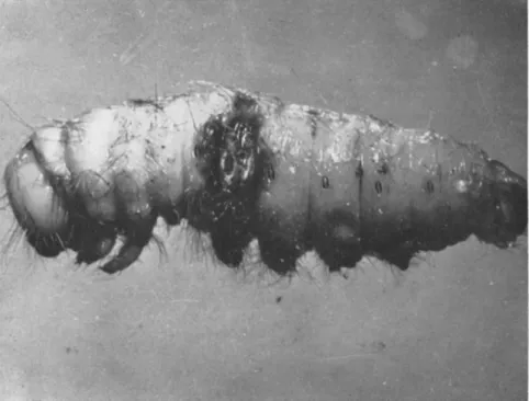
T h e physiological functions of the host can be affected also by some bacterial infections. T h i s has been shown by the analysis of a chronic Bacillus thuringiensis var. alesti infection in Bombyx mori (Vago, 1956a). When very small doses of this bacillus are ingested (oligoinfec- tion) the usually rapid toxemia, caused by the crystalline toxin of this group of microbes, does not take place. There is only a localized toxic effect on the digestive organs or a very slow effect on the hemolymph.
No deaths occur, but feeding decreases and after several days the affected larvae become atrophied; they lose two-thirds of their weight and nearly half their length. T h e r e may be tertiary consequences leading to dis
orders in ecdysis and in the development of the pupa or butterfly (Fig. 3 ) .
A similar observation has been recently made by Yamvrias (1961) in Anagasta kühniella (Zeller). Feeding also decreases in this case and there is a considerable reduction in weight, development is prolonged, fertility is reduced, and the development of the eggs is altered. T h e latter disorder is connected with disturbances in the food reserves built up in the larva.
It is probable that virus diseases may cause physiological effects in later stages of the affected insect. However, to obtain evidence of such
effects a more thorough knowledge of subacute forms of viroses is re
quired. Vago (1950a, 1956b) has noted that nuclear polyhedrosis at the incipient stage of the pathogenesis may lead to molting disturbances.
Benign forms of viroses may render insects sensitive to different eco
logical influences (Vago and Kremky, in press). Larvae of Bombyx mori in the initial stage of nuclear polyhedrosis (during which no deaths have yet occurred) die in considerable numbers under the influence of heat ( 3 2 ° C ) , heat (30°C) with high humidity, sun rays, or cold ( 3 ° C ) .
100J MOULTING LARVAE
1 2 3 4 5 6 7 8 9 10 11 1213 14 15 16 17 18 19 20
FIG. 3. Abnormal fourth ecdysis of Bombyx mori larvae after oligoinfection with Bacillus thuringiensis var. alesti. Solid line: control; broken line: infected. (From Vago, 1956a.)
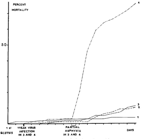
These conditions do not affect healthy larvae. In other experiments, most of the virus-infected larvae die from partial asphyxia, while the death rate is low in healthy caterpillars (Fig. 4). There is also increased sensibility to contact insecticides (HCH and D D T ) .
Virus and bacterial diseases can deeply disturb also wound healing and hemolymph clotting (Vago and Amargier, in press), and cause rapid mortality by hemorrhage.
T h e "shortworm disease/' recognized for several centuries in Bombyx mori, and which also occurs in other Lepidoptera that normally weave cocoons [Antheraea pernyi, Samia cynthia (Drury), Thaumetopoea pity- ocampa], results in the caterpillar forming a chrysalid without spinning
a cocoon. Frequently these chrysalids die, poisoned by the silk that has not been evacuated.
It was found (Vago, 1956a) that the primary cause of this anomaly, apart from physiological shock, may be of microbial origin. Normal chrysalids showed 0.5 percent polyhedrosis while of the "shortworms"
PERCENT / 4
MORTALITY · '
5 0
/ /
/
/ / / / / / /' /'
i I i I H I I
3
I f ' I I* T " I ι I I ' — ι « 1 ' 1 1 Γ 1 S\ WEAK VIRUS PARTIAL
ECDYSfS INFECTION ASPHYXIA DAYS IN 3 AND 4 IN 2 A NO 4
Fig. 4. Effect of sublethal nuclear polyhedrosis on the sensitivity of Bombyx mori larvae to partial asphyxia. (/) Control; (2) control with partial asphyxia during 12 hours; (3) weakly infected with nuclear polyhedrosis; (4) weakly infected with nuclear polyhedrosis and treated by partial asphyxia during 12 hours. (Original.)
of the same silkworm breeding, 60 percent were infected. T h e forma
tion of "shortworms" in which pebrine (Nosema bombycis) also occurs is not infrequent. T h e mechanism of this predisposition has been studied experimentally, and it appears to be due to lack of timing be
tween pupal metamorphosis and cocoon spinning. T h e first process, determined by the action of hormones, takes place normally, but if the larva does not start to spin its cocoon while it is still mobile it is unable
to do so at a later stage. Virosis and nosemosis are weakening factors that retard the second process.
In some cases, the physical impossibility of ejecting the silk may end in poisoning of the pupa. Buonocore (1937), and later Lipa (1957), in Aporia crataegi (Linnaeus) show that, alterations in the silk glands caused by Nosema may have that physiological consequence. Nosemosis can also increase sensibility of host to contact or ingestion insecticides as Rosicky (1951) showed in Otiorrhynchns ligustrici Linnaeus infected with Nosema otiorrhynchi Weiser.
Β. Effects on Microbial Diseases
For a long time in insect pathology, probably owing to the scarcity of observations on benign microbial diseases, it was considered that a morbid entity is composed of infection, pathogenesis, and final syndrome phases. However, it has now been ascertained that complications are liable to occur and that the final syndrome may not be caused by the microorganisms responsible for the first infection. This "complex," as defined by Vago (1956a), is the combination of usually slow and chronic microbial effects writh another more dynamic infection for which the ground has been prepared by the initial disease. Virus and protozoan diseases often play the part of predisposing factors, sometimes bacteriosis and mycosis may also be classified in this category.
1. Virus Diseases
This aspect of insect virology has been recently demonstrated. Vago (1956b) feels that the part played by insect viroses in preparing the way for other diseases is probably just as important as the acute infection caused by it. Nonacute virosis can be assumed to be a primary disease when unusual sensibility to different diseases is observed. In establish
ing pathological diagnosis it is useful to consider that the apparent syn
drome may be preceded by a primary virus disease. This author has analyzed (Vago, 1956a) the connection between nuclear polyhedrosis and subsequent bacteriosis (caused by Bacterium paracoli) in Vanessa urticae Linnaeus, Porthetria dispar, and Bombyx mori. T h e comparison of separate infections, occurring consecutively or simultaneously, proved that the virosis had a primary role and that the bacterial septicemia was a complication, caused partly by a decrease in phagocytosis in the af
fected larvae and by increased bacterial penetration through the midgut wall. It is also believed (Vago, 1950b) that a virus disease prepares Serratia marcescens epizootics in silkworm breedings. Another type of complex disease with a virus background is described by the same author in the lepidopteran Arctia caja. Polyhedrosis attacking peritracheal cells
disturbs the functions of the alimentary canal as explained above (Vago and Vasiljevic, 1961) and leads to a proliferation of enterococci in the midgut with acute dysentery. Steinhaus (1947) has referred to the role of bacteria as secondary invaders in a granulosis of the variegated cut
worm (Peridroma).
It is probable that under natural conditions bacterial insect diseases are frequently preceded by virosis. In higher animals and even from the medical point of view the concept of "exit microbes" (microbes de sortie) is now quite classical. However, care should be taken not to
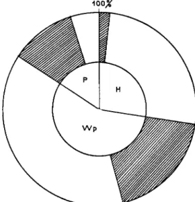
FIG. 5. Pathological analysis diagram of a population of Plusia gamma, showing the relation between nuclear polyhedrosis and bacteria in the hemolymph of living fourth instar larvae. (H) no polyhedra; (Wp) rare polyhedra in fat tissue; (P) heavy polyhedrosis; hatched area: bacteria (Pseudomonas, Aerobacter, Serratia). (Original.)
confuse "exit microbes" that play a part in the pathogenic action (and cause a real bacterial disease with a rapid course in the live insect) with portmortem saprophytic microflora. W e feel that "exit microbes"
following a virus disease belong mainly to the proteolytic group, which, according to Bucher (1960) play a preponderant role in hemolymph infections.
In some cases virus diseases seem to favor the appearance of other viroses. Bergold (1951) observed that granulosis of Cacoecia murinana
(Hübner) is not lethal to Choristoneura fumiferana (Clemens) ( = Ar- chips), but it increases nuclear polyhedrosis. Smith and Rivers (1956) noted in Operophtera brumata (Linnaeus) and in Bupalus piniarius
(Linnaeus) a high death rate caused by cytoplasmic polyhedrosis con
secutive to infection with nuclear-polyedrosis virus from Vanessa cardui (Linnaeus). T h e activation of latent cytoplasmic viruses was presumed to be caused by the introduction of a second virus although it was not possible to ascertain the mechanism of this action. Even when two viruses prove virulent for a particular species, the presence of one, for instance a granulosis in Pseudaletia unipuncta, can stimulate the other, a nuclear polyhedrosis in the same insect (Tanada, 1956).
2. Protozoan Diseases
In this group of diseases, the action of intestinal protozoa on cells seems to help the penetration of some bacteria into the hemocoel. In the honey bee, Apis mellifera, amoebiasis due to Malpighamoeba is con
sidered not as a serious disease, but rather as more of a weakening factor with regard to other diseases (Borchert, 1939). It may well prepare the ground for the development of Nosema apis and render this parasite more dangerous than it usually is (Morgenthaler, 1934). In addition, Weiser and Lysenko (1956) believe that the penetration of Pseudomonas noctuarum White through the gut wall may be favored by the presence of microsporidia and of helminths. Intestinal gregarines (Bucher, 1959) may cause wounds in the epithelium of the alimentary tract of grass
hoppers through which pass some species of proteolytic bacteria that cause septicemia.
3. Bacterial Diseases
T h e influence of a bacterial infection on the development of a second microbial disease appears (Vago, 1956a) in the case of an oral infection caused by a very weak dose of Bacillus thuringiensis var. alesti. This
"oligoinfection" has no direct effect, but secondary consequences follow after several days. Growth is retarded and at the end of the larval stage intestinal bacteria cause acute and often fatal dysentery. In this second syndrome there are no Bacillus alesti but only Enterococcus, Staphylococcus, or Pseudomonas. Experimental ingestion of purified toxic crystals of Bacillus alesti shows that the mechanism connecting the first disease to the second is set into action by a weak intoxication, at intestinal cell level, which favors the proliferation of bacteria in the ali
mentary tract. T h e same author describes similar sequences in other Lepidoptera and particularly in Pieris brassicae (Linnaeus).
T h e effect of Bacillus thuringiensis on another bacterium, Serratia marcescens, has been indicated recently by Steinhaus (1959) in Galleria mellonella. It is supposed to have an action similar to that mentioned in the preceding paragraph. Bucher (1957) notes that the secondary
flora appear following the intestinal infection of Malacosoma pluviale (Dyar) by an unidentified Bacillus.
4. Fungus Diseases
Few data are available on the part played by fungus diseases in pre
disposing hosts to other microbial diseases. T h i s may be due to the fact that most of the fungi pathogenic to insects tend to cause generalized diseases, and some may secrete antibiotic substances. However, it has been noted (Vago, in press) that some fungi (Aspergillus, Rhizopus, Fusarium) may have a toxic effect on the insect without killing it, and thus favor the proliferation of bacteria that are either septicemic or intestinal according to species and localization. T h e final syndrome is no longer a slowly developing mycosis but a rapid bacteriosis.
It should be mentioned that Serratia marcescens bacteria have often been found following different mycoses. T h e ease and frequency with which this bacterium is noticed is perhaps due to its characteristic and readily apparent red pigment.
V I . SIMULTANEOUS DISEASES
T h e study of atypical cases of diseases frequently shows the con
current presence of several microorganisms or of lesions peculiar to different pathological processes. However, even with the help of histo- pathological studies it is difficult to define the mechanism that leads to the coexistence of diseases. In fact, different diseases occurring in an organism may be entirely or partly independent processes that do not interfere with each other. They may also be interfering or antagonistic processes, or show inhibitory action, or be synergistic processes the ef
fects of which are cumulative and strengthen each other.
T h e concepts of "synergism" and of "predisposition" become dove
tailed in the latter category and it is likely that, with the development of technical methods of analysis, a certain number of cases considered today as representing "coexistence" will be recognized as a complex of diseases with a predisposition factor involved.
1. Participation of Protozoan Diseases
T h e development of a large number of protozoan diseases is known to be slow and benign during the entire life of the insect. In fact, among both microsporidia and gregarines we know species ranging from path
ogens to practically inoffensive forms. Because of this, in addition to the chronic protozoan infection, a second disease often develops.
T h e most classical example of the coexistence to which we refer can be found in the historical research wrork carried out by Pasteur (1870)
on the origin of the silkworm "disease" prevalent in the middle of the nineteenth century. T h e microsporidian nature of this particular dis
ease had been discovered when Pasteur proved that in reality there were several diseases, including "pebrine" caused by Nosema bombycis, and bacterial flaccidity, or flacherie, which in many cases were so inter
mingled that they were difficult to separate. This author mentions the synergistic effect of both diseases noticed also by the silkworm breeders of the time.
Bee keepers have also observed (Morgenthaler, 1934) that bee amoebiasis due to Malpighamoeba became more serious when it occurred at the same time as attack by the sporozoan Nosema apis. These two diseases, which are somewhat chronic, develop in parallel; however, it is the amoebiasis that becomes the lethal syndrome. T h e same author also mentions joint Nosema apis and gregarine infections (Leydrana). Re
cent studies show to what extent diseases may occur simultaneously (Giordani, 1959). Between 1948 and 1957 in Italy amoebiasis was found alone in 64.5 percent of samples, associated with nosemosis in 15.3 per
cent, with acarine parasitism in 7.5 percent, with paralysis in 8.1 per
cent, and with "mai-disease" in 6.4 percent.
In the lepidopteran Sesamia cretica Lederer, Vago (1956b) found Nosema sp. and granulosis often together. Similar cases are known both in Pieris brassicae and Cacoecia murinana suffering from granulosis and unspecified Sporozoa.
T h e concomitant pathogenesis of nosemosis and nuclear polyhedrosis has been followed during the larval and pupal life of the Lepidoptera Samia cynthia (Drury) and Bombyx mori (Atger and Vago, 1960). T h e two diseases may develop in parallel, although intricately mixed, and Nosema spores and polyhedra appear simultaneous not only in the cells of the same lobe of tissues but in the same cell, there being an accumu
lation of polyhedra in the nucleus and of spores in the cytoplasm. This shows that there is quite a remarkable mutual tolerance; but obviously there is also a certain synergism since the death rate increases in the case of dual infections. Huger (1960) also noted this tolerance and this synergistic effect in polyhedrosis and nosemosis in Agrotis segetum
(Schiffermüller) with the simultaneous presence of polyhedra and spores in the same cytoplasm.
Two varieties of Nosema may coexist in the same insect Porthetria dispar; Nosema lymantriae Weiser attacks the adipose tissues, and Nose
ma muscularis Weiser the muscles (Weiser, 1957a). T h e mortality curve varies according to whether the disease is single or combined; and the two types of nosemosis can be transmitted at the same time. Further
more, Euproctis chrysorrhoea is not very vulnerable to Nosema lyman-