Narrative review
How to isolate, identify and determine antimicrobial susceptibility of anaerobic bacteria in routine laboratories?
E. Nagy
1,*, L. Boyanova
2, U.S. Justesen
3, on behalf of ESCMID Study Group of Anaerobic Infections
1)Institute of Clinical Microbiology, University of Szeged, Szeged, Hungary
2)Department of Medical Microbiology, Medical University of Sofia, Sofia, Bulgaria
3)Department of Clinical Microbiology, Odense University Hospital, Odense, Denmark
a r t i c l e i n f o
Article history:
Received 14 December 2017 Received in revised form 9 February 2018 Accepted 11 February 2018 Available online xxx Editor: L. Leibovici Keywords:
Anaerobic bacteria
Antimicrobial susceptibility determination Identification
Isolation
a b s t r a c t
Background: There has been increased interest in the study of anaerobic bacteria that cause human infection during the past decade. Many new genera and species have been described using 16S rRNA gene sequencing of clinical isolates obtained from different infection sites with commercially available special culture media to support the growth of anaerobes. Several systems, such as anaerobic pouches, boxes, jars and chambers provide suitable anaerobic culture conditions to isolate even strict anaerobic bacteria successfully from clinical specimens. Beside the classical, time-consuming identification methods and automated biochemical tests, the use of matrix-assisted laser desorption/ionization time- of-flight mass spectrometry has revolutionized identification of even unusual and slow-growing an- aerobes directly from culture plates, providing the possibility of providing timely information about anaerobic infections.
Aims: The aim of this review article is to present methods for routine laboratories, which carry out anaerobic diagnostics on different levels.
Sources: Relevant data from the literature mostly published during the last 7 years are encompassed and discussed.
Content: The review involves topics on the anaerobes that are members of the commensal microbiota and their role causing infection, the key requirements for collection and transport of specimens, pro- cessing of specimens in the laboratory, incubation techniques, identification and antimicrobial suscep- tibility testing of anaerobic bacteria. Advantages, drawbacks and specific benefits of the methods are highlighted.
Implications: The present review aims to update and improve anaerobic microbiology in laboratories with optimal conditions as well as encourage its routine implementation in laboratories with restricted resources.E. Nagy, Clin Microbiol Infect 2018;▪:1
©2018 European Society of Clinical Microbiology and Infectious Diseases. Published by Elsevier Ltd. All rights reserved.
Introduction
Human pathogenic anaerobic bacteria werefirst discovered and identified in the middle of the nineteenth century. However, it was difficult at that time to obtain pure cultures for many of these or- ganisms [1]. Even later, anaerobic infections were the most commonly overlooked of all bacterial infections, especially when
the anaerobic bacteria were present in mixed culture and one or more aerobic or facultative anaerobic pathogens were grown on the same media at the same time. Use of insufficient anaerobic incu- bation techniques will often permit only isolation of the most common anaerobic pathogens, the members of the Bacteroides fragilis group or Clostridium perfringens, which are known as
‘moderate’ anaerobes surviving oxygen levels up to 2%e8%. This may let the microbiologist think that his/her techniques for anaerobic culture are fully adequate, as they can regularly isolate
‘anaerobes’. It is difficult to provide a correct and practical defini- tion of the term ‘anaerobe’. Anaerobic bacteria may differ
*Corresponding author. E Nagy, Institute of Clinical Microbiology, University of Szeged, 6701 Szeged, P.O. Box 427, Hungary.
E-mail address:nagy.erzsebet@med.u-szeged.hu(E. Nagy).
Contents lists available atScienceDirect
Clinical Microbiology and Infection
j o u r n a l h o m e p a g e :w w w . c l i n i c a l m i c r o b i o l o g y a n d i n f e c t i o n . c o m
https://doi.org/10.1016/j.cmi.2018.02.008
1198-743X/©2018 European Society of Clinical Microbiology and Infectious Diseases. Published by Elsevier Ltd. All rights reserved.
significantly in their tolerance to atmospheric oxygen and in how low oxygen levels are needed for them to multiply. A practical definition was formulated by Professor Finegold in 1977:‘anaerobe is a bacterium that requires a reduced oxygen tension for growth and fails to grow (form colonies) on the surface of solid media in 10% CO2, in air (18% oxygen)’[1].
Various kinds of equipment to provide a suitable anaerobic environment and different commercially available media for culturing the wide range of anaerobic bacteria with different re- quirements have been introduced into routine laboratories. The application of 16S rRNA gene-sequencing-based identification for difficult to identify anaerobes, and the matrix-assisted laser desorption/ionization time-of-flight mass spectrometry (MALDI- TOF MS) for the direct identification of them from the primary culture plate or from the anaerobic subculture plate, have made it possible to provide accurate and timely information about their role in infections. New anaerobic species have been detected and identified through studies on the composition of the normal gut microbiota, using a new culture tool called‘culturomics’[2,3]. All of these achievements have increased the interest for anaerobes in clinical situations and beyond. It has become increasingly impor- tant in human medicine to identify anaerobic infections. One reason is that there is a growing evidence of infections caused by more virulent anaerobic species such asClostridioides(Clostridium) difficilePCR ribotype 027, which produces16-fold more toxin than classical strains[4], orFusobacterium necrophorum causing some cases of‘culture-negative’chronic tonsillitis[5], or enterotoxigenic B. fragilisin sepsis and colon carcinoma[6,7]. Another reason is the constant increase of immunocompromised patients such as pa- tients with diabetic foot ulcers, which often involve neglected an- aerobes [8,9]. Last, there are dynamic changes in antibiotic
resistance of anaerobes. Since 2000, increasing resistance rates in Bacteroides/Parabacteroidesspp. tob-lactam/b-lactamase inhibitor combinations, clindamycin and carbapenems, inPrevotellaspp. to b-lactams, in Gram-positive anaerobic cocci to clindamycin, and in C. difficile and Bacteroides/Parabacteroides spp. to metronidazole have been reported in Europe[10e12]. A special concern is metallo- b-lactamase production inB. fragilis(Division II isolates) conferred by thecfiA gene, which may lead to carbapenem resistance. In some studies, the cfiA gene has been detected in nearly 40% of the B. fragilisisolates[13]. Multidrug resistance has also been detected amongB. fragilisclinical isolates in different countries[14e16].
Surveillance studies about anaerobe culture techniques, identi- fication and susceptibility testing methods carried out in routine laboratories in the USA and Belgium have shown that there are great differences in the methods used in different laboratories, where diagnostics other than detecting the presence of toxigenicC. difficile in faeces is carried out in-house[17,18]. The aims of this review are to discuss the presence of anaerobes in commensal microbiota and in infectious processes, and to present optimal methodology for carrying out anaerobic diagnostics of human infections.
Anaerobes as members of commensal microbiota and risks of anaerobic infections
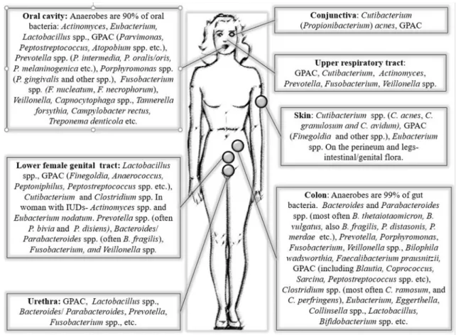
Anaerobes are abundant in commensal microbiota throughout different body sites being the dominating or less dominating part of the microbiota (Fig. 1)[3,19e21]. Low oxygen concentration in the intestinal and urogenital tract favours the abundance of anaerobic bacteria in these sites. In more aerated sites such as the oral cavity and skin, the anaerobes inhabit protected sites where cohabiting aerobic/facultative bacteria consume the oxygen [20]. This is
Fig. 1.Most common anaerobic species/groups of the commensal microbiome at different body sites. GPAC, Gram-positive anaerobic cocci.
essential as most anaerobic infections are endogenous and fav- oured by damaged mucosal/cutaneous barriers, which allow their penetration into normally sterile tissues often together with aero- bic/facultative bacteria. Importantly, from most infections where anaerobes are present together with aerobic/facultative bacteria, it is difficult to isolate them in pure culture for identification. A great variety of species belonging to strict anaerobic genera have been proven to cause infections in almost all regions of the body and may lead to serious bloodstream infections as well (Table 1)[21,22].
Besides the well-known species, the spectrum of the anaerobic bacteria isolated from infections in humans is increasing, due to better isolation and identification possibilities. New anaerobic genera and species have been shown to be present in serious in- fections, such as the spore-forming, Gram-positive rod, Rob- insoniella peoriensis, the non-spore-forming Gram-positive rods Solobacterium moorei and Turicibacter sanguinis, Ruminococcus gnavusorOscillibacter ruminantium, causing bacteraemia and other infections, just to mention a few examples[23e25].
Anaerobic microbiology is not routinely performed in many laboratories because of technical andfinancial reasons. However, thefirst steps for cost reduction depend on the clinicians' knowl- edge of the characteristics of anaerobic infections and on laboratory policy to reject unacceptable specimens such as those from body sites containing anaerobic commensal microbiota (Table 2) [18,19,21,26e28].
Key requirements for collection and transport of specimens for anaerobic microbiology
To fully benefit from the anaerobic microbiology, clinicians should collect and send clinical specimens to the laboratory
according to the specific recommendations (Table 2) [19,21,26,27,29]. The specimens for anaerobe culture should be:
collected at an appropriate timedbefore the start of antibiotic therapy, or if this is not possible, just before the following drug administration;
collected at the infection sitede.g. for wound infections and open abscesses, deep aspirates/tissue biopsies at the advancing wound edge (where the bacteria multiply) rather than pus or superficial specimens, and for closed abscesses, deep needle aspirates of the lesion and close to the borders;
aspirates and tissue biopsies instead of swabsdcotton wool swabs are porous, prone to desiccation and contain inhibitory fatty acidseit is best to use swabs made of syntheticfibres;
free from contamination with commensal micro- biotadtherefore, debridement and skin decontamination are needed; sufficient volumes should be taken (e.g. 8e10 mL of blood/bottle,>1e2 mL aspirates,1 g tissues,5 mL of watery/
semi-formed stool specimens forC. difficile);
inserted in anaerobic transport media (ATMs)dthe best ATMs are commercially available oxygen-free transport tubes/vials with pre-reduced and anaerobically sterilized (PRAS) anaerobic media, if not available, Stuart's, CaryeBlair or Amies transport media can be prepared in the laboratory. Specimens should be deeply inserted into the ATMs or onto the agar surface (after ethanol cleaning of the ATM stopper) if PRAS ATMs are used and the specimens are liquid;
stored and transported at room temperature and not in a refrigeratordat low temperatures, oxygen diffusion is increased;
sent to the laboratory within 2 h in ATMsdalthough, if delay is unavoidable, they can be accepted even after 8e48 h. If no ATMs
Table 1
Common detection rates of anaerobes in some clinically important infections[1,19e21,26,28]
Prevalence of anaerobesa
Very high (70%e100%) High (40%e70%) Moderate (13%e45%) Low (4%e10%) Very low (1%)
Head and neck Most dental and oral infections, involving head and neck cellulitis and abscesses, root canal infections, peritonsillar abscess, chronic sinusitis, post-surgical infections
Chronic otitis media, mastoiditis
Ocular infections
(dacryocystitis, post-traumatic endophthalmitis, perforated corneal ulcers), cervical lymphadenitis, serous otitis media, tonsillopharyngitis
Acute sinusitis, acute otitis media
Blood Intra-abdominal sepsis, septic abortion Bacteraemia after oral surgery/tooth extraction
Bacteraemia due to endocarditis
CNS Brain abscess, subdural empyema CNS shunt
infections
Meningitis Pulmonary Lung abscess, aspiration pneumonia,
necrotizing pneumonia, pleural empyema
Bronchiectasis, nosocomial pneumonia,
Skin/soft tissue Gas gangrene, breast abscess, synergistic necrotizing cellulitis, perianal and perirectal abscess, infected diabetic gangrene, pilonidal abscess, infections after trauma, acne vulgaris
Wound infections, abscesses, cellulitis, necrotizing fasciitis, bite wound, diabetic foot infection, infected decubitus ulcers
Impetigo
Abdominal Most intra-abdominal infections, appendicular abscess, appendicitis with peritonitis, post-surgical abdominal infections
Liver abscess Biliary tract infections, ascites and hepatic abscess
AAD Pseudomembranous colitis (Clostridium difficile)
Overall AAD (Clostridium difficileandClostridium perfringens)
Bone/joint Orthopaedic device
infections
Osteomyelitis, infections without orthopedic devices
PJIs Urogenital Most female genital tract infections
(pelvic inflammatory disease, pelvic abscesses, endometritis, vaginal cuff abscess, bacterial vaginosis)
Urinary tract infections
Abbreviations: AAD, antibiotic-associated diarrhoea; CNS, central nervous system; PJIs, prosthetic joint infections.
aDetection rates vary according to the methods used.
are available, large volume (2 mL) aspirates should be placed into sterile containers and in such cases, prompt transport (within1 h) is needed.
Processing specimens in the laboratory for culturing anaerobes
All clinical microbiology laboratories that accept specimens for anaerobic culture have to have facilities to isolate anaerobes and screen for the major anaerobic groups (Level 1 and 2 anaerobic laboratories) [21]. For definitive identification and antimicrobial susceptibility testing (AST), isolated anaerobes should be referred to reference laboratories. Level 3 anaerobic laboratories should identify anaerobes to genus and species level using phenotypic and enzymatic tests and should determine some presumptive antibiotic sensitivity. Level 4 anaerobic laboratories should pro- videfinal identification using MALDI-TOF MS and, if needed, 16S rRNA gene sequencing and should perform quantitative AST [19,21].
After visual examination of the acceptable specimens (Table 2) all samples should be homogenized in liquid medium (e.g. thio- glycolate broth). Direct Gram-staining of the specimen is manda- tory for anaerobic diagnostics[30]. It can reveal the presumptive involvement of some anaerobic species with characteristic morphology, such as Fusobacterium nucleatum, B. fragilis or C. perfringens[19]. In the case of urinary tract infections, which persist with negative aerobic culture growth, the rare uropathogen Actinotignum (Actinobaculum) schaaliican be detected by Gram- staining, indicating the need for prolonged incubation in CO2 or inoculation on anaerobic media[31]. The direct Gram-staining also makes it easy to distinguish between wound contamination (showing abundance of squamous epithelial cells) and infection (exhibiting both bacteria and inflammatory cells)[26]. Moreover, the technique has shown higher sensitivity compared with culture in the case of actinomycosis, where branchingfilamentous Gram- positive rods may be observed in specimens from abscesses or si- nus tracts[32].
The initial anaerobic culture process should include careful plating on selective and non-selective blood agar plates (freshly prepared in house or commercially available), as well as in liquid anaerobic media (thioglycolate broth). The primary enriched (with horse or sheep blood, vitamin K1 and haemin) non-selective media should allow the growth of all clinically significant anaerobes.
Many laboratories in the USA prefer to use commercially available PRAS media to isolate fastidious anaerobic pathogens[21]; how- ever, homemade fresh anaerobic media can also be successfully used. Based on the Gram-staining results of the specimen, further selective media (Bacteroides bile esculin agar, kanamycine vancomycin laked blood agar, phenylethyl alcohol agar, egg yolk agar) can be inoculated[19]. If isolation ofC. difficileis needed from stool samples, the classical cycloserineecefoxitin fructose agar or other commercially available selective chromogenic media should be used[21,33]. Few laboratories process specimens in an anaerobic chamber. Usually, work is performed outside an anaerobic envi- ronment, but to limit oxygen exposure, it is important to place inoculated media into an anaerobic environment (such as plastic envelopes, boxes, jars, or automated gasflushing instruments, e.g.
Anoxomat or anaerobic chambers) within 15e20 min after inocu- lation. Suggested incubation time is usually 48 h, but some anaer- obes (such asC. perfringens) may form colonies earlier. Several slow-growing anaerobes, however, may need much longer primary incubation time (up to 3e5 days) to form colonies suitable for subculturing or direct identification by the MALDI-TOF MS. The anaerobic broth should be held for up to 14 days in special cases such as detection of Cutibacterium spp. from prostatic joint in- fections. Control of anaerobiosis during incubation is strongly rec- ommended to be sure that even strict anaerobes will form colonies [19,21,34].
Anaerobe incubation techniques
In routine laboratories, the classical Hungate role tube method was rapidly replaced by the easier-to-use anaerobic jars, boxes, pouches and chambers for isolation of anaerobic bacteria. Various Table 2
Acceptable and unacceptable specimens for anaerobic microbiology[18,19,21,26,27,29,33]
Infection site Acceptable specimens Unacceptable specimens
Blood Blood (8e10 mL/bottle or acc. to the weight) in anaerobic blood bottles Catheter/catheter-tip
samples Central nervous system Tissue biopsies or needle aspirates through intact decontaminated surface Surface swabs Head and neck Percutaneous needle aspirates, surgical specimens. For Lemierre syndrome aspirates, tissue
biopsies. For actinomycosis sulphur granules as well
Oral, nose and throat swabs except for Lemierre syndrome Periodontal Abscess aspirates, subgingival pocket samples (by periodontal curettes or sterile paper points in
the canal)
Surface gingival and oral swabs
Ear In otitis media: aspirates by tympanocentesis (with sterile micropipettes) Surface material
Eye Corneal scraping, vitreousfluid needle aspirates, conjunctival swabs As above
Abscesses Closed abscess: needle aspirates through intact decontaminated tissue, surgical samples;
fistulas/sinuses: deep plastic catheter aspirates after disinfected skin opening; open abscesses:
see Wounds
Skin/mucosal surface swabs, pus Pulmonary Pleuralfluid, lung and transtracheal aspirates, lung-tissue biopsies, deep bronchial secretions
taken with double-lumen (protected) catheter
Nasopharyngeal swabs, sputum Abdominal cavity Peritoneal and ascitesfluid aspirates, surgical biopsies, bile aspirates Ileostomy/colostomy
samples
Stool Only forClostridium difficileorClostridium botulinum For other anaerobes
Female genital tract Tissue biopsies, pelvic infection aspirates (by culdocentesis) peritonealfluid, endometrial specimens (by protected catheters), surgical specimens
Cervical/vaginal swabs (except for bacterial vaginosis)
Bone Aspirate, bone biopsies. Taking several biopsies is recommended Swabs, soft-tissue samples
Joint, prosthetic joint Synovialfluid aspirates in anaerobic blood culture bottles, periprosthetic biopsies in anaerobic broth media
Surface swabs of wounds/fistulas Wounds/soft tissues Deep biopsy sampling, deep wound aspirates at the advancing wound edge after debridement
and cleaning the surface with sterile saline or alcohol
Surface swabs, pus, sinus tracts Decubitus or skin ulcers Needle aspirates or tissue biopsies. Deep sampling from the base of lesion Surface material, swabs Diabetic foot ulcers Bone biopsies (surgical or after debridement and surface disinfection) Tissue samples less suitable
Urine Bladder urine (suprapubic bladder aspirates) Voided and catheterized urine
systems for anaerobiosis have different advantages and disadvan- tages[19,21]. The most expensive anaerobic chambers allow inoc- ulation, inspection and subculturing in a permanent anaerobic environment to ensure viability of fastidious, slow-growing an- aerobes and to check the plates daily for growth. Anaerobic pouches, boxes and jars need more organized specimen processing as culture media are inoculated in air. Jars and boxes should not be opened before 48 h of incubation to prevent premature death of some slow-growing anaerobes by exposure to air during their logarithmic growth phase. Today, automated gas flushing in- struments (Anoxomat) can also be used to shorten the exposure of inoculated plates to air because an anaerobic atmosphere can be achieved within minutes[34,35]. If the clinical situation (symptoms of gas gangrene) or initial Gram-staining result suggests the pres- ence ofC. perfringens, it may be prudent to incubate plates indi- vidually in anaerobic pouches so that the plate may be examined earlier. The reduced condition for any anaerobic system should be monitored by the addition of anaerobe indicator strips (with methylene blue or resazurin), which become colourless in the low concentration of oxygen needed for appropriate growth of most clinically important anaerobes. Cost-effective biological indicators such as a subculture ofPseudomonas aeruginosaon Simmon's cit- rate slant[19]or measuring the inhibition zone diameter of a 5-mg metronidazole disc on an aerotolerantC. perfringenscontrol strain [34], can also be used.
To simplify anaerobe bacteriology, a medium containing oxygen-removing enzymes such as oxyrase was recommended [36]. OxyDish™is a tightly sealed Petri dish with PRAS blood agar medium containing the enzyme (OxyRase™) that removes oxygen from the medium and the space above the agar after inoculation and maintains anaerobic conditions during the incubation without using anaerobic jars or chamber[36]. Another approach to elimi- nate the use of anaerobic incubation systems is adding antioxidant molecules such as ascorbic acid, glutathione and uric acid to the blood agar plates (series of R mediumd‘quasi-universal’media) to avoid oxygen toxicity during incubation of the plates in ambient air [37]. Both approaches may simplify laboratory procedures; how- ever, a careful evaluation of their performance is needed for isolation of strict anaerobic bacteria from specimens containing a mixed bacterial population in clinical practice.
Identification of anaerobes
Initial examination of colonies should be performed using a stereomicroscope or at least a strong magnifying glass. Colony morphologies that appear similar when observed at a distance, can be differentiated when magnified, and the presence of tiny colonies near larger ones can be discerned. All different colonies should be isolated and plated on an anaerobic blood agar plate, a chocolate agar plate and a spot on a glass slide for Gram-staining.
Care should be taken that the same colony goes onto both plates and the slide.
Classical identification of anaerobes was based on a series of biochemical tests in PRAS test tubes with different sugars and various other substrates incubated for 1e6 days depending on the growth rate of the isolate. Careful evaluation of cell morphology and detection of alcohol and short-chain fatty acids by gaseliquid chromatography later became the basis of the identification of a wide range of anaerobic bacteria [38]. Nowadays some routine laboratories still rely on a presumptive identification carried out after subculturing the colonies from the primary plate in an anaerobic environment[19,21]. Beside investigation of colony and Gram-stain morphology and motility, a combination of rapid biochemical spot tests (such as detection of indole, catalase, nitrite, urease positivity) and susceptibility to special potency antibiotic
discs such as kanamycin (1000mg), vancomycin (5mg) and colistin (10mg) can be used for the identification of major groups of an- aerobes with clinical relevance. This presumptive identification is cost-effective, but takes 24e48 h after isolation of the colonies on the primary plate. Growth in the presence of 20% bile and the pigmentation of the colonies (brown to black) of somePrevotella andPorphyromonasspp. is characteristic after4 days of incuba- tion. Long-wave UV light can be used to detect characteristicfluo- rescence of some colonies (e.g. C. difficiledchartreuse,Prevotella melaninogenica,Porphyromonas asaccharolyticadbrick red)[19,21].
More detailed identification can be carried out by commercially available manual or automated identification kits, using panels to assess the ability of the isolated anaerobes to react with a limited number of carbohydrates and other substrates after 24-h incuba- tion in an anaerobic environment (detection of inducible enzymes).
Other systems detect preformed enzymes and the kits should be incubated aerobically for 4e6 h; however, a high inoculum is needed that is difficult to achieve in the case of slow-growing anaerobic bacteria with tiny colonies. These systems are hindered by a limited number of substrates to provide proper differentiation for a wide range of anaerobic species; moreover, many anaerobic bacteria are non-reactive in biochemical tests. Many clinically important or newly described taxa may be lacking from the data- bases. Users must be aware that databases of these products are rarely updated[39,40].
Recently, MALDI-TOF MS based on soft ionization of large mol- ecules such as proteins, peptides, lipids, sugars and DNA, has been shown to perform very well for the identification of anaerobes [41e43]. Microorganisms are identified by comparing their mass spectrum with those of known reference strains. The initial cost of the instrument may seem prohibitive to many microbiological laboratories, but it is inexpensive to run and will identify anaer- obes, as well as most other microorganisms, quickly and accurately.
As MALDI-TOF MS is a very sensitive method, only a very small amount of biomass is needed for correct identification. This pro- vides an early identification for anaerobes, very often directly from the primary culture plates, without additional subculturing and testing for aerotolerance. However, very small colonies from mixed culture may need subculturing for a shorter or longer time before MALDI-TOF MS identification. Applying the latest updates of the databases associated with the two widely used MS systems (Bruker Biotyper - Bruker Daltonik, Germany, VITEK MS - bioMerieux, France) is the prerequisite for the successful use of this technique.
The data libraries for anaerobes must include not only reference strains, but also clinical strains corroborated by molecular sequencing methods[42,44,45]. During the past few years, several studies have proved the superiority of MALDI-TOF MS during routine identification of anaerobes compared with different auto- mated or manual biochemical identification kits[46e49]. Sample preparation, incubation time, but not culture media or, in most cases, exposure to oxygen can influence the quality of the identi- fication of anaerobes by MALDI-TOF MS[50,51]. However, the MS- based name of an isolate has to correlate with the colony and Gram- stain morphology. Further possibilities such as typing of anaerobic bacteria (B. fragilis,C. difficileorCutibacterium acnes) at the sub- species level, determination of resistance and direct identification of anaerobic blood culture isolates can also help in routine anaer- obic diagnostics[42,52,53].
When the above-mentioned methods fail to correctly identify a clinically important anaerobic strain to the genus or species level, sequencing of genetic markers such as a portion of the 16S rRNA gene or other genetic elements may be used for identification [22,54].Fig. 2and Table 3summarize the possibilities of species identification of anaerobic bacteria with pros and cons including the time needed for genus/species determination.
Antimicrobial susceptibility testing of anaerobic bacteria
Resistance patterns of many anaerobes have changed signifi- cantly over the last decades, both within and between countries.
This has made antimicrobial susceptibility of anaerobic bacteria increasingly unpredictable [11,55]. There are specific infections from which anaerobic isolates should be considered for AST including bacteraemia, brain abscesses, endocarditis, osteomyelitis, joint infections, infections of prosthetic devices and vascular grafts.
There are also some species ofClostridium,Bacteroides,Prevotella,
Fusobacterium,BilophilaandSutterella, which are highly virulent and have unpredictable susceptibility patterns. Persistence of se- vere infection despite a proper antibiotic therapy is also a major indication for the AST of anaerobic bacteria.
A methodology for easy, inexpensive andflexible routine AST of anaerobic bacteria is not readily available, but is very much needed.
Some anaerobic bacteria are extremely sensitive to oxygen and the impaired growth will often result in overcalling susceptibility. The activity of metronidazole is also dependent on strict anaerobic conditions and even small amounts of oxygen will greatly reduce Fig. 2.Species identification of anaerobic bacteria by different methods and the minimum time needed.
Table 3
The available methods for species identification of anaerobic bacteria[19,21,38,40,42,43]
Method Pros Cons Comments
Wide range of biochemical test in slants
Different substrates can be tested in PRAS medium
Time consuming, labour intensive, subjective reading
Classical method for identification anaerobes Presumptive identification Easy to perform,flexible, early result is possible Only limited number of genera, species (~25
e30) can be identified
If needed confirmation by more developed method is possible Manual identification kit with
24e48 h incubation
Easy to perform, small inoculum is enough Anaerobe environment is needed, no time gained till reporting
Limited database Automated systems using
preformed enzymes kits
Timely result, easy to perform, no anaerobic environment is needed
Special equipment is needed Limited database MALDI-TOF MS Rapid, reproducible, accurate identification,
cost effective to identify several isolates, little biomass is needed, direct identification from the primary plate without confirming anaerobiosis, direct identification from positive blood cultures
Initial cost is high, back up equipment is needed in high-throughput laboratories
Further developments in progress to use it for typing and antibiotic susceptibility testing
Sequencing Gold standard for species identification, description of new species, decreasing cost and time
Special knowledge and equipment are needed Molecular assessment capability is needed (bioinformatics) Abbreviations: MALDI-TOF MS, matrix-assisted laser desorption/ionization time-of-flight mass spectrometry; PRAS, pre-reduced and anaerobically sterilized.
the conversion of metronidazole to active metabolites, resulting in pseudo-resistance. The available methods for anaerobic AST (Table 4) with pros and cons and for detection of special resistance mechanisms are described below.
Agar dilution is considered as the reference standard for AST of anaerobic bacteria. A comprehensive description of the standard from the CLSI is available, with the most recent update from 2012 [56]. The method is most cost-effective if many isolates are exam- ined at the same time (>100) and is the method for use when carrying out surveillance studies (Fig. 3). For routine purposes, the method is not practical for most laboratories. A modified version is breakpoint testing with an agar containing the breakpoint con- centration, i.e. no growth on the agar would categorize an isolate as susceptible. This is a very rough estimate of susceptibility and does not include intermediate susceptibility.
A standard broth dilution method, adapted for anaerobic bac- teria with supplementedBrucellabroth has been described by the CLSI[56]. However, for several years a caveat has been included which states:‘Until further studies are performed to validate this procedure for testing other organisms, it should be used only for testing members of theB. fragilisgroup.’[56]. There are commercial micro-dilution trays with freeze-dried antibiotics available and only the broth has to be added; however, some anaerobes such as Gram-positive anaerobic cocci cannot grow well in broth. Very few studies have evaluated the commercial broth micro-dilution sys- tems for anaerobic bacteria including theB. fragilisgroup[57,58].
Gradient strips from several manufacturers are available for anaerobic AST and are frequently used in routine clinical microbi- ology laboratories. The standard medium for gradient strip AST recommended by the manufacturer is the supplemented (vitamin K1 and haemin)Brucellaagar with 5% sheep blood. The most recent and comprehensive evaluation of gradient strips from two different manufacturers in comparison with agar dilution was published by Rennie et al. in 2012[59]. The study applied US Food and Drug
Administration (FDA) criteria for comparison with the reference standard and included amoxicillineclavulanate, imipenem, metronidazole and penicillin. Overall none of the strips complied with the FDA requirements for essential agreement (>90%) and the rate of very major errors for metronidazole was>10% for both strips tested (FDA requirement<1.5%). In 2015, EUCAST issued a warning concerning problems with piperacillinetazobactam gradient strips from two manufacturers[60]. Gradient strips are the most conve- nient AST method for most routine laboratories at the moment;
however, it is imperative that the method is performed strictly according to the manufacturer's instructions and always include the relevant quality control strains, e.g.B. fragilisATCC 25285 and C. difficileATCC 700057.
Currently, neither EUCAST nor CLSI recommend the use of disc diffusion for AST of anaerobic bacteria. Disc diffusion has been investigated multiple times over the years with varying success, possible reasons being incomplete standardization of all AST in- gredients and conditions. For anaerobic bacteria, everything has to be carefully standardized, including medium, inoculum, disc po- tency, atmosphere (and this will vary depending on the method used), temperature and time of incubation. Furthermore, anaerobic bacteria have often been studied as one entity. However, they are just as diverse a group of bacteria as all the aerobes. It is very un- likely that one set of breakpoints across a wide range of anaerobic species will perform reproducibly. A few recent attempts with disc diffusion have been made with some success using EUCAST MIC breakpoints for anaerobic bacteria. The studies have focused on a single anaerobic species or group. In a study by Erikstrup et al., it was possible to establish zone diameter breakpoints (ECOFFs) for C. difficilefor vancomycin (5mg) and metronidazole (5mg)[61]. The study was performed in a standardized format using supplemented Brucellablood agar. However, zone diameters were compared with MICs from gradient strips and not by agar dilution. Nagy et al.
investigated the B. fragilis group using the same standardized
Fig. 3.Metronidazole susceptibility testing ofBacteroides fragilisgroup isolates by the agar dilution method (the metronidazole concentration is shown below the plate). Twenty isolates were tested. One spot is free of growth lower right (negative control). From the top left to the right: the metronidazole MIC of isolates 7 and 14 can be read from the plates as 0.25 and 0.5 mg/L, respectively.
Table 4
The available methods for antimicrobial susceptibility testing of anaerobic bacteria[19,56,58,62]
Method Pros Cons Comment
Agar dilution Validated method. Cost effective if many isolates are examined at the same time
Labour intensive Reference standard
Broth microdilution Commercial assays are available, multiple antibiotics in one microtiter tray, relatively inexpensive
Fixed antibiotics in commercial products, medium labour intensive, only suitable for theBacteroides fragilisgroup
Limited number of studies on commercial products Gradient strips Easy andflexible, can detect
heteroresistance to some antibiotics
Expensive Concerns about performance and
warnings on specific agents Disc diffusion Inexpensive, easy,flexible No validated method, studied mainly
fast-growing anaerobic species
EUCAST development project
format of disc diffusion in comparison with agar dilution[62]. For imipenem (10mg), metronidazole (5mg) and clindamycin (10mg) there was very good zone diameter separation between susceptible and resistant isolates, but for piperacillinetazobactam (30/6 mg) this was not the case with many intermediately susceptible isolates among the susceptible. EUCAST is currently working on the development of a disc diffusion method for anaerobic bacteria, which will grow on a special rich medium with an incubation time of 24e48 h in an anaerobic environment (personal communication from EUCAST Development Laboratory, V€axj€o, Sweden).
The use of antioxidants (1 mg/mL ascorbic acid and 0.1 mg/mL glutathione) in the Schaedler agar medium was tested as an easy method for metronidazole MIC determination of five anaerobic culture collection strains incubated in an aerobic atmosphere for 72 h[63]. The long incubation time used and the lack of further publications comparing the data obtained with this method with those by the CLSI standard agar dilution method with anaerobic incubation, are however limitations of its applicability in routine practice.
Apart from methods that categorize isolates as S, I or R, different methods for detection of specific resistance mechanisms are also available, although the clinical implications are not always clear.b- lactamase disc testing including a chromogenic cephalosporin is mentioned in many guidelines, but is probably of limited value [19,56]. It is performed in the same way as with aerobic bacteria but the reaction might be slower (up to 30 min). If positive, the isolates should be considered resistant towards penicillin and ampicillin/
amoxicillin. A negative test does not rule out penicillin or ampi- cillin/amoxicillin resistance and an MIC test should be performed.
Double gradient strips with meropenem ±EDTA can be used to detect metallo-b-lactamase production inB. fragilis, although this does not always result in R categorization according to MIC breakpoints. Schwensen et al. detected metallo-b-lactamase pro- duction in B. fragilis isolates with meropenem MICs as low as 0.5 mg/L, which is below the EUCAST clinical MIC breakpoint of 2 mg/L[64]. The clinical implications of low-level metallo-b-lac- tamase production are not known. However, single mutations resulting in high-level resistance have been described, i.e. the presence of the cfiA gene and/or low-level metallo-b-lactamase production could be considered a warning[14,16].Table 5 sum- marizes laboratory procedures including the specimen trans- portation, isolation, identification and antimicrobial susceptibility testing for different laboratories providing diagnosis of anaerobic infections according to the estimated cost.
Conclusions
With increasing knowledge on the wide variety of anaerobic bacteria living together with humans and potentially causing serious infections, the anaerobic bacteriology in routine labora- tories is becoming more and more challenging. These
microorganisms are sensitive to different oxygen levels, therefore, if we want to be sure that all possible pathogenic anaerobic bacteria will form colonies on the surface of the primary plates, we have to provide excellent culture conditions including media and anaerobic environment. The diagnostic process however, also has to differ- entiate infecting pathogens from those that are often just present as members of the commensal microbiota on mucosal surfaces. The clinicians' role is crucial in this process, besides considering an- aerobes in many infections; they have to take samples very care- fully, preventing contamination with commensal microbiota. The application of advanced DNA-based and protein-based diagnostic methods to identify known anaerobic species or to detect new genera or species, create the need for taxonomic changes. The question is how the many new names of earlier known anaerobes such asClostridioides (Clostridium) difficile orCutibacterium (Pro- pionibacterium) acnesand those which were recently described as pathogens isolated from normally sterile body sites (such asBac- teroides dorei, Oscillibacter ruminantium, Robinsoniella peoriensis, Ruminococcus gnavus,Sneathia sanguinegens,Solobacterium moorei, Turicibacter sanguinis, etc.) will be accepted by the clinically ori- ented scientists.
Institutions have to allocate resources to their routine microbi- ology laboratories to develop expertise and practice to work with anaerobes and to be able to select the suitable laboratory procedures to give timely and useful reports to the clinicians. The laboratory staff has to determine the level of the anaerobic bacteriology they will use and also needs to carry out a risk assessment of the consequences.
Thefinancial situation will determine what kind of transport sys- tems, media, incubation facilities and identification processes should be used to reduce potential patient harm as much as possible by neglected anaerobic infections. The ESCMID Study Group for Anaerobic Infections (ESGAI) is regularly organizing postgraduate technical workshops to provide knowledge and help for those who want to improve their service for clinicians in thisfield.
Funding
No funding was received.
Transparency declaration
The authors have no conflicts of interest to declare.
References
[1] Finegold SM. Anaerobic bacteria in human disease. New York: Academic Press; 1977.
[2] Lagier JC, Armougom F, Million M, Hugon P, Pagnier I, Robert C, et al. Microbial culturomics: paradigm shift in the human gut microbiome study. Clin Microbiol Infect 2012;12:1185e93.
[3] Rajilic-Stojanovic M, de Vos WM. Thefirst 1000 cultured species of the human gastrointestinal microbiota. FEMS Microbiol Rev 2014;38:996e1047.
Table 5
Laboratory procedures for anaerobe bacteriology according to the cost effectiveness[19,21]
Cost effective Costly (optimal)
Transport of the specimen In home-made anaerobe transport media, sterile specimen container
In PRAS anaerobic transport media in tube or vial
Primary plates Home-made fresh media Commercially available supplemented anaerobe media, PRAS media
Incubation Anaerobic pouches, jars, boxes Anoxomat jar system, anaerobic chamber
Identification Presumptive identification using Gram stain, special antibiotic potency discs, spot indole, urease, catalase tests, rapid ID strips
MALDI-TOF MS, if needed 16S rRNA gene sequencing
AST Not done, suggestion for therapy according to
published surveillance data,b-lactamase test
Gradient test, broth micro-dilution test For surveillance agar-dilution test
Abbreviations: AST, antimicrobial susceptibility testing; MALDI-TOF MS, matrix-assisted laser desorption/ionization time-of-flight mass spectrometry; PRAS, pre-reduced and anaerobically sterilized.
[4] Warny M, Pepin J, Fang A, Killgore G, Thompson A, Brazier J, et al. Toxin production by an emerging strain ofClostridium difficileassociated with out- breaks of severe disease in North America and Europe. Lancet 2005;366:
1079e84.
[5] Dapefrid A, Lundstr€om B, Tano K. Prevalence ofFusobacterium necrophorumin tonsils from patients with chronic tonsillitis. Acta Otolaryngol 2017;137:
297e301.
[6] Purcell RV, Pearson J, Frizelle FA, Keenan JI. Comparison of standard, quanti- tative and digital PCR in the detection of enterotoxigenicBacteroides fragilis.
Sci Rep 2016;6:34554.
[7] Sears CL. EnterotoxigenicBacteroides fragilis: a rogue among symbiotes. Clin Microbiol Rev 2009;22:349e69.
[8] Iraj B, Khorvash F, Ebneshahidi A, Askari G. Prevention of diabetic foot ulcer.
Int J Prev Med 2013;4:373e6.
[9] Charles PG, Uçkay I, Kressmann B, Emonet S, Lipsky BA. The role of anaerobes in diabetic foot infections. Anaerobe 2015;34:8e13.
[10] Nagy E, Urban E, Nord C-E. Antimicrobial susceptibility of Bacteroides fragilis group isolates in Europe: 20 years of experience. Clin Microbiol Infect 2011;17:371e9.
[11] Boyanova L, Kolarov R, Mitov I. Recent evolution of antibiotic resistance in the anaerobes as compared to previous decades. Anaerobe 2015;31:4e10.
[12] Veloo ACM, van Winkelhoff AJ. Antibiotic susceptibility profiles of anaerobic pathogens in The Netherlands. Anaerobe 2015;31:19e24.
[13] Toprak NU, Uzunkaya OD, Soki J, Soyletir G. Susceptibility profiles and resis- tance genes for carbapenems (cfiA) and metronidazole (nim) amongBacter- oidesspecies in a Turkish University Hospital. Anaerobe 2012;18:169e71.
[14] Hartmeyer GN, Soki J, Nagy E, Justesen US. Multidrug-resistantBacteroides fragilisgroup on the rise in Europe? J Med Microbiol 2012;61:1784e8.
[15] Sydenham TV, Soki J, Hasman H, Wang M, Justesen US, ESGAI (ESCMID Study Group on Anaerobic Infections). Identification of antimicrobial resistance genes in multidrug-resistant clinical Bacteroides fragilisisolates by whole genome shotgun sequencing. Anaerobe 2015;31:59e64.
[16] Soki J, Hedberg M, Patrick S, Balint B, Herczeg R, Nagy I, et al. Emergence and evolution of an international cluster of MDR Bacteroides fragilis isolates.
J Antimicrob Chemother 2016;71:2441e8.
[17] Goldstein EJC, Citron DM, Goldman P, Goldman RJ. National hospital survey of anaerobic culture and antibiotic susceptibility methods: III. Anaerobe 2008;14:68e72.
[18] Peeters B, Magerman K, Waumans L, Cartuyvels R. Laboratory survey and literature review of anaerobic bacteriology: foundations of a clinically orien- tated and evidence-based workup for anaerobic cultures. Diagn Microbiol Infect Dis 2016;86:15e22.
[19] Jousimies-Somer HR, Summanen P, Citron DM, Baron EJ, Wexler HM, Finegold SM. Wadsworth-KTL anaerobic bacteriology manual. Belmont, CA:
Star Publishing; 2002.
[20] Hentges DJ. Anaerobes as normalflora. In: Finegold SM, George WL, editors.
Anaerobic infections in humans. , San Diego: Elsevier; 2012. p. 37e53.
[21] Clinical and Laboratory Standards Institute. Principles and procedures for detection of anaerobes in clinical specimens. Document M56-A. CLSI, Approved Guideline. Wayne, PA: CLSI; 2014.
[22] Justesen US, Skov MN, Knudsen E, Holt HM, Sogaard P, Justesen T. 16S rRNA gene sequencing in routine identification of anaerobic bacteria isolated from blood cultures. J Clin Microbiol 2010;48:946e8.
[23] Justesen US, Holm A, Knudsen E, Andersen LB, Jensen TG, Kemp M, et al.
Species identification of clinical isolates of anaerobic bacteria: comparison of two matrix-assisted laser desorption ionization-time offlight mass spec- trometry systems. J Clin Microbiol 2011;49:4314e8.
[24] SG1 Hansen, Skov MN, Justesen US. Two cases of Ruminococcus gnavus bacteremia associated with diverticulitis. J Clin Microbiol 2013;51:1334e6.
[25] Sydenham TV, Arpi M, Klein K, Justesen US. Four cases of bacteremia caused by Oscillibacter ruminantium, a newly described species. J Clin Microbiol 2014;52:1304e7.
[26] Baron EJ, Miller JM, Weinstein MP, Richter SS, Gilligan PH, Thomson Jr RB, et al. A guide to utilization of the microbiology laboratory for diagnosis of infectious diseases: 2013 recommendations by the Infectious Diseases Society of America (IDSA) and the American Society for Microbiology (ASM)(a). Clin Infect Dis 2013;57. e22e21.
[27] Edelstein MAC. Laboratory diagnosis of anaerobic infections in humans. In:
Finegold SM, George WL, editors. Anaerobic infections in humans. San Diego:
Elsevier; 2012. p. 111e36.
[28] Brook I. Spectrum and treatment of anaerobic infections. J Infect Chemother 2016;22:1e13.
[29] Harrington SM. If specimen collection and processing guidelines fall, does anyone hear them? Pre-analytical conundrums in clinical microbiology. CMN 2014;36:105e14.
[30] Boyanova L. Direct Gram staining and its various benefits in the diagnosis of bacterial infections. Postgrad Med 2017:1e6. https://doi.org/10.1080/
00325481.2018.
[31] Cattoir V.Actinobaculum schaalii: review of an emerging uropathogen. J Infect 2012;64:260e7.
[32] Valour F, Senechal A, Dupieux C, Karsenty J, Lustig S, Breton P, et al. Actino- mycosis: etiology, clinical features, diagnosis, treatment, and management.
Infect Drug Resist 2014;7:183e97.
[33] Crobach MJT, Planche T, Eckert C, Barbut F, Terveer EM, Dekkers OM, et al.
European Society of Clinical Microbiology and Infectious Diseases: update of
the diagnostic guidance document for Clostridium difficile infection. Clin Microbiol Infect 2016;22:S63e81.
[34] Justesen T, Justesen US. A simple and sensitive quality control method of the anaerobic atmosphere for identification and antimicrobial susceptibility testing of anaerobic bacteria. Diagn Microbiol Infect Dis 2013;76:138e40.
[35] Dubreuil L, Nagy E. In: Cornalgia G, Courcol R, Herrmann J-L, Khalmeter G, Peidue-Lafeuille H, Vila J, editors. Anaerobes. European manual of clinical microbiology. 1st ed. CFM/ESCMID; 2012. p. 197e201.
[36] Wiggs LS, Cavallaro JJ, Miller JM. Evaluation of the oxyrase OxyPlate anaerobe incubation system. J Clin Microbiol 2000;38:499e507.
[37] Dione N, Khelaifia S, La Scola B, Lagier JC, Raoult D. A quasi-universal medium to break the aerobic/anaerobic bacterial culture dichotomy in clinical micro- biology. Clin Microbiol Infect 2016;22:53e8.
[38] Holdeman LV, Cato EP, Moore WEC. Anaerobe laboratory manual. 4th ed.
Blacksburg, VA: Virginia Polytechnic Institute and State University; 1977.
[39] Lee EH, Degener JE, Welling GW, Veloo AC. Evaluation of the new Vitek 2 ANC card for identification of clinical isolates of anaerobic bacteria. J Clin Microbiol 2011;49:1745e9.
[40] Blairon L, Maza ML, Wybo I, Pierard D, Dediste A, Vanderberg O. Vitek 2 ANC card versus BBL crystal anaerobe and RapID ANA II for identification of clinical anaerobic bacteria. Anaerobe 2010;16:355e61.
[41] Holland RD, Wilkes JG, Rafii F, Sutherland JB, Persons CC, Voorhees KJ, et al.
Rapid identification of intact whole bacteria based on spectral patterns using matrix-assisted laser desorption/ionization with time-of-flight mass spec- trometry. Rapid Commun Mass Spectrom 1996;10:1227e32.
[42] Nagy E. Matrix assisted laser desorption/ionization time-of-flight mass spec- trometry: a new possibility for the identification and typing of anaerobic bacteria. Future Microbiol 2014;9:217e33.
[43] Garner O, Mochon A, Branda J, Burnham CA, Bythrow M, Ferraro M, et al.
Multi-centre evaluation of mass spectrometric identification of anaerobic bacteria using the VITEK® MS system. Clin Microbiol Infect 2014;20:
335e9.
[44] Veloo ACM, de Vries ED, Jean-Pierre H, Justesen US, Morris T, Urban E, et al., on behalf of the ENRIA workgroup. The optimization and validation of the Bio- typer MALDI-TOF MS database for the identification of Gram-positive anaer- obic cocci. Clin Microbiol Infect 2016;22:793e8.
[45] Wybo I, Soetens O, De Bel A, Echahidi F, Vancutsem E, Vandoorslaer K, et al.
Species identification of clinicalPrevotellaisolates by matrix assisted laser desorption ionization time-of-flight mass spectrometry. J Clin Microbiol 2012;50:1415e8.
[46] Kierzkowska M, Majewska A, Kuthan RT, Sawicka-Grzelak A, Młynarczyk G.
A comparison of Api 20A vs MALDI-TOF MS for routine identification of clinically significant anaerobic bacterial strains to the species level. J Microbiol Meth 2013;92:209e12.
[47] Li Y, Gu B, Liu G, Xia W, Fan K, Mei Y, et al. MALDI-TOF MS versus VITEK 2 ANC card for identification of anaerobic bacteria. J Thorac Dis 2014;6:
517e23.
[48] Nagy E, Becker S, Kostrewa M, Barta N, Urban E. The value of MALDI-TOF MS for the identification of clinically relevant anaerobic bacteria in routine lab- oratories. J Med Microbiol 2012;61:1393e400.
[49] Federko DP, Drake SL, Stock F, Murray PR. Identification of clinical isolates of anaerobic bacteria using matrix-assisted laser desorption ionization- time offlight mass spectrometry. Eur J Clin Microbiol Infect Dis 2012;31:
2257e62.
[50] Veloo ACM, Elgersma PE, Fridrich AW, Nagy E, von Winkelhoff AJ. The influ- ence of incubation time, sample preparation and exposure to oxygen on the quality of the MALDI-TOF MS spectrum of anaerobic bacteria. Clin Microbiol Infect 2014;20:O1091e7.
[51] Hsu YMS, Burhnam CAD. MALDI-TOF MS identification of anaerobic bacteria:
assessment of pre-analytical variables and specimen preparation techniques.
Diagn Microbiol Infect Dis 2014;79:144e8.
[52] Rizzardi K, Akerlund T. High molecular weight typing with MALDI-TOF MSda novel method for rapid typing ofClostridium difficile. PLoS One 2015;10:
e0122457.
[53] Almuhayawi M, Altun O, Abdulmajeed AD, Ullberg M,Ozenci V. The perfor-€ mance of the four anaerobic blood culture bottles BacT/ALERT-FN, -FN Plus, BACTEC-Plus and -Lytic in detection of anaerobic bacteria and identification by direct MALDI-TOF MS. PLoS One 2015;10(11):e0142398.
[54] Jensen A, Fago-Olsen H, Sorensen CH, Kilian M. Molecular mapping to species level of the tonsillar crypt microbiota associated with health and recurrent tonsillitis. PLoS One 2013;8:e56418.
[55] Claros M, Citron DM, Goldstein EJ, Merriam CV, Tyrrell KL. Differences in distribution and antimicrobial susceptibility of anaerobes isolated from complicated intra-abdominal infections versus diabetic foot infections. Diagn Microbiol Infect Dis 2013;76:546e8.
[56] Clinical and Laboratory Standards Institute. Methods for antimicrobial sus- ceptibility testing of anaerobic bacteria. Document M11eA8. Approved Standard. Wayne, PA: CLSI; 2012.
[57] Heizmann W, Werner H, Herb B. Comparison of four commercial micro- dilution systems for susceptibility testing of anaerobic bacteria. Eur J Clin Microbiol Infect Dis 1988;7:758e63.
[58] Wexler HM. Susceptibility testing of anaerobic bacteria: myth, magic, or method? Clin Microbiol Rev 1991;4:470e84.
[59] Rennie RP, Turnbull L, Brosnikoff C, Cloke J. First comprehensive evaluation of the M.I.C. evaluator device compared to Etest and CLSI reference dilution
methods for antimicrobial susceptibility testing of clinical strains of anaerobes and other fastidious bacterial species. J Clin Microbiol 2012;50:1153e7.
[60] EUCAST. Problems with piperacillin-tazobactam gradient tests from two manufacturers. 2015. Available at:www.eucast.org/ast_of_bacteria/warnings/
#c13111. [Accessed 22 November 2017].
[61] Erikstrup LT, Danielsen TK, Hall V, Olsen KE, Kristensen B, Kahlmeter G, et al.
Antimicrobial susceptibility testing of Clostridium difficile using EUCAST epidemiological cut-off values and disk diffusion correlates. Clin Microbiol Infect 2012;18:E266e72.
[62] Nagy E, Justesen US, Eitel Z, Urban E, ESCMID Study Group on Anaerobic Infection. Development of EUCAST disk diffusion method for susceptibility testing of theBacteroides fragilisgroup isolates. Anaerobe 2015;31:65e71.
[63] Dione N, Khelaifia S, Lagier JC, Raoult D. The aerobic activity of metronidazole against anaerobic bacteria. Int J Antimicrob Agents 2015;45:537e40.
[64] Schwensen SA, Acar Z, Sydenham TV, Johansson ÅC Justesen US. Phenotypic detection of the cfiA metallo-b-lactamase inBacteroides fragilis with the meropenem-EDTA double-ended Etest and the ROSCO KPC/MBL confirm kit.
J Antimicrob Chemother 2017;72:437e40.