Case report
Six cases of Solobacterium moorei isolated alone or in mixed culture in Hungary and comparison with previously published cases
K aroly P eter S arv ari
a,*, D ora S antha
b, R eka Kov acs
c, S andor K€ orm€ ondi
d, Zolt an Pet} o
e, Tam as Vereb
f, Bal azs Sztan o
gaInstitute of Clinical Microbiology, University of Szeged, Szeged, Hungary
bDepartment of Oncotherapy, University of Szeged, Szeged, Hungary
cDepartment of Dermatology and Allergology, University of Szeged, Szeged, Hungary
dDepartment of Traumatology, University of Szeged, Szeged, Hungary
eDepartment of Emergency Medicine, University of Szeged, Szeged, Hungary
fDepartment of Oral and Maxillofacial Surgery, University of Szeged, Szeged, Hungary
gDepartment of Oto-Rhino-Laryngology and Head-Neck Surgery, University of Szeged, Szeged, Hungary
a r t i c l e i n f o
Article history:
Received 10 May 2020 Accepted 13 July 2020 Available online 7 August 2020 Handling Editor: Hanna Pituch Keywords:
Solobacterium moorei MALDI-TOF MS Case report
a b s t r a c t
Solobacterium mooreiis a strict anaerobic gram-positive rod. It is found in the human microbiota in different parts of the body, but it also appears to be an opportunistic pathogen in some infectious processes. We describe six cases of severe infections identified in 2016 in whichS. mooreiwas isolated alone or in mixed culture involving other anaerobes or both aerobic and anaerobic bacteria. Three cases were associated with the oral cavity, including a middle ear infection, a wound infection after total laryngectomy, and a mandibular abscess as a result of bisphosphonate therapy. In the other three pa- tients, the sites of infection had no connections with the oral cavity and included chronic osteomyelitis of the tibia, a superinfection of cutaneous tuberculosis associated with hidradenitis suppurativa, and the isolation ofS. mooreifrom the blood culture of a cachectic man with several comorbidities. Based on our findings,S. mooreidoes not appear to be that virulent of a bacterium; except for the case with bacter- aemia,S. mooreiwas recovered as a co-pathogen in patients with several immunosuppressive predis- posing factors. We highlight the finding that the routine use of MALDI-TOF MS in microbiology laboratories can in a timely and detailed manner identify members of mixed infections involving different anaerobic bacteria that may be rare and difficult-to-culture and identify species, such as S. moorei.
©2020 Elsevier Ltd. All rights reserved.
1. Introduction
Solobacterium mooreiis an infrequently isolated strict anaerobic gram-positive rod that is approximately 0.20.4e0.7mm in size. It typically appears as a straight or slightly curved rod in pairs or in short chains without anyflagella. It is relatively inactive biochem- ically, which makes it difficult to identify by classical methods.
S. mooreiwasfirst described by Kageyama and Benno in 2000 [1], when it was isolated from human faeces and was considered a member of the intestinal microbiome. ThisEubacterium-like ba- cillus was originally known asBulleida extructaorBulleida moorei. It
differed from other gram-positive anaerobic genera such asBifi- dobacterium, Lactobacillus, andPropionibacterium, and according to 16S rRNA gene sequencing, it had only 86% sequence homology with Erysipelothrix rhusiopathiae and 87% with Holdemania fili- formis.Thus, this species was classified as a member of the new genusSolobacterium[1]. AlthoughS. mooreiwasfirst isolated from human faeces, this bacterium is a constant member of the micro- biota of the human oropharynx [2]. The involvement ofS. mooreias an opportunistic pathogen in different infections has been reported in recent years. It is associated with different types of serious underlining diseases or predisposing factors, such as cancers, immunosuppressive status, intravenous drug abuse, septic pul- monary embolism, and thrombosis [3e5].S. mooreihas also been reported as a possible pathogen in oral infections, such as peri- implantitis, periradicular lesions, or other endodontic infections
*Corresponding author. Institute of Clinical Microbiology, University of Szeged, Semmelweis Str. 6, H-6725, Szeged, Hungary.
E-mail address:sarvari.karoly.peter@med.u-szeged.hu(K.P. Sarvari).
Contents lists available atScienceDirect
Anaerobe
j o u r n a l h o m e p a g e : w w w . e l s e v i e r . c o m / l o c a t e / a n a e r o b e
https://doi.org/10.1016/j.anaerobe.2020.102241 1075-9964/©2020 Elsevier Ltd. All rights reserved.
[6e11]. Haraszthy et al. reported on the possible association of S. mooreiwith halitosis as a causative agent [2]. In most reported cases,S. mooreiwas isolated from mixed infections: three to eleven different aerobic and anaerobic species have been cultured in combination withS. moorei[4,5]. Few publications have described S. mooreias a causative agent of bacteraemia when isolated as a single pathogen or together with one or two other species [12,13].
Here we report six cases from 2016 in which S. moorei was isolated alone or together with different aerobic and anaerobic species from patients treated in various departments of the Uni- versity Clinic, Szeged, Hungary. This study was approved by the Ethics Committee of the University of Szeged, according to the Declaration of Helsinki (1975) and its revision in 2002.
2. Materials and methods
Samples from the patients were transferred to the laboratory in anaerobic transport vials, except for the septic patient, from whom two sets of blood culture bottles (aerobic and anaerobic) were submitted. The samples were processed by standard methods [14]
using anaerobic Schaedler blood agar (bioMerieux, Marcy l'Etoile, France) for the isolation of anaerobes by incubating the plates at 37C for 48e72 h in an anaerobic chamber (Perkin Elmer, Bea- consfield, UK) (85% N2, 10% CO2, 5% H2). Simultaneously, the aerobic bacteria were cultured on Columbia (with 5% sheep's blood), chocolate, and eosin methylene blue agar plates (bioMerieux, France), and the fungi were cultured on Sabouraud agar plates (bioMerieux, France) at 35C for 24 h. The identification was per- formed with Matrix-Assisted Laser Desorption/Ionisation Time-of- Flight Mass Spectrometry (MALDI-TOF MS, Bruker Daltonik, Ger- many) using Biotyper Version 3.0 software. When the log (score) value of the strains was2.000, we accepted it as a reliable species- level identification result for all strains isolated from these patients.
All S. moorei strains were identified with a log (score) value of
>2.000 (between 2.04 and 2.288). The antibiotic susceptibility of the aerobic bacteria was determined by the disc diffusion method according to the EUCAST guidelines (http://www.eucast.org).
Antibiotic susceptibility to penicillin, amoxicillin/clavulanic acid, clindamycin, imipenem, and metronidazole was assessed using the E-test (bioMerieux, France) on theS. mooreiisolates obtained from the blood culture. For mixed infections involving anaerobic bacte- ria, including S. moorei, antibiotic therapy was determined ac- cording to known literature data [14,15].
3. Case reports 3.1. Case 1
A 58-year-old cachectic man with a previous medical record of alcoholic polyneuropathy, encephalopathy, chronic obstructive pulmonary disease, a broken left femoral neck, atherosclerosis, and neuromuscular dysfunction of the urinary bladder was admitted to the emergency department with dehydration, unproductive cough, and pain in the left kidney area. Chest CT diagnosed a right central lung carcinoma (384872 mm) with hilar lymph node metas- tases, and a histopathological examination identified it as an anaplastic solid carcinoma. As sepsis developed (white blood cells:
29.40 G/l, C-reactive protein: 361.2 mg/dl, body temperature:
36.0C, blood pressure: 80e101/41e62 mmHg, pulse: 92e98 beats/
min), blood cultures were taken. A pure culture ofS. mooreiwas isolated from one anaerobic bottle after 63 h of incubation. Anti- biotic susceptibility testing was performed, and the minimum inhibitory concentration (MIC) values were as follows: penicillin, 0.016ml/ml; amoxicillin/clavulanic acid, 0.016ml/ml; clindamycin, 0.016ml/ml; imipenem, 0.002ml/ml; metronidazole, 0.064ml/ml.
The patient's clinical status deteriorated, he refused any type of therapy (including antibiotics), and he was transported to the Department of Internal Medicine. After 18 h of observation, he died of sepsis, accompanied by bronchiolitis, pulmonary oedema, and abscess followed by pyothorax in the upper lobe of the right lung (Table 1).
3.2. Case 2
Case 2 was a 71-year-old female patient with colorectal and facial skin cancers in her past medical history who had broken her right tibia twice, which had been treated with conservative therapy.
Although her wound from the previously broken tibia had secreted serousfluid since 2010, she did not accept conventional health care;
instead, she used alternative medical treatment over the following years. In 2016, her right leg was broken again, and she was admitted to the Department of Traumatology, where the injured tibia was plastered. Chronic osteomyelitis and a finger-sized fistula in the middle third of the tibia were diagnosed. Serousfluid from the fistula was sent to the microbiological laboratory for culturing, and large numbers of colonies ofS. moorei, Actinomyces odontolyticus, Actinomyces turicensis, Veillonella atypical, and Atopobium rimae were isolated anaerobically. During the aerobic cultivation, a few colonies ofStaphylococcus aureusandKlebsiella oxytocawere also isolated. The patient refused both operation for her right tibia and antibiotic drug administration. After local disinfection with povidone-iodine, she was dismissed from the Department of Traumatology. The outcome of this infection is unknown (Table 1).
3.3. Case 3
A 49-year-old man with a previous medical record of diabetes mellitus and hypertonia has been suffering from chronic hidrade- nitis suppurativa in the axillary, inguinal, and gluteal regions since 2004. In 2008, on the posterior part of the limbs, painful verrucous knots appeared, which were treated with antibiotics, but he did not recover. After two years, a punch biopsy from the arms identified tuberculosis verrucosa cutis, and his Mycobacterium tuberculosis complex PCR was positive. He received long-term antituberculotic therapy (isoniazid þpyrazinamide þ ethambutol þrifampicin) until 2016, when his clinical status improved and the discharge disappeared. The crusted skin of the gluteal region was operated on many times. After recovery from the operations, signs of hidrade- nitis suppurativa appeared on the left gluteal region. A pus sample was taken and sent to the microbiological laboratory.S. moorei, Anaerococcus vaginalis, Prevotella timonensis, Prevotella nigrescens, Porphyromonas somerae, Anaerococcus hydrogenalis, and Strepto- coccus constellatuswere isolated. All these bacteria were present with high colony counts in the sample. Due to this mixed infection, the patient received clindamycin per os (2300 mg per day) therapy for one month. The patient recovered and has been controlled regularly through the present day (Table 1).
3.4. Case 4
In September 2015, a 53-year-old man with a previous medical history of chronic parodontopathy, hypertension, smoking, alcohol abuse, and cachexia was admitted to the Department of Oral and Maxillofacial Surgery. He suffered from mild swelling of the right cheek and experienced pain caused by the pressure. He had lost 15 kg of weight over the previous two months. A right maxillary sinus wall destructing cancer was found on a chest and neck CT, and histopathological examination identified invasive basaloid squa- mous cell carcinoma, stage III (T4N0M0). Two months later, a right hemi-maxillectomy with cervical block dissection was performed.
ari et al. / Anaerobe 65 (2020) 102241 2
In the Department of Oncotherapy, the patient received combined weekly scheduled chemotherapy (30 mg/m2 cisplatin intrave- nously) and radiotherapy (2.81.8 Gyþ121.8 Gy). In February 2016, he complained of double vision, radiating pain in the right ear, and hearing loss. Purulent inflammation of the right middle ear was diagnosed. Puncture was performed, and from the gained pus, S. moorei and Actinomyces randingae were isolated from the enrichment broth. S. aureus and Pseudomonas aeruginosa were isolated in large numbers of colonies after 24 h of aerobic incuba- tion. Because both aerobic strains were fully susceptible to cipro- floxacin, the patient receivedi.v.ciprofloxacin therapy (2400 mg daily) forfive days. He did not receive any anti-anaerobic drugs becauseS. mooreiandA. randingaewere considered contaminants, and the patientfinally recovered (Table 1).
3.5. Case 5
A 63-year-old man had been suffering from mitral insufficiency, ischaemic heart disease, and recurrent metastatic urinary bladder cancer since 2008. He received palliative chemotherapy (Tegafur) for bone metastasis between September and November 2005 (which was stopped due to gastric complaints), and bisphospho- nate (sodium clodronate) therapy was administered between 2008 and 2011. Presumably due to the side effects of the bisphosphonate therapy, the patient lost all his teeth. In 2011, due to osteonecrosis, his mandible had to be resected marginally, and a sequestrectomy was performed. In 2016, he was admitted to the Department of Oral and Maxillofacial Surgery with a submandibular abscess because of suspected medication-related osteonecrosis of the jaw as a result of the bisphosphonate therapy. The panoramic X-ray showed osteo- penia between the lower premolar regions. The CT scan revealed a definitive abscess in the left mandibular region and also raised the suspicion of chronic osteomyelitis. The abscess ruptured sponta- neously, and pus was sent in anaerobic transport media to the microbiological laboratory.S. mooreiandFusobacterium necropho- rumwere isolated with high colony counts. The patient receivedi.v.
amoxicillin/clavulanic acid (31.2 g daily for five days) and metronidazole (2500 mg daily for five days). Irrigation with povidone-iodine and hydrogen peroxide was recommended when he was discharged. After afive-day-long stay at the Department of Oral and Maxillofacial Surgery, he was transferred to the Depart- ment of Oncotherapy, and amoxicillin/clavulanic acid was
administered for seven more daysper os. He recovered from the infection (Table 1).
3.6. Case 6
A 43-year-old man with a previous medical history of heavy smoking and alcohol abuse was admitted to the Department of Oto- Rhino-Laryngology and Head-Neck Surgery with stage II laryngeal squamous cell carcinoma. Despite undergoing radiotherapy, hem- ilaryngectomy, urgent tracheostomy, and finally, total laryngec- tomy were performed because of the cancer progression. A sample from the surgical wound during the total laryngectomy was sent to the microbiological laboratory, and S. moorei, Fusobacterium nucleatum, Prevotella nanceiensis, Lactobacillus orale,andPrevotella buccaewere isolated in large numbers under anaerobic conditions.
The patient receivedi.v.metronidazole (2500 mg daily) therapy for seven days. No further infectious process was observed in this patient after the laryngectomy (Table 1).
4. Discussion
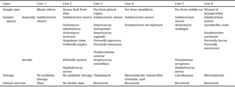
After the introduction of MALDI-TOF MS for the routine Table 1
Microbiological results of the six cases whereSolobacterium mooreiwas present alone or in mixed culture in the patient's sample.
Cases Case 1 Case 2 Case 3 Case 4 Case 5 Case 6
Sample type Blood culture Serousfluid from tibia
Pus from gluteal region
Pus from mandibula Pus from middle ear Wound of laryngectomy Isolated
species
AnaerobicSolobacterium moorei
Solobacterium moorei Solobacterium moorei Solobacterium moorei Solobacterium moorei
Solobacterium moorei Actinomyces
odontolyticus
Anaerococcus hydrogenalis
Fusobacterium necrophorum Actinomyces randingae
Lactobacillus orale Actinomyces
turicensis
Anaerococcus vaginalis
Fusobacterium nucleatum
Atopobium rimae Prevotella nigrescens Prevotella buccae
Veillonella atypica Prevotella timonensis Prevotella
nanceiensis Porphyromonas
somerae Aerobic Klebsiella oxytoca Streptococcus
constellatus
Pseudomonas aeruginosa Staphylococcus
aureus
Staphylococcus aureus
Therapy No antibiotic
therapy
No antibiotic therapy Clindamycin Metronidazole, Amoxicillin/
clavulanic acid
Ciprofloxacin Metronidazole
Clinical outcome Died No further data Recovered Recovered Recovered Recovered
Fig. 1.S. mooreicolonies of the subculture on Schaedler anaerobic blood agar after 48 hours of incubation in anaerobic chamber.
ari et al. / Anaerobe 65 (2020) 102241 3
Table 2
Overview of earlier published case reports of infections whenS. mooreiwas isolated.
Ref. Sample type Patient Medical history Culture results Antimicrobial therapy Clinical
outcome [13] Blood culture 43-year-old
woman
Cervix carcinoma Solobacterium moorei Cefuroxime/Piperacillin/tazobactam Recovered [3] Blood culture 67-year -old
man
Multiple myeloma Solobacterium moorei Cefepime Amikacin Vancomycin Recovered
[12] Blood culture 43-year-old man
Lymphoma, kidney transplantation Solobacterium moorei Phenoxymethyl-penicillin/ Benzylpenicillin Metronidazole
Recovered [12] Blood culture 64-year-old
woman
Colon cancer Solobacterium moorei Cefuroxime Metronidazole Recovered
[17] Blood culture 61-year-old man
Diabetes mellitus, rectal cancer Solobacterium moorei Streptococcus mitis
Cefuroxime Moxifloxacin/Vancomycin Meropenem
Died [12] Blood culture 66-year-old
woman
Non-small cell lung carcinoma Solobacterium moorei Eikenella corrodens
Meropenem/Ciprofloxacin Metronidazole
Recovered [12] Blood culture 33-year-old
woman
IVDA Hepatitis B infection Solobacterium moorei Actinomyces meyeri
Cefuroxime/Benzylpenicillin Metronidazole
Recovered [12] Blood culture 77-year-old
man
Ischemic heart disease Prostate cancer Solobacterium moorei Porphyromonas uenonis
Benzylpenicillin/Phenoxy- methylpenicillin
Recovered
[18] Blood culture 70-year-old man
HIV- positivity Solobacterium moorei
Campylobcater rectus
Amoxicillin/clavulanic acid Recovered [4] Blood culture 37-year-old
man
Femoral vein thrombophlebitis with septic pulmonary embolism
Solobacterium moorei Fusobacterium nucleatum
Bacteroides urealyticus
Penicillin Metronidazole Recovered
[5] Thigh abscess ND ND Solobacterium moorei
Actinomyces europeus Enterococcus faecalis Stenotrophomonas maltophilia Comamonas testosterone Corynebacteriumsp.
Corynebacterium urealyticum
Not reported Recovereda
[5] Abdominal wound abscess
ND Perforated appendix Solobacterium moorei
Peptosterptococcus micros
Slackia exigua Bacteroides fragilis Bilophila wadsworthia Enterococcus avium Eikenella corrodens Escherichia coli
Not reported Recovereda
[5] Axilla furuncle ND ND Solobacterium moorei
Stphylococcus aureus Propionibacteriumsp.
Corynebacteriumsp.
amycolatum
Not reported Recovereda
[5] Abdominal wound ND ND Solobacterium moorei
Prevotella nigrescens Parvimonas micra Streptococcus constellatus Fusobacterium nucleatum
Actinomyces turicensis Dialister pneumosintes Bacteroides urealyticus Pseudomonas aeruginosa Bacteroides fragilis Bacteroides ovatus
Not reported Recovereda
[5] Perirectal abscess ND ND Solobacterium moorei
Actinomyces turicensis Streptococcus anginosus Bacteroidessp.
Prevotella disiens Megasperasp.
Fusobacterium nucleatum Porphyromonassp.
Not reported Recovereda
[5] Perirectal abscess ND Diabetes mellitus Solobacterium moorei
Parvimonas micra
Not reported Recovereda
ari et al. / Anaerobe 65 (2020) 102241 4
identification of clinical isolates in our laboratory in 2013, a total of 32S. mooreistrains were isolated and identified by the end of 2019.
Most of theS. mooreicases (six) were discovered in 2016; these cases are presented in detail in this case report. During subcultur- ing, the clinical isolates ofS. mooreiform small, grey, non-hemolytic colonies with a 0.5e1 mm diameter on Schaedler blood agar after being incubated for 48 h in a strict anaerobic environment (Fig. 1).
Because of their small colony size, S. moorei colonies are often overlooked in mixed culture if the sample is not carefully spread on the original plate. In previous studies that primarily used molecular methods,S. moorei was mentioned as a member of a complex microbiome associated with different infections of the oral cavity such as periimplantitis [6], root canal [7], periradicular lesions [8], dentoalveolar abscess [9], apical endodontic abscess [10], and re- fractory periodontitis [11] together with other anaerobic and aer- obic bacteria. Furthermore, it has been suggested that S. moorei, which produces volatile sulfur compounds (e.g., hydrogen sulfide and methyl mercaptan) and organic acids (e.g., amines such as cadaverine, etc.), together with other oral anaerobic bacteria, may play role in the formation of halitosis [2,16].
Altogether, 19 clinical cases have been described in detail since 2006 in whichS. mooreiwas isolated alone or in mixed culture (Table 2). The reported cases ofS. mooreibacteraemia were mostly associated with cancers: cervix carcinoma with acute proctitis [13], kidney transplantation with lymphoma, lung carcinoma, colon cancer, prostate cancer with pneumonia and ischaemic heart dis- ease [12], and multiple myeloma [3]. Pedersen et al. reported on the isolation ofS. mooreifrom the blood culture of a patient who used intravenous drugs and was infected with hepatitis B [12]. Martin et al. reported onS. mooreibacteraemia in a case of femoral vein thrombosis with septic pulmonary embolism [4]. In four case
reports, S. moorei was isolated from blood culture as the only pathogen, but in some patients, in addition to S. moorei, other aerobic or anaerobic bacteria were present in the blood sample (Table 2). Our patient withS. mooreibacteraemia also had several debilitating conditions such as cancer and pyothorax. He died without any antibiotic therapy to treat the sepsis, despite the fact that the blood culture isolate was fully susceptible to all tested antibiotics (Table 1).
Zhen et al. [5] described nine cases of polymicrobial infections that had no clear connection with the oral cavity. These cases were obtained from wound infections, including abdominal abscesses, infected pilonidal cyst and abscess, axilla furuncle, perirectal ab- scesses, and abdominal wound infections, in whichS. mooreiwas isolated with higher or lower colony counts or was detected by 16S rRNA gene sequence analysis [5] (Table 2). The antibiotic suscep- tibility was also determined in a study on sixS. mooreiisolates. All isolates were fully susceptible to antibiotics usually used for the empirical treatment of mixed infections involving anaerobic bac- teria [5]. Thefive additional polymicrobial cases presented in this report in whichS. mooreiwas isolated, together with one tofive other different types of bacteria, were also associated with cancers (e.g., colorectal, facial, skin, laryngeal, lung) and immunosuppres- sive status (e.g., diabetes mellitus, chemotherapy, radiotherapy). In three patients, the infection had a direct connection with the oral cavity (including a middle ear infection, mandibular abscess, and wound infection after total laryngectomy) (Table 1). Except ourfifth case, in whichS. mooreiandA. randingaewere isolated only from the enrichment broth and were not considered real pathogens, the empirically initiated antibiotic therapy covered the mixedflora of the infection (Table 1). In the majority of published bloodstream infections involving S. moorei, the patients recovered, but in our Table 2(continued)
Ref. Sample type Patient Medical history Culture results Antimicrobial therapy Clinical
outcome Actinomyces turicensis
Bacteroides fragilis Fusobacterium equinum Streptococcus constellatus Staphylococcus aureus Escherichia coli [5] Infected pilonidal
cyst
ND ND Solobacterium moorei
Parvimonas micra Streptococcus anginosus
Bilophila wadsworthia Peptostreptococcussp.
Not reported Recovereda
[5] Thigh abscess ND IVDA, heroin Solobacterium moorei
Streptococcus anginosus Parvimonas micra Atopobiumsp.
Streptococcus constellatus
Not reported Recovereda
[5] Pilonidal abscess ND ND Solobacterium moorei
Parvimonas micra Fusobacterium gonidiaformans Atopobium minutum Slackia exigua Prevotella intermedia Tannerella forsythia Prevotella stomatis Bacteroides vulgatus Actinomyces turicensis
Not reported Recovereda
ND: No data IVDA: intravenous drug abuse.
aRecovered after surgical intervention and routine antibiotic treatment.
ari et al. / Anaerobe 65 (2020) 102241 5
case, and in a case recently reported by Liu et al. [17], the pro- gression of the infection due to the underlining diseases was rapid with [17] or without antibiotic therapy (ourfirst case), and the patient died.
The introduction of the MALDI-TOF MS method for the identi- fication of slow-growing and difficult-to-identify culturable bac- teria or fungi broadened our view about the possible pathogens of mixed infections. Conversely, the more time-consuming PCR-based method to identify culturable or non-culturable members of mixed infections has also been used by several authors [6e9]. The MALDI Biotyper system with various database developments focuses on different, difficult-to-identify species including anaerobic bacteria present in the normalflora or in different infections [19,20]. Ac- cording to our laboratory database, since 2013, we have identified 32S. mooreistrains on the species level with log (score) values of 2.000, including the isolates of the presented cases using the actual database developments of the MALDI Biotyper. Liu W-J. et al.
reported that they could not identify theirS. mooreiisolate with commercial biochemical tests or the MALDI-TOF MS method, but they could identify it with 16S rRNA gene sequencing [17]. There is no information on the version of the software applied in that study.
Several publications mention the isolation of S. mooreifrom mixed infections associated with the oral cavity or suggest the possible translocation of it from the gastrointestinal tract. However, the clinical significance of this rarely identified gram-positive anaerobic bacterium remains largely unknown because it is fully susceptible to antibiotics regularly used in mixed infections involving aerobic and anaerobic bacteria. Only one patient died due to sepsis whenS. mooreiwas the causative agent with the usage of targeted antibiotic therapy (Table 2). All other bacteraemia patients in whom S. moorei was present in at least one anaerobic blood culture bottle alone or together with some other anaerobic bacteria survived the infection because of appropriate antibiotic therapy.
The role ofS. mooreiin other mixed infections with abscess for- mation in different parts of the body, occasionally involving up to 10 different and mostly anaerobic bacteria, is very difficult to judge [5]. According to the published data and our experience, wound infections involvingS. mooreiresolved after routine surgical man- agement and various anti-anaerobic antibiotic regimens.
S. moorei, which is part of the human microbiota, seems to be a less virulent germ; however, in infectious processes that are con- nected with several debilitating predisposing factors, the patient may even develop bacteraemia involvingS. moorei.Our case reports demonstrate the value of the carefully used MALDI-TOF MS method for the identification of anaerobic bacteria includingS. moorei, the pathogenic role of which should be carefully evaluated in every case.
Ethical approval No. 4594.
Funding
This research received no external funding.
Author contributions
K.P.S. conceived and designed the study, Z.P. wrote case 1, S.K.
wrote case 2, R.K. wrote case 3, D.S. wrote case 4, T.V. wrote case 5, B.Sz. wrote case 6, and K.B. revised the full paper. All authors have read and agreed on the published version of the manuscript.
Declaration of competing interest
The authors declare no conflicts of interest to disclose, monetary or otherwise.
Acknowledgements
The authors would like to thank Prof. Dr. Elisabeth Nagy for her valuable proposals and advice during the writing of this manuscript.
References
[1] A. Kageyama, Y. Benno, Phylogenetic and phenotypic characterization of some Eubacterium-like isolates from human feces: description of Solobacterium mooreigen. nov., sp. nov, Microbiol. Immunol. 44 (2000) 223e227.
[2] V.I. Haraszthy, J.J. Zambon, P.K. Sreenivasan, M.M. Zambon, D. Gerber, R. Rego, C. Parker, Identification of oral bacterial species associated with halitosis, JADA 138 (8) (2007) 1113e1120.
[3] G. Detry, D. Pierard, K. Vandoorslaer, G. Wauters, V. Avesani, Y. Glupczynski, Septicemia due toSolobacterium mooreiin a patient with multiple myeloma, Anaerobe 12 (2006) 160e162.
[4] C.A. Martin, R.S. Wijesurendra, C.D.R. Borland, J.A. Karas, Femoral vein thrombophlebitis and septic pulmonary embolism due to a mixed anaerobic infection includingSolobacterium moorei: a case report, J. Med. Case Rep. 1 (40) (2007) 1e3,https://doi.org/10.1186/1752-1947-1-40. Open Access.
[5] G. Zheng, P.H. Summanen, D. Talan, R. Bennion, M.-C. Rowlinson, S.M. Finegold, Phenotypic and molecular characterization ofSolobacterium mooreiisolates from patients with wound infection, J. Clin. Microbiol. 48 (3) (2010) 873e876.
[6] T. Koyanagi, M. Sakamoto, Y. Takeuchi, M. Ohkuma, Y. Izumi, Analysis of microbiota associated with peri-implantitis using 16S rRNA gene clone library, J. Oral Microbiol. 2 (2010) 5104,https://doi.org/10.3402/jom.v2i0.5104. Open Access.
[7] H.J.A. Rolph, A. Lennon, M.P. Riggio, W.P. Saunders, D. McKenzie, L. Coldero, J. Bragg, Molecular identification of microorganisms from endodontic in- fections, J. Clin. Microbiol. 39 (2001) 3282e3289.
[8] J.F. Schirrmeister, A.-L. Liebenow, K. Pelz, A. Wittmer, A. Serr, E. Hellwig, A. Al- Ahmad, New bacterial compositions in root-filled teeth with periradicular lesions, J. Endod. 35 (2009) 169e174.
[9] J. Downes, M.A. Munson, D.A. Spratt, E. Kononen, E. Tarkka, H. Jousimies- Somer, W.G. Wade, Characterisation ofEubacterium-like strains isolated from oral infections, J. Med. Microbiol. 50 (2001) 947e951.
[10] N. George, E. Flamiatos, K. Kawasaki, N. Kim, Ch Carriere, B. Phan, R. Joseph, Sh Strauss, R. Kohli, D. Choi, J.C. Baumgartner, Ch Sedgely, T. Maier, C.A. Machida, Oral microbiota species in acute apical endodontic abscesses, J. Oral Microbiol. 8 (2016) 30989,https://doi.org/10.3402/jom.v8.30989. Open Access.
[11] A. Colombo, V. Paula, S. Boches, L. Sean, J. Goodson, R. Kent, A. Haffajee, S. Socransky, Comparison of subgingival microbial profiles of refractory periodontitis, severe periodontitis and periodontal health using the human oral microbe identification microarray, J. Periodontol. 80 (2009) 1421e1432.
[12] R.M. Pedersen, H.M. Holt, U.S. Justesen, Solobacterium mooreibacteremia:
identification, antimicrobial susceptibility and clinical characteristics, J. Clin.
Microbiol. 49 (7) (2011) 2766e2768.
[13] S.K.P. Lau, J.L.L. Teng, K.-W. Leung, N.K.H. Li, K.H.L. Ng, K.-Y. Chau, T.-L. Que, P.C.Y. Woo, K.-Y. Yuen, Bacteremia caused bySolobacterium mooreiin a pa- tient with acute proctitis and carcinoma of the cervix, J. Clin. Microbiol. 44 (8) (2006) 3031e3034.
[14] H. Jousimies-Somer, P. Summanen, D.M. Citron, E.J. Baron, H.M. Wexler, S.M. Finegold, in: Wadsworth-KTL. Anaerobic Bacteriology Manual, sixth ed., Star Publishing Company, Belmont, CA, USA, 2003.
[15] E. Nagy, A. Schuetz, Is there a need for the antibiotic susceptibility testing of anaerobic bacteria? Anaerobe 31 (2015) 2e3.
[16] F. Vancauwenberghe, J. Dadamio, I. Laleman, M. Van Tornout, W. Teughels, W. Coucke, M. Quirynen, The role ofSolobacterium mooreiin oral malodour, J. Breath Res. 7 (4) (2013), 046006,https://doi.org/10.1088/1752-7155/7/4/046006.
[17] W.-J. Liu, M. Xiao, J. Yi, T. Kudinha, Y-Ch Xu, First case report of bacteremia caused bySolobacterium mooreiin China, and literature review, BMC Infect.
Dis. 19 (2019) 730,https://doi.org/10.1186/s12879-019-4359-7.
[18] F.G. Genderini, D. Martiny, F. Ponthieux, M.A. Argudín, M. Gomez Galdon, A. Zaarour, C. Garcia, A. Libois, M. Gerard, N. Dauby, First case ofCampylo- bacter rectusandSolobacterium mooreimixed bacteraemia successfully iden- tified by MALDI TOF-MS, NMNI 31 (2019), 100587.
[19] M. Kostrzewa, E. Nagy, P. Schr€ottner, A.B. Pranada, How MALDI-TOF mass spectrometry can aid the diagnosis of hard-to-identify pathogenic bacteriae the rare and the unknown, Expert Rev. Mol. Diagn. 19 (8) (2019) 667e682.
[20] A.C.M. Veloo, H. Jean-Pierre, U.S. Justesen, T. Morris, E. Urban, I. Wybo, H.N. Shah, A.W. Fridrich, E. Nagy, M. Kostrewa, A multi-center ring trial for the identification of anaerobic bacteria using MALDI-TOF MS, Anaerobe 48 (2017) 94e97.
ari et al. / Anaerobe 65 (2020) 102241 6