Cytochrome Systems in Anaerobic Electron Transport
JACK W . N E W T O N ! AND MARTIN D . KAMENJ
I. Introduction 397 II. General Characteristics of Electron Transport in Anaerobes 398
A. Thermodynamic Considerations 398 B. Phosphorylation Systems of Anaerobes 401
III. Hematin-Protein Components 404 A. Nomenclature and Variation of the Hemoproteins 404
B. Specific Compounds 406 IV. Function of Heme Proteins 413
A. Spectrophotometric Studies on Purple Bacteria 413 B. Observations on Nitrate-Reducing Bacteria 416
C. Sulfate Reduction 418
V. Conclusions 419 References 420
I. Introduction
T h e division of t h e bacteria into aerobes a n d anaerobes constitutes an i m p o r t a n t nutritional criterion in bacteriology. I t is well k n o w n from com
p a r a t i v e biochemistry, however, t h a t this division cannot be rigidly m a i n tained.
T h e oxidoreduction mechanisms of chemosynthetic a n d photosynthetic bacteria can be considered analogous, although m a n y diverse bacterial genera exist grading from obligate anaerobes t o aerobes. Among t h e chem
osynthetic bacteria, those which reduce compounds of sulfur in various oxidation s t a t e s are anaerobes, those which reduce nitrogen compounds in various oxidation states are facultative anaerobes, and those oxidizing r e duced sulfur a n d nitrogen compounds are obligate aerobes. These bacteria grow either b y using energy derived from t h e oxidation of reduced s u b -
* The term "electron transport" is used in this chapter in its general sense without reference to any specific oxidoreduction mechanism.1
t Present address: Pioneering Laboratory for Microbiological Chemistry, Northern Utilization Research and Development Division, U. S. Dept. of Agriculture, Peoria, 111.
ί Researches by the authors referred to in this chapter have been supported by grants form the National Science Foundation, the C.F. Kettering Foundation, and the National Institutes of Health. This chapter is Communication N o . 44 in the series, Publications of the Graduate Department of Biochemistry, Brandeis Univer
sity. General reviews of interest which further elaborate topics discussed in this chapter are cited in the list of references.
397
strates with preformed electron acceptors or by oxidation of preformed electron donors with specific exogenous oxidants.
Among the photosynthetic bacteria there are obligate anaerobes, such as the green and purple sulfur bacteria and the facultative photoheterotrophic nonsulfur purple bacteria. Again, the photosynthetic bacteria can oxidize reduced substrates only by means of specific photooxidants generated by the absorption of light energy.
All of these bacteria differ basically from the strictly fermentative a n - aerobes in t h a t they cannot form electron acceptors solely by dismutation of substrate. They require t h a t both electron acceptors and donors be ex- ogenously supplied. I n the interaction between donor and acceptor, elec- tron transport systems are required which contain a variety of hematin compounds, t h a t is, heme proteins which are analogous to, b u t apparently not identical with, those found in aerobic tissues. Little similarity is found with fermentative microorganisms such as the Clostridia, which appear to lack electron carrier systems containing heme proteins.
Virtually all of our knowledge concerning the electron transport mech- anisms of these "oxidative anaerobes," as the chemosynthetic and photo- synthetic bacteria might be called, has only been obtained recently. H o w - ever, sufficient d a t a are now available to permit a discussion of some of the characteristic properties of cytochrome systems which occur in these groups of bacteria.
II. General Characteristics of Electron Transport in Anaerobes
A. THERMODYNAMIC CONSIDERATIONS
I t is informative to estimate t h e maximum energy available for biochem- ical use in the electron transport systems which m a y be encountered in anaerobes. I n the example of mammalian aerobic respiration a theoretical limit is set by the effective potential (2iV) of t h e oxidized-reduced di- phosphopyridine nucleotide ( D P N ) couple and those for typical substrates
(approximately —0.4 to —0.32 volt) and t h a t of t h e oxygen electrode ( a p - proximately + 0 . 8 volt). T h e flow of electrons between these two end points is mediated b y a series of oxidoreduction systems involving pyri- dine nucleotides, dehydrogenases, flavin enzymes, and cytochromes. T h e storage of energy released in this process is effected b y coupled phos- phorylation of inorganic phosphate, t h e biochemically useful energy a p - pearing eventually as adenosine triphosphate ( A T P ) . F o r each electron transferred across the transport chain t o oxygen, three phosphate esteri- fications can occur. Because the energy equivalent of approximately 1.2 electron volts is generated b y each electron transferred, and because each polyphosphate bond formed requires nearly 0.35 electron-volt equivalents,
practically all of the energy liberated by electron transport can be t r a n s - formed into phosphate-bond energy.
With the exception of the particles derived from photosynthetic bac- teria, there have been no demonstrations of phosphorylations coupled to electron transport in the anaerobic bacteria, although some preliminary attempts have been made with cells of nitrate reducers2 and clostridial extracts.3 , 3 a If it is assumed t h a t the starting potential for nitrate reduc- tion is near the D P N - D P N H system and t h a t the potential of the nitrate- nitrite couple is -f 0.5 volt,4 then the total potential change is reduced to approximately 0.9 volt, thus "shortening" the assumed electron transport chain and reducing the possible yield of ester phosphate, as A T P , per single electron transfer. However, as will be discussed later, it is not clear for w h a t hydrogen donor nitrate serves as terminal oxidant. T h e available evidence indicates t h a t several p a t h w a y s exist in different organisms for electron transport to nitrate. Therefore, the point of insertion of the ni- trate-nitrite system in the chain will determine the ester p h o s p h a t e / N 03~ obtained. Subsequent experiments m a y show t h a t this ratio does in fact v a r y from one carrier system to another.
I t is possible to demonstrate a phosphorylation coupled to the aerobic oxidation of ferrocytochrome c by m a m m a l i a n preparations, thereby prov- ing the availability for coupling to phosphorylating systems of electron transport above the cytochrome c level.5 T h e theoretical maximum poten- tial drop would be about 0.5 volt, the actual drop depending, of course, on the exact nature of the oxidant. T h e c-type cytochrome isolated from Pseudomonas denitrificans has a potential at p H 7 of +0.32 volt,6 and consequently, if coupled to the nitrate-nitrite system, could barely provide a one-step potential change adequate for synthesis of a pyrophosphate bond. As an oxidant for a cytochrome b, however, the nitrate system could provide an electron span adequate for one phosphorylation, assuming an oxidation potential of about zero volts for b cytochromes.7 - 9
When considering the sulfate system as oxidant, some special problems arise. Desulfovibrio desulfuncans contains a cytochrome " c " (so-called
"cytochrome c3") with an E0' of —0.205 volt, which can act as a carrier in the reduction of sulfite, thiosulfate, and tetrathionate or dithionite by hydrogen.1 0 Since hydrogen, or substrates at or near the same electrode potential as hydrogen, can serve as electron donors for growth, it is a p - parent t h a t the usable potential span extends only from E0' — —0.4 volt to E q ~ —0.188 volt. W i t h only approximately 0.2 volt per electron avail- able as a potential drop, insufficient energy is provided to produce one pyrophosphate linkage even with the whole span utilized. I t is necessary to assume t h a t at least two electrons are transferred. I t should be noted, as we will see in a later section, t h a t cytochrome c3 contains two heme moi-
eties per mole. An alternative mechanism—that of substrate level phos- phorylation such as encountered in the oxidation steps of glycolysis—may also be operative in addition to coupling through electron transport.
Active phosphorylating preparations are readily obtained from photo- synthetic a n a e r o b e s .1 1 - 1 4 All of the preparations obtained thus far require light, but otherwise are substrate-independent. I t is generally assumed t h a t an oxidant is formed in the light which oxidizes the terminal elec- tron acceptor and draws electrons through a "respiratory" chain coupled to phosphorylation. I n this system it is still not established whether the electrons are donated by bound substrates, or by photoreductant derived from water directly. T h e amount of phosphate which can be esterified in the absence of added substrates favors the latter alternative. This system is an anaerobic variant—it might be called "fermentation of water"—
since the water is apparently providing both electron donor and acceptor systems. So far, no q u a n t u m yield has been determined for photophospho- rylation by extracts of purple bacteria, but d a t a which will be discussed later suggest t h a t it m a y be a fairly efficient process.1 5
Pyridine nucleotides, flavin, and large amounts of cytochrome have been detected in particles from photosynthetic bacteria which carry out light-induced phosphorylation.1 6 Therefore, the minimum requirements of an electron transport system are present. A light-dependent oxidation of the particle-bound cytochrome has been demonstrated, both in whole cells and isolated particles,1 7"2 6 and evidence for the involvement of pyri- dine nucleotide and flavin has recently been obtained.2 6*' b
T h e anaerobic photosynthetic bacteria also appear to contain a "com- pressed" electron transport system. For, if the electron transport chain of Chromatium, for example, begins at the level of reduced pyridine nucle- otide, and ends at the bacterial cytochrome c (EQ = —0.040 volt) ,2 7 vir- tually all of the chain is required for synthesis of only one pyrophosphate bond. I t is possible t h a t the photooxidant provides an acceptor at a much more oxidizing potential, however, as is most probably the case with the facultative photoheterotroph, Rhodospirillum rubrum (see Table I ) .
F r o m this discussion it should be clear t h a t it is not possible to ex- trapolate directly from mammalian respiratory systems to those found in the microorganisms cited, for none of these electron transport systems contains all of the essential components of a " t y p i c a l " electron transport
"chain." As we shall see in a later section, none of these organisms contains cytochromes of the " a " type. While practically all contain c-type cyto- chromes, none of the bacterial " c " cytochromes are identical in physico- chemical and biological properties with the m a m m a l i a n compounds.
Whether the relatively slow growth rates of chemosynthetic anaerobes are a reflection of a " r u d i m e n t a r y " electron transport system is a t present an open question, and perhaps one of general biological significance.
T A B L E I
ELECTROCHEMICAL PROPERTIES OF CYTOCHROMES "C" AND SOME ACCEPTOR SYSTEMS UTILIZED FOR GROWTH
Chromatium D Pseudomonas denitrificans
Desulfovibrio desulfuricans Cytochrome oxidant "(OH)," photo- N 03-
oxidant*
Estimated potential (0.0 volt)* + 0 . 5 volt - 0 . 1 8 8 volt first step couple, E0'
Eo' heme compound - 0 . 0 4 volt + 0 . 3 2 volt - 0 . 2 0 5 volt
a See text.
B . PHOSPHORYLATION SYSTEMS OF ANAEROBES
I t is of historical interest t h a t the first demonstration of a phosphoryla- tion reaction in bacteria apparently "coupled" to electron transport was m a d e using an anaerobic system, "chromatophores" from the photosyn- thetic bacterium Rhodospinllum rubrum.11 Enough d a t a have now been obtained with such systems to w a r r a n t some statements about the general characteristics of these anaerobic phosphorylating preparations. Both t h e R. rubum s y s t e m1 2' 2 6 and the phosphorylating particles from the ob- ligately anaerobic photoautotroph Chromatium1* have the following prop- erties: (1) they are inhibited by oxygen; (2) they are very stable enzyme preparations, particulate in n a t u r e ; (3) they are relatively insensitive to cyanide and carbon monoxide, but inhibited by certain iron binding agents; (4) catalytic amounts of "poising" reagents stimulate the phos- phorylation reaction; (5) all require magnesium ions and are specific for purine nucleotides; T h e phosphate acceptor is adenosine diphosphate; (6) cytochromes and other electron transport components are tightly bound in the preparations, which contain large amounts of phospholipid.
As mentioned previously, phosphorylation coupled to the reduction of nitrate or sulfate has not y e t been demonstrated. However, if past ex- perience with other anaerobic systems serves as a guide, it is probable t h a t such phosphorylations will be found to be inhibited by oxygen. This seems especially probable for the sulfate-reducing system, because its low potential cytochrome is readily autoxidizable. I t has been known for some time t h a t oxygen and nitrate can compete with one another as terminal electron acceptor for growth of facultative anaerobes, this being com- pletely analogous to the competition between light and oxygen manifested by photosynthetic heterotrophs. I t seems likely therefore t h a t any phos- phorylating preparation which will be found coupled to nitrate reduction will also show oxygen competition, or perhaps inhibition, as is character- istic of the photophosphorylating preparations.
All of the bacterial phosphorylating systems studied thus far have been found to be relatively stable, particulate enzyme complexes. Particle size varies, depending on the organism and method of preparation.* I n the photosynthetic bacteria the site of A T P synthesis is the bacterial "chro- m a t o p h o r e , "2 8 a relatively large (400-1000 A.) subcellular macromole- cule which contains the photosynthetic pigments of the cell. T h e phos- phorylation system is not very dependent upon particle integrity, because chromatophore fragments prepared by a variety of procedures can carry out light-activated phosphorylation. The bacterial particles appear to be rather rigidly attached to the cell, because they are not immediately re- leased on cell r u p t u r e .1 6 Chromatophores, as usually prepared by differ- ential centrifugation, contain large amounts of polysaccharide which a p - pears to be derived from the bacterial cell wall. I n addition, they contain the bulk of the cellular heme protein and are very rich in an ethanolamine- containing phospholipid. These and other observations all suggest t h a t the site of electron transport and phosphorylation in the purple bacteria is in or near the bacterial cell surface, as it appears to be in aerobic bac- teria.2 9' 3°· 3 0 a
The bacterial phosphorylating system from the anaerobe Chromatium contains very large amounts of nonheme iron, and the phosphorylation reaction is very sensitive to iron-chelating r e a g e n t s .1 6 I t resembles in some ways parts of the mammalian electron transport system, especially the succinic dehydrogenase complex, because, for example, it is greatly stimulated by catalytic amounts of phenazine m e t h o s u l f a t e .2 6 , 1 3 P h o t o - phosphorylation is inhibited by 2,4-dinitrophenol, but at relatively high concentrations; the same is true for c y a n i d e .2 6'1 3
Since the bacterial myokinase is soluble and can be readily separated from the phosphorylating particles, it has been possible to show t h a t A D P (adenosine diphosphate), and not A M P (adenosine monophosphate), is the primary phosphate acceptor in the coupled phosphorylation reac- t i o n .1 2 Similar observations have been made with preparations from aerobic b a c t e r i a .3 1
Fragments of particulate phosphorylating particles from the anaerobes can be shown to be stimulated by protein fractions and soluble factors from the supernate of cell e x t r a c t s .1 3 , 3 2 > 3 3 Similar results of fractionation studies have been reported using preparations from aerobic b a c t e r i a .3 4
One of the more remarkable features of the anaerobic phosphorylating system found in the purple bacteria is its sensitivity to its electrochemical
* The reader will find detailed discussions of more recent work on bacterial chromatophores in the report of a symposium entitled "The Photochemical Appa- ratus, Its Structure and Function," published as #BNL512(C-28) by the Brookhaven National Laboratory, Upton, New York. This document is available from the Office of Technical Services, Department of Commerce, Washington, 25, D. C.
environment. Catalytic amounts of oxidizing and reducing agents can be shown to act synergistically and antagonistically in activating and in
hibiting light-activated phosphorylation.1 3 These effects are not very spe
cific ones, because a variety of oxidizing and reducing systems can be used to stimulate photophosphorylation. T h e reagents appear to act as
"poising" agents on the electron transport system. T h a t is, they bring it into an optimal electrochemical condition which permits the most ef
ficient electron flow through the presumed series of electron transport components. This effect appears to be analogous, only on a more complex level, to the effect of redox potential on the rate of metalloflavoprotein re
actions.3 5
W i t h the bacterial systems there are varying degrees of activation, de
pending on the redox couple used, but it seems significant t h a t the reagents most effective in activating the phosphorylation reaction are those with redox potentials near zero volts, such as ascorbate and phenazine metho- sulfate.1 3 I t is of interest in this connection t h a t the cytochrome which is firmly attached to the phosphorylating particle has an oxidation-reduction potential in this range (Ζ?0' = —0.040 v o l t ) ,2 7 which m a y explain why these reagents can couple so effectively to the redox systems involved in photophosphorylation. I t is easy to show by differential spectrophotome
t r y t h a t ascorbate only partially reduces the cytochrome system even when added in great excess.
As was mentioned earlier, the anaerobic photophosphorylation systems are inhibited by molecular oxygen. A clue to the mechanism m a y lie in an early observation3 6 t h a t photosynthetic bacteria, and cell-free "chro
matophores" from them, are capable of carrying out a light-activated photooxidation of ferrocytochrome and reduced dyes by molecular oxygen.
I t is generally thought t h a t oxidants, formed by a combination reaction between the reducing system generated in the light and oxygen, are re
sponsible for at least p a r t of the oxidizing power, and, in addition, the photochemical " ( O H ) " system is involved.
I t is pertinent t h a t of all substrates studied, the one which is photooxi- dized most rapidly consists of 2,6-dichlorophenolindophenol, reduced with ascorbate. Although the leuco dye can be photooxidized slowly when added alone, the rate of photooxidation is at least seventeen times as fast when an excess of ascorbate is present.3 7 Several other reducing agents are ef
fective, but none is as active as ascorbate. I t is significant t h a t , when the ascorbate-indophenol couple is added to phosphorylating Chromatium particles under anaerobic conditions, an inhibition of phosphorylation is observed, but only under conditions t h a t would permit maximum photo
oxidation if air were present, namely, an excess of ascorbate with the dye mediator.1 3
All of these d a t a lend support to the suggestion t h a t at least p a r t of the
electron transport pathway involved in photophosphorylation is also used for photooxidation when air is present, and can be made to "uncouple"
photophosphorylation—even under anaerobic conditions—merely by pro- viding a catalytic amount of a proper oxidation-reduction couple.
I t is worth considering further how ascorbate m a y function in activat- ing both the photooxidase system on one hand, and the photophosphoryla- tion system on the other. W e have referred to its "poising" effect on the heme proteins of the electron transport system. However, this property does not entirely explain the possible function of ascorbate, because other mild reducing agents are not as active in promoting photooxidation or phosphorylatiori activity. A suggestion of an additional function of as- corbate can be taken from studies3 8- 3 9 on the remarkable effect of enediols on heme protein reactions. Using dihydroxymaleic acid it is observed t h a t the enediol permits peroxidase preparations to simulate oxidase action.
In addition, it is possible to show oxygen uptake by cytochrome c in the presence of manganous ions and the enediol. Manganous ion forms H202 from the enediol. I n the presence of ferrous ion, which also stimulates oxygen uptake, no peroxide is formed because it is removed by reaction between the enediol and H202 . I t seems possible t h a t either ascorbate or an enediol analogous to the latter m a y be functioning in a similar manner in the bacterial preparation and causing photochemical oxygen u p t a k e cata- lyzed by photoperoxides and the bacterial heme proteins. The absence of any detectable peroxide formed by the photooxidase can be attributed to the large amounts of iron in the bacterial particles, which should lead to rapid peroxide decomposition and additional ascorbate oxidation. I t is of interest t h a t in addition to ascorbate, a variety of enediols, including dihydroxymaleic acid, stimulate photophosphorylation by Chromatium particles.6 3
Regardless of what the eventual chemical mechanism m a y be proven to be for coupling and uncoupling of the photophosphorylation system to terminal oxidant, available evidence supports the view t h a t anaerobic photooxidation of the cytochrome present in the photophosphorylating particles from purple bacteria is a consequence of the photochemical act and probably a necessary requisite for the phosphorylation reaction (see Section I V ) . T h a t this m a y be a general phenomenon in photosynthetic organisms is suggested by the finding of photooxidations of cytochromes in green plant cells and e x t r a c t s .4 0 - 4 2
III. Hematin-Protein Components
A . NOMENCLATURE AND VARIATION OF THE HEMOPROTEINS
Current concepts of hemoprotein structure come almost exclusively from studies of mammalian hematin compounds, particularly most recently
from elegant work on cytochrome c .4 3"4 5 From these studies it seems very probable t h a t the ferroheme group is surrounded by four peptide spirals and t h a t the iron is bound out of the porphyrin plane to histidine residues contributed by adjacent peptide spirals. T h e vinyl side chains of the heme are saturated by condensation and formation of thioether linkages to cysteine residues. In a study of the amino acid composition of a highly purified c-type cytochrome from the denitrifying bacterium Pseudomonas denitrificans,™ it was found t h a t the amino acid content differed radically from t h a t of the mammalian compound. However, sufficient histidine was present to account for a structure for the bacterial cytochrome c similar to t h a t postulated for beef heart cytochrome c. Nevertheless, it is a p parently not advisable to draw rigorous conclusions about structure of bacterial heme proteins from the studies with m a m m a l i a n preparations.
Cytochromes have been grouped into three general categories to ra
tionalize spectroscopic observations of respiring mammalian cells. These are cytochromes of the a type with absorption maxima for so-called
" a l p h a " bands in the spectral region around 600 τημ, b type cytochromes characterized by an alpha absorption band at about 560 τημ and a beta band at approximately 525 to 530 τημ, and cytochromes of the c type with alpha and beta band absorption maxima a t 550 and 520 τημ. I n addition, the so-called Soret absorption in the 400 τημ region, characteristic of the tetrapyrrol structure, is a property of these hemoproteins. Electron flow to oxygen appears, from spectroscopic and other evidence, to t a k e place in the order: b to c to a, the last cytochrome also constituting the terminal part of the electron transport system.
All anaerobic bacteria studied thus far which are capable either of sul
fate reduction, nitrate reduction, or photosynthesis contain cytochromes spectroscopically similar to the c t y p e with absorption maxima in the visible near 550 and 520 τημ. Beyond a similarity in absorption spectra, however, the isolated bacterial proteins have very little in common with the m a m m a l i a n components. They v a r y not only in enzyme specificity, but also in electrochemical potential, electrophoretic mobility, and a vari
ety of physicochemical properties. T h e greatest variations in properties are, as might be anticipated, in those associated with the protein moiety.
One of the major problems in the biochemistry of heme proteins, which could probably be attacked fruitfully through study of the bacterial heme proteins, is the manner in which great variation in electrochemical proper
ties is brought about by alterations in protein structures, which in turn are effective through interaction with t h e extraplanar electrons of the metal chelate. A clue to how some types of structural and functional variation m a y be accomplished might be taken from early w o r k4 7 on redox potentials of simple iron chelates. I t was found t h a t a gradual change in redox potential of iron complexes of organic acids occurred as the number
of carbon atoms of dicarboxylic acids was increased. I n the series from oxalate upward, changes in redox potential were ascribed to increasing molecular dimensions of the iron chelate. T h e iron atom was assumed to be forming p a r t of a ring system, the redox potential of which changed as the ring increased in size.
Ionization of protons from "heme-linked" acidic amino acid groups to which the prosthetic groups are attached has been known for some time to alter the affinity of the central iron atom for ligands ("Bohr effect").
I n addition, however, it has been s h o w n4 8 t h a t in the p H range 8.5 to 9.4, where ionization of carboxyl groups linked to the heme group is not likely to be involved, the ionization of other acidic groups on the protein can influence the reactivity of the central iron atom. A more detailed study of the protein constituents of bacterial heme proteins m a y give some insight into the types of groups responsible for the great a r r a y of electrochemical properties found among these compounds.
All of the c type cytochromes have been demonstrated to be oxidized in enzyme extracts and cells by their corresponding terminal oxidants—
sulfur compounds, nitrate, and the photooxidant of the purple bacteria.
No cytochromes of the a type have been found in any of these bacteria, al- though some other hemoproteins of varied nature are sometimes detected.
Consequently, the nature of any possible hemoprotein mediator between the c cytochromes and their oxidants remains to be clarified. I t seems pos- sible from in vitro studies t h a t enzymic mediators between the terminal oxidants and cytochrome c, if they exist, either are not hemoproteins, or are functional variants of cytochrome c.
T h e varied nature of bacterial cytochromes of the c type found in anaerobes makes it increasingly important to formulate nomenclature adequate to describe and identify the various compounds. A suggestion has been m a d e4 9 seeking to identify the various heme proteins by speci- fying isolation source and the position of the reduced alpha band, viz., Chromatium cytochrome 552. This has the advantage of readily identify- ing the heme protein source, and makes no commitment as to function.
However, the terminology is apt to become cumbersome. I t m a y be neces- sary, because of the great variation in properties of hemoproteins derived from closely similar organisms, t h a t the nomenclature adopted retain some reference to cellular origin. I t is clear t h a t the present terminology is inadequate for description of these spectroscopic entities from microor- ganisms.
B . SPECIFIC COMPOUNDS 1. CYTOCHROMES OF THE C T Y P E
Some of the heme proteins of anaerobes which have been isolated and investigated in some detail will now be considered. B y far the largest
group of cytochromes which have been studied are those of the c type.
These compounds have been detected in both cells and cell-free extracts.
Owing to their stability they can occasionally be extracted using proce
dures which usually denature most of the other cellular proteins, and con
sequently they are easily purified. W i t h some organisms, extraction of dried cells with cold dilute acids suffices. T h e heme protein, being soluble, is readily separated from precipitated cell debris. On the other hand, several of the obligate anaerobes do not yield their cytochromes when extracted directly with aqueous media; a preliminary defatting of the cell is required.1 0 , 2 7' 5 0 Sonic t r e a t m e n t6'1 6 appear to dissociate the cyto
chromes from their cellular origin, which appears to be near the cell surface.
Butanol solubilization has been u s e d5 1 , 5 2 as well as detergent t r e a t m e n t or alkaline extraction.5 3 Once the heme proteins are brought into solution, they can usually be purified by conventional means.
a. Compounds from Obligate Anaerobes. Table I I summarizes some of the properties of the c-type cytochromes which have been isolated from two obligate anaerobes, Chromatium, strain D , and Desulfovibrio desul- furicans. Chromatium is generally grown as a photoautotroph, e.g., it is grown in light on media containing reduced sulfur compounds as electron donor, and carbon dioxide as carbon source. Desulfovibrio, in contrast, is
T A B L E II
PROPERTIES OF C-TYPE CYTOCHROMES FROM OBLIGATE ANAEROBES Organism
Properties Desulfovibrio Chromatium D '7 2 7 a , , / . 1 0
aesulfuricans10 Spectra (πΐμ)
Oxidized Soret Reduced maxima
Reduced pyridine hemochromogen Reaction with acetone-acetic acid Silver salt cleavage of prosthetic group
(thiol linkage) Heat stability Autoxidizability CO complex
Isoelectric point, pH Molecular weight
Molecular weight per heme Iron content, %
Iron/heme Heme/mole E0', pH 7.0
410 410
416, 523, 552 419, 525, 553 412, 519, 551 408, 517, 546
None None
Yes Yes
Labile Stable
Rapid Rapid
Slight reaction None
5.4 —10.4
97,000 —12,000
—32,000 —6,000
— 0.9
— 1
3 2
+ 0 . 0 1 volt - 0 . 2 0 5 v o l t
usually grown on organic media containing sulfate. Direct potential meas- urements have indicated t h a t the organisms grow in a very reducing en- vironment, a range of —0.100 to —0.300 volt appearing best for growth initiation in cultures of marine sulfate reducing bacteria.5 4 Cells do not grow from small inocula unless media are supplemented with reducing agents.5 5' 5 6
Another cytochrome c has been isolated from the green sulfur bacteria Chlorobium limicola53 and Chlorobium thiosulfatophilum.57 I n these bac- teria also, low potential c cytochromes are found. T h e heme proteins m a k e up a large percentage of the total cellular protein of these organisms.
I t is of interest t h a t all of these anaerobic bacteria which possess cyto- chromes of low oxidation potential have also in common the ability to utilize reduced sulfur compounds in one way or another for growth. The terminal oxidants involved appear to be different, sulfate in the sulfate- reducers, and photooxidants in the photosynthetic sulfur bacteria. I t is difficult to say a t present whether the low values for the electrochemical potentials of cytochromes present in these organisms are simply reflections of their all living in reducing environments. Nor can the question whether the photoxidant acceptor generated in photosynthesis by the anaerobic bacteria is of relatively low potential be answered.
The cytochrome from Desulfovibrio has been named cytochrome c3 .1 0 I t has been brought to a purity of at least 94% by cellulose and ion-ex- change chromatography. Degradation studies show it to contain two heme groups per mole protein, which accounts for its high iron content of 0.9%.
Both heme groups appear to be attached to the protein by thioether link- ages as in mammalian cytochrome c. T h u s this remarkable heme protein is another example of a cytochrome c variant, but the changes in proper- ties from t h a t of the mammalian c are accompanied by addition of a second prosthetic group to the protein molecule.
The Chromatium cytochrome has recently been brought to a state of high purity by cellulose chromatography.2 7* Studies with intact cells which will be discussed later indicate, however, t h a t Chromatium m a y contain an array of cytochromes of the c type, which m a y represent func- tionally different bound forms of the hemoproteins. Precise characteriza- tion of the electron transport system of Chromatium m a y require some improvements in isolation methods for the tightly bound cellular hemo- proteins.*
* Recent studies2 7* have demonstrated the soluble preparation described in Table II for Chromatium heme protein to be a mixture of two heme proteins. One of these, a cytochrome c-type, has a molecular weight of~95,000 and contains three heme groups per mole of protein. The other, a CO-binding protein is similar to the "RHP"- type protein of the faultative photoheterotrophs has a molecular weight of~35,000 and contains two heme groups per mole.
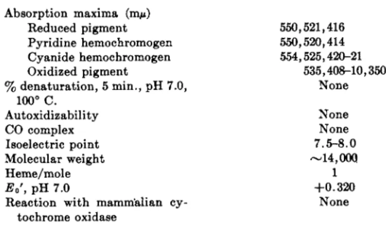
b. Compounds from Facultative Anaerobes. T h e first cytochrome of the c type isolated from bacteria was obtained from the facultatively anaerobic photoheterotroph Rhodospirillum rubrum in 1953.5 8 T h e compound was found to be present in high concentration, and occurred largely in the soluble fraction of the cell.5 9 I t could be readily extracted from R. rubrum cells with w a r m acid, and had an absorption spectrum almost identical with t h a t of mammalian cytochrome c. I n initial investigations on this pigment, it was believed to be indistinguishable from m a m m a l i a n cyto
chrome c. More extensive studies showed, however, t h a t its enzymic and electrochemical properties were quite different from m a m m a l i a n c .e o These observations provided the first clues leading to the revelation of the great variation in bacterial cytochromes c. Some of the properties of this pig
ment are listed in Table I I I .5 9
A distinguishing feature of the R. rubrum compound is its relatively high redox potential, some 80 to 100 millivolts more positive t h a n the mammalian compound. This m a y account for its inability to be oxidized by m a m m a l i a n cytochrome oxidase preparations,6 0' 6 1 and suggests a role for the pigment in photometabolism analogous to t h a t postulated for green plant cytochrome f.62
T h e R. rubrum cytochrome c has now been well characterized.5 9 I t is a typically small [M.W. (molecular weight) approximately 14,000], heat- stable hemoprotein, contains a prosthetic group apparently identical t o t h a t of mammalian c, and is not autoxidizable. Both in vitro and in vivo spectroscopic observations and enzymic studies suggest an important role for this pigment in the electron transport system of R. rubrum (see Section I V ) .
TABLE III
SOME PROPERTIES OF CYTOCHROME C FROM Rhodospirillum rubrum69 Absorption maxima (ιημ)
Reduced pigment Pyridine hemochromogen Cyanide hemochromogen Oxidized pigment
550,521,416 550,520,414 554,525,420-21
535,408-10,350 None
% denaturation, 5 min., pH 7.0, 100° C.
Molecular weight Autoxidizability CO complex Isoelectric point
None None 7.5-8.0
—14,000
Heme/mole 1
Eo', pH 7.0
Reaction with mammalian cy-
+ 0 . 3 2 0 None tochrome oxidase
T A B L E I V
CYTOCHROMES OF C TYPE FROM NITRATE-REDUCING BACTERIA Organism Properties Pseudomonas
denitrificans
Micrococcus denitrificans Absorption maxima (ιημ)
Reduced pigment
Reduced pyridine hemochromogen Reduced cyanide hemochromogen Oxidized pigment
552,525,418 551,520,415 556,527,422 412-13
550,522,416 550,520,414 554,525,420
410-11 Eo', pH 7.0
Oxidized by mammalian cytochrome
+ 0 . 3 2 volt No
+ 0 . 2 5 volt Yes oxidase
R. rubrum, as well as other photosynthetic bacteria, contain large amounts of heme proteins, especially of the cytochrome c type. I t seems rather unlikely, however, t h a t all of this heme protein is functional in t h e organisms, because t h e cytochrome content varies considerably with nu
tritional factors. Cells grown on media low in iron are especially low in cytochrome content, y e t are active metabolically.6 3
Cytochromes of the " c " type have been isolated from the denitrifying bacteria Micrococcus denitrificans and Pseudomonas denitrificans* Some properties of the pigments are given in Table IV. T h e cytochromes of M.
denitrificans and P. denitrificans are most easily obtained by homogeniza- tion of cell pastes with glass beads in a high-speed blendor.6 Other methods of extraction are relatively ineffective.
I t is of interest t h a t the cytochrome c from M. denitrificans is the first bacterial cytochrome of the c type which is indistinguishable spectroscopi- cally, electrochemically, and enzymically from mammalian cytochrome c.
Recent investigations have shown t h a t a series of soluble cytochromes of the c type can be isolated in high degree of purity from Pseudomonas aeruginosa™' 6 4 an aerobic denitrifier. These cytochromes are inactive enzymically when incubated in air with mammalian cytochrome oxidase, but can be oxidized rapidly by a soluble carbon monoxide-binding oxidase which can also be isolated from the organism. However, this bacterial oxi
dase, while a heme protein with a c-type spectrum, appears to be a mixture of c-type and an a2- t y p e cytochrome. M a n y examples of c cytochromes inactive toward cytochrome c oxidase are known in aerobic denitri- fiers51' 5 2' 6 5
2. CYTOCHROMES OF THE b T Y P E
Few compounds of the b type have been detected in the bacteria and a very limited number have been observed in the anaerobes. Among the obligate anaerobes, no b cytochromes have been detected thus far. A cyto- chrome b difference spectrum has been observed5 9 in particles obtained from an acetone powder of R. rubrum after removal of the cytochrome c, but the component responsible for the absorption bands has not been iso- lated. More r e c e n t l y6 6 cytochromes of the b type have been isolated from several nitrate-reducing bacteria. Both M. denitrificans and P. denitrifi- cans have been found to contain, in addition to cytochrome c, compounds with absorption maxima in the reduced state a t 559, 528, and 426 m^. T h e pigments appear t o be quite similar, and have been called cytochrome b .6 6 The cytochromes are autoxidizable, and chemical evidence indicates p r o - toheme to be t h e prosthetic group. Both pigments appear to have a low oxidation potential, and are reduced by D P N H rapidly in t h e presence of cell extracts. I n addition, t h e reduced pigments are rapidly oxidized by nitrate in cell extracts.
3. OTHER PIGMENTED PROTEINS OF ANAEROBES
a. RHP from Rhodospirillum rubrum and Other Photoheterotrophs.
During investigations on t h e hematin compounds of photosynthetic bac- teria, a pigmented protein was observed5 9 in R. rubrum extracts which resembled myoglobin spectroscopically, b u t which could not be placed wholly in any one of the known categories of hematin compounds. I t formed a carbon monoxide complex which, when reduced, h a d a myoglobin- carbon monoxide type spectrum, b u t reduced pyridine and cyanide hemo- chromogens formed by t h e pigment had spectra which were t h e same as those prepared from R. rubrum cytochrome c. T h e compound was shown to be reduced by D P N H and a m a m m a l i a n cytochrome c reductase prepa- ration, and was rapidly autoxidizable. T h e reduced pigment could be shown to reduce cytochrome c directly, without enzymic mediation. This heme compound has been termed "pseudohemoglobin," "yellow pigment,"
and, more recently, " R H P , " an abbreviation for Rhodospirillum heme protein.
I n a continuation of these studies6 the heme compound was found in a number of other photoheterotrophic bacteria. Later studies on the chemical properties of the p r o t e i n6 7 have established some of its more important properties, which are listed in Table V.*
From studies on the reversible binding of various reagents to the pros-
* For more recent information, see R. G. Bartsch and M. D . Kamen, J. Biol. Chem.
230 , 41(1958).
TABLE V
SOME PROPERTIES OF Rhodospirillum rubrum HEME PROTEIN1
Absorption maxima (πΐμ) Oxidized
Reduced
Reduced CO derivative
Reduced cyanide, pyridine derivatives Porphyrin,
390-95,640 423,550-60 416,535,565
Same as cytochrome c Same as cytochrome c
Rapid, entire physiological pH Autoxidizability
Reaction with CN, F~, N3", etc.
Stability to heat, aoid
range None
Relatively thermo- and acid- Isoelectric point
Eo', pH 7.0
Reaction with mammalian cytochrome c oxi
stable 5.0-5.1
- 0 . 0 1 volt None dase
Molecular weight Heme/mole' Iron/heme
28,000 =fc 2,000 1
1
thetic group of the heme protein, it has been inferred6 7 t h a t the compound contains the basic features of a cytochrome c type structure in t h a t thio- ether linkages are involved in prosthetic group attachment, but t h a t one or more of the imidazole bonds to the central iron atom are loosened, broken, or replaced. This compound m a y represent a widely distributed general group of respiratory catalysts, because its presence, or the presence of closely related compounds, in a number of bacteria has been indicated from spectroscopic d a t a (see review article by Smith, 1954). I t s behavior in the presence of oxidants and respiratory inhibitors suggest t h a t it m a y be the terminal respiratory pigment in these cells.1 8
The soluble oxidase found in Pseudomonas aeruginosa** and referred to in a previous section, resembles R H P in having a hematin band a t a p proximately 640 τημ but this band is not affected by reduction, as is the hematin band in R H P . I n certain "halotolerant" bacteria carbon mon
oxide-binding cytochromes combining both " R H P " hematin band-type spectra and c t y p e three-banded spectra have been reported.6 8
6. Desulfoviridin. A soluble, acidic porphyro protein, green in color, has been i s o l a t e d6 9'7 0 from the sulfate-reducing bacteria. T h e compound has absorption maxima at 630, 585, and 411 ηΐμ, and is stable over a limited p H range, decomposing to yield a fluorescent prosthetic group which is very photosensitive and water soluble. I t has been suggested6 9 t h a t the chromo- phore m a y be a highly carboxylated chlorin. T h u s far no metabolic func
tion has been ascertained for this compound.
c. The Blue Pigment from Pseudomonads. D u r i n g the course of puri
fication of a cytochrome c from Pseudomonas aeruginosa,71 it was noted t h a t there was a blue protein in cell extracts. T h e pigment had a broad absorption band at 630 ταμ which disappeared on reduction with hydro- sulfite, or by heating. I t did not seem to be a hematin compound, or re
lated to desulfoviridin, because it lacked strong Soret absorption. N o func
tional role was ascribed to the pigment. Later, in other s t u d i e s6 4 a similar blue protein was isolated from Pseudomonas aeruginosa, purified, and found to be a copper-containing protein. I t was shown it could participate in electron transport chains containing cytochromes of the c t y p e and the soluble carbon monoxide-binding heme protein oxidase, discussed previ
ously.
d. Other Compounds. N o cytochrome pigments have been detected in the strictly fermentative anaerobes, such as the Clostridia, b u t there are some claims t h a t heme compounds or related material m a y be present in small amounts in some of these strictly fermentative b a c t e r i a .3'7 2 I n studies on the hydrogenase system of Clostridium pasteurianum, a carbon monoxide-binding pigment with a broad Soret absorption and a slight peak in the 570-580 m/x region has been detected. C. pasteurianum is an active nitrogen-fixing organism, and it is of interest t h a t nitrogenase sys
tems are generally found to be inhibited by extremely low concentrations of carbon monoxide. Difference spectra of the C. pasteurianum prepara
tion in the presence of nitrogen have indicated an oxidation by molecular nitrogen of the component which absorbs in the Soret region. More re
cently, evidence has been presented showing t h a t molecular nitrogen can exert an oxidizing effect on the hemoglobin-like pigment from soybean nodules,7 3 thus reenforcing the long-suggested role of hemoproteins in nitrogen fixation.
IV. Function of Heme Proteins
A . SPECTROPHOTOMETRIC STUDIES ON PURPLE BACTERIA
Owing primarily to some elegant methods introduced in recent y e a r s ,7 4"7 7 it has been possible to obtain direct spectrophotometric evidence for the functional role of heme proteins in photometabolism by intact cells of a number of microorganisms. B y using double-beam or split-beam record
ing spectrophotometers, it is possible to eliminate from the recordings any interference which might be caused b y nonspecific changes arising from the light-scattering properties of the organisms. B y introducing light of the proper wavelength into the sample a t right angles to the measuring beam, it has been possible to measure changes in the steady state level of oxidation of the intracellular heme proteins induced by illumination.
I n early studies with Rhodospinllum rubrum it was found t h a t light and oxygen exerted similar effects on the heme proteins of the c e l l s .1 7 , 1 8 I t was concluded t h a t the entire cytochrome system of the bacteria be
came oxidized on illumination under anaerobic conditions.1 8 T h e change in steady state level of oxidation of the cytochromes was toward a more oxidized condition either if oxygen was introduced into the cell suspen
sion in the dark, or if the cells were illuminated anaerobically. The anaero
bic light effect was quantitatively less t h a n the effect of molecular oxygen, but the spectral shifts were remarkably similar under both conditions.
T h e anaerobic light oxidation increased in proportion to the extent of re
duction of the cellular cytochromes. As the suspension was incubated for longer periods of time in the dark, which caused extensive reduction of the cytochromes, the subsequent effect of illumination became greater in magnitude. T h e light oxidation of the cytochrome system was not in
hibited by either cyanide or carbon monoxide, compounds commonly used as respiratory inhibitors. Consequently, the photooxidant generated in the light which was responsible for oxidation of the cytochrome system was shown to differ from oxygen in the important respect t h a t its reaction with the terminal oxidant was both cyanide- and carbon monoxide-insen
sitive.
Later, similar spectrophotometric studies were reported on the hemo
proteins found in Chromatium cells.1 5 I t appeared, from light-dark and anaerobic-aerobic difference spectra, t h a t an array of four c cytochromes was present. These differed in the peaks of their difference spectra and the rates of these spectral changes under various conditions. T h e y were desig
nated cytochromes c553, c552, c555, and c560, based on the α-band maxima of the difference spectra: c553 was a component which shifted most rapidly to a more oxidized state on illumination, and presumably was closest in the chain to the photooxidant; c555 was also close to the photoactivated p a t h w a y ; c552 and c560 reacted most rapidly with air, c552 alone bind
ing carbon monoxide. T h e hemoproteins were reduced a t different rates in the dark, the reduction of c555 being accelerated by colloidal sulfur.
Three pathways to c553 were suggested as mediated by the other cyto
chromes, two responsible for transport from organic donors, and the third an "inorganic" p a t h w a y from reduced sulfur compounds. I t appeared t h a t the inorganic p a t h w a y was more intimately connected to the photooxidant in Chromatium, because the reaction between c553 and c555 was most rapid.
When the hemoproteins from Chromatium acetone powders were ex
tracted, using the techniques of previous studies, only c553 and c552 could be detected, as had been shown earlier,2 7 and the residue from the extrac
tions contained no other hemoproteins. Even when the preparations were
examined at —200° C. no additional hemoprotein bands could be ob
served spectroscopically. F r o m this finding it appeared t h a t either the other Chromatium hemoproteins were too labile to be isolated using these techniques, or the components responsible for the various spectral changes were hemoprotein complexes with the bacterial phospholipoprotein, the spectral properties of which were altered when extracted.
I n addition, the q u a n t u m yield for anaerobic photooxidation of the Chromatium hemoprotein c553 was estimated.1 5 I t was found t h a t a maximum of 2 q u a n t a of yellow light was required to remove 1 electron from the cytochrome via light oxidation, permitting computation of the efficiency of energy conversion from bacteriochlorophyll to cytochrome oxidation. T h e results did not exclude t h a t possibly only 1 q u a n t u m of yellow light per electron was required. From the potential of the hydrogen couple (—0.41 volt) and t h a t of the Chromatium cytochrome (—0.040 volt), &F° for cytochrome oxidation was estimated as 8.5 kcal. Since the utilizable energy from 1 einstein of 590 τημ light, corrected for losses in transfer to bacteriochlorophyll7 7 would be 32 kcal./einstein, the efficiency of conversion of energy from excited bacteriochlorophyll to oxidation of the cytochrome in a 2-quantum process could be estimated to be about 13%. An even higher efficiency would be expected in the infrared where the energy per q u a n t u m is less. T h e value of 2 q u a n t a per electron re
moved from the Chromatium cytochrome is in reasonable agreement with the q u a n t u m yields of about 12 usually found for carbon dioxide reduc
tion by purple b a c t e r i a .7 8
I t is evident t h a t the major observed effect of light on the metabolism of intact purple bacteria, as revealed by the components of the electron transport system, is to shift the steady state level of the heme proteins present toward a greater degree of oxidation. Recent studies (M. Nishi- m u r a and B . Chance, private communication) have shown t h a t anaerobic photochemical cytochrome oxidation can occur a t very low temperatures (—170° C ) , which indicates close coupling between chlorophyll and cyto
chromes of the photochemical unit. However, until the photochemistry of the various components involved in electron transport and energy storage is better understood, it will not be possible to discuss with any confidence the mechanisms which are operative.7 8*
T h e generally accepted postulates of Van N i e l7 9 ascribed the initial consequences of light absorption in bacterial photosynthesis to generation of photooxidants from water anaerobically, with resultant competition be
tween these photooxidants and oxygen for reducing systems utilized for d a r k aerobic growth, on the one hand, and light anaerobic growth, on the other. These postulates t a k e on new meaning with the demonstration of the participation of a carrier electron transport system, in particular heme
proteins, in light and dark metabolism of the photosynthetic nonsulfur bacteria.
B . OBSERVATIONS ON NITRATE-REDUCING BACTERIA
Much evidence has been obtained t h a t nitrate can serve as ultimate oxidant in the cytochrome systems of Escherichia coli, Pseudomonas de- nitrificans, and Micrococcus denitrificans.80*81 in E. coli, there are d a t a implicating cytochrome b in the major p a t h w a y of nitrate reduction. I t has been shown by differential spectrophotometry of both intact cells and cell e x t r a c t s8 0'8 1 t h a t nitrate causes a rapid anaerobic oxidation of cyto- chrome b, and further, t h a t addition of the specific inhibitor of cytochrome b oxidation, 2-heptyl-4-hydroxyquinoline-N-oxide, causes a 70% inhibi- tion of nitrate reduction by E. coli sonic extracts. T h a t the inhibitor, even at high concentration, never completely inhibits nitrate reduction is t a k e n as evidence for an alternate nitrate-reducing p a t h w a y in E. coli, perhaps of the flavoprotein type as elaborated in studies with Neurospora.82'8* I t seems likely t h a t nitrate, as well as O H in the photosynthetic bacteria, can oxidize the entire electron transport system of organisms capable of reducing nitrate via reactions coupled to the nitrate reductase enzyme, and t h a t eventual identity of the nitrate-reducing enzyme with any spe- cific electron transport component will require extensive purification of the enzyme.
A nitrate reductase has been isolated in relatively pure f o r m6 3 from Achromobacter fisheri, a luminescent bacterium, which reduces nitrate only to nitrite. The enzyme has been obtained essentially free of several components of the electron transport system and purified 250-fold. T h e preparation at highest state of purity contains a cytochrome of the c type which is oxidized by nitrate. A specific D P N H - c y t o c h r o m e reductase can be separated and purified. This enzyme is required for reduction of nitrate by D P N H and the nitrate reductase preparation; neither cytochrome b from yeast or liver can reduce nitrate when added to the enzyme. I n the absence of the D P N H - c y t o c h r o m e reductase, it is possible, however, to reduce nitrate with the nitrate reductase and reduced benzyl viologen, al- though leucomethylene blue or indophenol is inactive.
The nitrate-reducing systems of Neurospora and soybean leaves have been extensively studied and characterized as metalloflavoproteins.8 2"8 4 However, the potential difference between the donor system, T P N - T P N H , and the nitrate-nitrite couple is inordinately large for a single step oxido- reduction, and the possibility of intermediary carrier systems needs con- sideration. I t appears t h a t the nitrate reductase enzyme is rather unspe- cific for its electron donor and t h a t , when isolated, it can be coupled in a variety of ways to electron donors and mimics the presence of different enzymes from various sources.
TABLE V I
VARIATION IN CYTOCHROME C CONTENT OF DENITRIFIERS WITH CONDITIONS OF CULTIVATION71
Cytochrome c Organism Cultural conditions content 0*moles X
10~4/mg. protein)
Pseudomonas aeruginosa High-speed shaking 0.74
Pseudomonas aeruginosa Low-speed shaking 1.03
Pseudomonas aeruginosa Anaerobic 2.90
Micrococcus denitrificans High-speed shaking 0.37 Micrococcus denitrificans Low-speed shaking 0.99
Micrococcus denitrificans Anaerobic 2.14
Pseudomonas fiuorescensf non- High-speed shaking 0.19
denitrifying strain Low-speed shaking 0.37
Additional evidence for a functional role of cytochromes in nitrate re- duction comes from physiological studies. T h e cytochrome c content of a number of nitrate-reducing organisms has been studied as a function of varying oxygen tension, including complete anaerobiosis with n i t r a t e as oxidant.7 1 D a t a typical of the results obtained are shown in T a b l e V I . I t is seen t h a t by far the largest cytochrome c content is obtained from cells grown under conditions which permit the nitrate-reducing system to func- tion optimally.
I t has been shown in earlier work, using P. fluoresceins?* t h a t the cyto- chrome content of pseudomonads decreases in response to strong aeration.
Similar results are not obtained with certain yeasts, however, which can be shown to synthesize large amounts of cytochrome in response to a e r a t i o n .8 6 There have been a number of investigations demonstrating great v a r i a - tion in the cytochrome content of bacteria grown under different cultural conditions. N o general correlation can be made between degree of anaero- biosis and cytochrome content. T h e fact t h a t Azotobacter chroococcum can be shown to form large amounts of cytochrome, detected spectroscopically, merely by allowing washed resting cells to stand overnight in the cold (see review by L. Smith, 1954), is suggestive t h a t the heme proteins can be synthesized from endogenous reserves, or alternatively t h a t p a t h w a y s exist for enzymic interconversion of heme proteins and their prosthetic groups. This latter process might provide a useful regulatory mechanism for a crossover between various types of anaerobic and aerobic electron transport. I n this connection, it would be desirable to conduct studies on variation in content or composition of various heme proteins as a function of change from d a r k aerobic to light anaerobic metabolism in R. rubrum.
C. SULFATE REDUCTION
A number of studies1 0* 8 7« 8 8 have implicated a cytochrome c in sulfate reduction by D. desulfuricans. If precautions are taken to remove the H2S formed on reduction of the substrates, it is possible to show t h a t the puri- fied cytochrome can act as a carrier in the reduction of thiosulfate, t e t r a - thionate, and dithionite by molecular hydrogen and either detergent- treated cells or cell-free extracts of D. desulfuricans. I n addition, these preparations are capable of carrying out a rapid "knallgas" (oxy-hydro- gen) reaction mediated by the hydrogenase and cytochrome c3. Since D.
desulfuricans is an obligate anaerobe, it is of interest t h a t the rate of this reaction is more rapid t h a n the rate of sulfate reduction. This suggests a mechanism for growth inhibition in these organisms analogous to t h a t which is implicated by results with photoautotrophic bacteria, namely, t h a t a nonphosphorylative oxygen reaction can interfere with the normally used electron transport system.
T h e low potential dye, benzyl viologen, can replace the cytochrome in the system coupling hydrogenase to reduction of the various sulfur com- pounds. Hence some caution must be observed in assigning a specific role for the cytochrome in the sulfate-reducing system.
Recently, H . D . Peck, J r .9 0 has obtained a cell-free system from hetero- trophically-grown Desulfovibrio, which can reduce both sulfates and sul- fites, using molecular hydrogen as reductant. A T P is required and evidence is presented implicating adenosine-5'-phosphosulfate as substrate for the sulfate reduction. As with other systems from the anaerobes, the electron transport system from D. desulfuricans appears to be both cyanide- and carbon monoxide-insensitive. The cytochrome c in this organism accounts for 0.3% of the weight of dried bacterial cells.
When D. desulfuricans is grown in iron-deficient p y r u v a t e media, it contains no detectable cytochrome c3 and in addition is unable to reduce sulfate. T h e content of hydrogenase and of the green pigment, desulfo- viridin, is reduced five- and twofold, respectively. I n sharp contrast to D.
desulfuricans, no cytochromes are found in D. thermodesulfuricans, the thermophilic sulfate reducer. Furthermore, when D. thermodesulfuricans strains are selected for growth at 30° C , they still contain no cytochrome.
Analogous results are obtained with strains of D. desulfuricans selected for growth at 50° C.; under these circumstances the original cytochrome complement of the cells is retained. F r o m these results, then, it appears t h a t although cytochrome deficiency in D. desulfuricans is associated with the inability to reduce sulfate, this is not true for the thermophilic strains.
T h e finding t h a t certain strains of D. desulfuricans can grow in sulfate- deficient pyruvate media indicates t h a t the sulfate p a t h w a y for electron transport is not an obligatory one in these organisms, in contrast to the
photosynthetic autotrophs, which so far have never been grown fermenta- tively in t h e dark.*
T h e presence of large amounts of nonheme iron in t h e photophosphoryl- ating particles from t h e photosynthetic autotroph Chromatium h a s been mentioned (see Section I I , B ) . I t is noteworthy t h a t cells of D. desulfuri- cans have also been found to contain appreciable amounts of nonheme iron. When t h e u p t a k e of iron b y certain strains was studied it was found t h a t although over 9 0 % of t h e added iron w a s assimilated, less t h a n 1%
of it could be accounted for as cytochrome, a n d no other hematin com- pounds could be detected in t h e cells. This additional iron cannot be a s - cribed t o t h e presence of metalloflavins, because t h e flavin content of t h e Chromatium p a r t i c l e s1 6 or of D. desulfuricans89 is not high enough t o bind t h e very large amounts of iron in t h e organisms. I t seems likely t h a t a search for additional iron-binding compounds in these anaerobes would be rewarding. Some preliminary s t u d i e s6 3 in which growth media for R.
rubrum were supplemented with F e5 9- l a b e l e d iron have shown the presence of large amounts of nondialyzable, nonheme iron in soluble protein frac- tions of t h e cells.
V. Conclusions
So far, there is not enough evidence t o justify t h e conclusion t h a t t h e electron transport systems of m a n y anaerobic bacteria differ in a n y major w a y from those of aerobes, either structurally or functionally. A large number of heme proteins have now been isolated from various anaerobic bacteria, and although none of these compounds h a s been shown capable of reacting directly with its physiological oxidant, t h e evidence in favor of functional roles for heme protein mediators of electron transport in these organisms is very convincing.
I t is worth considering w h a t evidence exists for heme protein catalysis of oxygen reactions of m a m m a l i a n cells. Generally, "cytochrome oxidase"
is regarded as an enzyme capable of reacting with molecular oxygen; how- ever, this reaction has y e t t o be demonstrated unequivocally. T h e enzyme of aerobic tissue capable of activating molecular oxygen remains t o be identified conclusively as a heme protein, just as do those enzymes which activate nitrate, sulfate, and other anaerobic oxidants.
T h e photoautotrophic bacteria are extreme obligate anaerobes and will not grow under relatively anaerobic conditions normally used t o grow other anaerobic bacteria, such as t h e Clostridia, for example. This leads to t h e interesting suggestion t h a t t h e presence of a cytochrome system in
* The anomalous status of the thermophilic sulfate-reducers has been clarified by confirmation (J. Postgate, private communication) of findings by Campbell et al.89* that the so-called thermophilic strains of Desulfovibno are in reality clostridial species.