Selective Inhibitors of Photosynthesis
Manuel Losada and Daniel I. Arnon
I. Introduction 559
II. Inhibitors of C 02 Assimilation 561
A. Sites of A T P and P N H2 Utilization in Photosynthesis 561
B. Phosphoribulokinase 562 C. The Carboxylation Enzyme (Carboxydismutase) 564
III. Cyclic and Noncyclic Photophosphorylation 565 A. Mechanism of Cyclic Photophosphorylation 566 B. Mechanism of Noncyclic Photophosphorylation 569
IV. Inhibitors of Oxygen Evolution 571 A. Effect of Phenylurethan and Substituted Phenylureas 572
B. Insensitivity of Cyclic and Noncyclic Photophosphorylation to
Inhibitors of Oxygen Evolution 574 V. Inhibitors of Photosynthetic Electron Transport 577
A. Inhibitors of Cyclic Electron Flow 577 B. Inhibitors of Noncyclic Electron Flow 582
VI. Inhibitors of A T P Formation 584 A. Dinitrophenol and Other Substituted Phenols 585
B. Arsenate 586 C. Ammonia 587 D. Atebrin 587 E . Valinomycin 588 VII. Concluding Remarks 589
References 590
I. INTRODUCTION
1Inhibitors of photosynthesis have been discussed in monographs and
1 The following abbreviations will be used: A T P , adenosine triphosphate;
P N , P N H2, oxidized and reduced di- or triphosphopyridine nucleotide; D P N , D P N H2, oxidized and reduced forms of diphosphopyridine nucleotide; T P N , T P N H2, oxidized and reduced forms of triphosphopyridine nucleotide; PGA, 3-phosphoglyceric acid; RuDP, ribulose diphosphate; F M N , flavin mononu- cleotide; Pi, orthophosphate; A D P , adenosine diphosphate.
5 5 9
5 6 0 μ . l o s a d a a n d d. i. a r n o n recent reviews2 (1-3). Our purpose in undertaking this review is not to survey once more the extensive literature of the subject but to discuss selected investigations, mainly of subcellular systems, in which inhibitors were used for the study of component reactions of photosynthesis. We will attempt to show how the effects of inhibitors have been traced to com
ponent reactions of photosynthesis and how these effects support the unified concept of the mechanism of this process that has recently emerged from investigations of photosynthesis in subcellular particles (4).
The emphasis in our discussion will be placed on inhibitors of those reactions that are directly concerned with the unique feat of photosyn
thesis, the conversion of radiant energy into chemical energy. These re
actions have long been the subject of conjecture, but in the last decade work with subcellular particles (isolated chloroplasts and bacterial chromatophores) has provided experimental evidence that the conversion of solar radiant energy into cellular energy involves the formation of adenosine triphosphate (ATP) and reduced pyridine nucleotide ( P N H2) . These two compounds are now known to be the first stable, chemically defined molecular species formed by photosynthetic cells at the expense of absorbed radiant energy (cf. 5).
The reduction of C 02 to carbohydrates consists exclusively of dark reactions that are driven by A T P and P N H2. The same reactions are now known to occur widely in nonphotosynthetic cells (6-8). The distinction between photosynthetic and nonphotosynthetic cells seems to lie, there
fore, in the manner in which A T P and P N H2 are formed. Photosynthetic cells form these compounds at the expense of radiant energy, whereas nonphotosynthetic cells form them at the expense of energy released by dark chemical reactions.
Although the photochemically generated A T P and P N H2 (jointly termed assimilatory power) are most often used for the reduction of C 02 to carbohydrates, they may also be used for other types of cellular syn
theses that proceed at the expense of A T P or P N H2, or both. Thus, in photosynthetic cells, inhibitors of photochemically generated assimilatory power may also indirectly inhibit the whole gamut of those synthetic reactions of cellular metabolism that are driven by A T P or P N H2. Since no review of this scope is intended here, we will mainly stress the inhibi
tors of those reactions in which A T P and P N H2 are formed and consider only a few of the reactions in which they are used by photosynthetic cells.
Inhibitors of oxygen evolution will also be included, since oxygen evolu-
2 An extensive survey by Ν. E . Good of inhibitors of the Hill Reaction (Plant Physiol. 36, 788, 1961) has recently appeared after the review of litera
ture pertaining to this article w a s concluded.
tion is an important part of the over-all photosynthetic process in green plants, even though it is now known to be fundamentally independent of the photochemical formation of A T P and P N H2 (9-12).
Apart from inhibitors of photochemical reactions, we will discuss inhibi- tors of those dark enzymic reactions that are involved in the initial incor- poration of C 02 in photosynthesis. These enzymes were first found in photosynthetic tissues and, for a time, were thought to be peculiar to photosynthesis. As already stated, they are now known, along with all other reactions of C 02 assimilation, to function also in nonphotosynthetic cells (cf. reviews 7, 8), but they continue to hold special interest for students of photosynthesis.
II. INHIBITORS OF CO2 ASSIMILATION
A. Sites of ATP a n d P N H2 Utilization in Photosynthesis
The notion that A T P and P N H2 represent the sum total of the photo- chemically generated assimilatory power sprang from the realization that, fundamentally, the synthesis of carbohydrate in photosynthesis may proceed by a reversal of the reactions of carbohydrate breakdown. These degradative reactions, particularly the glycolytic pathway, were mapped out in detail in the 1930's and the early 1940's. The main energy barrier in the reversal of the glycolytic pathway would be the reversal of the oxidation of glyceraldehyde-3-phosphate to 3-phosphoglyceric acid, a reaction which in the forward direction generates A T P and P N H2. Thus, to reverse this reaction an input of A T P and P N H2 would be required.
The first complete hypothesis of this sort was put forward in 1943 by Ruben (13), who, elaborating on earlier suggestions by Thimann (14) and Lipmann (15), proposed that sugar formation in photosynthesis is a completely dark, chemosynthetic process which depends on only two products formed by light reactions: P N H2 and ATP. Ruben's theoretical proposal (there was then no experimental evidence to support it) distin- guished two phases of sugar synthesis in the dark: a special carboxylative phase, dependent on A T P only, in which C 02 enters cellular metabolism by carboxylating an acceptor molecule, and a reductive phase, analogous to the reversal of the glyceraldehyde-3-phosphate oxidation in glycolysis, in which a carboxyl group is reduced by P N H2 with the aid of ATP.
In the ensuing years, Ruben's scheme received experimental support from several directions: (a) Calvin, Horecker, Ochoa, Racker, and their associates (16-19) have identified the ATP-dependent carboxylative
562 Μ. LOSADA AND D. I. ARNON phase in C 02 assimilation. They have shown that the entry of C 02 into the metabolism of photosynthetic cells depends on the phosphorylation of ribulose monophosphate by A T P to ribulose diphosphate, which is then carboxylated by C 02 and cleaved to give 2 molecules of PGA. Thus, the consumption of 1 mole of A T P at this stage would result in the incorpora
tion of 1 mole of C 02 and the formation of 2 moles of PGA. (b) The kinetic studies of Calvin's group (20) suggested that the reductive phase of C 02 assimilation is indeed the reduction of PGA to triose phosphate by a reversal of the glyceraldehyde-3-phosphate oxidation reaction of glycolysis. Since 1 mole of A T P and 1 mole of P N H2 are required for the reduction of 1 mole of PGA to triose, 2 moles of A T P and 2 moles of P N H2 are required to reduce the 2 moles of PGA that are formed for each mole of C 02 taken up in phase (a), (c) In addition to a carboxylative and a reductive phase, the finding in photosynthetic tissues of components of the pentose cycle (cf. review 7) afforded a mechanism for the regen
eration of the C 02 acceptor in photosynthesis, in what might be desig
nated as a third, regenerative phase of C 02 assimilation. N o A T P or P N H2 are required here since this phase proceeds without any additional input of energy.
The carboxylative, reductive, and regenerative phases constitute a cyclic sequence of dark reactions (20) which Racker has termed a reduc
tive pentose phosphate cycle (19). A complete turn of the cycle requires 3 moles of A T P and 2 moles of T P N H2 per mole of C 02. The distinctive reactions of this cycle, whether it operates in photosynthetic or non
photosynthetic cells, are those of the carboxylative phase, i.e., the phos
phorylation of ribulose monophosphate to a diphosphate, followed by the carboxylation and subsequent cleavage of the adduct to give two moles of PGA (cf. 7, 8). It will be shown that some of the inhibitors of the carboxylative phase of C 02 assimilation are among the best-known inhibi
tors of photosynthesis at the cellular level, for example, cyanide. Only the inhibitors of this phase of the carbon cycle will be discussed here, since the other two phases, the reductive and the regenerative, comprise the well-known reactions of glycolysis and of the pentose cycle—two topics that are discussed elsewhere in the volume.
B. Phosphoribulokinase
Phosphoribulokinase (18, 19, 21) catalyzes the phosphorylation of Ru-5-P in accordance with Eq. (1).
Ru-5-Ρ + ATP > RuDP + ADP (1)
The enzyme is activated by divalent metal ions, of which M g + + is the most effective. Phosphoribulokinase appears to be a sulfhydryl enzyme.
It is inhibited by the addition of heavy metals (Cu++ or H g + + ) and by p-chloromercuribenzoate. Hurwitz et al. (21) showed that the inhibition by metals and by p-chloromercuribenzoate was completely reversed by cysteine.
In an investigation of C 02 assimilation by a reconstituted chloroplast system, which yielded evidence for the operation of the reductive pentose phosphate cycle in isolated chloroplasts, Trebst, Losada, and Arnon (22) found that iodoacetamide strongly inhibited the carboxylative phase, but only mildly inhibited the reductive phase, i.e., that phase which involves the reduction of 1,3-PGA by glyceraldehyde-3-phosphate dehydrogenase, an enzyme known to be inhibited by iodoacetamide. Trebst et al. (22) interpreted their results as probably due to an inhibition of phosphoribulo- kinase, although they did not exclude the possibility that iodoacetamide may have inhibited pentose phosphate isomerase, the enzyme which catalyzes the conversion of ribose-5-P to ribulose-5-P.
Similar conclusions were reached by Calo and Gibbs (23), who found that low concentrations of iodoacetamide (5 X 1 0 ~5 M), which inhibit C 02 fixation by isolated chloroplasts, are also inhibitory to phosphoribulo- kinase but not to pentose phosphate isomerase or to glyceraldehyde- 3-phosphate dehydrogenase. It would appear, therefore, that in isolated chloroplasts, iodoacetamide affects photosynthetic C 02 fixation by in- hibiting phosphoribulokinase.
Different conclusions, however, were reached by investigators of the effects of iodoacetate on photosynthetic C 02 assimilation by intact cells.
Simonis and Weichart (24) found that iodoacetate inhibition of photo- synthesis in Elodea leaves resulted in an accumulation of PGA and phosphoenolpyruvate and a decrease in sugar phosphates. They concluded from this that the site of the inhibition was the glyceraldehyde-3-phos- phate dehydrogenase reaction. Likewise, Kandler, Liesenkotter, and Oaks
(25) observed an accumulation of PGA and a decrease in sugar phosphates in iodoacetate-inhibited Chlorella cells.
Kandler et al. (25) also ascribed the effect of iodoacetate to the inhibi- tion of the reductive step in C 02 assimilation, but they postulated that the reductive step is not the well-known reduction of phosphoglyceric acid by glyceraldehydephosphate dehydrogenase but a reduction, directly to sugar (by an as yet unknown enzyme), of a keto acid formed by the carboxylation of ribulose diphosphate. Kandler et al. suggested that their postulated pathway is the physiological one in whole cells. The reduction of 1,3-PGA to triosephosphate by reversal of the glyceraldehyde-3-
564 Μ. LOSADA AND D. I. ARNON phosphate dehydrogenase reaction, is, according to Kandler et al., an artifact occurring only in isolated chloroplasts (22, 25a). N o meaningful evaluation of Kandler's suggestions can be made without more direct experimental evidence for the postulated enzyme and its substrate (the keto acid) in photosynthetic cells.
C. The Carboxylation Enzyme (Carboxydismutase)
The carboxylation enzyme (ribulosediphosphate carboxylase) was first purified from spinach leaves (17-19) and was found to catalyze the re
action shown in Eq. (2).
RuDP + C 02 2 PGA (2)
The carboxylation enzyme is activated by sulfhydryl compounds (cysteine, glutathione) and is inhibited by sulfhydryl-binding agents.
HgCl2 at 2 X 1 0 ~4 Μ completely inhibited the activity of the enzyme, whereas p-chloromercuribenzoate at the same concentration inhibited 50%. Arsenite at Ι Ο- 3 Μ was without effect, showing that a dithiol is not involved (85). The enzyme requires M g + + , but this may be replaced by other divalent ions such as N i + + or C o + + .
The carboxylation enzyme was reported to be inhibited by arsenate and by relatively low concentrations of phosphate. In the presence of 0.03 Μ and 0.01 Μ phosphate, Weissbach, Horecker, and Hurwitz (17) found that the activity of the enzyme was only 10 and 30% of the control value, respectively. Arsenate at 0.02 Μ gave complete inhibition.
B y contrast, Trebst et al. (22) found little inhibition by arsenate or phosphate of the carboxylative phase of C 02 assimilation in a reconsti
tuted chloroplast system. However, the reductive phase of C 02 assimila
tion by chloroplasts was strongly inhibited in their experiments by the addition of arsenate (but not of phosphate). In the presence of arsenate, C 02 assimilation did not go beyond PGA, which accumulated as the main product.
Gibbs and Calo (26, 27) observed a difference in the effect of phosphate on C 02 assimilation between intact and fragmented and reconstituted chloroplasts. In the reconstituted chloroplast system, increasing the con
centration of phosphate from 5 Χ 1 0 ~β Μ to Ι Ο- 2 Μ stimulated C 02 fixation approximately fourteenfold. N o inhibition occurred until the phosphate concentration exceeded 5 Χ Ι Ο- 2 Μ. B y contrast, C 02 fixation by intact chloroplasts was strongly inhibited (60%) by 2 χ Ι Ο- 3 Μ phosphate. Gibbs and Calo (27) found also an inhibition of C 02 fixation
by arsenate, but again at high concentrations, viz., 0.03 Μ and 0.05 M.
Arsenate did not inhibit at concentrations below 1 0 ~2 Μ.
The high concentrations of phosphate and arsenate needed to demon
strate inhibition of C 02 fixation by the reconstituted chloroplast system, and the reported differences between intact and reconstituted chloroplasts, make it difficult to assess the significance of the reported phosphate and arsenate inhibition of the purified carboxylation enzyme (17).
Cyanide inhibition of photosynthesis has been the subject of many investigations (cf. 1) ever since it was first observed by Warburg (28).
Trebst, Tsujimoto, and Arnon (29) found that cyanide inhibited the dark C 02 assimilation in isolated chloroplasts, but the site of this inhibition was not known. It was of special interest to find, therefore, that cyanide was a strong and, so far, the only known effective inhibitor of the carboxyla
tion enzyme in chloroplasts (22). This effect of cyanide is in harmony with other considerations (cf. 1) which indicate that cyanide inhibition of photosynthesis results from inhibition of C 02 assimilation proper.
III. CYCLIC A N D NONCYCLIC PHOTOPHOSPHORYLATION
As already stated, the contribution of the photochemical reactions of photosynthesis to carbon assimilation is the production of A T P and P N H2. Since these reactions and their presently envisaged mechanisms will form the main basis for our interpretation of inhibitor action in photosynthesis, it might be useful first to discuss them briefly.
Photosynthetic phosphorylation (photophosphorylation) is a term in
troduced by Arnon, Allen, and Whatley (SO) to describe a light-induced A T P formation which they discovered in isolated chloroplasts. Photo
synthetic phosphorylation was distinct from oxidative phosphorylation, since it occurred without the aid of mitochondria and without a net con
sumption of molecular oxygen or chemical substrate. A similar reaction was found in subcellular preparations of photosynthetic bacteria by Frenkel (SI). The over-all reaction is represented by Eq. (3).
ADP + Ρ L i 8 h t> ATP (3)
Reaction (3), in which the sole product is ATP, was subsequently renamed cyclic photophosphorylation (9) to distinguish it from noncyclic photophosphorylation, a second photophosphorylation reaction, which Arnon, Whatley, and Allen (9) found later in chloroplasts and which Nozaki, Tagawa, and Arnon (32) recently demonstrated in chromato-
566 Μ. LOSADA AND D. I. ARNON phores of photosynthetic bacteria. A generalized representation of non- cyclic photophosphorylation is given by Eq. ( 4 ) . (The donors of the electrons shown on the left side of the equation will be discussed later.)
ADP + Ρ + PN + 2e~ + 2H+ ATP + P N H2 (4)
We still retain the term photosynthetic phosphorylation, but we use it now more broadly as a collective term for both cyclic and noncyclic photophosphorylation. In cyclic photophosphorylation all of the biochemi
cally effective radiant energy is used for A T P formation. In noncyclic photophosphorylation only a portion of the biochemically effective radi
ant energy is used for A T P formation; the remainder is used for the formation of a strong reductant, P N H2, and, in green plants, for oxygen evolution. There is evidence (33) to support the view that both cyclic and noncyclic photophosphorylation are needed for the synthesis of carbo
hydrates from C02. The noncyclic process by itself does not supply enough A T P to run the reductive carbon cycle.
Photosynthetic phosphorylation was first discovered in a single species of green plants (spinach). The process has now been observed in every major class of photosynthetic organisms. The discovery of cyclic and non- cyclic photophosphorylation by isolated chloroplasts was confirmed and extended in other laboratories, notably those of Jagendorf (34-38), Wes- sels (39, 40), Vennesland (41, 42), and Hill (43). Whatley et al. (44) have demonstrated both cyclic and noncyclic photophosphorylation in chloroplasts isolated from several other species of plants. FrenkePs (31) finding of cyclic photophosphorylation in subcellular preparations of a purple nonsulfur photosynthetic bacterium, R. rubrum, was confirmed by Geller (45) and was followed by Williams' demonstration of the same process in the anaerobic photosynthetic sulfur bacteria, Chromatium and Chlorobium (46). Cyclic photophosphorylation by Chromatium particles was further investigated by Kamen and Newton (47) and Anderson and Fuller (48). In algal preparations, cyclic photophosphorylation was dem
onstrated by Thomas and Haans (49) and Petrack and Lipmann (50).
The widespread occurrence of photosynthetic phosphorylation has em
phasized the fundamental nature of this process in energy conversion during photosynthesis. It is, therefore, of considerable interest to examine the effects of various inhibitors on this process.
A. Mechanism of Cyclic Photophosphorylation
A mechanism for cyclic photophosphorylation must account for the unique features of this process that distinguish it from oxidative phos-
phorylation by mitochondria: A T P is formed without the consumption of either an external electron donor (substrate) or an electron acceptor
(oxygen) and hence cannot be formed at the expense of free energy re- leased during electron transport from substrate to oxygen. The mechanism postulated in 1959 by Arnon and his associates (5, 51, 52) envisages that the formation of the high-energy pyrophosphate bond of A T P in cyclic photophosphorylation occurs at the expense of free energy released during an endogenous electron transfer that is set in motion by the capture of radiant energy by the chlorophyll pigment system.
According to this theory, during the primary photochemical act a chlorophyll molecule within the chloroplast or chromatophore system becomes excited by an absorbed photon. This excited state leads to an
"expulsion" of an electron that has been raised to a high energy level.
The excited chlorophyll thus becomes the electron donor. On losing an electron, chlorophyll becomes also the electron acceptor (Chl)+ to which the expelled electron returns with a resultant release of free energy. This
"downhill" return of the electron proceeds in a stepwise manner, via a
"closed circuit" of electron carriers, the terminal member of which is a cytochrome that adjoins the chlorophyll molecule. In the final step, the electron is transferred from reduced cytochrome to the adjoining "elec- tron-deficient" chlorophyll molecule (Chl)+, which is thereby restored to the ground state and becomes ready to accept another photon to initiate the cyclic electron flow again.
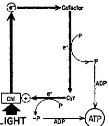
This theory (51, 5) envisages a phosphorylation step coupled with electron transfer within the chloroplast from a reduced cytochrome to (Chl)+. The phosphorylation that accompanies the oxidation of the cytochrome in chloroplasts would be analogous to the phosphorylation that accompanies the oxidation of the cytochromes in mitochondria by oxygen. Thus, chlorophyll with the aid of light is the ultimate oxidant in photophosphorylation and plays a part which corresponds to molecu- lar oxygen in oxidative phosphorylation. Earlier members of the photo- synthetic electron carrier system, those that precede the cytochromes, are quinones (vitamin K ) , flavins ( F M N ) , or related physiological equivalents. Additional phosphorylations are probably coupled with the oxidation-reduction of these cofactors (51, 5). A diagram illustrating this mechanism is shown in Fig. 1.
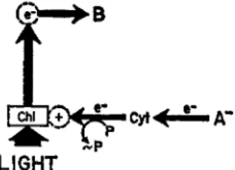
These physiological cofactors (electron carriers) can be replaced by certain nonphysiological agents, such as phenazine methosulfate, which apparently act by providing an artificial "shortcut" that bypasses one or more phosphorylation sites (45). A diagram illustrating a shortened cyclic electron flow mechanism of this type is shown in Fig. 2.
5 6 8 Μ. LOSADA AND D. I. ARNON
^ • C o f a c t o r
e"
IADP
LIGHT
-P ADP >( A T PFIG. 1 . Scheme for cyclic photophosphorylation catalyzed by a physiological cofactor (a quinone or a flavin). The chlorophyll (Chi) molecule (bound to a protein in the chloroplast) becomes excited by the absorption of a photon.
The excited chlorophyll donates its high energy electron to the oxidized co- factor and accepts an electron from reduced cytochrome ( C y t ) . The cycle is completed when the cofactor, after becoming reduced, in turn transfers an electron to cytochrome, which has become oxidized by its transfer of an elec
tron to chlorophyll. The phosphorylation steps are envisaged as being linked with the transfer of electrons from cofactor to cytochrome and from the cyto
chrome chain to chlorophyll. [From ( 5 ) . ]
FIG. 2 . Scheme for cyclic photophosphorylation catalyzed by phenazine methosulfate ( P M S ) . The envisaged mechanism here differs from that in Fig.
1, in that P M S serves as a shortcut bypassing the site of the physiological cofactor. This bypass is not coupled with phosphorylation. The phosphoryla
tion coupled with electron transfer in the sector between cytochromes and chlorophyll remains the same as in Fig. 1 [From ( 5 ) . ]
The salient feature of the proposed cyclic mechanism (Figs. 1 and 2) is that electrons, "activated" by light energy, travel in a "closed circuit"
from which they are not removed. The stepwise interaction of the high-
PMS
β"
energy electron with the intermediate electron acceptors and the coupled phosphorylating system constitutes the energy conversion process in cyclic photophosphorylation.
Granted the validity of this mechanism [for a review of supporting evidence see (5)], an inhibitor of cyclic photophosphorylation may be effective by impeding one of the three main phases of the process: (a) the light-induced generation of a high-energy electron and the ultimate electron acceptor (Chl)+, (b) electron transport by the photosynthetic electron transport chain, and (c) phosphorylation reactions coupled to electron transport. Phases (b) and (c) are analogous to, and at some points possibly identical with, their counterparts in oxidative phos- phorylation, and hence there is considerable interest in comparing the inhibitors of these two processes. Phase (a) is peculiar to photosynthetic phosphorylation and involves physical events for which our knowledge of inhibitor action is only now beginning to emerge. Arnold and Clayton {52a) observed that phase (a) could be isolated from subsequent photo- synthetic events by the use of sodium azide or hydroxylamine. In the presence of these inhibitors, intact cells of a carotenoidless mutant Rhodopseudomonas spheroides showed on illumination spectral changes near the absorption maxima of bacteriochlorophyll, similar to spectral changes seen on illuminating of chromatophores, either in suspension or as dried films. The spectral changes may result from an accumulation of a pool of electrons expelled from bacteriochlorophyll in the primary photochemical act [phase ( a ) ] ; the use of these electrons in phase (b) is prevented by the inhibitor.
B. Mechanism of Noncyclic Photophosphorylation
As summarized in Eq. (4), noncyclic photophosphorylation differs from the cyclic type in that the formation of A T P accompanies the reduction of pyridine nucleotide and the oxidation of an external electron donor. In the mechanisms for noncyclic photophosphorylation proposed by Arnon and his co-workers (11, 12, 51), the primary photochemical act remains the same as in cyclic photophosphorylation. Photon capture by chloro- phyll results in the generation of a high-energy electron and of an electron acceptor (Chl)+. However, the cyclic path of the electron is interrupted;
the high-energy electron removed from the excited chlorophyll molecule is transferred to pyridine nucleotide which, in the intact cell, is used for C 02 assimilation.
As the electrons from excited chlorophyll are transferred to pyridine nucleotide, they must be replaced by an external electron donor. The
570 Μ. LOSADA AND D. I. ARNON
"closed" circuit of cyclic photophosphorylation thus gives way in non- cyclic photophosphorylation to an "open" circuit for the transport of electrons from an external electron donor to pyridine nucleotide. This noncyclic electron transport also requires an input of light energy, since the electron donor is at a potential less reducing than the electron acceptor. A diagrammatic representation of this concept is given in Fig. 3.
FIG. 3. Scheme for noncyclic photophosphorylation of the bacterial type.
The chlorophyll molecule (Chi) becomes excited by the absorption of a quan
tum of light. The excited chlorophyll donates its high energy electron (e~) to the electron acceptor (B) and accepts, via the cytochrome system ( C y t ) , an electron from an external electron donor (A~). The phosphorylation step is envisaged as linked with the transfer of the electron from cytochrome to chloro
phyll. Β represents pyridine nucleotide. [From (11).]
Noncyclic and cyclic photophosphorylation are thus envisaged as sharing the primary photochemical act, i.e., chlorophyll excitation by photon capture, and the related oxidation of cytochromes with an accom
panying phosphorylation step. Noncyclic photophosphorylation would, therefore, be expected to be equally sensitive to inhibitors which inhibit these steps in cyclic photophosphorylation. But the proposed mechanism for noncyclic photophosphorylation provides two additional sites for inhibitor action: (a) the transfer of electrons from the external electron donor to the photosynthetic apparatus and (b) the transfer of electrons from excited chlorophyll to pyridine nucleotides.
Noncyclic phosphorylation in green plants is distinguished from that in photosynthetic bacteria by its electron donor system (11, 52). In green plants, water,3 i.e., O H- ions, supplies the electrons required in reaction (4). However, photosynthetic bacteria cannot use hydroxyl ions but use inorganic or organic electron donors, such as thiosulfate or succinate. It now appears that plants, but not photosynthetic bacteria, are able to use
3 OH" will be used here interchangeably with water and represents hydroxyl ions at neutral pH as in the reaction:
LIGHT
4 OH" (ΙΟ"7 M) •>02 2 H20 + 4e~; Ε*· - 0.815F (pH 7)
hydroxyl ions as electron donors because plants have evolved an addi
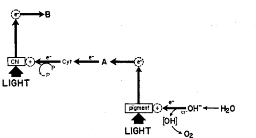
tional pigment system {11,12, 53)—chlorophyll b or equivalent accessory pigment—that catalyzes a special light-dependent reaction [Eq. (5) ] for the photooxidation of hydroxyl ions (Fig. 4 ) .
A
i
1 ifj^ ©<7^?.0ΓΓ<— rfeO
* [OH]
LIGHT \ > o2
FIG. 4. Scheme for photooxidation of water by chloroplasts. This reaction is now visualized (Cf. 11,12) as an auxiliary photochemical reaction in chloro
plasts, catalyzed by a photosynthetic pigment (chlorophyll b or equivalent) that does not occur in photosynthetic bacteria. The pigment molecule, when it becomes excited by the absorption of a photon, donates a high-energy elec
tron (e~) to an intermediate electron acceptor (A) and accepts an electron from an hydroxyl ion. The oxidation product (OH) of the hydroxyl ion is the precursor of molecular oxygen. [From (11).]
4 OH- 4e- + 02 + 2 H20 (5)
To recapitulate, the proposed mechanisms for photosynthetic phos
phorylation provide for several groups of inhibitors, each capable of in
hibiting the over-all process of photosynthesis by affecting one of the following: (a) oxygen evolution (plants only), (b) electron transfer steps involved in cyclic and noncyclic photophosphorylation (plants or bacteria), or (c) the phosphorylation reactions themselves (plants or bac
teria).
IV. INHIBITORS OF OXYGEN EVOLUTION
Prior to the recognition of oxygen evolution as a component of noncyclic photophosphorylation in chloroplasts, evidence for a specific action of certain inhibitors on oxygen evolution in photosynthesis came from ex
periments on hydrogen-adapted algae and on the Hill reaction. Gaffron (54) has shown that certain green algae can be adapted to hydrogen gas to perform a type of light-dependent C 02 assimilation, resembling bac
terial photosynthesis, in which hydrogen consumption replaces oxygen
572 Μ. L0SADA AND D, I. ARNON
evolution. This kind of photosynthesis, which he called photoreduction, was much more resistant to hydroxylamine, o-phenanthroline, vitamin Κ derivatives, and hydrogen sulfide inhibition than the conventional photo
synthesis in which oxygen was evolved, leading to the conclusion that these agents affect primarily the reaction (s) responsible for oxygen evolu
tion (55, 56).
This conclusion was supported by evidence that the same inhibitors also inhibit the Hill reaction, under conditions when C 02 assimilation or photophosphorylation are excluded. Warburg found (57) that o-phenan
throline was a powerful inhibitor of oxygen evolution in the Hill reaction, and this was confirmed by other investigators (see review 3). Among other inhibitors of the Hill reaction are hydroxylamine, sodium azide, and phenylurethan; cyanide was found to be without effect (cf. 58).
A. Effect of Phenylurethan a n d Substituted Phenylureas
Urethans have been widely used as poisons ("narcotics") of photo
synthesis since they were first introduced by Warburg in 1919. In 1956, Wessels and van der Veen (59), investigating the mode of action of the herbicide C M U [3-(4-chlorophenyl)-l,l-dimethylurea], discovered that this substance and other substituted ureas were extremely powerful in
hibitors of the Hill reaction (as measured by the photoreduction of 2,6-dichlorophenolindophenol), exceeding greatly in effectiveness the structurally related phenylurethan (Table I ) .
These findings were confirmed by Spikes (60) and extended by Bishop (61) to hydrogen-adapted cells. Bishop compared the effect of D C M U (for abbreviations see Table I) on photosynthesis and photoreduction in Scenedesmus. Photosynthesis was reduced to half by 5 χ 1 0 ~7 Μ D C M U and became completely inhibited in the presence of 3 Χ Ι Ο- 6 Μ D C M U . The inhibitory effect of D C M U disappeared if the alga was adapted to hydrogen; photoreduction was not inhibited.
High light intensities are known to "deadapt" algae adapted to hydrogen gas and to restore conventional photosynthesis in them (54-56). This reversion of photoreduction to photosynthesis by the use of higher light intensities was prevented at 3 Χ Ι Ο- 6 Μ D C M U , indicating again that conventional photosynthesis of green plants of which oxygen evolution is an essential part, cannot proceed in the presence of D C M U (61).
The substituted urea compounds have provided for the first time photo
synthetic inhibitors which are effective at concentrations lower (by a hundredfold) than that of chlorophyll (59, 60). Moreover, this powerful inhibitory action seems to be affecting a specific photochemical reaction,
T A B L E I
PHENYLURETHAN AND SUBSTITUTED PHENYLUREAS
Name Formula OC2H5
Phenylurethan
3 - P h e n y l - 1 , 1 -dlmethylur ea
/ C H , N—CH3
3-(4'-Chlorophe*nyl)-l, 1 - dlmethylurea (CMU)
3-(3i4-Diehlorophenyl)-l, 1- dimethylurea (DCMU)
3-(3-Chlorophenyl)-l, 1-dimethylurea
CH3
Ν—CH3
CI
574 Μ . LOS ADA A N D D . I . A R N O N
the photooxidation of O H - that gives rise to oxygen evolution (Fig. 4 ) , a reaction which Losada et al. (11, 12) have experimentally separated in isolated chloroplasts from the other photochemical reactions of photo
synthesis.
If the proposed reaction mechanisms were valid, it would be expected that substituted ureas, and other inhibitors of oxygen evolution, such as o-phenanthroline, would not inhibit those photosynthetic reactions in which O H ~ does not serve as an electron donor, viz., (a) an experi
mentally modified noncyclic photophosphorylation in chloroplasts in which the physiological electron donor O H - is replaced {11) by an ascorbate-dye system (A~ in Fig. 3), (b) noncyclic photophosphorylation in bacterial chromatophores (Fig. 3 ) , and (c) cyclic photophosphoryla
tion, either in chloroplasts or in chromatophores, since in both these cases an endogenous electron flow is being set in motion by light without the involvement of any external electron donor (Fig. 1). As will be shown below, these expectations have been experimentally verified.
B. Insensitivity of Cyclic a n d Noncyclic Photophosphorylation to Inhibitors of O x y g e n Evolution
1. N O N C Y C L I C P H O T O P H O S P H O R Y L A T I O N
Vernon and Zaugg (62) have shown that in the presence of 1 0 ~5 Μ D C M U , freshly prepared chloroplasts, which have lost completely (98%) the capacity to reduce T P N with electrons donated by O H - , are never
theless able to reduce T P N photochemically by electrons donated by a reduced dye, 2,6-dichlorophenolindophenol. (In practice, the dye is sup
plied in catalytic amounts and kept in a reduced form by substrate amounts of added ascorbate.) Similar effects were observed in the pres
ence of Ι Ο- 3 Μ hydroxylamine (62).
Losada, Whatley, and Arnon (11) have recently shown that in isolated chloroplasts the light-dependent noncyclic electron transfer from ascor
bate to T P N (via 2,6-dichlorophenolindophenol) is coupled with the formation of ATP. The phosphorylation accompanying this T P N reduc
tion was not accompanied by oxygen evolution, and hence, as predicted by the working hypothesis, it was not inhibited by C M U (Table 1 in ref. 11). This provided evidence that in chloroplasts, as in photosynthetic bacteria, (82) the essential events of noncyclic photophosphorylation,
(i.e., A T P formation coupled with photoreduction of P N ) are basically independent of oxygen evolution. B y blocking the photoproduction of
oxygen, Losada et al. (11) have shown that chloroplasts can be experi
mentally induced to give the bacterial type of noncyclic photophos
phorylation described by Nozaki et al. (82).
These findings are in agreement with the earlier ones of Vernon and Ash (63) with chromatophores of R. rubrum, that D C M U had little effect on bacterial photoreduction of pyridine nucleotide, photophosphorylation, or photooxidation of ascorbate.
2. C Y C L I C P H O T O P H O S P H O R Y L A T I O N
There is general agreement among different investigators (12, 64-69) that cyclic photophosphorylation in chloroplasts catalyzed by phenazine methosulfate (or pyocanin) is a truly anaerobic process in which no oxygen is produced or consumed. This view is supported by the consistent evidence from different laboratories that this type of cyclic photophos
phorylation is resistant to inhibition by inhibitors of oxygen evolution.
However, in the case of cyclic photophosphorylations catalyzed by vita
min Κ or F M N , the effects of inhibitors of oxygen evolution seemed less clear-cut.
Whatley et al. (65) and Trebst and Eck (66) found that cyclic photo
phosphorylation catalyzed by F M N was more sensitive to o-phenan- throline than that catalyzed by vitamin Κ or phenazine methosulfate, and this observation served, among others, to support a case for two parallel pathways of cyclic photophosphorylation: one catalyzed by F M N and another, by vitamin Κ or phenazine methosulfate (65). Jagen- dorf and Margulies (67) found that, under their experimental conditions, cyclic photophosphorylation catalyzed either by vitamin Κ or F M N was equally inhibited by CMU. They therefore put both these photophos
phorylations in the same category and suggested that both depend on an oxygen-evolving step that is missing in the CMU-resistant cyclic photo
phosphorylation catalyzed by phenazine methosulfate. Similar conclu
sions were reached by Vennesland (68) and Krall et al. (69).
The effect of C M U on cyclic photophosphorylation by chloroplasts has recently been reinvestigated by Arnon et al. (5, 12). They confirmed the resistance of cyclic photophosphorylation catalyzed by phenazine metho
sulfate to CMU, but, in addition, they have shown that under appro
priately controlled experimental conditions cyclic photophosphorylation catalyzed by vitamin Κ or F M N is also resistant to inhibition by CMU.
The required experimental conditions include an absence of oxygen, higher concentrations (catalytic versus "microcatalytic") of vitamin Κ and F M N , low light intensity, and, particularly for the F M N system.
576 Μ . L O S A D A A N D D . I . A R N O N
high concentrations of the chloroplast material. The need for a relatively high concentration of chloroplast material suggested that the CMI5- resistant, anaerobic F M N and vitamin Κ systems require more chloro
plast factor (s) than the anaerobic phenazine methosulfate system.
Arnon et al. (12) found that, unless the necessary experimental condi
tions were maintained, cyclic photophosphorylation catalyzed by F M N or vitamin Κ became converted into a pseudocyclic type, i.e., a special case of noncyclic photophosphorylation in which molecular oxygen re
places pyridine nucleotide as the electron acceptor [cf. Eq. ( 4 ) ] . The pseudocyclic process is dependent on oxygen evolution by chloroplasts [Eq. (5)] and hence is sensitive to CMU, o-phenanthroline, and other oxygen inhibitors. Thus, these inhibitors of oxygen evolution provide an experimental device for distinguishing the pseudocyclic, oxygen-dependent photophosphorylation catalyzed by vitamin Κ or F M N from the corre
sponding cyclic type (also catalyzed by vitamin Κ or F M N ) which is truly anaerobic and involves neither the consumption nor the evolution of oxygen (see Table 2 in ref. 12).
Similar conclusions about the anaerobic nature of cyclic photophos
phorylation by chloroplasts were independently reached by Trebst and Eck (66). In their experiments, cyclic photophosphorylation catalyzed either by vitamin Κ or by phenazine methosulfate was independent of molecular oxygen and resistant to inhibition by D C M U and o-phenan
throline. In agreement with the earlier results of Whatley et al. (65), Trebst and Eck (66) found that cyclic photophosphorylation catalyzed by F M N was sensitive to these inhibitors and thus seemingly dependent on oxygen. However, the experimental conditions that are now known to be necessary for demonstrating the anaerobic nature of cyclic photo
phosphorylation catalyzed by F M N (5, 12) were not used in the experi
ments of Whatley et al. (65) and Trebst and Eck (66).
In bacterial preparations, the independence of photosynthetic phos
phorylation from oxygen evolution and its inhibitors was to be expected in view of the strictly anaerobic nature of bacterial photosynthesis. In isolated chloroplasts, where oxygen evolution is normally an integral part of photosynthesis, special experimental techniques, including the use of inhibitors, have been used to demonstrate the basic independence of cyclic and noncyclic photophosphorylation from oxygen evolution or consump
tion. The elucidation of the important but nevertheless special role of oxygen evolution in noncyclic photophosphorylation in chloroplasts has revealed the basic similarity of this process in green plants and photo
synthetic bacteria; this basic similarity was also shown for cyclic photophosphorylation.
V. INHIBITORS OF PHOTOSYNTHETIC ELECTRON TRANSPORT
We will now consider inhibitors of photosynthetic electron transport pathways unrelated to oxygen evolution. The discussion will include (a) electron flow in cyclic photophosphorylation by chloroplasts and bacterial chromatophores and (b) electron flow in noncyclic photophosphorylation of the bacterial type, i.e., the type in which oxygen evolution does not occur. As already stated, the bacterial type of noncyclic photophos
phorylation has recently been demonstrated in chromatophores of R. rubrum (32) and has also been shown to be an experimentally separa
ble component of noncyclic photophosphorylation in isolated chloroplasts (11,1*).
A. Inhibitors of Cyclic Electron Flow
In bacterial preparations, cyclic photophosphorylation proceeds without the addition of external cofactors. Chromatophores of Chromatium, when prepared under anaerobic conditions, give a vigorous cyclic photophos
phorylation which is not increased by the addition of cofactors. On aging the isolated chromatophores, this "physiological" cyclic photophosphoryla
tion progressively decreases but is restorable by the addition of phenazine methosulfate and vitamin Κ analogues (see Table 2 in ref. 5).
These results suggest that aging of chromatophores brings about a pro
gressive inactivation of some physiological cofactor of electron transport and that this may be overcome by the addition of exogenous cofactors.
Phenazine methosulfate and vitamin Κ analogues are also effective in catalyzing cyclic photophosphorylation in isolated chloroplasts, but here they must be added even to freshly prepared chloroplasts if a vigorous rate of cyclic photophosphorylation is to be obtained. It seems likely that loss or inactivation of the endogenous cofactor (s) of cyclic photophos
phorylation is inherent in the present methods for isolating chloroplasts but is avoided in preparing bacterial chromatophores.
With bacterial chromatophores the effect of inhibitors on cyclic photo
phosphorylation may, therefore, be investigated either in an endogenous (physiological) system to which no cofactors are added or in a system fortified by the addition of exogenous cofactors. In isolated chloroplasts investigations of inhibitors have usually been carried out not in endoge
nous systems but in the presence of added cofactors because, as already mentioned, without them the rate of cyclic photophosphorylation is very low. These experimental differences between chloroplasts and chromato-
578 Μ . LOSADA A N D D. I. A R N O N
phores must be kept in mind when comparing the effects of inhibitors on the two systems.
1. E F F E C T OF F E R R I C Y A N I D E
In oxidative phosphorylation by mitochondria oxygen uptake gives a measure of electron transport both under conditions when A T P is formed or when it is experimentally abolished (uncoupled). There is no similar direct procedure for measuring electron flow in cyclic photophosphoryla
tion independent of A T P formation. A T P formation is a measure of both electron flow and phosphorylation. If phosphorylation is abolished, a cyclic electron flow would, by definition, produce no other measurable chemical change in the system. Special experimental devices have, there
fore, been used for distinguishing between the effect of a given treatment on the phosphorylation reaction proper and on cyclic electron flow. Some of these, going beyond conventional inhibitors, will now be reviewed.
The key premise in the proposed mechanisms for cyclic photophos
phorylation is that the electron expelled from the chloroplast molecule in the primary photochemical act is not removed from the closed circuit within which it travels before it returns to the chlorophyll (Chl)+. If this basic postulation is correct, it follows that cyclic photophosphorylation should be abolished if electrons are prevented from completing the cycle because of capture by an external electron acceptor. To be convincing, such an experiment should be carried out with an electron "trap" which would be free from the suspicion that it prevented A T P formation by acting as an uncoupler or an inhibitor of phosphorylation.
An electron acceptor that fulfills these requirements is ferricyanide.
As shown by Avron and Jagendorf (37) and confirmed in this laboratory, ferricyanide has a great affinity for trapping electrons during cyclic photo
phosphorylation without acting as an uncoupler or inhibitor of ATP formation. Because of these properties it may be used as a terminal electron acceptor in noncyclic photophosphorylation by chloroplasts
(9, 10) to replace T P N (see Β in Fig. 5). In the conventional noncyclic photophosphorylation by chloroplasts, electrons trapped by ferricyanide are continuously replaced by electrons donated by O H-, with a resultant evolution of oxygen that is accompanied by photoreduction of substrate amounts of ferricyanide (Fig. 5). When the flow of electrons from O H - is prevented by the omission of chloride (5, 51), ferricyanide ceases to act as a terminal electron acceptor in noncyclic photophosphorylation and acts instead as an electron trap which breaks the closed circuit of cyclic electron flow. In chromatophores ferricyanide always acts as an electron
Cyt β"
A L I G H T
FIG. 5. Scheme for noncyclic photophosphorylation of the green plant type.
This scheme combines the photooxidation of water ( F i g . 4 ) , a s the first light reaction, with the noncyclic photophosphorylation of the bacterial type (Fig.
3 ) , as the second light reaction (cf. 11, 12). The intermediate electron acceptor (A) from the first light reaction serves, in its reduced form (A"), as the electron donor for the second light reaction. [From (11).1
trap, since O H ~ cannot serve as an electron donor, whether chloride is present or not.
The addition of ferricyanide (in the absence of chloride) had indeed abolished cyclic photophosphorylation both in chloroplasts and in chromatophores (see Table 2 in ref. 51). Adding this ion in its reduced form as ferrocyanide was without effect. The reduction of ferricyanide with ascorbate either prior to or during illumination of chloroplasts or chromatophores restored in full their capacity for cyclic photophos
phorylation. The conclusion seemed justified, therefore, that the inhibitory effect of ferricyanide resulted from the capture by this ion (in its oxidized form) of electrons which would normally have traveled the cyclic electron transport route. This conclusion was strengthened by the finding that the inhibition was produced by very low concentrations of ferricyanide. This would be expected if, as demanded by the hypothesis, the quantity of ferricyanide necessary to capture electrons from the cyclic system need only be sufficient to leave the catalytic components of the system in an oxidized form.
2. E F F E C T OF I N H I B I T O R S O F R E S P I R A T O R Y E L E C T R O N T R A N S P O R T
From the standpoint of comparative biochemistry, special interest centers on the effects on photosynthetic phosphorylation of inhibitors of
580 Μ. LOSADA AND D. I. ARNON oxidative phosphorylation in mitochondria, especially those that have been identified with a particular site in the electron transport chain.
a. Antimycin A and HOQNO. Antimycin A and 2-heptyl-4-hydroxy- quinoline-iV-oxide (HOQNO) are two inhibitors of electron transport in oxidative phosphorylation whose site of inhibition is generally con
sidered to lie between cytochromes b and c (70-72). Geller (45, 73) and Smith and Baltscheffsky (74) found, respectively, that antimycin A and HOQNO inhibit cyclic photophosphorylation in chromatophores of R. rubrum. Similar effects of these two inhibitors were observed by Nozaki et al. on cyclic photophosphorylation with chromatophores of Chromatium
(32). In the experiments of Nozaki et al. (82) the inhibition was particu
larly pronounced in washed chromatophores.
The inhibition of cyclic photophosphorylation in chromatophores does not occur in the presence of catalytic amounts of phenazine methosulfate (45, 78, 75), which evidently serves as a bypass around the site of inhibi
tion. These findings are of interest, since cyclic photophosphorylation in chloroplasts—measured, as will be recalled, in the presence of added cofactors—was found to be resistant to antimycin A [Arnon et al. (76), Whatley etal. (65)] and to HOQNO [Avron (77) ]. Baltscheffsky (78,79) reported that the inhibition of cyclic photophosphorylation in chloroplasts required a 1000 times higher concentration of each of these two inhibitors than in chromatophores.
On present evidence, it remains unsettled whether chloroplasts have or have not an endogenous cyclic photophosphorylation that is sensitive to these two inhibitors. The possibility cannot be ruled out that chloroplasts also have an antimycin- and HOQNO-sensitive site (i.e., one lying be
tween cytochromes of the b and c types) which is lost by the present methods of isolating chloroplasts and is experimentally bypassed by the addition of exogenous cofactors of cyclic photophosphorylation.
Further support for the "bypass" effect of phenazine methosulfate in cyclic electron transport in chromatophores, and a suggestion that the site of inhibition of antimycin A in the photosynthetic electron transport chain lies between cytochromes b and c, has come from a recent report of Rudney (80). He found that chromatophores obtained from R. rubrum cells grown in the presence of diphenylamine ( D P A ) , an inhibitor of carotenoid synthesis, had a lower cyclic photophosphorylation activity than chromatophores obtained from normal cells. Phosphorylation in both types of chromatophores (from normal or DPA-treated cells) was equally stimulated by the addition of phenazine methosulfate. Moreover, the phosphorylating activity of chromatophores from the DPA-treated cells, but not from normal cells, was increased by the addition of isoprenologues
of coenzyme Q. Antimycin A blocked completely the increased phos
phorylation obtained by adding isoprenologues of coenzyme Q, whereas the phosphorylation in the phenazine methosulfate treatment was rela
tively insensitive to antimycin A. These observations again support the view that antimycin A inhibits a site between cytochromes b and c. This is the region of the respiratory electron transport chain in which coenzyme Q is placed [Green (81)].
b. Gramicidin, Oligomycin, Methylene Blue and Other Dyes. Gramici
din was found to inhibit cyclic photophosphorylation in isolated chloro
plasts (65, 79) and in extracts of R. rubrum (82) but not in Chromatium chromatophores (see Table 3 in ref. 5). Baltscheffsky and Baltscheffsky (82) found that oligomycin inhibited cyclic photophosphorylation in extracts of R. rubrum in concentrations similar to those necessary to inhibit oxidative phosphorylation in mitochondria.
Methylene blue, a dye that is known to be a strong inhibitor of oxida
tive phosphorylation (83), was found by Arnon et al. to be also a strong inhibitor of cyclic photophosphorylation in chloroplasts (65, 76) but not in Chromatium chromatophores (see Table 3 in ref. 5). However, Geller and Lipmann (73) obtained a strong inhibition of photophosphorylation in R. rubrum by 1 0 ~4 Μ methylene blue as well as by other dyes, namely, brilliant blue, thionine, and dichlorophenolindophenol.
c. Arsenite and the Question of Lipoic Acid. Interest in the effect of arsenite on photosynthetic phosphorylation stems from the proposal of Calvin and his associates (84) that lipoic (thioctic) acid is a key com
pound in photosynthesis, the one concerned in the primary conversion into chemical energy of the radiant energy absorbed by chlorophyll. This suggestion has recently been brought forward again by Racker (71).
Reactions in which lipoic acid is a cofactor are very sensitive to arsenite inhibition (85). For example, the pyruvic oxidation system, in which lipoic acid is a cofactor, is inhibited by concentrations of arsenite of the order of 3 Χ 1 0 ~5 Μ (85, 86). In chloroplasts, Arnon et al. (76) and Whatley et al. (65) found that Ι Ο- 3 Μ arsenite failed to inhibit either cyclic or noncyclic photophosphorylation. The improbability of lipoic acid as a participant in photosynthetic phosphorylation is also indicated by the results of Geller and Lipmann (73); Ι Ο- 2 Μ arsenite failed to inhibit phosphorylation by illuminated particles of R. rubrum.
d. p-Chloromercuribenzoate. Cyclic photophosphorylation by chloro
plasts, whether catalyzed by F M N or vitamin Κ is sensitive to p-chloro- mercuribenzoate inhibition (64, 65, 76). Cyclic photophosphorylation in chloroplasts, when catalyzed by phenazine methosulfate, is also inhibited
582 Μ , LOSADA AND D. I. ARNON by p-chloromercuribenzoate (#4). The inhibition of cyclic photophos
phorylation was reversed by the addition of glutathione (65, 76). These results suggest that sulfhydryl compounds, in enzymes or cofactors, or both, participate in photosynthetic phosphorylation.
e. Cyanide. Cyanide, the classic inhibitor of respiration, [and of the carboxylation enzyme (see Section II, B ) ] is noted for its inhibition of cytochrome oxidase (see review, 87). Since this enzyme is not involved in the strictly anaerobic reactions of bacterial photosynthesis and is absent from chloroplasts (88, 89), the effect of cyanide on the photosyn
thetic electron transport chain would be expected to differ from that on the respiratory electron transport chain. Thus, Smith and Baltscheffsky (74) found that in cell-free extracts of R. rubrum cyanide inhibited respiration but had no inhibitory effect on photophosphorylation.
Geller and Lipmann (73) observed that photophosphorylation in R. rubrum was not appreciably affected by Ι Ο- 2 Μ cyanide or azide. In chloroplasts, Wessels (39) found the addition of 1 0 ~3 Μ K C N increased by over 100% cyclic photophosphorylation catalyzed by vitamin K3 but severely inhibited cyclic photophosphorylation catalyzed by F M N . Whatley et al. (65) have also observed a cyanide inhibition of an F M N - catalyzed cyclic photophosphorylation in chloroplasts, but no stimula
tion of the vitamin Κ system. In fact, Whatley et al. (65) found that, depending on the pH and the form of vitamin Κ used, cyanide also inhibited the vitamin Κ system. These conflicting observations cannot, as yet, be reconciled by any cogent theory as to a possible mode of action of cyanide in photosynthetic phosphorylation.
/. Other Inhibitors. Among other inhibitors, Baltscheffsky (79) found that Amytal gave only a weak inhibition of cyclic photophosphorylation in chloroplasts, even at concentrations several times higher than those which inhibit oxidative photophosphorylation in mitochondria. Geller and Lipmann (73) obtained inhibition of cyclic photophosphorylation in R. rubrum by butyl-3,5-diodo-4-hydroxybenzoate and the alkylated naphthoquinone, Compound S N 5949.
B. Inhibitors of Noncyclic Electron Flow
As was already stated (Section III, B ) , the proposed mechanism of noncyclic photophosphorylation provides for two sites of inhibitor action which are not encountered in cyclic photophosphorylation: (a) the trans
fer of electrons from the external electron donor to the photosynthetic apparatus and (b) transfer of electrons from excited chlorophyll to pyri-
dine nucleotide. In both isolated chloroplasts and chromatophores the physiological electron acceptor is pyridine nucleotide; experimentally, in isolated chloroplasts pyridine nucleotide may be replaced by ferricyanide or some other Hill reagent.
The physiological external electron donor for chloroplasts is O H ~ (water); and since oxidation of this ion must be accompanied by oxygen evolution, the previously discussed (Section IV) inhibitors of oxygen evolution necessarily prevent electron transfer from the external donor, O H-, to the photosynthetic apparatus. These inhibitors do not affect either the bacterial type of noncyclic photophosphorylation where O H " is not the electron donor or the strictly anaerobic cyclic electron flow (either in chromatophores or in isolated chloroplasts) in which molecular oxygen does not participate {12).
The use of inhibitors provides an experimental technique for comparing the electron transport chain in cyclic and noncyclic photophosphorylation.
Similar sensitivity to the inhibitors would point to similar components in the two pathways. Thus, in the case of R. rubrum chromatophores, Nozaki et al. {82) found that the noncyclic electron flow from succinate (a physi- ological electron donor for this organism) was also inhibited by antimycin A and HOQNO, two inhibitors of the cyclic electron flow. Nozaki et al.
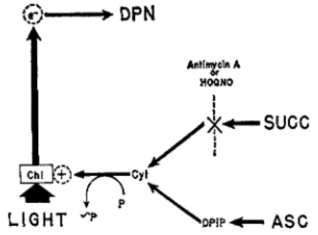
{82) have, therefore, concluded that the cyclic and noncyclic electron pathways in R. rubrum share the same site of inhibition (X in Fig. 6 ) , lying between cytochromes b and c, which is bypassed by using ascorbate as the electron donor.
FIG. 6. Site of antimycin A or H O Q N O inhibition in noncyclic photophos- phorylation of R. rubrum chromatophores, with succinate as the electron donor. Electrons from ascorbate bypass [via 2,6-dichlorophenolindophenol
( D P I P ) ] the site of inhibition. [From (32).]
1. P H E N Y L M E R C U R I C A C E T A T E
Smith and Baltscheffsky (74) found that phenylmercuric acetate ( P M A ) inhibited photophosphorylation by R. rubrum in the presence of