Clinical anatomy of partial liver transplantation, a modified corrosion cast method
PhD thesis
Mátyás Kiss MD.
Semmelweis University
Doctoral School of Basic Medicine
Consultants: Prof. Zoltán Máthé MD, Ph.D., professor, head of department Ágnes Nemeskéri MD, Ph.D., associate professor
Official reviewers: Prof. László Entz MD, Ph.D., professor
András Bálint MD, Ph.D., Med. Habil., private professor
Head of the Final Examination Board:
Prof. József Sándor MD, Ph.D., professor
Members of the Final Examination Board:
Zsolt Csapó MD, Ph.D., chief physician and head of department Ákos Szűcs MD, Ph.D., senior lecturer
Budapest
2017
1
1. Table of Contents
1 Table of Contents...1
2 List of abbreviations...3
3 Introduction...4
3.1 History of liver transplantation...4
3.2 Segmental anatomy of the human liver...6
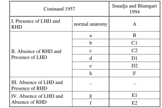
3.2.1 Couinaud’s terminology...7
3.2.2 “The Brisbane 2000 Terminology”………....13
3.3 Technical aspects of split liver transplantation………...22
4 Objectives...30
5 Methods...32
5.1 Working out of the corrosion cast technique………...32
5.2 New corrosion cast technique to study the hilar variations of the hepatic duct system………..42
5.3 Vessel lumen filling without corrosion technique to study the optimal line of hepatotomy for left lateral living donor liver transplantation………...43
6 Results...44
6.1 Hilar variations of the hepatic duct system……….…44
6.1.1 Group I: Presence of left and right hepatic ducts, normal biliary anatomy: Type "A" (54.74%) ………...……… 45
6.1.2 Group II: Absence of the right hepatic duct - presence of the left hepatic duct (41.49%)……….…46
Variation Type "B"………..46
Variation Type "C1"………..………..47
Variation Type "C2"……… ………...48
Subvariant of Type "C2"………..49
Variation Type "D1"………50
Variation Type "D2"………51
Variation Type "F"………...52
Newly described variation Type „G”………...53
2
6.1.3 Group III: Absence of the left hepatic duct - presence of the right
hepatic duct (0 %)………...…55
6.1.4 Group IV: Absence of the left hepatic duct - absence of the right hepatic duct: Type "E" (3.75 %)……… 55
Variation Type "E1"……….………55
Type "E1a" subvariant……….55
Type "E1b" subvariant……….………56
Variation Type "E2" ………57
Subvariant of Type "E2"………..………58
6.2 Variations of the left hepatic duct, optimal line of hepatotomy for left lateral living donor liver transplantation……….61
6.2.1 Branching patterns of the left hepatic duct………….……….61
6.2.2 Topographical relationship of the falciform ligament and the confluence of segment II and III ducts………...………64
6.2.3 Evaluation of the number of bile ducts in three different division lines…66 7 Discussion...68
7.1 Hilar variations of the hepatic duct system……….…68
7.2 Variations of the left hepatic duct, optimal line of hepatotomy for left lateral living donor liver transplantation……….…………72
8 Conclusions...77
9 Summary...79
10 Összefoglalás... 81
11 Bibliography...83
12 Bibliography of the candidate’s publications...101
13 Acknowledgements...103
3
2 List of abbreviations
CHD - common hepatic duct CT - computed tomography EU - European Union FL - falciform ligament HCC - hepatocellular carcinoma HPB - hepato-pancreato-biliary HU - Hounsfield unit
IHPBA - International Hepato-Pancreato-Biliary Associaton IVC - inferior vena cava
KOH - potassium hydroxide LHA - left hepatic artery LHD - left hepatic duct LHV - left hepatic vein
LDLT - living donor liver transplantation
LLS - left lateral segment (segments II and III) LPV - left portal vein
MHV - middle hepatic vein
OLT - orthotopic liver transplantation RAHD - right anterior hepatic duct RAPV - right anterior portal vein RHA - right hepatic artery RHD - right hepatic duct RHV - right hepatic vein
RPHD - right posterior hepatic duct RPPV - right posterior portal vein RPV - right portal vein
SLT - split liver transplantation 3D - three dimension
4
3 Introduction
3.1 History of liver transplantation
In the USA in 1968 Starzl performed the first successful orthotopic liver transplantation (OLT) using total hepatectomy. Calne and Williams (1968) announced similar achievements in the United Kingdom. Both teams greatly contributed to the development of liver transplantation and immunosuppressive therapy. The report of Fortner and co-workers (1970) on the first successful heterotopic (auxillary) liver homograft, was the forerunner of the oncoming development of heterotopic liver transplantation, split liver (SLT) and living donor related (LDLT) transplants. Liver transplantation has undergone continuous evolution although initially there was only one moderately effective immunosuppressive therapy within reach. The survival rates after the first year slowly approached 50% by 1979 [1, 2]. However, due to chronic rejection, infection and surgical infections there were only a small group of people among the long- term survivors. In 1979 Calne reported cyclosporine as immunosuppressive, which made a fast transformation [3]. A consensus conference held in 1983 announced liver transplantation to be an acceptable therapy from now on and ceased to be an experimental treatment. In 1987 Tacrolimus was declared to be another powerful immunosuppressive agent by Zeevi and colleagues [4]. Furthermore, de novo malignancy and other serious complications of immunosuppressive therapy for liver and other organ transplantations have been detailed [5, 6, 7]. In 1988, according to Iwatsuki, 54% of the patients survived the first five years [8]. There was a fast development and by 1992, more than 3000 OLT were carried out yearly in the United States (source: United Network for Organ Sharing, www.unos.org).
Treating liver cancer with total hepatectomy and liver transplantation was a disappointment at the beginning. Iwatsuki (1988) presented that three out of every four patients surviving the first two month had a recurrence of cancer [8]. Adjuvant chemotherapy made no demonstrable benefit. Ringe (1989) reported a 15.2% 5 year graft survival for such patients [9] and Calne and co-workers (1986) had come to similar conclusions 10]. The best results and apparent cure could be achieved when cancer was an incidental finding during the removal of the liver taken out for noncancerous disease
5
(e.g., alcoholic cirrhosis). Geevarghese (1998) reported an 85% 1-year survival and 78%
5-year survival for such cases [11]. Olthoff (1995) using fluorouracil, doxorubicin, and cisplatin declared a 45% 3-year survival, however, the size of the cancer was larger than 5cm only three cases [12]. Nowadays after liver transplantation no adjuvant chemotherapy is given. In 1996 Mazzaferro and co-workers established the so-called
“Milan Criteria” for patients with hepatocellular carcinoma (HCC). A 75% survival at 4 years with a recurrence rate 17% after liver transplantation was unfolded in the case of patients with HCC 5 cm or less or with a maximum of three nodules each 3 cm or less without extrahepatic manifestations or vascular invasion. The case of the presence of vascular invasion or lymph node involvement went along with the increase at the risk of recurrence. Milan criteria was argued to be too restrictive considering liver transplantation, however a more lenient tumor criteria still would be beneficial. In 2002 Yao and the co-workers of the liver transplant group at the University of California at San Francisco (UCSF) has championed larger tumor sizes as criteria for liver transplantation and the result resembled to those ones which had been carried out while following the Milan criteria (UCSF criteria: single lesion ≤ 6.5 cm; multiple lesions ≤ 3 cm; largest tumor diameter if multiple ≤ 4.5 cm; total tumor diameter if multiple ≤ 8 cm).
The improvement of immunosuppression led to the boom of surgical methods.
Reduced size orthotopic liver grafts in young patients were announced by Bismuth and Houssin (1984) [13]. In 1988 Pichlmayer used one donor liver for two recipients (SLT) [14]. Raia (1989) [15] and Strong (1990) [16] were the volunteers to produce LDLT with the usage of segments II and III. Yamaoka (1994) used the right liver lobe [17]. In Hungary it was Zoltán Máthé who carried out the first successful adult right-lobe LDLT on 19th November, 2007 [18].
Cultural restrictions and the shortage of donor organs greatly motivated the usage of split liver grafts and living related donors for both lobes. These techniques have been the main improvements in the field of liver surgery. The main ethical worry was the risk of mortality and morbidity for the recipients and also for the donors. Applying liver transplantation for tumors has begun significant with primary hepatocellular cancer.
Although liver resection stays to be a treatment of choice, for HCC in the case of good liver function, or compromised liver function, and patients with hepatitis C and a little sized tumor in a unpropitious part of the liver (for resection) are presently thought to be
6
cured with the greatest effectiveness by liver transplantation. For patients with small HCC and patients with a wide range of benign diseases such as compromised liver function due to liver cirrhosis liver transplantation is widely used. Furthermore, it is accepted to treat the Budd-Chiari syndrome, the polycystic liver and kidney disease, sclerosing cholangitis, and other parenchymal and metabolic liver diseases. It is still being investigated if liver transplantation is the effective treatment for hilar cholangiocarcinoma. Time and again it is applied for patients with widespread neuroendocrine metastatic liver disease. Since people with metastatic disease from adenocarcinoma have shown insufficient outcomes, transplantation is not applied for in these cases [19].
So that the safety of the donors would improve in LDLT, Cherqui announced the first full laparoscopic left lateral segment (LLS) transplantation for adult-child LDLT in 2002 [20]. From that time on, numerous centres practice the laparoscopic technique which has a good standard by now, is linked to the decrease of the donors’ blood loss and hospital stays and produces similar quality grafts to the open approach [21, 22]. On the other hand, presently there is no standardization for laparoscopic major right or left hepatectomies for adult-adult LDLT and several other methods for example the full laparoscopic approach [23, 24, 25, 26], the hand assisted approach [27, 28, 29] and the hybrid approach [23, 30, 31, 32, 33, 34, 35] have been presented. There was a publication about robot-assisted right lobe donor hepatectomy [36]. Although several publications point out the feasibility of these procedures, the real advantage of laparoscopy over laparotomy still needs to be fully investigated. For achieving it standardization is necessary and international registries from the Eastern countries are need to be created where LDLT is prosperous [22].
3.2 Segmental anatomy of the human liver
The major topics of modern hepatic anatomy instead of the surface marks are internal vascular and biliary textures. The inside set-up has been revealed by McIndoe and Counseller (1927) [37], Ton That Tung (1939, 1979) [38, 39], Hjörtsjö (1931) [40], Healey and Scroy (1953) [41], Goldsmith and Woodburne (1957) [42], Couinaud (1957) [43] and Bismuth’s literary pieces (1982) [44]. Among these, it is Couinaud’s work,
7
which is the most useful and the most well-known for operations until 2000. The basis of Couinaid’s terminology is based on the three main hepatic veins (within the scissurae) which divide the liver into four sectors, each with a portal pedicle, with alterations between the hepatic veins and portal pedicles. In 2000, the International Hepato- Pancreato-Biliary Associaton (IHPBA) modernised the classification of the structure of the liver and named it “The Brisbane terminology” [45]. This new terminology is mostly founded on hepatic artery and bile duct ramifications rather than the portal and hepatic venous system. In order to make the two terminologies clear and comparable, I would like to pay great attention to both of them in this chapter.
3.2.1 Couinaud’s terminology
The inside structure of the liver is made of several parts which create sectors that are divided by scissurae containing the hepatic veins (Figure 1).
Figure 1: “The portal vein, the hepatic artery, and the draining bile ducts are distributed within the liver in a beautifully symmetric pedicular pattern, which belies the asymmetric external appearance. Each segment (I-VIII) is supplied by a portal triad composed of a branch of the portal vein and hepatic artery and drained by a tributary of the right or left main hepatic ducts. The four sectors demarcated by the three main hepatic veins are called the portal sectors; these portions of parenchyma are supplied by independent portal pedicles. The hepatic veins run between the sectors in the portal scissurae; the scissurae containing portal pedicles are called the hepatic scissurae. The umbilical
8
fissure corresponds to a hepatic scissura. The internal architecture of the liver consists of two livers, or hemilivers, the right and the left liver separated by the main portal scissura, also known as Cantlie’s line. It is preferable to call them the right and the left liver rather than the right and left lobes because the latter nomenclature is erroneous, there being no visible mark that permits identification of a true hemiliver.” (Source:
Blumgart LH, Hann LE. Surgical and Radiologic Anatomy of the Liver, Biliary Tract, and Pancreas. In: Blumgart LH. (ed.), Surgery of the Liver, Biliary Tract, and Pancreas.
Saunders, Philadelphia, 2007: 6.).
Basically, in the scissurae there are three main hepatic veins dividing the liver into four sectors, each has a portal pedicle, with alteration between the hepatic veins and portal pedicles. In the main portal scissura there is the middle hepatic vein (MHV) and goes to the left side of the cava from the centre of the gallbladder bed. The line of demarcation between the right and left parts of the liver is the main portal scissura, thus, these parts are self-contained in regards with of portal and arterial vascularization and of biliary drainage (Figure 2) [46].
Figure 2: “The functional division of the liver and of the liver segments according to Couinaud’s nomenclature. A, as seen in the patient. B, In the ex vivo position.” (Source:
Blumgart LH, Hann LE. Surgical and Radiologic Anatomy of the Liver, Biliary Tract,
9
and Pancreas. In: Blumgart LH. (ed.), Surgery of the Liver, Biliary Tract, and Pancreas.
Saunders, Philadelphia, 2007: 6.).
The right and left parts of the liver are apportioned into two by the remaining portal scissurae. These four subdivisions are named as segments in Goldsmith and Woodburne’s works [42] and called sectors in Couinaud’s nomenclature [43] (Figures 1- 2).
In his famous and worldwide extensively used peculiar book "Surgery of the Liver, Biliary Tract, and Pancreas" [46], Blumgart gives the most detailed anatomical description, based on Couinaud’s terminology: “The right portal scissura separating the right liver into two sectors - anteromedial or anterior and posterolateral or posterior - is almost in the frontal plane with the body supine. The right hepatic vein (RHV) progresses inside the right scissura. The left portal scissura separates two distinct parts in the left liver. The left portal scissura is not within the umbilical fissure which is not a portal scissura and involves a portal pedicle. The place of the left portal scissura is posterior to the ligamentum teres inside the left next to the left hepatic vein. The anterior sector of the left liver is composed of a part of the right lobe (segment IV) that is to the left of the main portal scissura and of the anterior part of the left lobe (segment III). The left posterior sector is the only sector composed of one segment (segment II). At the hilus of the liver, the right portal triad pursues a short course of approximately 1 to 1.5 cm before entering the substance of the liver. In some cases the right anterior and posterior pedicles arise independently, and their origins may be separated by 2 cm. In some cases, it appears as if the left portal vein (LPV) arises from the right anterior portal vein (RAPV) (Figure 3).
On the left side, however, the portal triad crosses over approximately 3 to 4 cm beneath the quadrate lobe embraced in a peritoneal sheath at the upper end of the gastrohepatic ligament and separated from the undersurface of the quadrate lobe by connective tissue (hilar plate). This prolongation of the left portal pedicle turns anteriorly and caudally within the umbilical fissure giving branches of supply to segments II and III and recurrent branches to segment IV. Beneath the quadrate lobe, the pedicle is composed of the left branch of the portal vein and the left hepatic duct (LHD), but it is joined at the base of the umbilical fissure by the left branch of the hepatic artery. The branching of the portal pedicle at the hilus, the distribution of the branches to the caudate lobe (segment I) on the
10
right and left side, and the distribution to the segments of the right (segments V-VIII) and left (segments II-IV) hemiliver follow a remarkably symmetric pattern and, as described by Scheele (1994) [47], allow separation of segment IV into segment IVa superiorly and segment IVb inferiorly. This arrangement of subsegments mimics the distributions to segments V and VIII on the right side. The umbilical vein provides drainage of, at least, parts of segment IVb after ligation of the middle hepatic vein and is important in the performance of segmental resection. The caudate lobe (segment I) is the dorsal portion of the liver lying posteriorly and embracing the retrohepatic inferior vena cava (IVC). The lobe lies between major vascular structures. On the left, the caudate lies between the IVC posteriorly and the left portal triad inferiorly and the IVC and the middle and left hepatic veins superiorly. This portion of the caudate is sometimes referred to as segment IX. The portion of the caudate on the right varies, but is usually quite small. The anterior surface within the parenchyma is covered by the posterior surface of segment IV, the limit being an oblique plane slanting from the LPV to the left hepatic vein (LHV). Thus, there is a caudate lobe (segment I) with a constantly present left portion and a right portion of variable size (Figure 4). The caudate lobe is supplied by blood vessels and drained by tributaries from the right and left portal triad. Small vessels from the portal vein and tributaries joining the biliary ducts also are found, usually two on the left side and one on the right. The right portion of the caudate lobe, including the caudate process, predominantly receives portal venous blood from the right portal vein (RPV) or the bifurcation of the main portal vein, whereas on the left side the portal supply arises from the left branch of the portal vein almost exclusively. Similarly, the arterial supply and biliary drainage of the right portion is most commonly associated with the right posterior sectoral vessels or pedicle and the left portion with the left main vessels. The hepatic venous drainage of the caudate is unique in that it is the only hepatic segment draining directly into the IVC. These veins sometimes can drain into the posterior aspect of the vena cava if there is a significant retrocaval caudate component. In the usual and common circumstance, the posterior edge of the caudate lobe on the left has a fibrous component, which fans out attaching lightly to the crural area of the diaphragm, but importantly extending posteriorly behind the vena cava to link with a similar component of fibrous tissue protruding from the posterior surface of segment VII and embracing the vena cava.
In 50% of patients, this ligament is replaced, in whole or in part, by hepatic tissue, and
11
the caudate may completely encircle the IVC and contact segment VII on the right side.
A significant retrocaval component may prevent a left-sided approach to the caudate veins. The caudal margin of the caudate lobe has a papillary process that occasionally may attach to the rest of the lobe via a narrow connection. It is bulky in 27% of cases and can be mistaken for an enlarged lymph node on computed tomography (CT) scan” [46].
Figure 3: An anatomical variation of the portal vein (PV) system. The left portal vein (LPV) arises from the right anterior portal vein (RAPV). (Source: author’s own work.
Co-workers: Ildikó Horti, Zsolt Pápai, Sándor Kovács, András Szuák).
Figure 4: “The caudate lobe (shaded)-segments II and III are rotated to the patient’s right. Superiorly, the left portion of the caudate lobe is linked by a deep anterior portion, which is embedded in the parenchyma immediately under the middle hepatic vein (MHV), reaching inferiorly to the posterior margin of the hilus of the liver and fusing anterolaterally to the IVC on the right side to segment VI and VII of the right liver. The
12
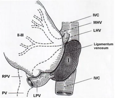
major blood supply arises from the left branch of the left portal vein (LPV) and the left hepatic artery close to the base of the umbilical fissure of the liver. The hepatic veins (MHV, LHV) are short in course and drain from the caudate directly into the anterior and left aspect of the vena cava. LHV, left hepatic vein; RPV, right portal vein; PV, main trunk of portal vein.” (Source: Blumgart LH, Hann LE. Surgical and Radiologic Anatomy of the Liver, Biliary Tract, and Pancreas. In: Blumgart LH. (ed.), Surgery of the Liver, Biliary Tract, and Pancreas. Saunders, Philadelphia, 2007: 8.).
To summarize:
1. The main hepatic scissura divides the liver into two hemilivers in which the MHV can be found.
2. The left portal scissura containing the LHV partitions the liver into two sectors (Figure 1). In the posterior sector there is only one segment (segment II). The umbilical fissure partitions the anterior part into two segments, a medial segment (the quadrate lobe- segment IV) and the lateral segment (segment III).
3. The right portal scissura containing the RHV splits the right liver into two parts.
Each sector is partitioned into two segments, an anterior sector (segment V inferiorly and segment VIII superiorly) and a posterior sector (segment VI inferiorly and segment VII superiorly) (Figures 1-2).
4. Segment I (the caudate lobe) is situated posteriorly to the IVC, it is adjacent with segments IV and VII [46] (Figures 4-5).
Figure 5: “Hepatic segmental anatomy as shown by CT. A, At the level of the hepatic veins. B, At the portal vein bifurcation. C, Below the hepatic hilus. Roman numerals stand for liver segments.” (Source: Blumgart LH, Hann LE. Surgical and Radiologic Anatomy of the Liver, Biliary Tract, and Pancreas. In: Blumgart LH. (ed.), Surgery of the Liver, Biliary Tract, and Pancreas. Saunders, Philadelphia, 2007: 9.).
13 3.2.2 “The Brisbane 2000 Terminology”
In December 1998, the Scientific Committee of the IHPBA had a meeting in Berne, Switzerland, to establish a Terminology Committee to address the confusion existing in the field of terminology of hepatic anatomy and liver resections. In the Committee there were eight hepato-pancreato-biliary (HPB) surgeons from all over the world. The Committee begun his work with seeking input from the IHPBA members, by publishing a survey questionnaire with 46 propositions in HPB. After almost 18 months’
work the Terminology Committee initiated its suggestions in the Scientific Committee at the World Congress of the IHPBA in Brisbane, Australia in May, 2000. These recommendations contained a modern terminology labelled as “The Brisbane 2000 Terminology” of liver anatomy and resections. It was assumed with one accord by the Scientific Committee of the IHPBA and were presented to the members as the official terminology of the IHPBA on the last day of the meeting. A description of the new terminology follows [45, 48].
The primary (first-order) partition divides the proper hepatic artery into the right (RHA) and left (LHA) hepatic arteries (Figure 6). The arterial inflow is provided by them to both hemilivers (Figure 7). The plane situated between the two distinct zones of vascular supply is named as a watershed the border of which at the first-order division is called the “midplane of the liver”. It intersects the gallbladder fossa and the fossa for the IVC. The right liver is generally expected to have a bigger size than the left one (60:40), although it might change [45, 48].
14
Figure 6: “Ramification of the hepatic artery in the liver. The prevailing pattern is shown. The first-order division of the proper hepatic artery is into the right (A) and left (B) hepatic arteries, which supply right and left hemilivers respectively (Figure 7). The second-order division of the hepatic arteries, supplies the four sections (c, d, e, f) (Figure 8). The third order-division supply the segments (II-VIII) (Figure 9). The left medial section and segment four are the same. The caudate lobe is supplied by branches from A and B. Bile duct anatomy and nomenclature is similar to that of the hepatic artery. © Washington University in St Louis.” (Source: Strasberg SM. Hepatic, biliary and pancreatic anatomy. In: Garden OJ, Parks RW. (eds.), Hepatobiliary and Pancreatic Surgery. A companion to specialist surgical practice. Fifth edition. Saunders, Edinburgh, 2014: 18.)
The second-order divisions (Figure 6 and 8) of the hepatic artery makes the liver into four distinct parts, which are referred to sections. The right liver has two sections, the right anterior and the right posterior section. The blood supply comes into these sections from the right anterior and from the right posterior sectional hepatic arteries. The plane between these sections is the right intersectional plane. The right intersectional plane is difficult to be found due to the fact that it lacks all surface markings which would indicate its position. The left liver has two sections, also which are the following; (1) the left medial section, and (2) the left lateral section, both of which are supplied by the left medial sectional hepatic artery and the left lateral sectional hepatic artery. The left intersectional plane can be found between these sections. It has visible surface marks
15
which show its position – the umbilical fissure and the attachment line of the falciform ligament (FL) to the anterior surface of the liver [45, 48].
The third-order partitions of the hepatic artery distinguish segments II-VIII (Figure 6 and 9) in the right and left hemilivers. Each segment has its own supply via a segmental artery. The left lateral section contains segment II and III. It is impossible to subdivide the left medial section into segments due to the pattern or ramification of the vessels within it. Since it has an own arterial blood supply, the left medial section and segment IV are synonymous. On the other hand, segment IV can be arbitrarily divided into superior (segment IVa) and inferior (segment IVb) parts without an exact anatomical plane of separation since it is based on the internal ramification of the vessels. Two segments, segment V and segment VIII belongs to the right anterior section whereas segment VI and segment VII belongs to the right posterior section. The planes between segments are labelled as intersegmental planes. The ramifications of the bile ducts are identical with that has already been described for the arteries, as are the areas of the liver drained by the respective ducts [45, 48].
Segment I (caudate lobe) is a clearly distinct part of the liver, disparate from the right and left hemilivers (Figure 10). Appropriately called a lobe, bordered by visible fissures, containing three parts (1) the bulbous left part (Spiegelian lobe), gripping the left side of the IVC and is clearly visible through the lesser omentus; (2) the paracaval portion lying anterior to the IVC; finally (3) the caudate process, on the right. The caudate process is inseparable from the right hemiliver. Posterior to the hilum and the portal veins the caudate lobe can be found. The hepatic veins which lie anterior and superior to the paracaval portion, put a limit to the upper extension of the caudate lobe [41, 43] (Figure 10). Both the right and the left hepatic arteries (and portal veins) offer vascular supply for the caudate lobe. Its bile ducts drain into both right and left hepatic ducts [43]. There are several short caudate veins entering the IVC directly from the caudate lobe which drain it. The number and size of which are changeable. Sometimes a careful isolation and division is needed since the caudate veins might be quite short and wide. Generally the entering point of these veins into the IVC can be on either side of the midplane of the vessel, providing a possibility for the creation of a tunnel behind the liver on the surface of the IVC without touching the caudate veins. The “hanging manoeuvre” means lifting it up on a tape which is put through the tunnel mentioned before [45, 48].
16
The basis of the terminology of hepatic resections is in complete accordance with the terminology of hepatic anatomy. When one side of the liver is resected it is called either a hepatectomy or hemihepatectomy (Figure 7). If it is a right or a left hepatectomy or hemihepatectomy it is decided by the side of the liver which is to be resected. When only a liver section is involved in the process it is called sectionectomy (Figure 8). When the liver is operated to the left side of the umbilical fissure it is a left lateral sectionectomy.
Other sectionectomies are labelled accordingly, e.g. right anterior sectionectomy. Right trisectionectomy is a procedure when the right hemiliver plus segment IV are involved (Figure 10). Similarly, resection of the left hemiliver plus the right anterior section is named as a left trisectionectomy. Resection of one of the numbered segments is referred to as a segmentectomy (Figure 9). Resection of the caudate lobe is labelled as a caudate lobectomy or resection of segment I. It is always adequate to refer to a resection by the numbered segments. For instance, it would be appropriate to call a left lateral sectionectomy as resection of segment II and III [45, 48].
17
Figure 7: First-order division (hemilivers, livers), nomenclature for anatomy and resections. (Source: Terminology Committee of the International Hepato-Pancreato- Biliary Association. (2000) The Brisbane 2000 Terminology of Liver Anatomy and Resections. HPB, 2: 333-339. https://www.ihpba.org/92_Liver-Resection- Guidelines.html).
18
Figure 8: Second-order division (sections), nomenclature for anatomy and resections.
(Source: Terminology Committee of the International Hepato-Pancreato-Biliary Association. (2000) The Brisbane 2000 Terminology of Liver Anatomy and Resections.
HPB, 2: 333-339. https://www.ihpba.org/92_Liver-Resection-Guidelines.html).
19
Figure 9: Third-order division (segments), nomenclature for anatomy and resections.
(Source: Terminology Committee of the International Hepato-Pancreato-Biliary Association. (2000) The Brisbane 2000 Terminology of Liver Anatomy and Resections.
HPB, 2: 333-339. https://www.ihpba.org/92_Liver-Resection-Guidelines.html).
20
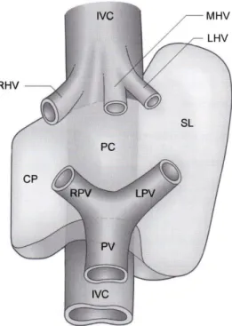
Figure 10: “Schematic representation of the anatomy of the caudate lobe. The caudate lobe consists of three parts: the caudate process (CP), on the right, the paracaval portion anterior to the vena cava (PC) and the bulbous left part (Spiegelian lobe, SL). IVC, inferior vena cava; RHV; right hepatic vein, MHV; middle hepatic vein, LHV; left hepatic vein, PV; portal vin, RPV; right portal vein, LPV; left portal vein. © Washington University in St Louis.” (Source: Strasberg SM. Hepatic, biliary and pancreatic anatomy.
In: Garden OJ, Parks RW. (eds.), Hepatobiliary and Pancreatic Surgery. A companion to specialist surgical practice. Fifth edition. Saunders, Edinburgh, 2014: 20.).
“The Brisbane Terminology” contains in the addendum of the original table an alternative and also adequate terminology for the second-order division. In the body of the table, the second-order partition is following Healey’s and Couinaud’s concept of apportionment of the artery and bile duct; in the addendum the second order rests on Couinaud’s idea of portal vein divisions. It was necessary to include it in the addendum because it maintains the ability of naming particular rare resections on the left side according to Couinaud’s concepts of the portal and hepatic veins, e.g. left paramedian sectorectomy [45] (Figure 11).
21
Figure 11: Alternative second-order division (sectors), nomenclature for anatomy and resections. (Source: Terminology Committee of the International Hepato-Pancreato- Biliary Association. (2000) The Brisbane 2000 Terminology of Liver Anatomy and Resections. HPB, 2: 333-339. https://www.ihpba.org/92_Liver-Resection- Guidelines.html).
22
In the rest of this study "The Brisbane 2000 Terminology" will be used.
3.3 Technical aspects of split liver transplantation
One of the greatest advantage of Split liver transplantation (SLT) is, that it maximises the use of available cadaver donor organs, both at the case of adults and children, owing to the fact that the full liver can be utilised. Previously, in the reduced size liver transplantation it was impossible, thus it means a major profit compared to the earlier technique. In 1989, 2 patients were reported to be transplanted with 1 donor liver [14]. The basis of SLT and its varieties are cutting the liver into parts, each of which has a sufficiently functioning hepatic mass, a bile duct, a venous outflow and a vascular pedicle. [49]. SLT has two main types. With applying the regularly used splitting technique a left lateral graft (segments II and III) and a right extended graft (segments I and IV-VIII) are achieved and can be transplanted into a young child plus an adult.
Whereas in the second type, the liver is cut along the Cantlie line, making two hemilivers - a left graft (segments I-IV) and a right graft (segments V-VIII) - which are sufficient for two grownup patients. The before mentioned splitting techniques however, show a great variety in the aspects of the challenges raised by anatomy, the required professional expertise and purpose [49].
Anatomical principles
As the result of dividing the hepatic parenchyma at the FL a segment II-III graft is obtained. In the case of pediatric recipients the size of the transplantable graft is about 250 cc in volume [50, 51], for adults it is one ‘right tri-segment’ graft, with the volume of 1100 cc, the rest of the Couinaud segment I, IV-VIII [49, 50]. Furthermore a ‘mono- segment graft’ (segment III) obtained from LLS graft is appropriate for new born babies and toddlers; for avoiding large-for size syndrome, we can apply a segment II mono- segment graft so that we can minimise the size of LLS grafts [49, 52, 53]. A fully developed cadaver liver may provide two grafts almost with the same size when cut along the MHV, suitable for giving them two adults with higher body mass. For people under the 60 kg weight, left-side 400-cc grafts are made from (segments I-IV) or without the caudate lobe (segments II-IV). Likewise, for patients who weigh 80 kg or more, right-
23
side grafts (segments I, V-VIII, or V-VIII) with the volume of 800-1000-cc are normally experienced to be adequate [49, 54, 55, 56, 57].
Donor selection
The efficiency and success of SLT has many factors out of which the most determining and critical one can be the adequate donor for the proper recipient. There is a criteria system for the donor selection. It involves age, serum sodium concentration, ABO match, liver function, similarity in size, no arrest period, vasopressor requirements, finally short donor hospitalization [49, 58, 59, 60]. Only hemodynamically stable cadaveric donors are suitable for SLT. Further requirements of the donors before left lateral splitting are that they have to be younger than 55, they should have not spent more than 5 days in intensive care, the fatty degeneration of their liver should be less than 30%, gamma-glutamyl transpeptidase is to be under 50 U/L, serum glutamic pyruvic transaminase less than 60 U/L, and serum Na less than 160 mmol/L [49, 61]. To get organs from adults with a full right - full left split, the donors have to be over 70kg. They are considered adequate for making grafts suitable for two adult recipients. Full left - full right split has greater requirements considering the quality of the organ and the donor should fulfil the oncoming requirements: younger than 40, should have not been in intensive care for more than 3 days, and fatty degeneration has to be less than 10% [62].
A liver biopsy has to be carried out the result of which, according to the macroscopic criteria, can be decisive considering the final decision if the quality of the graft is suitable for the splitting or not [49].
Recipient criteria
Before carrying out SLT on patients they need to be examined for some critical factors. The circumstances which are essential to be taken into account are the following;
age, the history of illicit drug usage and examination for drugs, alcohol agreement, treatment agreement, evaluation of the possibility of relapse, and checking two types of hepatitis (B and C). All these circumstances are to be taken into consideration in every general liver transplantation, even with greater emphasis if SLT is carried out [49, 63]. It is worthy to remember that right split-liver graft recipients are more advanced in age than the recipients of left split-liver graft [64]. The graft variables of the recipient involves the
24
fraction, the mass, and the type of the reconstruction of the hepatic artery ex vivo before the operation begins, cold and warm ischemia time with the use of Roux limb biliary drainage, and finally multiple-duct biliary anastomoses. For SLT extended dissection has to be carried out either on the back table ex vivo or within the heart-beating donor.
Increased blood loss is concerned when in situ SLT is made and the thoracic organ quality is to be taken into account as a result of volume replacement [49, 61]. A number of reports suggest that additional thoracic or abdominal organs with in situ SLT have no effect [49, 59, 65]. On average, in situ SLT for adults the extra time of the operation is 3 hours and one and half hours for children. Though, exceptions can happen and there has been a report of longer times [49, 66].
Left lateral splitting
In the case of small children who had end-stage liver problems there was an eager demand for the development of new techniques in the 1980s since the waiting list was unacceptably high, and the mortality rate almost reached the 40%. The evolution of left lateral splitting began when the first successful segmental graft was transplanted into a child from an adult through living donation and with the size reduction of the cadaver liver. Similarly, the transplantation of a whole adult organ to a child resulted in a significant reduction of the need for the living donations. It is essential to point out that the left lateral splitting does not compromises the adult graft pool and the remaining extended right graft is suitable for even large-sized adults, too without involving a small- for-size condition [67]. Due to its relatively great weight variability the LLS potentially can be given to recipients whose weight is less than 40 kg. Since the beginning of SLT, ex situ and in situ transplantation methods have been developed [68], which were comparably successful, if carried out in accordance with the logistical proportions.
However, the final result is greatly influenced by the selection of donor and recipients and the optimal technique, also. To identify the LHA, the hepatoduodenal ligament is cut from the left side. It is determined by the individual anatomy, if the right graft main arterial trunk may remain in continuity with the segment IV artery it may need to be anastomosed with the stump of gastroduodenal or LHA so as to minimize any risks of necrotizing of segment IV [49, 61]. Due to this, LPV is dissected down to the main bifurcation. Arising at segment I and IV portal branches, the main LPV, has to be
25
transected. Just right of the FL, the parenchyma is dissected. A flat surface is created out of the parenchyma when cutting it sharply into a single even plane in the ex situ technique, to create an efficient hemostasis. With the application of the already existing liver resection procedures combined with suture ligation of vessels and vessel clipping in the in situ technique the parenchyma is suitably transected. For the optimal hemostasis, the donor’s coagulation system is made use of in this technique. A LHV which surrounds the vessel loop, between the LHV and MHV can give a guidance to the surgeon while doing in situ splitting [49, 69]. With avoiding the isolation and dissection of the main LHD, the bile ducts and parabiliary vascular plexus of segments I and IV can be saved. The hilar plate, which includes the segment II and III hepatic duct(s), is to be divided sharply at the longitudinal part of the left portal vein (Rex Recessus). We have to divide the left side of the IVC right next to the LHV. The IVC to the right graft is kept in continuity with the MHV and RHV. The left lateral splitting is shown in Figure 12. All has to be done to prevent the possibility of right graft bile leakage risks. In case of necessity an intraoperative cholangiography might be included to save the segment I and IV draining bile ducts. Due to split-liver and LLS LDLT the waiting list fatalities in case of children has deeply fallen. Considering safety and surgery, the complete graft seems to be the safest; although, in young patients, successful outcomes were reported with SLT [49, 60, 70, 71, 72, 73]. LLS LDLT can lead to equal results with left lateral splitting of a cadaveric donor liver and transplantation of the created grafts [60, 74]. Therefore, in places where cadaveric livers are within easier reach SLT is suggested to be used to reduce the risks for the living donors. In the existing literature there is no mentioning of the availability of inferior graft or patient survival or higher surgical complication rates for right extended graft transplantation. As a result of this, considering security, when a right extended graft is transplanted it can be equal with the process when a whole organ is given. [49, 71, 75, 76].
26
Figure 12: Left lateral splitting: left lateral (segments II and III) and extended right (segments IV-VIII) liver grafts. IVC, inferior vena cava; LHV, left hepatic vein; SII-III HD, segment II and III hepatic duct; LPV, left portal vein; SII-III HA, segment II and III hepatic artery; CHA, common hepatic artery; PV, portal vein; CHD, common hepatic duct; Roman numerals stand for liver segments. (Original source:
http://accessmedicine.mhmedical.com/data/books/980/bru_ch11_f17.png).
Full left - full right splitting
As a result of routine use of left lateral splitting and using alive donors the lack of available organs in the cases of young patients has notably dropped. On the other hand, for adults and older children the want of organs was still pressing. In 1989, Bismuth and his colleagues [77] presented an operation in which one cadaveric liver was given to two adults for the first time. Since then, full left - full right splitting of liver became an essential factor for adult liver graft pool expansion since it doubles the grafts to be given to adult recipients (Figure 13), therefore decreasing the want for alive donors and the dangers involved in that method. The previous technique provides one graft (segments V-VIII) for an adult recipient with average size and one graft (segments I-IV) for a larger pediatric or for an adult with a smaller body weight. Full left - full right splitting, however, is such a complex liver transplantation form that it requires special knowledge of the
27
anatomic variations, furthermore high level of skills and expertise without which the success of the procedure cannot be granted. The complexities are the following; liver splitting for two adults should be done in places where skills and knowledge are based on the performance of many liver transplantations yearly. Where there is an ongoing hepatobiliary surgical program deeply experienced in left lateral splitting. The aspects of SLT for two grownup recipients has two critical issues: managing to ensure the safe biliary drainage for every implanted segments and sharing of blood vessels, especially the IVC and hepatic veins. The main alterations in left lateral splitting are that, the transection plane is larger, bile drainage of the implanted parts, higher possibility of the disturbance of the vascularization, and where to set the resection line, since there is no indicating anatomic structure (such as the FL). In these circumstances, the in situ splitting has a further advantage, which is the ability of identifying the ideal dissection plane. It can be identified by blocking the inflow of one of the hemilivers. Prior to perfusion, adequate venous outflow and arterial/portal inflow (especially at segments V and VIII) is to be guaranteed after the parenchymal transection. Finally, the donor’s own coagulation system is to be applied in this method to achieve a biliostasis and hemostasis.
Intraoperative cholangiography is used to identify the anatomical hepatic duct variations, which can completely prohibit liver splitting or can point out a left lateral splitting [49, 61]. The place of common hepatic duct (CHD) either to the right or left of the hemiliver is determined by individual anatomy. The CHD regularly belongs to the right graft since frequent anatomical variations have been noted with the right lobe where the RHD is shorter than in the left. To assure sufficient perfusion the hepatic ducts are to be shortened to every possible extent. Arteriography is advised in some literature [64], but hilar dissection can be applied for the safe identification of the arterial anatomy for the most part. The arterial trunk sharing, especially segment IV artery origin is specified by individual donor anatomy. In general, the left graft is considered with the main arterial trunks. When using traditional methods, the IVC remains with the right graft and the MHV stays with the left graft. The viability of the liver segment I is in question when the division of the caudate lobe veins is needed. In this case a resection can be necessary [49].
The split vena cava method [78] has been introduced to provide both hemiliver grafts with optimal venous drainage. This method includes IVC division, and the sustenance of venous drainage of dorsal parts and segment I of right lobe through retrohepatic veins.
28
On the other hand, the venous congestion of segment V and VIII is impossible to be averted by this technique when the left graft contains the MHV. Thus, there is a requirement of these veins on the cut surface to undergo further venous reconstruction.
The literature published about full left - full right splitting is much less than on splitting for an adult and a child. This procedure, however, has not been widely accepted since it resulted in poor success at the beginning and the fatalities were high, especially after left hemiliver grafting [49, 64, 79, 80, 81]. The main barriers of the expansion of this technique are that of logic and technic, furthermore, there is normally a risk for the development of a small-for-size problem when a full left - full right splitting for two adults is carried out. The essential factors of having positive outcomes with full left - full right SLT compared to whole-size organ transplantation are: proper technical abilities ideal size match of graft recipients, and suitable graft quality. Graft quality has to be evaluated by an experienced surgeon with SLT knowledge, and when possible, a liver biopsy should help. If we compare it to left lateral splitting, donor selection has much higher requirements. Not only the individual recipient’s needs but also an absolutely transplantable functioning liver mass has to be assessed during the selection of the recipient. The transplanted graft should reach the minimum of 1.0% of the recipient’s body mass weight [49, 59]. There can be a possibility of increasing the functional liver mass if general worsening condition of the recipient may occur. This limit can only be exceeded in individually evaluated elective cases where there is no portal hypertension.
Therefore no compromises can be made when selecting the donor and the recipient since they are essential for the most successful outcomes [49].
29
Figure 13: Schematic illustration of full left - full right liver grafts. IVC, inferior vena cava; RHV, right hepatic vein; LHV, left hepatic vein; MHV, middle hepatic vein; LHD, left hepatic duct; LPV, left portal vein; LHA, left hepatic artery; RPV, right portal vein;
CHA, common hepatic artery; CHD, common hepatic duct; PV, portal vein; FL, falciform ligament; RL, round ligament; Roman numerals stand for liver segments. (Original source: https://optn.transplant.hrsa.gov/media/2016/fig2_split_liver.jpg?width=359px&
height=233px).
30
4 Objectives
The detailed surgical anatomy is one of the basics of major liver surgery including liver resection and transplantation. Since the lack of cadaveric organs, all over the world the number of liver transplantation was limited, Pichlmayr (1988) and Raia (1989) carried out the first successful SLT and LDLT [14, 15] two decades after the first human liver transplantation. Subsequently, Broelsch initiated the idea of LLS LDLT for transplantation in young patients [82].
Partial liver graft transplantation is a process in which the surgeon creates a liver graft from a living-donor [82, 83, 84], that reduces a larger cadaveric graft [13, 85], or divides an adult cadaveric liver during SLT [68, 86, 87, 88]. The use of partial liver graft transplantation techniques led to a rise in the number of pediatric donor organs and lessened the pretransplant complications and mortality [82, 88, 89] but the SLT predisposes to specific complications [84, 91, 92, 93, 94]. Biliary complications can be the following (1) biliary stricture and (2) anastomotic leakage which are still considered to be stressing problems to address during partial liver graft transplantation and are often originate from an ischemia of the biliary tract or the lack of complete anatomic expertise of the bile duct system [95, 96, 97, 98]. The reported incidence of the biliary complications’ rate is announced to be 5% - 38% which made some authors to consider biliary anastomoses as the "Achilles heel" of segmental liver transplantation [99]. Owing to the previously mentioned worries anatomical and clinical researchers are eager to make a deeper clarification of anatomic variations and surgical techniques to eliminate the risk of these complications [100, 101, 103, 104, 105, 106]. Owing to these reasons it is absolutely necessary that liver surgeons should be full-trained in the anatomy of the bile duct system and have the ability to realise the existence and the implications of the anatomical variations [107]. Since the data published on the biliary anatomy and statistics display significant differences, altered incidences in different countries and/or limitations of different methods cannot be excluded. Therefore we aimed to investigate the incidence of bile duct variations in the Hungarian population. With the increasing prevalence of partial liver transplantation and liver resections a detailed preoperative assessment of biliary anatomy is mandatory. More and more sophisticated high resolution diagnostic imaging methods provide accurate preoperative evaluation of hepatobiliary anatomy.
31
Moreover, new data are of special importance since the development of a new system of common transplantable organ pool in Europe is in progress. It is known that the European Union (EU) started already the implementation of EU directives including safety and quality standards in transplantation. The investigation and findings of the anatomical variations can improve the level of education and can help the standardization of surgical techniques.
Since the incidence of biliary variations is far more numerous in the right lobe, the right lobe LDLT is widely accepted for adult patients, the complications are concomitants of reconstruction of complex biliary routes after right lobe harvesting [108].
The detailed knowledge of hilar anatomy is also essential in case of full-left full-right, split or in case of left lobe living donor liver transplantation when the resection line goes through the LHD at the hilum. An unforeseen biliary variant may extend graft ischemia time and increase the risk of postoperative complications. Therefore the first aim of this study was to complete the data on the surgical anatomy of the hilar biliary tree by investigating the types and incidence of hilar biliary variants in the Hungarian population.
The most commonly used LDLT technique is LLS transplantation for children.
Recently, the survival rates have significantly improved, however, biliary complications are still the major source of morbidity after pediatric LDLT [109,110,111, 112]. The incidence of biliary complications after LLS transplantation is higher when multiple bile ducts are present [99], hence it is important to choose a line of hepatotomy that results in the fewest possible surface ducts that need to be anastomosed in the recipient. Therefore the second aim of this study was to investigate the optimal division line of hepatotomy for an LLS donation, based on the anatomical variations of left hepatic duct system.
32
5 Methods
5.1 Working out of the corrosion cast technique
Although several researcher have used corrosion cast techniques to study the anatomy of different vessels in humans or in animals we aimed to work out our own vessel lumen filling and modified corrosion technique for the best possible results. If a researcher decides to use corrosion technique to get the required data one will face a great number of questions, e.g.:
- Should conventional corrosion technique or vessel lumen filling technique without corrosion be used?
- Which resin is the best for the planned research work (investigation of the hepatic ducts system)?
- Is the use of CT scan wanted? – Is any extra contrast material needed?
- Does it need to be coloured?
- Special additives?
- Special circumstances during preparation?
We summarise here our answers for the above mentioned questions.
Conventional corrosion cast or vessel lumen filling without corrosion technique?
The main difference between the two techniques is that while the conventional corrosion technique results “just” in the resin cast without any organic tissue around the vessels or on the preparation, the vessel lumen filling technique does not use corrosive material to remove the organic tissue so by the end of the procedure the preparation does have the organic tissue with its vessels filled up with resin. To decide which technique is better to get the aimed data mainly depends on the followings:
If the resin cast analysis is simply enough and we do not plan to do any further preparations which require the original organic tissue, we need to choose the conventional corrosion technique (e.g. the study of the „hilar variations of the hepatic duct system” in this work) (Figure 14). However, if we need to keep the organic tissue e.g. we need it in order to be able to perform surgical procedure on the preparation (e.g. LLS hepatectomy)
33
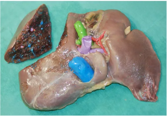
or to be able to determine the exact place of an intra organic anatomical structure in the view of surface markings on a CT scan (e.g. the study of “optimal line of hepatotomy for left lateral living donor liver transplantation” in this current work), in these cases the lumen filling technique without removing the organic tissue is the choice (Figure 15).
Both techniques are suitable for CT scans but if there are soft tissues around the resin cast, more investigation will be needed to find the optimal CT density because of the higher background density of the organic tissue.
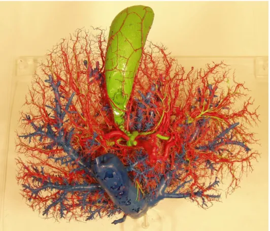
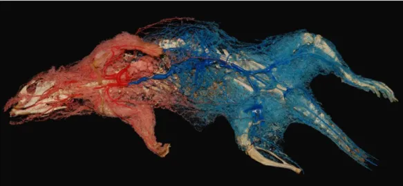
Figure 14: Corrosion cast preparation of human liver. Liver parenchyma is removed with potassium hydroxide. IVC and hepatic veins - blue, bile ducts and gallbladder – green, hepatic artery - red, (Source: author’s own work).
34
Figure 15: Vessel lumen filling technique. Liver parenchyma is fixed around the resin cast. IVC and hepatic veins - blue, portal vein – purple, bile duct – green, hepatic artery – red. (Source: author’s own work. Co-workers: Zsuzsanna Kürti, Zsolt Pápai, András Szuák).
Find the best resin
Synthetic resins are materials which possess the qualities of the natural plant resins: they are viscous liquids which are able to harden permanently. From a chemical point of view they show a great alteration from the different resinous compounds secreted by plants.
In the time of chemical inventions numerous different types of synthetic resins are available to perform corrosion cast studies. These resins have been mainly in use for industrial flooring, tool-making, car and boat making and repairing purposes since the 1960’s (source: https://en.wikipedia.org/wiki/Synthetic_resin). There are at least five main types of resins to be considered for corrosion casts:
1. Polyester 2. Vinyl ester 3. Epoxy ester 4. Polyurethane 5. Acrylic resin
35
From the corrosion cast study point of view the main physicochemical parameters must be considered are:
1. viscosity of the liquid resin mixture 2. flexibility of the hardened resin 3. durability of the hardened resin 4. acid resistance of the hardened resin 5. CT density of the hardened resin
The viscosity is one of the most important features of the resin. If it is too high, the resin is not able to be injected deeply enough into the small vessels or ducts. On the other hand, if the viscosity is too low researchers have to be very careful not to overfill the structures make the preparations inaccessibly dens (e.g. in case of injecting too low viscosity resin injection into the hepatic veins the resin can go as deep as the liver sinusoids without staying just in the level of the hepatic veins). In 2014, in collaboration with the 1st Department of Surgery, Semmelweis University, Károly Németh from our Clinical Anatomy Research Laboratory, took part in the study of the „Collateral circulation of the rat lower limb and its significance in ischemia - reperfusion studies”.
For this unique work, the research team needed to use an extreme low viscosity resin with extreme durability and perfect acid resistance. After the trials of different resins, an
acrylic resin, Methyl Methacrylate (UZIN KR 416,
http://www.uzin.com/products/product-search/details/uzin-kr-416-219/) was chosen that was worked out by Bence Dorogi. This resin was found very useful for the study of the smallest vessels and their anastomoses but was not ideal for wider diameter structures since it always filled up even the smallest vessels making the casts unnecessarily dense for the planned investigations of the ramification of bile duct system (Figure 16).
36
Figure 16: Corrosion cast of a male Wistar rat’s arterial system for the study of the
„Collateral circulation of the rat lower limb and its significance in ischemia - reperfusion studies” by Rosero et al. In this study an extreme low viscosity Methyl Methacrylate resin mixture was used by Németh Károly’s guide, as advised by Bence Dorogi. The arterial system of the rat’s head, upper limbs and upper trunk is red, while the lower limbs, lower trunk and tail are blue. Anastomoses can be identified between the two systems. (Source:
Rosero O, Nemeth K, Turoczi Z, Fulop A, Garbaisz D, Gyorffy A, Szuak A, Dorogi B, Kiss M, Nemeskeri A, Harsanyi L, Szijarto A. (2014) Collateral circulation of the rat lower limb and its significance in ischemia - reperfusion studies. Surg Today, 44: 2345-2353.).
As regards flexibility, generally speaking it can be said that for a conventional corrosion cast technique high flexibility is a disadvantage because the cast will not have a stable frame and will lose its original shape without a supporting organic tissue around the flexible resin. However, if the aim is to perform/simulate surgical procedures on the preparations, a hard framed cast can be easily broken in the soft organic tissue during the simulation or the hard resin cast cannot be cut with a conventional surgical scalpel. That is why a flexible resin generally is more preferred for a vessel lumen filling technique (Figure 15 and 17).
37
Figure 17: Left lateral segment graft of human liver vessel lumen filling technique preparation. High flexibility resins were used at this preparation (liquid urethane rubber- Vytaflex by Smooth-on was used for the artery and bile ducts; Köraform, a two- component silicone mould-making compound by Alpina Technische Produkte GmbH was used for the portal and hepatic veins) which kept the liver soft and easy to cut, while creating a graft from the whole size liver on hands on course. Preparation from the „First Donor Surgery Masterclass” Hungary, Budapest 2014.01.30-31. IVC and hepatic veins - blue, portal vein - purple, hepatic duct - green, hepatic artery - red (Source: author’s own work. Co-workers: András Szuák, Zsuzsanna Kürti, Zsolt Pápai, Sándor Kovács).
If there is a need for further preparation of a hard resin filled liver, super durable resin is essential (Figure 18).
Figure 18: Human liver vessel lumen filling technique preparation after further preparation of the hepatic duct system. This technique requires „super durable” resin (Source: Zsolt Pápai).
38
For a vessel lumen filling technique there is no need for acid resistance since the parenchyma will not be removed by acid from the cast, but this is a really important feature of the resin in case of conventional corrosion cast technique. We have tried many different resins from this aspect and the Novolac-based Epoxy Vinyl Ester Resin (Derekane 470-300 by Ashland) was proved to be the most acid resistant.
For CT scan examination we need to know the density of the resin itself to make sure we can set up the optimal density of the resin mixtures that go into the different vessels or bile ducts. The average density of the different resins is about 200 Hounsfield unit (HU) (e.g. Derekane 470-300), but occasionally some resins has a higher value (e.g.
Köraform, a two-component silicone mould-making compound by Alpina Technische Produkte GmbH has a CT density of 400HU). Naturally, higher density resins require less contrast materials in the resin mixture.
Contrast material
If a certain research work calls for the CT scan of the cast, the proper CT density of the resin mixture needs to be set up in advance in order to be able to make difference between the resin in the different vessels and the surrounding organic tissues on the CT scan.
Hounsfield unit is a standard form of quantity widespread in CT scanning to express CT. Hounsfield units, labelled after their creator Sir Godfrey Hounsfield, originate from the measured attenuation coefficients, which undergoes a linear transformation. This transformation is founded on the peremptory definitions of water (0 HU) and air (-1000 HU) [113]. In our series of formaldehyde fixed human liver preparations the average CT density of the liver parenchyma was 100 HU. If the aim is to fill up more than one vessels on a certain preparation (and we want to visualise all of them separately on the CT scan), we need to keep at least 300-400 HU difference between the different structures since the density of the resin drops in the vessels from proximal to distal. On the other hand, the highest density of any of the structures must not be more than 1900-2000 HU because it would cause severe secondary products. The preparation shown on Figure 19, we set up the density of the resin mixtures as follow: hepatic vein - 600 HU, portal vein - 1000 HU, hepatic duct – 1400 HU, hepatic artery – 1800 HU. Since the density of formaldehyde fixed liver parenchyma is about 100 HU we could keep the
39
required 400 HU difference with these densities. As a result of this, the different vessels could be visualised (and colour coded) separately or all together (with or without the parenchyma) on the CT pictures (Figure 19).
Figure 19: CT scans of a human liver preparation with specially adjusted CT density for the different vessels and bile ducts. The different structures can be visualised (and colour coded) separately or all together (with or without the parenchyma) because of the appropriately different CT density. IVC and hepatic veins - blue, portal vein - purple, hepatic duct - green, hepatic artery - red (Source: author’s own work. Co-workers:
Zsuzsanna Kürti, Ibolyka Dudás, Zsolt Pápai, András Szuák).
40
Various contrast materials could be considered to set up the required CT density if those meet the following requirements:
- compose a homogenous resin mixture - reproducible with the same density
- available on the market (on acceptable price)
In our studies we practically used Lipiodol, Gastrographin or Barium powder. While the first two are liquid, barium is a powder. We made dilution series with the different contrast materials to check their homogeneity, reproducibility and their enhancement of CT density. It was found, that 1% m/m Lipiodol adds 150 HU extra density to the resin while 0.25% m/m barium powder gives the same enhancement (Figure20).
Figure 20: Dilution series with the different contrast materials to check the homogeneity, reproducibility and enhancement of CT density (Source: author’s own work. CT scan was done by Dr. Ibolyka Dudás)
Additives
1. Most resin requires a Catalyst for the polymerization.
2. Many resin (e.g. Derekane 470-300 by Ashland) requires accelerator to speed the polymerization time up.
3. In most studies there is a need for pigments to set the required colours of the resin mixtures. After we tried various colourants, Pigments FP 6018 yellow-