Clinical anatomy of partial liver transplantation, a modified corrosion cast method
PhD thesis
Mátyás Kiss MD.
Semmelweis University Doctoral School of Basic Medicine
Consultants: Prof. Zoltán Máthé MD, Ph.D., professor, head of department Ágnes Nemeskéri MD, Ph.D., associate professor
Official reviewers: Prof. László Entz MD, Ph.D., professor
András Bálint MD, Ph.D., Med. Habil., private professor Head of the Final Examination Board:
Prof. József Sándor MD, Ph.D., professor Members of the Final Examination Board:
Zsolt Csapó MD, Ph.D., chief physician and head of department Ákos Szűcs MD, Ph.D., senior lecturer
Budapest 2017
1
1 Introduction
Two decades after the first human liver transplantation, Pichlmayr (1988) and Raia (1989) performed the first successful split- and living donor liver transplantation (SLT and LDLT). Partial liver graft transplantation techniques have increased the supply of donor organs and reduced the pretransplant complications and death but the SLT predisposes to specific complications. Biliary complications such as biliary stricture and anastomotic leakage reportedly remain serious problems in partial liver graft transplantation and are caused by an ischemia of the biliary tract or the incomplete understanding of the surgical anatomy of the bile duct system.
2 Objectives
The detailed knowledge of hilar anatomy is essential in case of full-left full- right SLT and LDLT since the resection line goes through the hilar hepatic ducts. The first aim of this study was to complete the data on the surgical anatomy of the hilar biliary tree by investigating the types and incidence of hilar biliary variants in the Hungarian population.
The most commonly used LDLT technique is left lateral segment (LLS) transplantation for children. The incidence of biliary complications after LLS transplantation is higher when multiple bile ducts are present, hence it is important to choose a line of hepatotomy that results in the fewest possible surface ducts that need to be anastomosed in the recipient. Therefore the second aim of this study was to investigate the optimal division line of hepatotomy for an LLS donation, based on the anatomical variations of left hepatic duct system.
3 Methods
We worked out new vessel lumen filling and modified corrosion techniques for the best possible results.
2
New corrosion cast technique to study the hilar variations of the hepatic duct system
A total of 106 fresh human adult livers were removed then the common hepatic duct (CHD) were cannulated and injected with Vinyl Ester resin mixture (1.4 ml resin / 100gr liver tissue). Liver parenchyma was corroded by potassium hydroxide, casts were macroscopically analyzed and CT scanned. The confluence of left and right hepatic ducts and those of segmental ducts participating in the formation of common hepatic duct in the absence of left or right hepatic ducts were analyzed and categorized according to the modified Couinaud’s classification both in the liver casts and their CT scans.
Vessel lumen filling without corrosion technique to study the optimal line of hepatotomy for left lateral living donor liver transplantation
Thirty human liver preparations were made for this experiment using a flexible resin mixture with colorant and CT contrast agents. After polymerization, the liver parenchyma was not corroded but was fixed with 8% of formaldehyde solution. CT scans were performed and the branching pattern of the left hepatic duct and the distance between the falciform ligament (FL) and the confluence of segment II and III ducts was analyzed using 3D CT reconstruction. The number of bile ducts on the surface of virtual hepatotomy was estimated for three different virtual division lines.
4 Results
4.1 Hilar variations of the hepatic duct system
We prepared 106 high quality human liver casts with our newly developed corrosion technique. The casts were durable enough for complete analysis, and provided excellent density for the CT scans. The classification presented in the current study is based on the segmental anatomy described by Couinaud according to the absence or presence of the left (LHD) and right (RHD) hepatic duct (1957) and modified by Smadja and Blumgart in 1994 (Figures 1-6). Table 1. Summerises the classification and
3
frequencies of hilar biliary variations in the present study and those of variations reported by other authors.
Group I: Presence of left and right hepatic ducts, normal biliary anatomy: Type
"A" (54,74%)
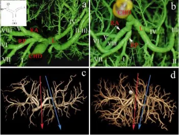
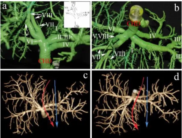
The right and left lobes are drained by the RHD and LHD, respectively; this configuration is commonly considered as normal biliary anatomy (Figure 1). The right anterior hepatic duct (RAHD) draining segments V and VIII, join the right posterior hepatic duct (RPHD) draining the segments VI and VII. Their confluence gives rise to the right hepatic duct. The left hepatic duct (LHD) drains segments II, III and IV. In full left - full right split, the cutting plane is on the right side of the joining segmental duct IV, through the LHD.
Figure 1: Normal biliary confluence: Type “A”. a) Biliary cast; antero-inferior view. The insert shows the schematic illustration of Type “A” configuration. b) postero-inferior view of the cast. c) 3D volume rendering reconstruction; antero- superior view. d) 3D volume rendering reconstruction; postero-inferior view. On the CT images the blue arrow indicates the plane of the left lateral split while the red arrow shows the plane of the full left - full right split. CHD, common hepatic duct;
RA, right anterior hepatic duct; RP, right posterior hepatic duct; L, left hepatic duct. Roman numerals stand for the segmental ducts. (Source: author’s own work. Co-workers: András Szuák, Zsolt Pápai, Sándor Kovács. CT pictures made by Ibolyka Dudás).
4
Group II: Absence of the right hepatic duct - presence of the left hepatic duct (41,49%)
Variation Type "B" (8.49%). A triple confluence forming the common hepatic duct is the main feature of variation Type “B”. The RAHD and RPHD join to the LHD without forming a considerable length of the RHD. In full left - full right split, the optimal transection line runs through the LHD just before the LHD joins CHD.
Variation Type "C1" (5.66%). In type “C1” the RAHD drains directly into the CHD as its continuation, and the RPHD crosses the RAHD on reaching the confluence. It is easy to perform full left - full right split in this variation just before the LHD drains into the CHD.
Variation Type "C2" (1.87%). An ectopic drainage of RPHD into the CHD characterizes this variant. In full left - full right split, the resection is just before the LHD merges into the RAHP.
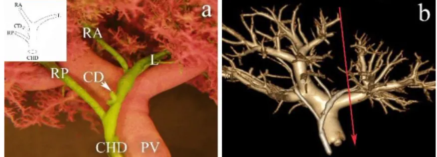
Subvariant of Type "C2” (0.94%). The cystic duct drains into the bile duct between the merging site of RPHD distally and the union of RAHP with the LHD proximally (Figure 2). This subvariation is also ideal for full left - full right split.
Figure 2: Configuration of Type "C2" subvariant. The right posterior hepatic duct (RP) drains into the common hepatic duct (CHD) distally to the confluence of cystic duct (CD). a) Biliary cast; anterior view of the hilum. The insert shows the schematic illustration of Type “C2” subvariant configuration. Beside the biliary tree, the portal vein (PV) was also injected with purple coloured resin. b) 3D volume rendering reconstruction; anterior view. On the CT image the red arrow shows the site of the full left - full right split. RA, right anterior hepatic duct; L, left hepatic duct. (Source: author’s own work. Co- workers: András Szuák, Zsolt Pápai, Sándor Kovács. CT pictures made by Ibolyka Dudás).
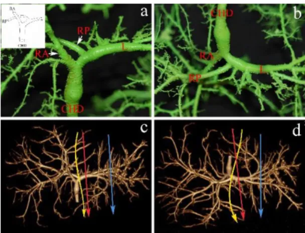
Variation Type "D1" (22.64%). In type “D1” variation the RPHD drains into the LHD. In full left - full right split, the adequate site of transection is through the
5
LHD before the RPHD drains into it. Since several ducts drain segment II and III on this particular cast, the left lateral split may result in more than two ducts on the surface of resection to be reconstructed (Figure 3). Out of the total of 106 casts 24 cases displaying “D1” variation. The distance between the origin of the right posterior and the right anterior ducts was less than 9 mm in 95.83%, in one case (4.17%) it was 24.15 mm.
Figure 3: Type “D1” configuration: The right posterior duct (RP) drains into the left hepatic duct (L). a) Biliary cast; antero- superior view. The insert shows the schematic illustration of Type „D1” configuration. b) Infero-posterior view of the cast.
c) 3D volume rendering reconstruction; antero-superior view. d) 3D volume rendering reconstruction; postero-inferior visceral view. On the CT images the blue arrow indicates the site of the left lateral split while the red arrow shows the plane of the full left - full right split. The yellow arrow shows the site of the erroneously designed full left - full right split. CHD, common hepatic duct; RA, right anterior hepatic duct. (Source: author’s own work. Co-workers: András Szuák, Zsolt Pápai, Sándor Kovács. CT pictures made by Ibolyka Dudás).
Variation Type "D2" (0.94%). The RAHD collecting the bile from segments V and VIII drains into the LHD.
Variation Type "F" (0.94%). The RPHD drains into the common trunk of the RAHD and LHD. There is a confluence of the RPHD and cystic ducts.
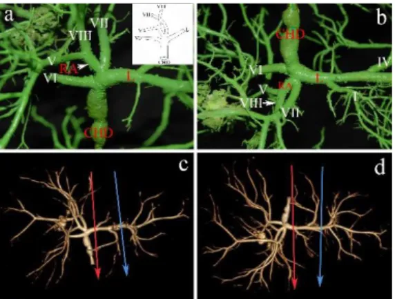
Newly described variation Type “G” (0.94%). Revealing a biliary configuration that has not yet been recorded until present, we have further extended the Couinaud’s classification modified by Blumgart and Smadja by a new category of variation: “Type G”. On the right side, only the RAHD can be identified, while the duct from segment VI drains separately into the main confluence. Moreover, the duct from
6
the segment VII has a common entry with the RAHD into the terminal part of the LHD (Figure 4). Performing full left - full right split, the LHD can be transected before the duct from segment VII drains into it.
Figure 4: Type “G” configuration: Presence of left hepatic duct and absence of right posterior hepatic duct and right hepatic duct. Note the common ostium of the right anterior hepatic duct (RA) and the duct of segment VII into the left hepatic duct (L). The duct of segment VI joins the common hepatic duct (CHD). a) Biliary cast; antero-superior view. The insert shows the schematic illustration of Type „G” configuration. b) Postero-inferior view of the cast. c) 3D volume rendering reconstruction; antero-superior view. d) 3D volume rendering reconstruction; postero-inferior view. On the CT images the blue arrow indicates the site of the left lateral split while the red arrow shows the site of the full left - full right split. Roman numerals stand for the segmental ducts. (Source: author’s own work. Co-workers: András Szuák, Zsolt Pápai, Sándor Kovács. CT pictures made by Ibolyka Dudás).
Group III: Absence of the left hepatic duct - presence of the right hepatic duct (0
%). Of the 106 biliary casts none had such biliary configuration.
Group IV: Absence of the left hepatic duct - absence of the right hepatic duct: Type
"E" (3.75 %)
Variation Type "E1". In this configuration the ducts from segments II and III after receiving the segmental duct I and IV, respectively, separately drain into the RPHD; while the RAHD joins these merged ducts. None of our casts showed this variation; however, we found two preparations that fit into this group, except, the ducts from segments II and III do not join separately to form the CHD with the right sectional ducts. We extended the Couinaud’s classification modified by Blumgart and Smadja by two subvariations.
7
Type "E1a" subvariant (1.87%). In Type “E1a” configuration the ducts from segment II and III form a common trunk with the RPHD which receives then the duct from segment IV. Most distally the RAHD joins into this common trunk resulting in the formation of the CHD. This variation is not ideal for full left - full right split since there would be two separated bile duct on the surface of resection, instead of one LHD.
Type "E1b" subvariant (0.94%). Type “E1b” biliary anomaly exhibits one particular dissimilarity compared to variation “E1a”, namely the entry of the duct of segment IV is distal to that of the RAHD. This variation is not ideal for full left - full right split since there would be two separated bile duct on the surface of resection, instead of one LHD.
Variation Type "E2". Biliary tree in this group has also double hepatic duct on the left side (IV - III and II - I) like in group “E1” and the right sectional ducts join the CHD separately at the same level. Of 106 casts none of them bears this variation;
however, we observed a configuration that fits into this group, except, the double hepatic ducts on the left side are formed by the segmental duct IV and by a duct from segments III, II, and I, respectively. Moreover, right sectional ducts (RAHD and RPHD) are also absent. We inserted this variant into the Couinaud’s classification modified by Blumgart and Smadja as subvariant of type “E2”.
Subvariant of type "E2" (0.94%). Instead of the right anterior and posterior hepatic ducts a highly complex drainage pattern is present in this variant. One branch from segment VIII forms a common trunk with the segmental duct VII, while the other branch from segment VIII forms a common trunk with the segmental duct V.
Furthermore, these two common trunks and the duct from segment VI form a trifurcation. On the left side, there is a common stem of segmental ducts II and III that receives duct from segment I, while the duct from segment IV joins independently and most distally into the CHD (Figure 5). This variation is not ideal for full left - full right split since there would be two separated bile ducts on the surface of resection, instead of one left hepatic duct.
8
Figure 5: Subvariant of type "E2" configuration: There are neither RAHD (RA) nor RPHD (RP) and LHD.. a) Biliary cast;
antero-superior view. The insert shows the schematic illustration of subvariant of type “E2” configuration. b) Postero- inferior view of the cast. c) 3D volume rendering reconstruction; antero-superior view. d) 3D volume rendering reconstruction; postero-inferior view. On the CT images the blue arrow indicates the site of the left lateral split while the red arrow shows the site of the full left - full right split. CHD, common hepatic duct; Roman numerals stand for the segmental ducts. (Source: author’s own work. Co-workers: András Szuák, Zsolt Pápai, Sándor Kovács. CT pictures made by Ibolyka Dudás).
Figure 6: Branching patterns of hepatic ducts in 106 human biliary casts classified according to Couinaud’s classification modified by Smadja and Blumgart and by the present author. Newly inserted variants: C2 subvariant, E1a subvariant, E1b
9
subvariant, E2 subvariant. "G" a recently observed new variant. CHD, common hepatic duct; RA, right anterior hepatic duct; RP, right posterior hepatic duct; L, left hepatic duct; Roman numerals stand for the segmental ducts.
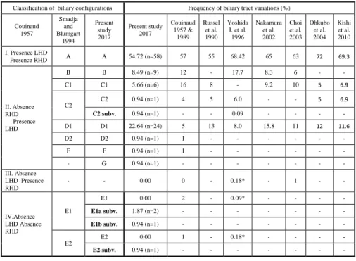
Table 1: Classification and frequencies of biliary variations in the present study and those of variations reported by other authors
Classification of biliary configurations Frequency of biliary tract variations (%) Couinaud
1957
Smadja and Blumgart
1994
Present study 2017
Present study 2017
Couinaud 1957 &
1989 Russel
et al.
1990 Yoshida
J. et al.
1996
Nakamura et al.
2002 Choi et al.
2003 Ohkubo
et al.
2004 Kishi et al.
2010 I. Presence LHD
Presence RHD A A 54.72 (n=58) 57 55 68.42 65 63 72 69.3
II. Absence RHD Presence LHD
B B 8.49 (n=9) 12 - 17.7 8.3 6 - -
C1 C1 5.66 (n=6) 16 8 - 9.2 10 5 6.9
C2
C2 0.94 (n=1) 4 5 6.0 - - 5 6.9
C2 subv. 0.94 (n=1) - - 0.09 - - - -
D1 D1 22.64 (n=24) 5 13 8.0 15.8 11 12 11.6
D2 D2 0.94 (n=1) 1 - - - - - -
F F 0.94 (n=1) 1 - - - - - -
- G 0.94 (n=1) - - - - - - -
III. Absence LHD Presence RHD
- - 0.00 0 - 0.18* - 1 - -
IV.Absence LHD Absence RHD
E1
E1 0.00 2 - 0.09* - - - -
E1a subv. 1.87 (n=2) - - - - - - -
E1b subv. 0.94 (n=1) - - - - - - -
E2
E2 0.00 1 - 0.18* - - - -
E2 subv. 0.94 (n=1) - - - - - - -
RHD, right hepatic duct; L, left hepatic duct; -: no data; *: calculated on the basis of percentages and numbers of cases presented by the authors
4.2 Variations of the left hepatic duct, optimal line of hepatotomy for left lateral living donor liver transplantation
With our newly developed vessel lumen filling technique with preserved liver parenchyma we made 30 high quality human liver preparations to study the anatomical variations of the LHD and the optimal line of hepatotomy for the simulation of the left lateral LDLT.
10
Branching patterns of the left hepatic duct
According to the confluences of bile ducts segment II, III and IV, three different main types with subtypes were found (Figures 7-9).
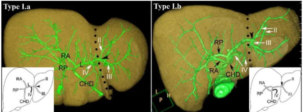
In variation Type I. (76.77%) the bile ducts from segments II and III form a common trunk. The duct from segment IV joins into this common trunk in the subtype named: Type I.a (66.67%) (Figure 7). In another subtype, segment IV duct merges into the common hepatic duct/one of the hilar ducts; we have designated it as Type I.b (10%) (Figure 7).
Figure 7: Type I.a and Type I.b. FL is indicated by the black dotted line, while a black arrowhead shows the junction of segment II and III ducts. RA, right anterior hepatic duct; RP, right posterior hepatic duct; L, left hepatic duct; CHD, common hepatic duct; Roman numerals stand for the segmental ducts. (Source: author’s own work. Co-workers: András Szuák, Tien Nguyen. CT pictures made by Ibolyka Dudás).
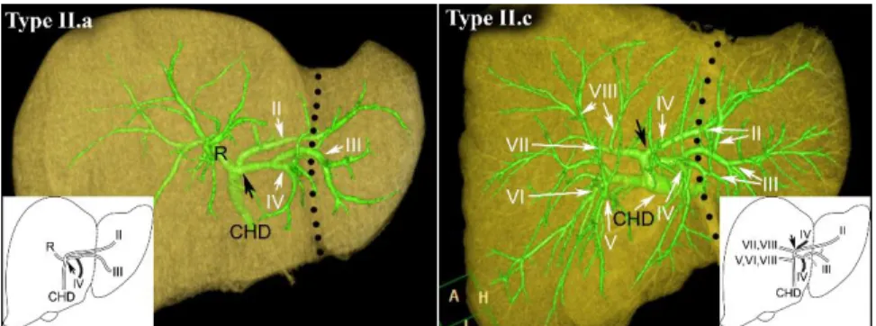
In variation Type II. (20%) the duct of segment IV drains into segment II or segment III or both ducts proximally from the confluence of segment II and III ducts.
When segment IV duct drains into segment III duct, we named it as Type II.a (16.67%) (Figure 8). Theoretically segment IV duct could drain into segment II duct, it should be named as Type II.b in this classification, however there was no liver found in our series like this. When segment IV duct drained into both segment II and segment III ducts, we labeled it as Type II.c (3.33%) (Figure 8).
11
Figure 8: Type II.a and Type II.c. FL is indicated by the black dotted line, while a black arrow shows the junction of segment II and III ducts. R, right hepatic duct, CHD, common hepatic duct. Roman numerals stand for the segmental ducts. (Source:
author’s own work. Co-workers: András Szuák, Tien Nguyen. CT pictures made by Ibolyka Dudás).
Variation Type III. denotes a trifurcating confluence of segment II, III and IV ducts forming the LHD (3.33%) (Figure 9).
Figure 9: Type III. FL is indicated by the black dotted line, while a black arrow shows the junction of segment II and III ducts. R, right hepatic duct; L, left hepatic duct; CHD, common hepatic duct. Roman numerals stand for the segmental ducts.
(Source: author’s own work. Co-workers: András Szuák, Tien Nguyen. CT pictures made by Ibolyka Dudás).
Evaluation of the number of bile ducts in three different division lines
The surgical relevance of the LHD variations described above was evaluated by counting the number of bile ducts on the surface of virtual hepatotomy in three different division lines. When the virtual division line was on the FL, there was a single duct for anastomosis in just 30% of cases and there were 2, 3 or 4 ducts in 53.3%, 10.0%, and 3.3%, respectively. The optimal line of division was achieved when virtual
12
hepatotomy was performed one cm to the right of the FL resulting in one hepatic duct only to be anastomosed in about two thirds (70%) of the investigated livers (Table 2).
Table 2: Number of bile ducts on the surface of virtual hepatotomy in case of three different division lines Number of bile ducts
on the surface of virtual hepatotomy
Division line: 1 cm to the right of FL
Division line: 0.5 cm
to the right of FL Division line: on FL
1 21/30 (70%) 15/30 (50%) 9/30 (30%)
2 8/30 (26.6%) 11/30 (36.7%) 16/30 (53.3%)
3 1/30 (3.3%) 3/30 (10%) 3/30 (10%)
4 0/30 (0%) 1/30 (3.3%) 1/30 (3.3%)
FL, falciform ligament
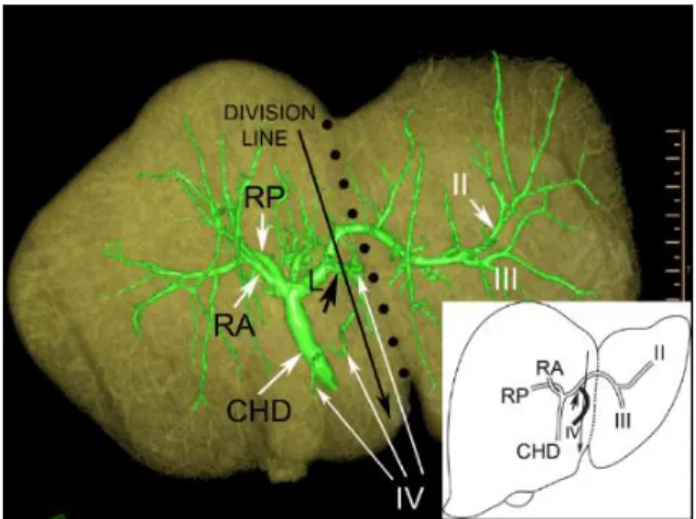
However, dividing the liver 1 cm to the right of the FL, impairs segment IV duct in 46.7% of cases (Figure 10).
Figure 10: Dividing the liver 1 cm to the right of the FL may impair the bile drainage of segment IV in the remaining liver.
Resection line is shown by the long black arrow, and FL is indicated by the black dotted line, while ducts draining segment IV are indicated by long white arrows. Short black arrow shows the joining of segment IV duct into the LHD. RA, right anterior hepatic duct; RP, right posterior hepatic duct; L, left hepatic duct; CHD, common hepatic duct. Roman numerals stand for the segmental ducts. (Source: author’s own work. Co-workers: András Szuák, Tien Nguyen. CT pictures made by Ibolyka Dudás).
13
5 Conclusions
Both of our newly developed corrosion cast- and vessel lumen filling without corrosion techniques were perfectly suitable to perform our planned studies and get the aimed data. The resin mixture could fill up even the small subsegmental ducts, after the polimerization the liver preparations were hard enough to keep their shape, provided excellent density for CT scans and also kept their coloures.
Hilar variations of the hepatic duct system, according to the absence or presence of the LHD and RHD four different groups could be identified: Our results in a series of 106 livers showed 45.28% (58/106) perihilar biliary variants. These data confirm the supposition that in categorizations, instead of “normal biliary anatomy” the use of “most frequent variation” would be reasonable.
Among the biliary duct variations recorded in our study, the incidence of “D1”
configuration exceeds all “D1” figures reported by others (22.4% versus 5%, 13%, 8%, 15.8%, 11%, 12%, 11.6%). In full left–full right split or in case of left lobe LDLT, the LHD needs to be dissected from the CHD for the left liver graft. The last 9 mm of the LHD should be preserved for the right liver lobe in cases of hilar variation type ”D1”, since according to this current study the distance between the ostia of the RAHD (joining the CHD) and the RPHD (joining the LHD) is less than 9 mm in 95.83% in variation type ”D1”. In view of the fact that a considerable number of variations in the hepatic ducts persist in the hilar region, it is necessary to have a profound knowledge of the actual variations in the hepatic ducts in the hilar area around the hilar confluence to perform safe right or left liver lobe transplantation. Our data stress on the high incidence of perihilar biliary variations in the livers taken from Hungarian deceased.
The current study strongly supports the view that if the division line of LLS hepatectomy is precisely on the FL, the implantable graft will have a single bile duct for the anastomoses in only 30% of cases and in 70 % the surgeon should prepare multiple ductal anastomoses during implantation. Contrary to our expectations, the division surface of standard hepatotomy just at the FL displayed high percentage of two (53,3%), three (10%) and moreover four (3,3%) biliary ducts which must be
14
anastomosed individually. Only a single bile duct for anastomosis was present when we performed the virtual hepatotomy one cm to the right of the attachment of FL on the diaphragmatic surface and equally one cm to the right of umbilical fissure on the visceral surface, in 70% of the investigated livers. However, dividing the liver well to the right of the falciform ligament, can cause accidental damage of segment IV ducts in the remaining liver in 46.7% of our cases. Our conclusion is, that ideally the division line of LLS hepatectomy for LDLT is one cm to the right of the FL. However, if there are related vascular or other contraindications or if it is absolutely essential to preserve segment IV in the donor, then the hepatotomy should be performed at the FL, accepting the relatively higher chance of needing multi-ductal anastomoses in the recipient.
Statistical analysis of the observed anomalous branching patterns in the hepatic duct system and comparison of that to the literary data together with the recognition of new biliary variants may help to make easier the preoperative preparations for transplantations. We believe that our hereby presented data and new techniques can contribute to the more perfect knowledge of the biliary duct system of the human liver and therefore may lead to the reduction of post-transplantation complications in the partial liver transplantation and in the LDLT.
15
6 Bibliography of the candidate’s publications
Publications related to the subject
Kiss M, Deshpande RR, Nemeskéri A, Nguyen TT, Kürti Z, Kovács S, Pápai Z, Németh K, Szuák A, Dudás I, Kóbori L. (2015) Optimal line of hepatotomy for left lateral living donor liver transplantation according to the anatomical variations of left hepatic duct system. Pediatr Transplant, 19: 510-516. IF: 1,284
Nemeth K, Deshpande R, Mathe Z, Szuak A, Kiss M, Korom C, Nemeskeri A, Kobori L. (2015) Extrahepatic arteries of the human liver - anatomical variants and surgical relevancies. Transpl Int, 28: 1216-1226. IF: 2,835
Törő K, Matlakovics B, Dudás I, Karlinger K, Kiss M, Molnár A, Nemeskéri A. (2014) The utility of the combination of the corrosion cast method and post mortem MSCT scans. Leg Med (Tokyo), 6: 283-289. IF: 1,238
Rosero O, Nemeth K, Turoczi Z, Fulop A, Garbaisz D, Gyorffy A, Szuak A, Dorogi B, Kiss M, Nemeskeri A, Harsanyi L, Szijarto A. (2014) Collateral circulation of the rat lower limb and its significance in ischemia - reperfusion studies. Surg Today, 44: 2345- 2353. IF: 1,526
Nemeskéri A, Matlakovics B, Dudás I, Molnár B, Bartykowski A, Kiss M, Kristóf I, Törő K, Karlinger K. (2009) Combination of post mortem coronary angiography, corrosion cast method and multi-slice computed tomography (MSCT) for diagnostic improvement in pathology and forensics. Interv Med Appl Sci, 1: 20-34.
16
Töro K, Kiss M, Szarvas V, Nemeskéri A, Kristóf I, Magyar L, Keller E. (2007) Post mortem introduction of corrosion cast method after coronary stent implantation.
Forensic Sci Int, 171(2-3): 208-211. IF: 2,015
Other Publications
Dezső K, Rókusz A, Bugyik E, Szücs A, Szuák A, Dorogi B, Kiss M, Nemeskéri Á, Nagy P, Paku S. (2017) Human liver regeneration in the late, irreversible phase of cirrhosis is driven and organized by the portal tree. J Hepatol. 66: 778-786. IF:10,58
Kóbori L, Máthé Z, Fazakas J, Gerlei Z, Doros A, Fehérvári I, Sárváry E, Hartmann E, Németh A, Mándli T, Tóth S, Szonyi L, Korponay Z, Kiss M, Görög D, Járay J. (2008) Surgical aspects of pediatric liver transplantation. Living donor liver transplant program in Hungary. Orv Hetil, 149: 1271-1275.