OF HUMAN FIBROBLASTS IN CULTURE
JAMES C. LACEYf JR.
JANNA D. STROBEL DANIEL P. STEPHENS, JR.
Laboratory of Molecular Biology University of Alabama in Birmingham
Birmingham, Alabama
I. SUMMARY
We found brief rinses with phosphate buffered saline or 0,9%
NaCl stimulated the proliferation of density inhibited human fi- broblasts. Additions of trypsin or pronase to phosphate buffered saline did not increase this effect. Systematic elimination of the components of phosphate buffered saline showed none of these components was essential to the overgrowth phenomenon. Part of the stimulation by phosphate buffered saline was probably due to a transient increase in pH. However, because unbuffered 0.9%
NaCl, which assumes the pH of the cells, also caused stimulation, we believe other factors in addition to pH are involved. Since additions of dibutyryl cyclic AMP to phosphate buffered saline did not reduce the overgrowth effect, we believe a reduction in
541
542 JAMES C. LACE Y, JR. et al.
intracellular cyclic AMP levels is not responsible. The evidence does, however, suggest to us that a negative substance, perhaps a chalone or cell surface glycoprotein, is being extracted from the cell.
II. INTRODUCTION
Much experimental effort has been directed toward understand- ing the basic mechanisms controlling the proliferation of cells in multicellular organisms. Many experiments have been carried out using in vitro procedures involving cells in culture. Sta- tionary cell cultures maintained under fixed conditions grow to a particular density and then their growth rate slows. Most re- searchers assume that the in vitro processes which lead to this reduction in growth are qualitatively the same as those operating in the intact creature. In addition to serum (Temin, 1971), a number of diverse factors have been reported to stimulate the overgrowth of such density inhibited cells. These factors in- clude insulin (Temin, 1967), proteases (Burger, 1970; Burger et al., 1972; Noonan and Burger, 1973; Rubin, 1970; Sefton and Rubin, 1970), guanosine 3'5' monophosphate (cyclic GMP) (Rudland et al., 1974), low levels of cyclic AMP (Burger et al., 1972), hyaluroni- dase, ribonuclease (Vasiliev et al., 1970) and neuraminidase
(Vaheri et al., 1972). There is considerable controversy regard- ing the ability of several of these factors to reinitiate pro- liferation. We are particularly interested in a number of re- ports regarding the stimulation of overgrowth by various pro- teases. These include the reports from Burger's laboratory, us- ing 3T3 cells (Burger, 1970; Burger et al., 1972; Noonan and Burger, 1973) and Rubin's laboratory (Rubin, 1970; Sefton and Rubin, 1970), using chick embryo cells. There is ample reason to question whether 3T3 cells are normal since they are heteroploid and undergo a more rigorous density dependent inhibition than
normal cells. Chick embryo cells, while they can be grown under conditions giving density dependent inhibition, might not be con- trolled by the same mechanisms as cells from either born chickens or mammals. Because of these considerations, we wanted to deter- mine if proteases would cause the overgrowth of normal human fi- broblasts in culture.
III. MATERIALS AND METHODS
A. Cell Cultures
Secondary cultures of normal human diploid foreskin fibro- blasts (HF-6 and HF-7) from two different individuals* were grown at 37°C in 32 oz prescription flats (Brockway) containing 50 ml of medium. The medium consisted of MEM (50-50 Hanks1-Earle1 s salts) supplemented with 10% fetal calf serum, penicillin, strep- tomycin and fungizone purchased from GIBCO. Flats were innocu- lated at half confluency and required 5 days at 37°C to become confluent giving a cell density of about 90,000 cells/cm or ap- proximately 1.3 x 1 07 cells/flat. For experimental samples, flats were trypsinized with 0.25% trypsin (GIBCO). The detached cells were suspended in the same medium supplemented with 3%, in- stead of 10%, fetal calf serum. Borosilicate scintillation vials with rubber lined caps were innoculated with 2.0-2.5 χ 10^ cells in 2.0 ml of medium. Under these conditions, the cells reached a plateau level of about 4.0-4.5 χ 1 05 cells/vial in 5 to 7 days.
Passage numbers are given in the figure legends.
B. Cell Counts
Cell counts were made by removing the medium from vials by aspiration and adding 2.0 ml of 0.25% trypsin. After 20 min at 37°C, the cells were aspirated up and down in a disposable pipette 12 times to break up clumps. Eight ml of 0.9% NaCl were added
*See note added in proof.
544 JAMES C. LACEY, JR. et al.
and a Coulter Counter, Model B, probe was inserted directly into the vial. Routinely, triplicate counts were made on each of trip- licate samples. When cell counts were approximately 20,000
counts/0,5 ml, the variation among a set of triplicate samples was about 1000 counts.
C. Washing Procedures
Our experimental procedure usually consisted of asceptically pouring off the medium from confluent vials at 00 time. The pH of the medium at 7 days was about 6.8. This conditioned medium was stored sterilly while not in use. Last traces of medium were carefully removed from the cells by aspiration using a sterile disposable pipette. One ml of the solution being tested (pH 7.2 except for the unbuffered 0.9% NaCl) was added to the cells and allowed to stand for 15 min. The solution was then carefully re- moved by aspiration and 2.0 ml of the conditioned medium was read- ded to each vial. Control samples were treated in the same man- ner only conditioned medium was used instead of a test solution.
In the protease experiments, crystalline trypsin (Sigma) or pro- nase (Calbiochem) was dissolved in Dulbecco's phosphate buffered saline (PBS). When trypsin was used in an experiment, the cell layer was prewashed for 5 min with 1.0 ml of PBS buffer. This was removed and then the trypsin put on. The prewashing was done to remove trypsin inhibitors present in the medium. When pronase was used the prewash was omitted and replaced by a post-treatment wash because there is no pronase inhibitor present in the medium.
After each washing experiment triplicate samples were counted to determine the amount of cell loss due to the washing (0 time).
Subsequently cell counts were taken at intervals up to seven days.
IV. RESULTS
In studying the effects of proteases on cell proliferation, we ran two types of controls in parallel with the test samples;
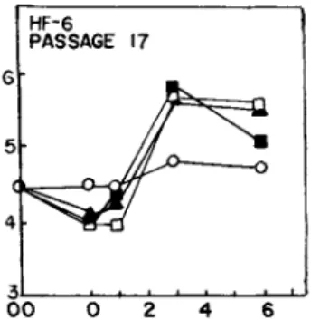
one set consisted of samples washed with PBS (Dulbecco's phosphate buffered saline) alone, the other set was exposed to the medium in which the cells were grown (conditioned medium). We were sur- prised to find that the control washes with PBS alone resulted in overgrowth above the levels observed with samples in which condi- tioned medium was used. Figure 1 shows that additions of up to 3 yg/ml of trypsin to the PBS did not cause additional overgrowth.
In fact, 3 yg/ml resulted in disruption of the cell layer and early death of the cells. Higher levels of trypsin caused great- er cell loss. Similarly, in Fig. 2, up to 5 yg pronase/ml did not stimulate overgrowth any more than PBS alone. Higher levels of pronase also caused cell loss.
Using the Student t Test for independent samples, we examined our samples for significant differences in cell numbers. In both protease experiments, the arithmetic means of PBS and the protease treated samples were significantly greater (P < 0.05) than those of the conditioned medium samples by the second day after washing.
There were no significant differences between the PBS and protease treated samples (P > 0.05).
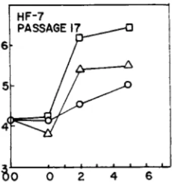
After observing overgrowth with PBS in these and many other experiments, (not shown) we wondered which component, if any, in the PBS might be responsible for the stimulation. We systemati- cally eliminated every component in the PBS including the replace- ment of phosphate with HEPES (N-2-hydroxy-ethylpiperazine-N1-2- ethanesulfonic acid) buffer. We still got significant overgrowth in every case. Since the stimulation did not appear to depend on any factor in the PBS solution we then tried plain isotonic NaCl
(0.9%), unbuffered, as shown in Fig. 3. There was still signi- ficant overgrowth, but reduced about 50% from that obtained with PBS. The pH of unbuffered 0.9% NaCl was 6.0 before putting it on
546 JAMES C. LACEY, JR. et ai
HF-6
PASSAGE 17
3l . — J
0 0 0 2 4 6
FIGURE 1 Abscissa: days after washing; ordinate: cells/vial x 10~5. Effect of trypsin on proliferation of confluent human fibroblasts (HF-6; Passage 17). At time 00, medium was removed from cells and saved. Last traces of medium were carefully as- pirated. Samples to receive trypsin were washed with 1.0 ml of PBS for 5 min. PBS was carefully aspirated and then we added 1.0 ml of •, PBS; k, PBS with 1.5 ]ig/ml cryst. trypsin; M, PBS with 3.0 \\g/ml cryst. trypsin; for 15 min. Test solutions were aspi- rated and 2.0 ml of conditioned (cond.) medium readded to each sample. Control samples of cond. medium, o, were treated in an analogous manner with cond. medium. Cell counts were made at intervals shown.
5 HF-7 PASSAGE 17
—ι 1 1 1 ,.
»0 0 2 4
FIGURE 2 Abscissa: days after washing; ordinate: cells/vial x 10"^. Effect of pronase on proliferation of confluent human fibroblasts (HF-6; Passage 17). At time 00, medium was removed and saved. Last traces of medium were carefully aspirated. Cells were exposed for 15 min to 1.0 ml: D, PBS; k, PBS with 1 vg/ml pronase; •, PBS with 5 \ig/ml pronase; o, cond. medium control.
All solutions were aspirated. Samples were washed with 1.0 ml of PBS for 5 min to remove pronase except controls which were exposed to cond. medium. All solutions were aspirated and cond. medium readded. Cell counts were made at intervals shown.
HF-7
*J É 1 1 1 · 1 1 i-J 0 0 0 2 4 6
FIGURE 3 Abscissa: days after washing; ordinate: cells/vial x 10~5. Effect of PBS (pH 7.2) and 0.9% NaCl (unbuffered) on proliferation of confluent human fibroblasts (HF-7; Passage 17).
At time 00, medium was removed from cells and saved. Last traces of medium were carefully aspirated and cells were exposed for 15 min to 1.0 ml of either D, PBS: Δ, 0.9% NaCl; or o, cond. medium.
Solutions were carefully aspirated and cond. medium replaced.
Cell counts were made at intervals shown.
8o 0 2 4 6
FIGURE 4 Abscissa: days after washing; ordinate: cells/vial x 10~5. Effect of small amount of residual medium on PBS stimu- lation of proliferation of confluent human fibroblasts (HF-7; Pas- sage 17). At time 00, medium was poured off all vials. Last traces of medium were removed from 2 sets of vials by aspiration.
A 3rd set of vials was not aspirated, leaving an estimated 0.1 ml of medium in each vial. 1.0 ml of PBS was added to the unaspi- rated vials, ·, and to one set of aspirated vials, •. 1.0 ml of cond. medium was added to the remaining set of aspirated vials, o.
After 15 min all solutions were aspirated. 2.0 ml of cond. medi- um was added to each vial. Samples were counted at intervals shown.
548 JAMES C. LACEY, JR. et al.
7 6 5
4
3 L 00 0 2 4 6
FIGURE 5 Abscissa: days after washing; ordinate; cells/vial x 10~5. Effect of dibutyryl cyclic AMP on PBS stimulation of proliferation of confluent human fibroblasts (KF-7; Passage 14).
At time 00, medium was removed from cells and saved. Last traces of medium were carefully aspirated. Samples were exposed for 15 min to 1.0 ml: PBS with 1 x 1 0 ~5 M dibutyryl cyclic AMP, , PBS,
; and cond. medium, o. All solutions were aspirated and cond.
medium readded. Cell counts were made at intervals shown.
the cells, while the pH of PBS was 7.2. The NaCl solution probab- ly assumed the pH of the medium (6.8) just removed from the cells.
In attempting to arrive at a mechanism which is consistent with our results, our first feeling was that the stimulation was caused by cell loss since washing the cell layers with either PBS or 0.9% NaCl caused a cell loss of up to 10 per cent. In some cases, however, there was no loss and stimulation still resulted.
Multiple washes did not increase cell loss or growth. There was some time dependence, 15 min washes caused slightly more over- growth than 5 min washes. However, simply putting PBS on the cells and aspirating immediately also resulted in overgrowth.
To our knowledge, stimulation of cell proliferation by simple saline extraction has not been previously reported. Burger (1970) washed 3T3 cells with PBS but reported no stimulation. We won- dered if our careful removal of last traces of medium might have made a difference. In one experiment we simply poured the medium out of one set of vials, and did not aspirate the last traces of medium. This procedure left about 0.1 ml of medium in each vial.
The PBS was then added to this set of vials and also to a control set which had been carefully aspirated. The results are shown in Fig. 4. Residual medium caused about a 50% reduction in over- growth.
A number of workers have shown that intracellular cyclic AMP levels are low in proliferating cells and high in confluent mono- layers (Anderson et al., 1973; Burger et al., 1972). One possible explanation of our observed overgrowth phenomenon is that saline washing causes a drop in the level of cyclic AMP. To test this possibility we repeated the PBS wash experiment including a set of vials washed with a PBS solution containing 1 χ 10~5 M dibutyrl cyclic AMP. As Fig. 5 shows, there was no statistically signifi- cant decrease in overgrowth.
V. DISCUSSION
These experimental results leave us with several important conclusions regarding the control of proliferation in culture:
(1) Washing of human fibroblasts with saline solutions (PBS or 0.9% NaCl) can induce proliferation in confluent cells and the induction does not seem dependent on any factor present in the solution. Washing confluent monolayers with saline solutions usually results in the loss of up to 10% of the cells. The stimu- lation may be partly dependent on a transient pH increase since unbuffered 0.9% NaCl caused less proliferation.
(2) We find no additional stimulation of proliferation by adding trypsin or pronase to the PBS washes. Our results do not support the reports for protease induced overgrowth in 3T3 cells and chick embryo fibroblasts. Glynn and associates (1973) also failed to find increases in cell number by exposing 3T3 cells to various pronase concentrations, although they did find that pro- nase treatment increases cell agglutinability with concanavalin A. However, we cannot rule out the possibility that in our
550 JAMES C. LACE Y, JR. et al.
experiments the proteases may cause some overgrowth that is masked by the PBS effect.
Reich (1973f 1974} recently suggested that increased protease activity leading to the ability to lyse fibrin is a characteristic of transformed cells. This contention has been challenged by Mott and his co-workers (1974) who find that some normal cells have this ability and some transformed cells do not. Similarly, Chou et al., (1974a) showed that suppression of fibrinolysin activity did not restore density dependent inhibition to SV40 transformed 3T3 cells. While some protease inhibitors do cause a reduction in growth of transformed cells. Chou and his associates (1974b) are of the opinion that this effect is due to an inhibition of protein synthesis rather than an inhibition of protease activity.
On the other hand, since some growth factors (Jones and Ashwood- Smith, 1970; Greene et al., 1971) have been shown to have proteo- lytic activity, it is probable that proteolysis is a part of the process leading to mitosis but is probably not the trigger.
Anderson and his collaborators (1973) found high levels of intracellular cyclic AMP in confluent monolayers but low levels during proliferation. These and other data (Burger et al., 1972) suggest that low levels of cyclic AMP trigger cell proliferation.
However, more recent experiments by Rudland et al. (1974) show high levels of cyclic GMP are more important than low cyclic AMP levels. Since the stimulatory effect of PBS in our experiments was not inhibited by exogenously added dibutyryl cyclic AMP, our inference is that cyclic AMP is not a part of the mechanism in- ducing proliferation by PBS.
Part of the stimulation by pH 7.2 PBS could be due to in- creased pH. As Ceccarini and Eagle (1971) have shown, pH plays an intimate role in controlling the growth of cells. Before washing with salt solutions our cells had a pH of 6.8-6.9, and 15 min exposure to pH 7.2 PBS stimulated considerable overgrowth.
Unbuffered 0.9% NaCl, which would assume the pH of the cells, also caused overgrowth but only about 50% of that caused by PBS.
Therefore, the overgrowth due to the PBS wash probably results only in part from a pH effect. Furthermore, the pH elevation due to PBS was only brief and after replacing the conditioned medium on the cells there was no difference in the pH of the PBS washed cells and the control cells washed with conditioned medium. Con- sequently, whatever mechanisms were initiated during the brief exposure to the salt solutions were not reversed by the condi- tioned medium.
There are several other possible effects that could be adding to the pH effect. Ceccarini and Eagle (1971) showed that ex- posure of human fibroblasts to Earle1s salt solution could in- crease uridine uptake, but they did not observe an increase in the synthesis of RNA, DNA or protein. More recently, Jimenez de Asua and his co-workers (1974) reported that additions of phos- phate to the medium of cells in culture caused increased uridine transport and also that one of the primary results of serum addi- tion to medium was an increase in phosphate uptake by cells. In- creased phosphate uptake in turn resulted in an increased uridine uptake. These authors propose that an increase in phosphate up- take is a primary event in the initiation of the mitotic sequence.
However, they did not report an increase in cell density. Our results with PBS are probably not related to this phenomenon since replacement of phosphate with HEPES still gave stimulation.
Another of the possibilities is that the saline wash is re- moving a negative factor from the cell layer. There are several possible candidates.
(a) Removal of cyclic GMP phosphodiesterase from the cell could cause elevation of cyclic GMP levels.
(b) Extraction of cell surface factors could decrease cell- cell adhesion.
(c) Removal of intercellular materials might act as a micro wound.
552 JAMES C. LACEY, JR. et ai
Cd) Another possibility is that our saline washes are removing a fibroblast cell surface glycoprotein which is extractable in salt solutions (Yamada and Weston, 1974) or with 0.25% trypsin
(Rouslahti and Vaheri, 1974) and is markedly diminshed or absent in transformed cells. However, it is not clear why removal of the glycoprotein would stimulate proliferation.
Ce) Since fibroblast mitotic inhibitor Cchalone) has been isolated and partially identified CHouck et al., 1972; Houck et al., 1973a; Houck et al., 1973b), one of the most likely mechan- isms is that the saline solution is extracting chalone from the cell surface. Chalone is found in conditioned medium from fibro- blasts and retention of cells in a resting state probably requires a particular level of chalone in or on the cell. It is possible that chalone is equilibrium bound to the cell, and because PBS contains no chalone, PBS washing rapidly releases chalone which results in proliferation.
We are presently attempting to isolate fibroblast chalone from the PBS solutions which were used to wash cells. If we identify chalone in the PBS wash and can show that addition of exogenous chalone can inhibit PBS stimulation of overgrowth, we may conclude that the stimulation is due to chalone extraction.
ACKNOWLEDGMENTS
We thank Dr. Charles Alford for the gift of the fibroblasts and Dr. William Wingo for the use of his Coulter Counter. This work was supported by National Institute of Health General Re- search Support No. 5-501-RR-05300-10 and by Public Health Service Research Grant No. CA-13148 from the National Cancer Institute.
Note added in proof: The cell culture specimens HF-6 and HF-7 used in these experiments were obtained from another laboratory
(Dr. Charles Alford of our university). We were under the impres- sion that the cells had been derived from a single individual.
Long after this manuscript was prepared, we found that the cul- tures were actually derived from multiple individuals, HF-7 from seven individuals. Furthermore, cells derived from a single in- dividual required a double PBS wash to show the stimulation, and by preparing cultures from various numbers of individuals, we found the ease of stimulation generally increased with the number of individuals represented. The fact that cells derived from a single individual require a double wash for stimulation probably accounts for this phenomenon not being previously observed by other workers.
Furthermore, pH has been ruled out as being responsible for the PBS stimulation, since PBS at pH 6.8 stimulates the same as PBS at pH 7.2.
We have also shown, using SDS gel electrophoresis, that the PBS extract contains, predominantly a protein with the same mobili- ty as the so-called LETS protein and another of about 50,000 daltons, which is approximately equal to the molecular weight of the fibroblast chalone (Houck et al., 1973a).
REFERENCES
Anderson, W. B., Russell, T. R., Carchman, R. A. and Pastan, I.
C1973). Proc. Nat. Acad. Sei. U.S.A. 70, 3802-3805.
Burger, M. M. C1970). Nature 227, 170-171.
Burger, M. M., Bombik, Â. Ì., Breckenridge, Â. M. and Sheppard, J. R. (1972). Nature New Biol. 239, 161-163.
Ceccarini, C. and Eagle, H. (1971). Proc. Nat. Acad. Sei. U.S.A.
68, 229-233.
554 JAMES C. LACEY, JR. et al.
Chou, I., Black, P. H. and Roblin, R. O. (1974a). Nature 250, 739-741.
Chou, I., Black, P. H. and Roblin, R. 0. (1974b). In "Control of Proliferation in Animal Cells." (B. Clarkson and R. Baserga, eds.) Vol. I, pp. 339-350. Cold Spring Harbor Laboratory.
Glynn, R. D., Thrash, C. R. and Cunningham, D. D. (1973). Proc.
Nat. Acad. Sei. U.S.A. 70, 2676-2677.
Greene, L. Á., Tomita, J. T. and Varon, S. (1971). Exp. Cell Res.
64, 387-395.
Houck, J. C , Weil, R. L. and Sharma, V. K. (1972). Nature New Biol. 240, 210-211.
Houck, J. C , Cheng, R. F. and Sharma, V. K. (1973a). Nat. Cancer Inst. Mono. 38, 161-170.
Houck, J. C , Sharma, V. K. and Cheng, R. F. (1973b). Nature New Biol. 246, 111-113.
Jimenez de Asua, L., Rosengurt, Å. and Dulbecco, R. (1974). Proc.
Nat. Acad. Sei. U.S.A. 71, 96-98.
Jones, R. 0. and Ashwood-Smith, M. J. (1970). Exp. Cell Res. 59, 161-163.
Mott, D. M., Fabisch, P. Ç., Sani, B. P. and Sorof, S. (1974).
Biochem. Biophys. Res. Commun. 61, 571-577.
Noonan, K. D. and Burger, M. M. (1973). Exp. Cell Res. 80, 405- 414.
Reich, E. (1973). Fed. Proc. 32, 2174-2175.
Reich, E. (1974). In "Control of Proliferation in Animal Cells."
(B. Clarkson and R. Baserga, eds.) Vol. I, pp. 351-355. Cold Spring Harbor Laboratory.
Rouslahti, E. and Vaheri, A. (1974). Nature 248, 790-791.
Rubin, H. (1970). Science 167, 1271-1272.
Rudland, P. S., Gospodarowicz, D. and Seifert, W. (1974). Nature 250, 741-742.
Sefton, Â. M. and Rubin, H. (1970). Nature 227, 843-845.
Temin, H. M. (1967). J. Cell. Physiol. 69, 377-384.
Teminf H. M. (1971). J. Cell. Physiol. 78, 161-170.
Vaheri, A. E., Rouslahti, E. and Nordling, S. (1972). Nature New Biol. 238, 211-212.
Vasiliev, Ju. M., Gelfand, I. M., Guelstein, V. P. and Fetisova, E. K. (1970). J. Cell. Physiol. 75, 302-314.
Yamada, K. M. and Weston, J. A. (1974), Proc. Nat. Acad. Sei.
U.S.A. 71, 3492-3496.