Original Paper
Copyright © 2014 S. Karger AG, Basel NonCommercial 3.0 Unported license (CC BY-NC) (www.karger.com/OA-license), applicable to the online version of the article only. Distribution permitted for non-commercial purposes only.
István Wittmann M.D., Ph.D. 2nd Department of Medicine and Nephrological Center, Faculty of Medicine University of Pécs, 1. Pacsirta Str., Pécs, 7624 (Hungary)
Tel. +36-72/536-050 Fax +36-72/536-051, E-Mail istvan.wittmann@aok.pte.hu
Incorporation of Ortho- and Meta-
Tyrosine Into Cellular Proteins Leads to Erythropoietin-Resistance in an
Erythroid Cell Line
Esztella Mikolása Szilárd Kuna Boglárka Laczya*HUJę$0ROQiUa Eszter Sélleya7DPiV.ęV]HJLb István Wittmanna
a2nd Department of Medicine and Nephrological Center; bInstitute of Laboratory Medicine, University of Pécs, Pécs, Hungary
Key Words
(U\WKURSRLHWLQUHVLVWDQFH&HOOVLJQDOLQJ2[LGDWLYHVWUHVV7\URVLQHLVRPHUV
Abstract
Background/Aims: Erythropoietin-resistance is an unsolved concern in the treatment of renal anaemia. We aimed to investigate the possible role of ortho- and meta-tyrosine – the hydroxyl free radical products of L-phenylalanine – in the development of erythropoietin-resistance.
Methods: TF-1 erythroblast cell line was used. Cell concentration was determined on day 1; 2 and 3 by two independent observers simultaneously in Bürker cell counting chambers.
Protein concentration was determined with colorimetric method. Para-, ortho- and meta- W\URVLQHOHYHOVZHUHPHDVXUHGXVLQJUHYHUVHSKDVH+3/&ZLWKÁXRUHVFHQFHGHWHFWLRQ8VLQJ Western blot method activating phosphorylation of STAT5 and ERK1/2 were investigated.
Results: We found a time- and concentration-dependent decrease of erythropoietin-induced proliferative activity in case of ortho- and meta-tyrosine treated TF-1 erythroblasts, compared to the para-tyrosine cultured cells. Decreased erythropoietin-response could be regained with a competitive dose of para-tyrosine. Proteins of erythroblasts treated by ortho- or meta- tyrosine had lower para-tyrosine and higher ortho- or meta-tyrosine content. Activating phosphorylation of ERK and STAT5 due to erythropoietin was practically prevented by ortho- or meta-tyrosine treatment. Conclusion: According to this study elevated ortho- and meta-tyrosine content of erythroblasts may lead to the dysfunction of intracellular signaling, resulting in erythropoietin-hyporesponsiveness.
y: omanyi 4/29/2014 9:26:54 AM
Introduction
Erythropoietin (EPO) is a hematopoietic growth factor playing major role in the proliferation and differentiation of erythroid cells. Renal anaemia is present in patients suffering in chronic kidney disease (CKD), resulting in the impairment of quality of life. Approximately 15 % of the recombinant-human-EPO (rh-EPO) receiving subjects are hyporesponsive [1]. Several possible pathomechanisms are discussed, such as iron
ϐ ǡϐǡ ǡ ǡ
ϐ ǡ ȏʹȐǤǦ
study, performed by Solomon et al., the decreased response to rh-EPO was associated with higher risk of mortality [3, 4].
Ǧ ȋǦȌ ǡ
ȏͷǦ ͳʹȐǤǦǦǡʹǦǡ
ǦͷȋͷȌ Ɉ [13]. The stimulus also results in the activation of the Ras/mitogen-activated protein kinase (MAPK) pathway. Erythroid cell proliferation and differentiation is dependent on both the
ͶʹȀͶͶǦȏͳͶȐǤ
The imbalance between pro- and antioxidant processes leads to the formation of reactive oxygen species (ROS). When excessive amount of hydroxyl radical is present, L-phenylalanine is converted into meta-tyrosine and ortho-tyrosiǡ
formation of the physiological isomer, para-tyrosine [15]. Gurer-Orhan et al. proved that the concentration-dependent integration of meta-tyrosine into cellular proteins may be a mechanism of cytotoxicity [16]. Ehrlich observed in 1906 the phenomenon, known as concomitant tumor resistance, where a tumor-bearing host inhibits the growth of secondary
ǤǤϐ
for this phenomenon containing a mixture of the three isoforms of tyrosine [17]. According to their in vitro and in vivo studies ortho- and meta-tyrosine inhibits tumor growth in a dose dependent manner. Using immunoblot analysis, they found impaired ERK and STAT3
Ǧ ȏͳͺȐǤ Ǥ ϐ
ǦǡǦϐ ǡǦ
level in patients with CKD [19]. According to the recent work of our group, on the one hand
Ǧ ϐ ǡ Ǧ Ǧ
ϐ ȋ
publication).
ǡ Ǧ Ǧ tyrosine into cellular proteins may result in the alteration of signal transduction, leading to EPO-hyporesponsiveness.
Materials and Methods
Materials
Unless otherwise noted, chemicals were purchased from Sigma Aldrich (St. Louis, MO, USA). Culture medium products were purchased from Life Technologies (Carlsbad, CA, USA).
Cell culture and treatments
ǦͳȋǦʹͲͲ͵Ȍȋ ǡ ǡǡȌǤ
ǦͳͶͲǡ ʹȀǦ ǡͳͲΨ
of antibiotics. Prior to experiments, cells were cultured in medium containing indicated amount of para- , ortho-, or meta-tyrosine for 3 days. Tyrosine content of the culture media was stable; no considerable alteration of para-, ortho- or meta-tyrosine concentration could be detected in the absence of cells, during 3 days (data not shown).
yi 014 9:26:54 AM
Ǧ ǡ͵ȀǦ Ǥ
ǡͳʹ ǡ
͵ȀǦͳͲǤ
Analysis of cell proliferation
ȋʹͳͲ5 /ml) was planted onto 60 mm plates in culture medium containing
ʹͲȀǦǡǦǡǦǡ Ǧ ǡ͵Ȁ
Ǧ͵Ǥ ȋ ȀρȌͳǢʹ͵
ǡ ò Ǥ
ǤǡǦͺͲ
oC for one night protein concentration was determined as described below. For concentration-dependence experiments cells were cultured in medium containing para-, ortho-, or meta-tyrosine with the addition of ͲǢʹͲǢͶͲͺͲȀǦ͵ǦǤǦͳͶͲ ʹͲȀǦǤ
HPLC (Investigating tyrosine incorporation into cellular proteins)
ϐ HPLC-method. After three-day incubation cell culture was terminated. Medium was removed by centrifugation (1000 rpm; 10 min), then cells were washed three times by addition of 1 ml physiological
Ǧ ȋͳͲͲͲ Ǣ ͷ ȌǤ ʹͲͲ ρ
the cells, followed by resuspendation and ultrasound treatment. Thereafter 100 µl 60% trichloro-acetic acid (TCA) was added. Mixture was then vortexed and centrifuged (4000 rpm; 10 min). After removing
ǡʹͲͲρͳΨǡǡͳͲͲρͲΨ Ǥ ȋͶͲͲͲǢͳͲȌǡʹͲͲρͳΨ
added. Samples were then resuspendated, treated by ultrasound and added by 100 µl 60% TCA, followed by vortexing. Subsequently precipitate was separated by centrifugation (4000 rpm; 10 min) and 40 µl
ȋǢͷͲͲȌǡͶρȋͶͲͲȌͶͲͲρͳʹǤ
ͳʹͲιǤ ȋͷͲͲͲǢ ͳͷ ȌǤ ϐ ϐ ȋͲǤʹ ρȌ Ǥ para-, ortho- and meta-tyrosine levels were determined using reverse phase-HPLC (C18 ǡʹͷͲͶȌ
ϐ ȋɉEXαʹͷǢɉEM=305 nm) as described earlier [19]. Concentrations were calculated using an external standard. We calculated the ratios of para-tyrosine and total-tyrosine, ortho-tyrosine and total-tyrosine and meta-tyrosine and total-tyrosine.
Immunoblot analysis
Ǧ ǡ Ǧ ȋͳ Ǧǡ
ǤͶǡͳǤͳͷȋȀȌΨǦͳͲͲǡͷͲͲǡʹͲͲǡͳͲͲ
ͳͲͲͲǤͷȋȀȌΨϐǡǡ
ʹȀ͵Ȍ ͵ͲǤǡ ȋͳͶͲͲͲǡ for 15 min, at 4oȌǡ Ǧ
ȋ ǡ ǡ Ȍ ȋȌ Ǥ
ȋʹȌǡ denaturated (for 5 min, at 90oC), then were separated on 10% SDS-PAGE
ϐ ȋǡ ǡ ǡ ȌǤ
ȋͷͲǦͷρȌ ϐ ǦǤ
ͷȋȀȌΨͲǤͳȋȀȌΨǡ
ȋͳǣͳͲͲͲǢǡǡǡȌǦȋʹͲʹȌȀȋʹͲͶȌ
ȋͶͶȀͶʹȌǢǦȋͻͶȌͷͷéǦ ͶoC. After being washed, the membranes were incubated with appropriate, horseradish peroxidase-conjugated secondary
ȋͳǣʹͲͲͲǢǦǡȌͲǤǡ
ȋǦ ǡ
ϐ ǡ ǡ Ȍ Ǧ ϐ ȋ ȌǤ ͷ
Ǥ
ȋ ǡ ǡ ȌǤ ͳȀʹ
ͷǡ éǦ Ǥ
y: omanyi 4/29/2014 9:26:54 AM
Statistical analysis
Data are expressed as means±SE or means±SD, as indicated. Analyses were performed as appropriate
ͳǤͲȋ Ǥǡ ǡǡȌǤ ϐ ϐ
ϐǤϐ Ȃ
Ǥ ǡ
ǯǦ Ǥ Ǧǡ
was taken as 100% and one sample t-test was performed for the comparison versus controls. To compare
ǯǦ
used.
Results
Cell proliferation
Panel A of Figure 1 shows the EPO-induced, time-dependent proliferation of TF-1 erythroblasts grown in para-, ortho- or meta-tyrosine containing medium. Culturing TF-1 cells in the presence of ortho- and meta-tyrosine EPO-induced proliferative activity was found to be decreased compared with para-tyrosine cultured cells. Maximal difference between cell counts was observed at day 3 (time curve).
Ͳǡ ʹͲǡ ͶͲǡ ͺͲ Ȁ Ǧ
into the ortho- or meta-tyrosine supplemented medium on the cell counts at the 3rd day
A C
B
Fig. 1. Effect of para-, ortho-, or meta-tyrosine supplementation on the proliferation of cells cultured in me- dium with or without EPO. Panel A shows the time dependent rise of cell count in para-tyrosine (open bars), ortho-tyrosine (black bars) or meta-tyrosine (grey bars) supplemented medium. (*, P<0.05 vs. para-tyro- sine cultured cells on the same day; n=10). Panel Bshows the alterations of cell counts after incubation for 3-days in media containing ortho- or meta-tyrosine and the indicated additional amount of para-tyrosine (*,P<0.001 vs. para-tyrosine containing medium; n=5). Panel C shows the effect of supplementation by or- tho- or meta-tyrosine on the increment of cellular protein induced by EPO. Results are expressed as the ratio of protein content of EPO and non-EPO (control) cells. (*, P<0.05 vs. para-tyrosine cultured cells; n=10).
yi 014 9:26:54 AM
para-tyrosine in case of ortho-tyrosine cultured control cells (Panel A of Figure 2, left part).
Cells grown in medium containing ortho- or meta-tyrosine and EPO showed less para- tyrosine compared to the para-tyrosine supplemented cells (Panel A of Figure 2, right part).
ʹǡ Ǧ
the proteins of ortho-tyrosine cultured erythroblasts compared to the cells cultured with
ǦǦǡ Ǧȋ ǡϐȌȋ
ϐȌ ǡǤǡǦ ϐ Ǧ cultured cells than that of non-EPO-treated (control) cells, when both were grown in ortho-
ȋ ϐȌǤ
Ǧ ϐ Ǧ
erythroblasts both in non-EPO (control) and in EPO groups compared to cells cultured in
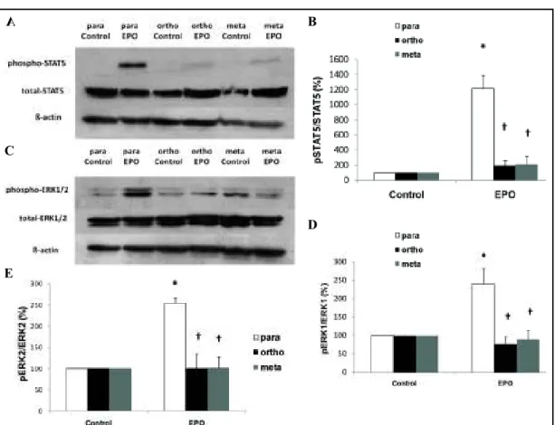
ǦǦȋ ʹǡȌǤ Analyses of STAT5 and ERK activation
ǦǦ
the increase of STAT5-phosphorylation induced by EPO in para-tyrosine cultured cells (Panel
͵ȌǤǦǦ
ͳʹȋǦ ͵ȌǤ A
C B Fig. 2.Relative para-, ortho- and meta-tyrosi- ne content (i.e. ratios of para-, ortho-, and me- ta-tyrosine/total tyrosine) of cellular proteins
Ǧͳ ȋǡ ȌǤȗǡ P=0.003 vs para-tyrosine cultured erythrob- lasts; §, P<0.001 vs para-tyrosine cultured cells;
†, P<0.001 vs. para- and meta-tyrosine cultured ǢᓐǡδͲǤͲͲͳǤǦ - trol cells; +, P<0.001 vs. para- and ortho-tyrosi- ne cultured erythroblasts; n=10.
of growing with or without rh-EPO (concentration dependence). The ortho- tyrosine induced impairment of EPO- response could be competed by 40 mg/l of para-tyrosine, while in the case of meta-tyrosine at least 60 mg/l of para- tyrosine concentration was necessary.
As shown in Panel C of Figure 1 relative protein content (EPO/non- EPO) of meta- or ortho-tyrosine treated ϐ ǡ
that grown on para-tyrosine.
Tyrosine incorporation into cellular proteins
Treatment of the cells with meta-
Ȃ
ǦȂ
para-tyrosine content of cellular proteins in non-EPO (control) experiments. No
ϐ
y: omanyi 4/29/2014 9:26:54 AM
A
C
B
D
E
Fig. 3. Representative immunoblots (Panel A) and densitometric analyses of phosphorylation of STAT5 (Pa-
Ǣα͵ȌǢͳʹȋȌ ͳȋǢαͶȌ
ʹȋǢαͶȌǤ ȋ Ȍ ǤȗǡδͲǤͲͷǢ
ȋǦǦȌǢȘǡδͲǤͲͷȋǦȌǤ
Discussion
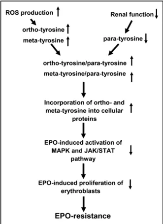
ǡǦǦ
ǡȂ ǦȂ
(0.10-0.15%), with a consequent decrease in activation of antiapoptotic and mitogenic
Ǥ ͷ ϐ
proliferative response to EPO, leading to EPO-hyporesponsiveness of erythroid progenitor cells (Figure 4).
Oxidative stress is proven to play a major role in the pathogenesis of several morbidities,
ǡ ȏʹͲǦʹʹȐǤ
plasma level or urinary excretion of ortho- and meta-tyrosine, as hydroxylated phenylalanine derivatives, is associated with altered oxidative state, thus they serve as markers of oxidative stress. On the other hand, Ruggiero et al. proved, that ortho- and meta-tyrosine decreases tumor proliferation due to the inhibition of ERK and STAT3 activation. They used meta- and
Ǧ ͺʹPmol/l. Similarly, we detected decreased ERK and STAT5 phosphorylation in the presence of ortho- and meta-tyrosine.
ǤȏͳʹȐǡ
with EPO for 3 days resulted in an approximate 1.7 fold increase in cell counts. Addition of meta- or ortho-tyrosine into the culture medium caused a markedly decreased proliferative activity, leading to lower cell counts. Since, according to the source, duplication time of this ʹʹǡ ʹ
(Panel A of Figure 1). yi 014 9
:26:54 AM
Ǥǡ Ǧ
ʹͲͲͲρȀȏʹͶȐǤ
As EPO-treatment is widely used among patients suffering in renal anaemia, EPO-
Ǥ
ǡǦ ȏʹͷȐǤVanesse and Berliner ȏʹȐǤ
ǡǤǡ
electron transport chain and through the leakage of electrons, leads to increased production
ȏʹȐǤϐ ȂȂ
overproduction may play a role in the decrease of EPO-sensitivity.
ǡ ϐǦ
ȏʹͺȐǡ
in hormone resistances via incorporation of ortho- and meta-tyrosine into the proteins.
ǡMcCullogh et al. observed increased risk for cardiovascular events related
Ǧǡ ȏʹͻȐǤ
ϐ Ǧ ǡǡ that this is an epiphenomenon, and higher cardiovascular risk is not triggered by higher plasma EPO-level itself, but rather by the incorporation of the ortho- and meta-tyrosine into
Ǥϐ
to the role of insulin resistance in mortality instead of role of the necessarily high insulin dosage [30].
Fig. 4. Flowchart of the scheme of the possible subcellu- lar process, how ortho- and meta-tyrosine incorporation leads to the dysfunction of signal pathway mechanisms, resulting in the hyporesponsiveness of erythroid progeni- tor cells.
Ruggiero et al., inhibition by meta-tyrosine could be reversed with excessive phenylalanine, glutamine, aspartic acid, histidine and glutamate, but not with para-tyrosine itself. On the contrary, we could break through the EPO-resistance by para- tyrosine in a dose dependent manner ȋ ͳȌǤ
suggest that para-tyrosine competitively inhibits the integration of ortho- and meta-tyrosine into cellular proteins.
Ǧ ǡ 1:1.8 ortho- to para-tyrosine ratio was necessary to regain 50 % of EPO-
Ǥ Ǧǡ
ͳǣʹǤǤ
ǦͳͶͲ
contains para-tyrosine at twice as high concentration than the normal human serum does. Consequently, the same concentration had to be used in case of ortho- and meta-tyrosine, in order to block EPO-response. We performed a model experiment, demonstrating a total block of the effect of EPO, therefore high doses of tyrosine isomers were
Ǥ
Ǧ ȋʹͲǦ ʹͲɊȀȌ
ȏʹ͵ȐǤ ǡ
et al. examined the role of meta-tyrosine incorporation into cellular proteins and
y: omanyi 4/29/2014 9:26:54 AM
Conclusion
ϐ Ǧ Ǧ Ǧ resistance. Furthermore, inhibiting their cellular integration with the physiological
Ȃ Ǧ Ȃ ǡ
ǤǡȂ
Ȃ Ǧ
treatment in the management of EPO-resistance.
Disclosure Statement
ϐ Ǥ
Acknowledgements
This research was supported by the European Union and the State of Hungary, co- ϐ ͶǤʹǤͶǤȀʹǦͳͳǦͳǦʹͲͳʹǦ ͲͲͲͳǮ ǯǤ
References
ͳ ǡǦǡ ǡ ǣ
ǤʹͲͲǢʹʹǣͻͶǦͺͲͲǤ ʹ ǣ Ǧǣǡǡ
ǤʹͲͳʹǢʹǣͳͻͷǦʹͲͷǤ
͵ ǡǡǡǡ ǡ ǡÚǡ ǡ ǡ
ǡǣ guidelines for the management of anaemia in patients with chronic renal failure. Nephrol Dial Transplant ʹͲͲͶǢʹǣͳǦͶǤ
Ͷ ǡǡ ǡ ǡǡǡǡ ǡǡ
ǡǡǡǡ ǡǡ ǡǣ
ȋȌǤ
ʹǤʹͲͳͲǢ͵͵ǣͳͳͶǦͳͳͷͷǤ
ͷ ǡǦǡǡǡǡǡǣ
associated with dose response of erythropoiesis-stimulating agents in hemodialysis versus peritoneal
ǤʹͲͳʹǢ͵ͷǣͳͻͺǦʹͲͺǤ
ǡǡ ǣ
ǦǤ
ʹͲͲͻǢ͵ͻǣͻǦͲͳǤ
ǡǡǡǡǡǡǣ
Ǥ ͳͻͻǢʹ͵ͻǣͶͻͶȂͷͲͲǤ 8 Morishita E, Masuda S, Nagao M, Yasuda Y, Sasaki R: Erythropoietin receptor is expressed in rat
hippocampal and cerebral cortical neurons, and erythropoietin prevents in vitro glutamate-induced
Ǥ ͳͻͻǢǣͳͲͷȂͳͳǤ
ͻ ǡǡǡ×ǡǡ ǡÝǡ ǡǡ±ǡ±ǡ
ǣ ǤʹͲͳʹǢͶͶǣʹͻǦʹͺͷǤ ͳͲ ǡ ǡǡǡǡ ǡǡǣ
Ǥ ͳͻͺͺǢͳͷͶǣͻͲʹǦ 909.
yi 014 9:26:54 AM
ͳͳ ǡǡǡǡǡǦ ǡǡ±ǡ
ǡǣͺͳʹǣ
ǤͳͻͺͻǢ͵ǣʹͲͲ͵ǦʹͲͳ͵Ǥ
ͳʹ ǡǡǡǡǡǡ ǣϐ
Ǧ ǡ ǦͳǤͳͻͺͻǢ͵ǣ͵ͷǦ͵ͺͲǤ ͳ͵ ǡ ǡǡǡǡǡǣʹ
erythropoietin receptor and is tyrosine phosphorylated and activated following stimulation with
Ǥͳͻͻ͵ǢͶǣʹʹǦʹ͵Ǥ
ͳͶ ǣ ǣ Ǥ
ʹͲͳͳǢͷͻǣͳͲǦͳͲʹǤ
ͳͷ ǡ ǡǦǣǦ
ʹǦǦͶǦǦǡǦǦͷǡǡǡͺǦǤ ͳͻͳǢͳʹͷǣͷͻǦͷͶǤ
ͳ Ǧǡ ǡǡǡǡ ǣ Ǧ
ǣ Ǥ ʹͲͲǢ͵ͻͷǣʹǦ ʹͺͶǤ
ͳ ǡǡǡǡǡǣ ǣ
Ǥ ʹͲͳʹǢʹǣͳͲͶ͵ǦͳͲͷͲǤ ͳͺ ǡǡǡǡǡǡǡǡ
OD, Pasqualini CD: Tyrosine isomers mediate the classical phenomenon of concomitant tumor resistance.
ʹͲͳͳǢͳǣͳͳ͵ǦͳʹͶǤ
ͳͻ ǡǡ×ǡ×ǡǡ ǡǡǡ×ǡ
ǡ ǡǡǣǦ ǣ
ǤʹͲͲͷǢͺǣʹʹͺͳǦʹʹͺǤ
ʹͲ ǣϐ ǣ
ǤʹͲͳ͵ǢʹͲͳ͵ǣʹͶͺͷ͵Ǥ
ʹͳ ǡ ǡǡ ǣ Ǥ
ʹͲͳ͵ǢͳͶǣͳ͵ͳͻǦͳ͵ͶǤ
ʹʹ ǡǡǡǡǣ Ǥ
ʹͲͲͻǢͳͷǣʹͲͻǦʹͳͻǤ
ʹ͵ ǡǡǡǡǡǡ ǣ ǣǦ
ǡ Ǥ ʹͲͲǢͳͲͶǣͳͻͶǦͳͻͻǤ
ʹͶ ǡǡ ǡǣ
Ǥ ʹͲͲʹǢ͵ʹǣǦͷǤ
ʹͷ ǡǡ ǡǡǡ ǡǡǡ ǡǣ
ǣǤ ʹͲͲͷǢͷ͵ǣͳ͵ͲǦͳ͵ͷǤ
ʹ ǡǣǣʹͳ Ǥ
ʹͲͳͲǢʹͲͳͲǣʹͳǦʹͷǤ
ʹ ǡ ǡǣ Ǥ ʹͲͳʹǢͳͳͲǣͳͳͲͻǦͳͳʹͶǤ ʹͺ ǡǡ ǡǡǡǣ
Ǥ ʹͲͲǢ͵ͻǣͶͶǦͺͶǤ
ʹͻ ǡǡǡǡǡǡǡ ǡǣ
Ǧ ǤʹͲͳ͵Ǣ͵ǣͷͶͻǦ 558.
͵Ͳ ǡǡǣ ǤǤ
ʹͲͳͲǢ͵͵ǣͳͳͻǦͳͳͺͷǤ
y: omanyi 4/29/2014 9:26:54 AM