Novel kinase inhibitor compounds for combination cancer therapy
Thesis for doctoral degree (Ph.D.)
Gyulavári, Pál
Doctoral School of Pharmaceutical Sciences Semmelweis University
Supervisor: Vántus, Tibor Ph.D.
Opponents: Tábi, Tamás Ph.D.
Wunderlich, Lívius Ph.D.
Chair of examination board: Tretter, László Ph.D., D.Sc.
Members of examination board: Balla, András Ph.D.
Molnár, Tamás Ph.D.
Budapest
2019
1 Contents
List of abbreviations ... 2
1. Introduction ... 4
1.1. Cancer ... 4
1.1.1. On cancer in general ... 4
1.1.2. Cancer as a genetic disease ... 5
1.1.3. Comprehending the genetic heterogeneity of cancers ... 8
1.2. Kinases ... 10
1.2.1. Kinases as part of signal transduction pathways... 10
1.2.2. Kinases as drivers of cancers ... 11
1.2.2.1. Aurora kinases ... 12
1.2.2.2. EGFR ... 18
1.2.2.3. c-Met ... 20
1.3. Targeted cancer therapy ... 21
1.3.1. Types of targeted agents ... 21
1.3.2. Properties of small molecule KIs ... 22
1.3.3. Development of small molecule KIs ... 24
1.3.4. Examples of small molecule KIs ... 27
1.3.4.1. Aurora KIs ... 27
1.3.4.2. EGFR inhibitors ... 30
1.3.4.3. c-Met inhibitors ... 31
1.3.5. Combinatorial therapy ... 32
2. Aims of the Thesis ... 35
3. Materials & Methods... 36
4. Results ... 44
5. Discussion ... 65
6. Conclusions ... 77
7. Summary... 78
8. Összefoglalás ... 79
9. List of references... 80
10. List of the candidate’s publications... 125
11. Acknowledgement ... 127
2 List of abbreviations
ADME absorption, distribution, metabolism, excretion
AKI(s) Aurora kinase inhibitor(s)
ALL acute lymphoblastic leukaemia
AML acute myeloid leukaemia
ATCC American Type Culture Collection
BRCA1 breast cancer 1 (protein)
CI combination index
CIN chromosomal instability
CPC chromosomal passenger complex
c-Met hepatocyte growth factor receptor
DMSO dimethyl sulfoxide
DNA desoxyribonucleic acid
DTT dithiothreitol
EGF epidermal growth factor
EGFR epidermal growth factor receptor
EMT Epithelial-mesenchymal transition
EVL Extended Validation Library™
Fa fraction affected (=activity)
FAK focal adhesion kinase
FBS foetal bovine serum
FDA food and drug administration (US office)
GnRH gonadotropin-releasing hormone
GPCR G protein-coupled receptor
HEPES 4-(2-hydroxyethyl)-1-piperazine-1-
ethanesulfonic acid (buffer)
HER2 human epidermal growth factor receptor 2
HGF hepatocyte growth factor
HRP horseradish peroxidase
IC50 inhibitory concentration at 50% effect
INCENP inner centromere protein
3
KI(s) kinase inhibitor(s)
KM Michaelis-Menten constant
KMN Knl1 - Mis12 - Ndc80 (protein complex)
MAPK mitogen-activated protein kinase
MC mitotic checkpoint
MR master regulator (protein)
MTT 3-(4,5-dimethylthiazol-2-yl)-
2,5-diphenyltetrazolium bromide
NCL Nested Chemical Library™
NSCLC non-small cell lung cancer
PARP poly (ADP-ribose) polymerase
PBS phosphate-buffered saline (solution)
PDGF platelet-derived growth factor
PDGFR platelet-derived growth factor receptor
PI3K Phosphoinositide 3-kinase
PI propidium-iodide
PMSF phenylmethylsulfonyl fluoride
PVDF polyvinylidene-difluoride
rt room temperature
RTKs receptor tyrosine kinase(s)
SAR structure-activity relationship
TAMRA carboxytetramethylrhodamine
TBST tris buffered saline (solution) with 0.1% TWEEN 20
TKI(s) tyrosine kinase inhibitor(s)
VEGF vascular-endothelial growth factor
VEGFR vascular-endothelial growth factor receptor
4 1. Introduction
1.1. Cancer
1.1.1. On cancer in general
Abnormal tissue growth – tumour – is a common phenomenon in multicellular organisms. While benign tumours usually cause no harm, malignant tumours – cancers (from the Greek word for crab) – have the ability to detach from the originating tissue.
These invading cancer cells then spread across the body via the blood and lymphatic current and create new tumours elsewhere – called metastases.1 These cancerous bodies crowd out normal cells and cause dysfunction of the invaded organs which eventually leads to the death of the patient. Nowadays cancer became one of the most prevalent cause of death in both developing (mainly because of environmental risks) and developed countries (mainly because of longer lifespan).2,3,4
Cancers are highly heterogeneous in many aspects:
- Regarding the tissue of origin, cancers can be classified into numerous histological types (carcinoma, leukaemia, melanoma etc.) that gives valuable information at the first glance.5
- Cancer cells from different parts of a given tumour tend to utilize different metabolism.6
- Like healthy cells, cancer cells might also differ in their potency to differentiate into specialised (cancer) cell types. This notion is an ever hotter topic – see the theory of cancer stem cells.7
- Furthermore, there are always non-cancer cells in a tumour: endothelial cells build up the well-known amorphous vasculature of tumours,8 cancer-associated fibroblasts surround and interact with epithelial cancer cells9,10 also immune cells infiltrate tumours and either hinder or promote cancer progression.11 These cells constitute the tumour microenvironment that also able to modify drug response.12
- However, in the last thirty years it has became clear, that various genetic alterations and mainly the resulted malfunctioning proteins – increased or lost activity – stimulate cancer cells to divide ceaselessly.13 The heterogeneity set up by these diverse genetic alterations not only explains the development of cancers but also conveys firm clues for therapy as well.14,15 Targeted pharmacological inhibition of the malfunctioning proteins
5
has already improved many cancer patient’s life expectancy.16 However, the list of potential target proteins is far from complete, and also many proved targets still lack an approved drug.
1.1.2. Cancer as a genetic disease
Genetic alterations occur naturally (mainly during DNA replication)17 but certain environmental factors – carcinogens (like viral infections, certain chemical compounds and high-frequency electromagnetic radiation (gamma and UVB rays)) – are able to rise their number through damaging the DNA. This way carcinogens raise the probability that a normal cell turns into a cancer cell.18 Some cancer-promoting genetic alterations can be inherited as well.19
If too many DNA damages are accumulated, normal cells commit suicide – called apoptosis – or cease dividing for ever – called senescence –, in order to prevent the formation of genetic alterations that might lead to uncontrolled cell proliferation.20 If that does not happen and the DNA damage causes permanent genetic alteration that is able to abnormally stimulate, “drive” the proliferation of a cell, a cancer cell is born.21 Regarding their origin, cancer driver genetic alterations can be:
a) Mutations, alterations in the DNA sequence affecting rather few nucleotides (substitution, insertion, deletion). The majority of them affect no regulator or protein coding regions. A silent mutation does so, but without altering any function or protein sequence. Even mutation of a protein coding region that leads to amino acid change usually does not alter the function of the encoded protein – they are passenger mutations. However, if a controller region is spoiled the mutation results in an over- or under-expressed protein (and so increased or decreased activity of the pool of that protein).22 Similarly, if the swapped amino acid is crucial for protein function the mutation results in a malfunctioning translated protein with increased or decreased activity on its own.23 In both cases the affected gene becomes a mut-driver gene.
b) chromosomal abnormalities of various scale are alterations in the DNA sequence affecting larger chromosomal segments (translocation, duplication or deletion) or even loss/gain of whole chromosomes compared to normal number – later phenomenon called aneuploidy. They might cause gene copy-number alterations (amplification or deletion) and new fusion genes.24 These alterations are also a source of over-, under- expressed or fusion proteins with increased activity.21
6
c) Epigenetic alterations are actually not genetic phenomena since they affect no DNA sequence25 but usually affect the regulator regions of genes – resulting in over- or under-expressed proteins. The affected genes are epi-driver genes.26
d) Aberrant RNA processing and splicing might also result in over- or under-expressed proteins.27
Point c) and d) constitute the most elusive kind of genetic alterations which are hard to analyse by conventional sequencing methods. Therefore, most studies focus on sheer sequence alterations of DNA, especially mutations.
Regarding function, driver genes fall into two groups:
I) many genes and proteins stimulating normal cell growth, division and differentiation are proto-oncogenes. Provided a genetic alteration affects them, their activity increases and they become oncogenes. Oncogenes endow the cell with selective growth advantage compared to normal cells of the same tissue.21
II) genes and proteins hindering cell growth, division and differentiation are tumour suppressors. Many of them induce apoptosis or senescence and their loss of function is which endows the affected cell with selective growth advantage.21
Regarding effect, drivers concert seven important hallmarks of cancer cells.28 The first two are fundamental – so called enabling – characteristic in the progression of a cancer.
The following five phenomena usually occur later but there is no invariant order of them and not every cancer cell displays all of them – e.g. benign tumours typically lack point 5).29
1) sustained proliferative signalling is usually a result of increased activity of an oncogene, e.g. due to gene mutation or protein overexpression in case of EGFR (epidermal growth factor receptor) or due to gene amplification in case of c-Met (hepatocyte growth factor receptor).
2) evasion of apoptosis – mainly due to decreased activity of tumour suppressors.
However, as cancer cells continue to divide and the tumour mass grows, new challenges immediately arise. These obstacles surely eliminate most incipient cancers – or at least keep them in a few-cell, undetectable and harmless state:
3) preserving telomeres. Telomere sequences protect chromosomes from stochastic breakage and fusion while shortening with each cell division.30 A critical length of telomeres induce apoptosis or senescence. For incipient cancer cells these repeated
7
breakage-fusion cycles create new – sometimes driver – genetic alterations.31 This is the well-known CIN (chromosomal instability) that further fosters – and eventually becomes the major source of – genetic heterogeneity found in cancers32 and indicates poor prognosis.33 Of course high CIN can be detrimental to cancer cells,34 so after a while telomere restoring enzymes – telomerases – are acitvated in about 90% of cancers.35
4) induction of angiogenesis. As the tumour mass reaches a critical volume, it needs blood vessels to efficiently obtain nutrients and oxygen – similarly to healthy tissues.
For this purpose, cancer cells need to express or increase the activity of pro-angiogenic molecules like: VEGFR (vascular-endothelial growth factor receptor), PDGFR (platelet- derived growth factor receptor) and their natural ligands VEGF (vascular-endothelial growth factor) and PDGF (platelet-derived growth factor).36
5) invasion and formation of metastasis. EMT (epithelial-mesenchymal transition) is the phenomenon when some differentiated epithelial cells break the cell-cell junctions and penetrate the basal membrane. The EMT program is normally active in embryonic cells37 or during wound healing.38 Cancers of epithelial origin often activate genes and proteins promoting EMT, detach from the basal membrane and invade neighbouring tissues.39 Apart from stochastic endogen cellular processes40 hypoxic tumour environment41 and certain drugs can also induce EMT.42 Unfortunately in cancer cells with active EMT program also anti-apoptotic signals are evoked and they become more resilient to treatment.43 However, many cancer cells do not survive amidst the shearing forces of blood or lymphatic current and only a fraction or them manage to colonise distant tissues.44 Established metastases then independently evolve to a new tumour.45 According to the latest studies, there are no solid “metastasis genes or mutations” and metastasis occurs at a very early stage during cancer development.46
6) Evading the immune system – solid cancers are known to be infiltrated by cells of innate and adaptive immunity: “tumours are wounds that never heal”.47 In fact, evading immune destruction by selection of less-immunogenic clones (displaying altered cell membrane proteins) might be an important step in cancer development, at which many incipient cancers fail.48 Paradoxically, immune destruction of cancer cells and the accompanying inflammation have tumour-promoting effect as well, because of the
8
secretion of angiogenic and survival factors and the breaking down of intracellular matrix (easing invasion).11
7) Reprogramming energy metabolism. Incipient cancers often lack oxygen (see point 4), thus they switch off oxidative phosphorylation and use only glycolysis to ferment glucose to lactate. Surprisingly, very often well-oxygenised cancers behave the same way – the phenomenon is called aerob glycolysis or Wartburg effect.6 The rationale is that upregulated glycolysis produces more intermediers (“building blocks”) for intensive cell growth. Also proliferation-inducing oncogenes are known to activate aerob glycolysis.49
1.1.3. Comprehending the genetic heterogeneity of cancers
The elevated number of genetic alterations found in a tumour mass originates from increased cell proliferation and CIN. It is important to note that there are no invariant or consensus genetic alterations in cancers of any histological type, rather more abundant ones in a given sample.50 Different parts of the same tumour (intratumoral heterogeneity) and even metastases of the very same primordial tumour (intrametastatic heterogeneity) harbour different genetic alterations.51 This genetic heterogeneity provides the pool for the “natural selection” of cancer cells by the physiological obstacles mentioned in chapter 2.1.2. (hallmarks 1-7). Only those cancer cells form a life-threatening metastatic cancer in the long run, which acquire enough drivers to overcome most of these obstacles and continue proliferating – this is a real evolutionary process. Unfortunately recent pharmacological therapies cannot exterminate 100% of cancer cells, there are always survivors. So drugs further stimulate this evolutionary process,52 selecting drug resistant cancer clones which eventually results in the relapse of the patient in most of the cases.53
Vogelstein et al. defined 138 mut-driver genes according to mutation frequencies (54 oncogenes and 71 tumour suppressors) which are responsible for the growth of most human cancers. A regular cancer accumulates 0-6 driver mutations during several years or even decades before the diagnosis.21 Besides, an average cancer harbours a huge number of passenger mutations, cancers from fast-renewing tissues the most (e.g. up to 80000 in melanoma54).55 Actually, >99% of all genetic alterations detected in human cancers are merely passengers.21
9
While this model is clear and well-corroborated, it does not really address other genetic alterations than mutations (see points b), c) and d) in chapter 1.1.2.).56 For example, 20% of all human cancers express malfunctioning proteins that regulate epigenetic modifications.57 Since epigenetic alterations are early phenomena in cancer58 their therapeutic reversal is very enticing.59
The notion that huge percentage of cancers do not harbour unambiguous drivers led to the theory of mini-drivers. Instead of a few drivers with great impact, a multi-step, continuous model of cancer development has been proposed by Castro-Giner et al.60 They say that many functions of a cancer cell are the result of numerous, redundant mini-drivers. Rather than occasionally gaining a major driver mutation, perpetual accumulation of mutations with modest effect provide the selective growth advantage eventually.61
According to the concept of mutator mutations, the malfunction of DNA replication and damage repair machinery might be the primary alterations which rise the number of genetic alterations and drive cancer progression.62 Upon a selection pressure (like any anti-cancer drug) a possibly advantageous mutation arises sooner in cells with elevated mutation rate. The notion that cancers sometimes harbour the advantageous mutations, e.g. drug-resistant clones, already prior to therapy seems to corroborate this.63 This theory is based on the preconception that normal mutation rate in rapidly dividing cells is not enough to gather so many mutations. On the other hand, it is known that there are slow-dividing cancer cells.64 Furthermore, erroneous DNA damage repair increases CIN (which is a double-edged sword for cancer cells), so its therapeutic inhibition may be beneficial.65
According to another aspect, a few MR (master regulator) proteins form small, autoregulated modules called cancer checkpoints.66 These checkpoints integrate the effect of heterogeneous genetic alterations (drivers) to a more defined cancerous cell homeostasis. Moreover, MRs themselves can malfunction due to post-translational modifications and drive cancer formation, indicating that rather protein abundance and activity data are needed instead of DNA mutation analysis to understand cancer function.67
10
These seemingly irreconcilable theories well represent that we have just began to untangle the roots of the most complex human disease. Nevertheless, the central role of genetic alterations seems to be fundamental in every model so far.
1.2. Kinases
1.2.1. Kinases as part of signal transduction pathways
In living cells information flow is nothing more, but induced conformational alteration upon the physical interaction of molecules. Kinases are proteins with an enzymatic activity that are able to transfer a phosphoryl group (PO32-) from ATP to their substrates (lipids, carbohydrates or proteins).68 The phosphorylation reaction is highly substrate- specific: in case of protein kinases, there is a consensus amino acid sequence in the protein substrate that should surround the phosphorylatable residue (a tyrosine, serine or threonine).69 This transfer then alters the conformation of the substrate protein and activates or impedes a specific function of it.70 Kinases are not active all the time – in fact they are mostly switched off. Furthermore the “on” and “off” states are non-binary, rather multi-step.71 The level of kinase activity might be influenced by phosphorylation (by another kinase or by themselves – later called autophosphorylation), or by binding a ligand molecule, a scaffold protein or another kinase domain of the same type.71 Finally, the effect of kinases is compensated by phosphatase enzymes which constantly remove the phosphoryl groups from the substrate molecules – so the conformation altering effect of kinases is mostly transient.72
Every protein kinase has a similar conserved structural module – called domain – that possesses kinase activity: it consists of a smaller amino-terminal and a larger carboxy- terminal lobe connected by a so called hinge. These two lobes form the MgATP-binding cleft (for ATP to coordinate its β and γ phosphate groups a Mg2+ ion is always needed) while the protein substrate bounds mainly to the carboxy-terminal lobe.71 Inside the amino-terminal lobe there is the αC helix – an important inner switch of activity – and inside the carboxy-terminal lobe there is the activation segment.73 The activation segment of the carboxy-terminal lobe has extended or closed conformation which is one factor influencing the activity of the kinase. It begins with a “DFG” motif – the aspartate residue D binds the crucial Mg2+ ion68– and its end interacts with the phosphorylatable serine / threonine / tyrosine residue of the substrate protein.74 There is
11
also a phosphorylatable residue in the activation segment, the phosphorylation of which is usually needed for enzyme activation.71 The exception is EGFR family kinases.75 If the protein substrate of a kinase is also a kinase, a kinase cascade formed. Kinase cascades with multiple members are common types of signalling pathways. Signalling pathways are the means of signal transduction from receptors in the plasma membrane to transcription factors inside the nucleus.76 Signalling pathways amplify the signal up to a ~hundred fold77 and by cross-talking they form an elaborate information processing network inside every cell.78 In the end transcription factors regulate transcription of genes and the resulting proteins influence various cell functions: transcription of further genes and metabolism, growth, division, motility or apoptosis of cells.68
Unprovoked increased activity of certain protein kinases – so corrupted information flow – is common in many human illnesses (diabetes, cardiovascular-, nervous- and inflammatory diseases and cancer) and their inhibition proved to efficiently mitigate the symptoms, so they have become the leading drug targets in the past two decades.79 1.2.2. Kinases as drivers of cancers
Given their central role in the regulation of so many cellular functions it is not surprising that many protein kinases are common drivers of cancers.80 As far back as 1952 Williams-Ashman and Kennedy noticed that cancer cells usually more actively phosphorylate than healthy ones.81 Increased activity of a protein kinase overdrives the signal transduction pathway in which it is situated or – in case of the effector kinases – directly stimulates oncogenic cellular functions.82 Increased activity can be the result of a) various genetic alterations mentioned in chapter 1.1.2., b) increased paracrine or autocrine stimulation by ligands – in case of receptor-kinases, c) decreased phosphatase activity and d) increased structural stability due to elevated amount of chaperones (like HSP90).83
The human genome encodes 538 protein kinases84 and of the 54 oncogenes in Vogelstein’s model 31 are protein kinases.21 Unfortunately, only mut-driver kinases or kinase fusion genes can be detected by DNA sequencing, the aforementioned other reasons of increased activity are by proteome analysis only.66
12 1.2.2.1. Aurora kinases
Every cell is the result of a previous cell division. Cells that are not in a quiescent state (phase G0) continuously synthesize all their components and grow in volume (phase G1). During phase S also the DNA content (chromosomes) and the centrosome are duplicated. In phase G2 the cell continues to grow and prepares to the division itself, phase M (mitosis). The most delicate process in mitosis is the equal distribution of the duplicated chromosomes to the daughter cells (Figure 1). Centrosome contains centrioles and is the centrum of the microtubule scaffold system of cells.85 During mitosis microtubule spindles build up to connect the two centrosomes (polar microtubules) while some run to the cell membrane (astral microtubules) – these will excert the force that physically separates daughter cells. Other microtubule spindle fibers reach the pinch of the duplicated and condensed chromosomes – called centromeres – and join to the complex network of proteins there – called kinetochores.86 When correctly aligned, these spindles pull sister chromatids evenly into distinct daughter cells.87 Ideally only one microtubule spindle should bind to one kinetochore and each sister chromatid to ones emanating from opposite centrosomes.88 Every other possibilities – if not corrected – cause aneuploidy, that is one form of CIN.89,90
Three cell cycle checkpoints – intricate systems of feedback signalling at important phase transitions – assess the condition of the cell and let continue cell cycle only when certain progresses are completed.91 They are the G1/S checkpoint, the G2/M or DNA damage checkpoint and the MC (mitotic checkpoint).92 The MC ensures equal distribution of chromosomes into daughter cells: since microtubule-kinetochore bonds created and break stochastically, the MC hinders sister chromatid segregation until all attachments are normal.93
Aurora kinases are key effector kinases of cell division.94 They regulate maturation, duplication and separation of the centrosome, likewise proper mitotic spindle assembly and microtubule-chromosome attachment, furthermore separation of daughter cells – cytokinesis – itself.95
In humans the centrosome-associated Aurora kinase is denoted A, while the chromosome-associated paralogue B.96
The third Aurora kinase ‘C’ orchestrates cell division of gametocytes.97 Aurora C has similar role to B98 and is overexpressed in several cancer cell lines.99 However, data
13
regarding its real significance in cancer is scarce, so it will not be discussed in this study.
Figure 1. Scheme of a cell in metaphase. Duplicated chromosomes are arranged to the midsection of the dividing cell – called metaphase plate. Modification of picture from:
[https://www.emedicalprep.com/study-material/biology/cell-structure-functions/cell- cycle-cell-division]
Aurora A
The serine-threonine kinase Aurora A is expressed predominantly during mitosis in every human cell where it is localized at the centrosomes100 and transiently along the spindle microtubules.101
Basically, function of Aurora A is regulated by expression and autophosphorylation but several other signals also impact its activity: hypoxic conditions102 or well-known driver kinases like PI3K (Phosphoinositide-3-kinase)103, BCR-ABL104 and HER2105 (human epidermal growth factor receptor 2) activate Aurora A kinase whereas Chrf106 and p53107 tumour suppressors promote its degradation.
14
The activation of Aurora A is a multi-step process, and besides (auto-)phosphorylation, it requires the interaction of protein TPX2.108 Activated Aurora A – directly or indirectly – stimulates all major intracellular signalling pathways: MAPK,109 PI3K/Akt110 and NF-κB.111 However, the most important role of Aurora A is to facilitate the G2/M phase transition:112 it phosphorylates the aforementioned p53 tumour suppressor113 and negatively regulates its function. In turn, p53 represses transcription of AURKA.114 Aurora A also phosphorylates the PLK kinase115 and activates key structure proteins that orchestrate maturation, duplication116 and separation of the centrosome.117 Later, during mitosis the main role of Aurora A is to stabilize microtubule spindles118 and indirectly to ensure the stabile biorientation of chromosomes.119 At the end of mitosis Aurora A also triggers the completion of M/G1 transition, the “mitotic exit”.120 What is more, its degradation is crucial for the proper separation of daughter cells – called cytokinesis.121
Role of Aurora A kinase in cancer
Evidences point Aurora A as a biomarker of cancerous cell growth. The AURKA gene is located in a chromosome region that is frequently amplified in cancer.122 Indeed, besides AURKA gene amplification (that means Aurora A protein overexpression and so increased activity) transcriptional and posttranslational modifications all can increase Aurora A activity.123 Elevated Aurora A activity is a common phenomenon in several cancers like ones of the digestive tract,124 head and neck squamous cell carcinomas,125 ovarian cancer,126 bladder cancer,127 cervical cancer128 and is associated with shorter cancer patient survival.
Indeed, increased Aurora A activity influences many hallmarks of cancer formation:
Hallmark 1), proliferation. Aurora A has some non-mitotic functions: it is able to phosphorylate important signalling proteins which relay proliferation signal.129 Unfortunately cancer cells might express Aurora A in any phase130 in which case it fosters cell proliferation and induces resistance to cytotoxic therapy.131
Hallmark 2), anti-apoptosis. Aurora A directly activates anti-apoptotic signalling111 and so confers resistance to many anti-cancer drugs.132
Hallmark 3), genomic instability. As mentioned above, Aurora A facilitates cell phase transitions. Provided Aurora A has increased activity, it indirectly abrogates the G2/M DNA damage checkpoint.133 More importantly, increased Aurora A activity might result
15
in more than two centrosomes (and so multipolar spindles) and cytokinesis failure134 all of those leading to aneuploidy. This way Aurora A directly contributes to CIN and confers resistance to drugs which interfere with microtubule dynamics.135
Hallmark 4) As a consequence of Aurora A activity VEGF expression is upregulated and angiogenesis is stimulated in the tumour mass.136
Hallmark 5) Aurora A promotes EMT as well,105 through activation of several proto- oncogenes like AKT,137 MAPK (mitogen-activated protein kinase),138 Coffilin-F- actin,103 Src,139,140 FAK (focal adhesion kinase),140 Rap-1A141 and NM23-H1.142 Aurora A-induced PI3K/Akt signalling also confers resistance to many cytotoxic drugs.132 While overexpressed Aurora A protein causes multipolar spindles, cytokinesis failure, thus chromosomal aberrations, its transcriptional silencing impairs centrosome maturation and separation, leading to monopolar spindles, delayed mitotic entry,143 activation of the MC and thus inhibition of cell proliferation. Silencing of Aurora A induced apoptosis in some experiments,144 it is still not clear whether Aurora A is a bona fide driver.134 However, since inhibition of Aurora A kinase activity hinders cell division it might be a useful therapeutic target in cancer.145
Aurora B
After the discovery of Aurora A, a paralogue serine-threonine kinase was identified in many organisms attached to the condensed chromosomes.146 The new kinase, Aurora B, is also activated by autophosphorylation and regulated by a complex network:147 for example BubR1,148 or Mad2 – if overexpressed149– counteracts Aurora B function while Bub1150 and the MAPK pathway151 activates Aurora B.
Together with proteins INCENP (inner centromere protein),152 Survivin and Borealin,153 Aurora B constitutes the highly important CPC (chromosomal passenger complex).147 The CPC is located at the kinetochores during the first part of mitosis, and then relocates to the microtubule spindle during the last steps.154 The CPC ensures three delicate tasks during mitosis:
1) condensation of the chromosomes through phosphorylation of histone H3 by Aurora B155,156
2) correct sister chromatid segregation.157 Aurora B phosphorylates the KMN (Knl1 - Mis12 - Ndc80) protein network, that part of the kinetochore which directly connects to
16
microtubules.158,159 Phosphorylation destabilizes and breaks up erroneous microtubule- chromosome connections which are always weaker than correct ones.160 On the freed kinetochores new, stabile attachments can build up and in the end only functional connections remain (each sister chromatid is connected to only one of the two centrosomes) that ensures equal segregation of chromosomes. This way Aurora B is an important constituent of the MC.161 Furthermore, Aurora B directly facilitates MC and chromosome segregation through activation of Mps1 kinase162 and Hec1 protein,163 as well.
3) Cytokinesis. As the dividing cell is pulled apart by bipolar microtubule spindles, tension increases on kinetochores of bioriented chromosomes that separate CPC and thus Aurora B from there.164 The CPC then migrates to the half-section of the microtubule spindle – called midzone – and concerts cytokinesis.165 Assembly of the midzone protein complex on the microtubule spindle will mark the point where cytokinesis will occur.166, 167 At the end of mitosis Aurora B protein is degraded just like Aurora A.168
Role of Aurora B kinase in cancer
Currently no mutation is known in any genes of the CPC proteins. AURKB gene amplification, or altered promoter methylation have not been reported either.169 In human cancer cells level of Aurora B protein is often reduced, e.g. by simultaneous deletion of AURKB and TP53 genes.170 Since p53protein is able to arrest cell-cycle at the G2/M checkpoint in case of genetic alterations, absence of these two central regulator proteins might contribute CIN.171 The apoptotic regulator Mad2 protein – if overexpressed – also able to reduce level of Aurora B protein.149
While reduced level of Aurora B is not linked to carcinogenesis, overexpression in many cancer cell lines and cancer types172 is explicitly associated with aneuploidy173 and poor prognosis.174 The reason is that overexpression means increased Aurora B activity, over-phosphorylation the aforementioned KMN network and histone H3.173 These false signals give rise to CIN through three mechanisms: a) accumulation of impaired microtubule-kinetochore connections leads to chromosome segregation problems and aneuploidy;175 b) cytokinesis failure gives rise to monstrous, multinucleated cells with amplified centrosomes which leads to mal-attachments in the
17
next mitosis and fosters aneuploidy even further;90 c) premature sister chromatid separation – that is poorly understood yet.176
It is possible though, that in many experiments the elevated level of Aurora B might have been rather the result of increased proliferation itself, since it is predominantly expressed during phase G2 and mitosis.100 Also, Aurora B is overexpressed together with many other proteins regulating cell division – so it is not entirely clear yet, to what extent increased Aurora B activity contributes to CIN.169 Although loss of INCENP, Borealin and Survivin also impairs error correction and cytokinesis, there is no strong evidence that omission of any CPC components indeed increase segregation errors in mouse models.177 Nevertheless, if overexpressed Aurora B is only an accompanyment phenomenon, it is still an important one because druggable by KIs (kinase inhibitors – see chapter 1.3.4.) – unlike Survivin or Borealin.
It is worth to note that loss of Aurora B function results in very similar phenomena to increased activity178 and can be also detrimental to cells,179 so there is an optimal level of increased Aurora B activity (see also point 3 in chapter 1.1.2.).34,175 Therefore it was hypothesised that further increasing the number of missegregations in cancer cell might be therapeutically favourable, but hard to carry out.180 On the other hand, depletion of Aurora B protein or inhibition of its kinase activity prevents cytokinesis, results in multi-nucleated polyploid cells and ultimately leads to apoptosis of normal and cancerous cells. Since kinase activity of Aurora B can be inhibited by designed small- molecules, it qualifies as a potential drug target in cancer.181,182
Still, it is not clear whether increased Aurora B activity is a cause or a consequence183, 170 – many claim that Aurora A is the better target.184, 185 Indeed, while Aurora A is overexpressed in rapidly proliferating glioblastoma186 and breast187 cancer cells and correlated with poor outcome, B is not. Furthermore, inhibition of Aurora B caused neutropenia in some clinical trials.188
Yet, since perpetual proliferation is the very essence of cancer and the number of druggable (see chapter 1.3.3.) proteins regulating it is limited, pharmacological inhibition of both Auroras remains a possible approach.189,190
18 1.2.2.2. EGFR
EGFR was the first RTK discovered,191 and is also one of the most studied kinase.192 It makes up the EGFR family with HER2, HER3 and HER4.193 The constitution of EGFR follows the standard build of RTKs (receptor tyrosine kinases): extracellular domains bind the ligand (receptor part) and facilitate dimerization. Linked to them through a short transmembrane segment the intracellular tyrosine kinase domain activates downstream proteins.194
EGFR exists as an inactive monomer in the cell membrane of most epithelial cells194 and activated when its extracellular domains bind one of its specific ligands e.g. EGF (epidermal growth factor).195 Two activated receptor monomers then able to form a dimer – called homodimer if two EGFRs, or heterodimers if different members of the EGFR family constitute it.196 Upon dimerization the two intracellular kinase domains get into proximity and form an asymmetric dimer, in which one kinase domain is the allosteric activator of the other.197 The activated kinase domain then phosphorylates the C-terminal cytoplasmic tail of its own (autophosphorylation) and of the other receptor’s (transphosphorylation) on several tyrosine residues.198
Activated EGFR dimers internalized by endocytosis and either degraded or recycled.199 However, simultaneously a signalling platform builds up on the phosphorylated C-terminal tails200 that serve as an origo for many signalling pathways201 encompassing circa 122 proteins:
a) the RAS-RAF-MEK-ERK (also called MAPK) pathway,202, 203 b) the PI3K-AKT-mTOR cascade,204
c) the PLC-γ1-PKC pathway,205 d) the Jak-STAT pathway,206 e) and the NOTCH pathway.207
Eventually most signalling pathway activates transcription factors that effectuate the signal coming from EGFR.196 This way EGFR is able to positively regulate most cellular processes: metabolism, growth, motility, differentiation, survival (anti- apoptosis), migration (EMT) and angiogenesis.208, 209 Nevertheless, the most striking effect of EGFR activity is the one on proliferation: it drives cells past the G1/S checkpoint during cell cycle.210 But how can a single receptor regulate so many pathways? First, different ligands of EGFR211 and the different pH of the internalised
19
vesicules212 both seem to trigger distinct downstream pathways.213 However, the main source of this diversity is heterodimerisation.214 EGFR family members are able to form heterodimers also with other RTKs, such as c-Met. It is worth to note though that vast majority of these observations happened in cancer cells with overexpressed RTKs,215 for example EGFR–c-Met heterodimers are present in hepatoma (liver cancer) cells but not in normal hepatocytes.216
Furthermore, under certain stimuli EGFR can translocate to the nucleus where it phosphorylates nuclear proteins like histone H4217 and directly associates with transcription factors218 and activates genes like AURKA.219 The effect of these functions is also enhanced cell proliferation.220
Role of EGFR in cancer
The first relationship of receptor overexpression and cancer formation was demonstrated with EGFR221 so EGFR is also one of the first proven drivers. Indeed, active EGFR promotes many processes, all favourable for cancer cells (see hallmarks in chapter 1.1.2.):
Hallmark 1) fosters continuous cell division,222 Hallmark 2) promotes cell survival,223
Hallmark 4) elevates the expression level of angiogenic factors and receptors,224
Hallmark 5) induces EMT which triggers metastasis but also confers resistance to EGFR TKIs (tyrosine kinase inhibitors – see chapter 1.3.4.2.).225
Of course, EGFR and the activated pathways also increase EGFR TKI drug resistance of cancer cells without activation of the EMT process.226, 227 E.g. heterodimerisation fosters TKI resistance, because a TKI-inhibited EGFR kinase domain is still able to act as an allosteric activator for c-Met.228 Another possible mechanism of EGFR TKI resistance is the increase of the activity of another signalling component that drives the same pathways as EGFR, like KRAS.229 In case of NSCLC (non-small cell lung cancer) the appearance of RTK c-Met can be such a phenomenon. Nuclear localisation of EGFR is also particularly common in cancer230 where it confers resistance to radio-, cytotoxic 231 and EGFR TKI therapy.232
20 Increased activity of EGFR can have many origins:
1) Ligands of EGFR are often overexpressed in human cancers, most prominently EGF that triggers increased EGFR activity.233 Then elevated EGFR activity further facilitates EGFR expression in a positive feedback loop.234
2) Methylation of the EGFR gene promoter increases translation and EGFR protein overexpression.235, 236
3) The EGFR gene is often amplificated (that leads to protein overexpression and increased activity)237, 238 or mutated.239 These mutations stabilize ligand-independent homo/heterodimers,208 facilitate evasion of endocytosis (and the frequency of degradation, so “switching off” of the receptor),240 or constitutively activate the kinase domain itself. The EGFR kinase domain mutations can be classified into activating and resistance mutations according their main impact on the cancer cell. Activating EGFR mutations (e.g. point mutation L858R or various deletions right before the αC helix (EGFRDel)) increase and sustain phosphorylation (thus activity) of the receptor without ligand stimulation.241 This way the cancer cell becomes addicted to the activity of EGFR but simultaneously more sensitive to EGFR TKIs. Therefore these mutations are also called sensitising mutations and their presence and inhibition greatly improves patient survival.242, 243 Unfortunately, cancer becomes resistant in time and most patients relapse. Among the various reasons244, 245 new, secondary EGFR mutations are often the cause,246 like T790M residue exchange (see chapter 1.3.4.2.) that does not reduces the affinity of EGFR to the TKI but enhances its catalytic activity.247 The T790M accounts for approximately half of all secondary, resistance mutations.248 Unfortunately the T790M mutation is sometimes present in the cancer before treatment;
moreover it can also be an inherited polymorphism.19
According to all these notions EGFR qualifies as a proto-oncogene249 in vitro250 and in many human cancer histotypes: carcinomas,251 sarcomas,252 gliomas253 and non-small cell lung cancer NSCLC.254
1.2.2.3. c-Met
C-Met is a RTK similar to EGFR and also situated in the plasma membrane. C-Met has one exclusive ligand, HGF (hepatocyte growth factor).255 When two c-Met monomers bind one HGF with their extracellular domains, they form a dimer and the intracellular kinase domains phosphorylate the C-terminal tails of each other.256 The active c-Met
21
(hetero- or homo-) dimer then activates signal transduction pathways, many common ones with EGFR.257
Whereas in adults c-Met is expressed by many tissue types (e.g. liver, pancreas, prostate, kidney), its function is more vital during embryonic development and wound healing where it drives cell migration and normal EMT process.258
Role of c-Met in cancer
Increased c-Met activity can be a result of stronger-than normal autocrine / paracrine HGF stimulus or c-Met protein overexpression – latter sometimes due to MET gene amplification259– and is present in many cancer types with poor prognosis.260 Selective inhibition of c-Met is able to beat some cancer cell lines, and MET amplificated gastric or NSCLC patients respond to the c-Met–ALK dual inhibitor crizotinib. So in these examples c-Met seems to function as a driver.261 However, it is hard to appropriately select patients for c-Met targeted therapy262 because activity of other RTKs (e.g. EGFR) are usually also increased257 and they are able to form heterodimers. In other words increased c-Met activity is rarely a standalone phenomenon.256 Furthermore, activating mutations of c-Met are rare.263
It is rather important that increased c-Met activity is a source of secondary resistance to EGFR TKIs264 and cytotoxic drugs,265 most of all in NSCLC.266 Similarly to resistance mutations of EGFR, erroneous presence and increased activity of c-Met sometimes occur before EGFR TKI treatment.267 In both of those cases, simultaneous inhibition of c-Met and EGFR restores sensitivity to EGFR TKIs in vitro.268, 269 This topic is still hot,270 since after a while cancer cell lines become resistant to c-Met inhibitors as well - which foreshadows the clinical fate of c-Met inhibitor drugs.271, 272
1.3. Targeted cancer therapy 1.3.1. Types of targeted agents
Surgery is the most obvious and also the oldest approach to cure cancer. It proves to be remarkably effective in case of some types of cancer, but has its limitations.273 In the past ~50 years conventional cytotoxic and radiotherapy have emerged and still represent an important force of anti-cancer efforts.274 Their common mechanism of action is to interrupt the division process at some point that induces apoptosis of the affected cells.
Applied systemically, cytotoxic therapies act on every dividing cell – regardless they
22
are healthy or cancerous.275 Therefore most of their side-effects are derived from the malfunction of fast-dividing tissues and are quite harsh: skin rush, hair loss, digestion problems, immunosuppression, myelosuppression, mucositis and hepatotoxicity.276 Unfortunately even this rude, generic intervention can’t keep cancer from acquiring resistance277 through various means e.g. overexpressing transporters that expel the cytotoxic drug.278 This principal problem facilitated the development of further alternatives: hormone,279 immuno-,280 gene281 and targeted therapy.
Targeted therapies are designed to interfere with the very driver oncogene(s) of the given cancer type, patient, or single tumour itself. Of course healthy cells are also affected since they harbour proto-oncogenes but they are not “addicted” to them and therefore less sensitive to their loss – as coined by Weinstein et al.282 Unfortunately, resistance occurs with targeted agents as well283 and they not necessarily increase survival much better than cytotoxic drugs.284 Furthermore the number of available approved drugs is very limited even against proven drivers. It is also important to note that targeted therapies are useless without equally developed diagnostic tools, since only those patients benefit from a targeted agent who harbour the given oncogene.285 Four types of targeted agents exist up to day: monoclonal antibodies,286 aptamers,287 immunotoxines288 and small molecule inhibitors. Regarding the topic of this Thesis the fourth type will be specified in the followings.
1.3.2. Properties of small molecule KIs
So called small molecule inhibitors are low molecular weight organic compounds that typically contain several heterocycles. They are not easily biodegradable, so can maintain an effective serum concentration for longer periods. Contrary to monoclonal antibodies which exclusively bind to extracellular domains of transmembrane proteins, small molecule inhibitors freely diffuse through cell membranes without active transport and can inhibit intracellular targets as well. The drawback of this is that small molecules are not targeted by themselves. That is, they can reach almost every protein in the body so inhibit their target no matter it is in a healthy cell or a cancerous one.
However, they can be conjugated to targeting moieties like GnRH (gonadotropin- releasing hormone),289 carbon nanotubes290 or embedded into liposomes291 to direct their spatial distribution in the body.
23
Most small molecules are developed to inhibit protein kinases because they are often drivers in cancers and relatively easily druggable (see chapter 1.3.3.) by structural analogues of ATP.292 Unfortunately many of the driver proteins e.g. most convergent nodes of pathways are transcription factors with no enzyme activity to inhibit.293 While the most widely used targeted agents against kinases are monoclonal antibodies, small molecule KIs are close second.294
Since ATP is a highly conserved energy currency of all living things, it came as a surprise that analogues of ATP can have enzyme specificity at all. Indeed, the ATP- binding pocket of protein kinases is highly conserved, but the surrounding (mostly hydrophobic) side-pockets are quite unique to the particular enzyme that enables remarkable selectivity of ATP analogue small molecule KIs.295
When small molecule KIs attach to the surface of the target protein they disturb conformation of the enzyme and block its activity. This can be achieved through several ways.73 Basically, KIs are classificated according to the activation state of the kinase target they bind:
Type I and II inhibitors are ATP competitive. They all reversibly occupy the ATP- binding pocket thus have to compete with high intracellular ATP concentration. They prefer different positions of the αC helix and DGF sequence – so active (DFG-in) or inactive (DFG-out) conformations, respectively – and utilize the back/front hydrophobic side-pockets depending on their type. Type I inhibitors bind without regard to the conformation of the kinase, but might induce either DFG-in or -out states. Whereas type II inhibitors specifically recognise the DFG-out state. Examples of Type I or II inhibitors are VX-680, MLN8054, MLN8237, erlotinib and crizotinib.296
Type III allosteric inhibitors occupy a pocket close to the ATP-binding pocket -thus they are uncompetitive or noncompetitive inhibitors of ATP.297
Type IV allosteric inhibitors occupy a pocket far from the ATP-binding pocket.
Type V inhibitors are bivalent inhibitors because they are able to bind to two different regions of the protein kinase domain at the same time.298
Type VI inhibitors are irreversible: they occupy the ATP-binding pocket like type I inhibitors but harbour a reactive moiety that binds covalently to a suitable residue of the kinase. This way the targeted kinase protein becomes permanently disabled.299
24
Protein KIs usually bind to their target enzyme by forming 1-3 hydrogen bonds with the hinge residues and also interacting with residues of the ATP-binding site and the hydrophobic pockets. A significant amino acid residue of the ATP-binding site is the gatekeeper residue (e.g. threonine 790 in case of EGFR) that usually shrinks a hydrophobic pocket and hinders the attachment of KIs.300 The effectiveness of a reversible inhibitor can be described with the dissociation constant and the IC50 value – later is the inhibitor concentration required to elicit half of the maximum effect.
The number of diseases targeted by KIs is increasing: inflammatory and autoimmune diseases,301 hypertension, Parkinson’s disease. However, most KIs are designed purposely for cancer treatment.302
Protein KIs have generally good toxicity profile303 but some patients experience quite harsh side-effects.304 Still, targeted agents do not prolong life greatly compared to conventional cytotoxic drugs.305 After the initial response306 resistance occurs to nearly all KIs in a few months or years.
1.3.3. Development of small molecule KIs
The two preconditions of anti-cancer drug development are:307
- a validated drug target, practically a malfunctioning protein that is a proven driver of cancer. Furthermore, it has to be druggable – that is it has to be accessible by e.g. small molecule drugs and should have a specific function that can be inhibited upon binding with the drug.
- finely adjusted, reliable assays that provide useable data.
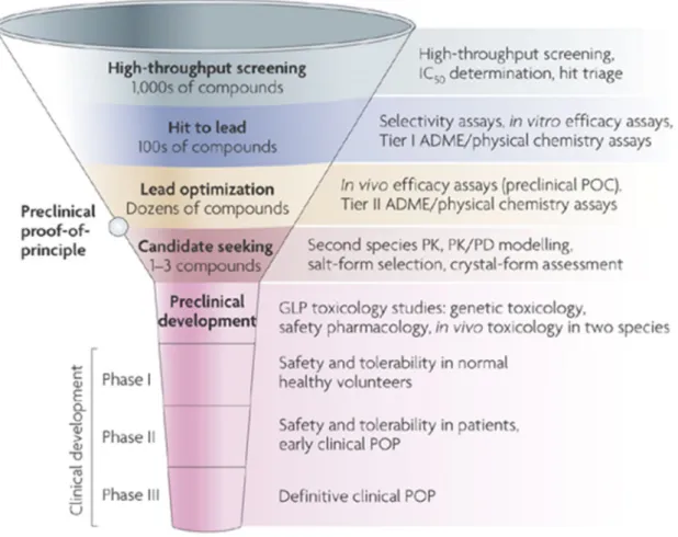
Provided these are given, the time-honoured first step of drug development is the screening of numerous compounds against the targeted kinase. Large molecule libraries contain several thousands of compounds and usually have a multi-layer structure – like the NCL™ (Nested Chemical Library™) of Vichem Ltd. (Figure 2). In case of focused or knowledge-based screening only a smaller subset of the molecule library is checked that is likely to have activity – like the EVL™ (Extended Validation Library™) of Vichem Ltd.308 There are of course more modern approaches to drug development, e.g.
in-silico modelling and design is getting more and more invaluable. Except molecular docking none of them were used in the Thesis, therefore not discussed here.
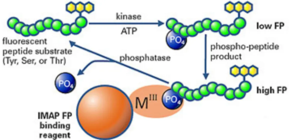
The tool of screening is mostly an in vitro assay where a recombinant kinase represents the target. This approach focuses only on the interaction of compound and kinase.
25
However, it is possible to utilize more expensive and time consuming cell-based assays on cell lines driven by the particular target. In that case some information is acquired also on the metabolism and secretion of the compound in a living cell.309 This is important because many compounds that effective in in vitro enzyme assay fail in cellular tests due to a number of conditions modifying their effect (e.g. enzymatic degradation or susceptibility to drug efflux pumps).310 However, in this case subsequent assays are even more important to confirm mechanism of action of compounds.311 Whether enzyme or cell-based assays are the best to begin is still an open question.312 Finally, the outputs of a screening process – called hits – have verified activity on the given target. Dose-response curves of hits usually obtained as soon as possible to get IC50 values which enable refined comparison of compounds.
Figure 2. Build-up of the Nested Chemical Library (NCL™) of Vichem Ltd.
[https://vichemchemie.com/nested-chemical-library-ncl]
The second step of drug development is the hit to lead phase. Hits undergo many further functional assays to test their “drug-likeness”: pharmacokinetic properties like the aforementioned membrane permeability, and ADME (absorption, distribution, metabolism, excretion) parameters. Also solubility and drug selectivity measurements commenced in this phase. The aim of SAR (structure-activity relationship) study is to
26
define essential substituents associated with activity. Small molecules usually designed according to Lipinski’s rule of five313 and considered drug-like if they possess these features:
- have molecular weights of less than 500 Dalton (g/mol)
- have a clogP value (a measure of membrane permeability) not greater than 5.
- have no more than 5 hydrogen bond donors - have a maximum of 10 hydrogen bond acceptors
Promising molecules are addressed to the next step: lead optimization. During this phase the aim is to maintain favourable properties while improving deficiencies through modification of the structure. For this purpose new analogues are synthetized. After additional rounds of pharmacokinetic and in vivo pharmacodynamic assays, a clinical candidate is declared.
Figure 3. Scheme of the drug discovery and development process. The diameter of the funnel represents the number of molecules involved at the particular level.
27
Up to 106 molecules have to be screened to find one or two clinical candidates. Attrition of compounds in the clinical phases is much lower – approximately 1 in 10 reaches the market. In turn, the cost of clinical trials is much higher than of the previous preclinical tests (Figure 3).
1.3.4. Examples for small molecule KIs 1.3.4.1. Aurora KIs
Inhibition of Aurora kinases affects all dividing cells like conventional cytotoxic drugs therefore similar systemic effects are expected. The rationale of aurora inhibition lies in the fact that their activity tends to be increased in cancer cells (see chapter 1.2.2.1.).
AKIs (Aurora kinase inhibitors) can be more-or-less paralogue selective or pan-AKIs.
The major cellular phenotypic response of dual Aurora A and B inhibitors is consistent with inhibition of Aurora B, in other words inhibition of Aurora B has dominant phenotype.314 So it was hypothesized for a long time that these dual inhibitors mediate their anti-cancer activity through inhibition of Aurora B activity.315, 316 Now there are quite selective Aurora A inhibitors that also able to induce apoptosis. The most notable AKIs that reached phase II up to date are:
- VX-680 (tozasertib, MK-0457, Figure 4/A) is a type I small molecule inhibitor that promotes DFG-out conformation of Aurora kinases317 A and B – so it is a pan-AKI.
VX-680 efficiently abrogated the growth of tumour xenografts in animal models318 but failed in clinical trial phase II due to frequent adverse events and low efficiacy.319
- MLN8054 (Figure 4/B) and MLN8237320 (alisertib, Figure 4/C) are both type I inhibitors, promoting DFG-out state. Since Aurora A is more likely a driver, so MLN 8054 was developed by Millennium Pharmaceuticals (now Takeda Oncology Company) in 2007 to be selective to Aurora A. MLN8054 decreases proliferation of cancer cell lines in in vitro cell culture and in xenografts.321 Unfortunately in phase I study MLN8054 caused somnolescence in patients with advanced solid cancers because of off-target GABAA receptor (GABAAR)-binding.322 After minimal modification of the structure of MLN8054 a new analogue, namely MLN8237 was developed. MLN8237 has similar pharmacokinetic properties to MLN8054 and quite the same GABAAR- binding but has increased affinity to Aurora A.320 Several clinical trials have been commenced with MLN8237 alone323, 324, 325 or in combination with other
28
drugs326, 327, 328, 325 but only one proceeded to phase III so far, and even that one was terminated in 2015329 because of harsh general cytotoxicity. However, applying it more carefully for the treatment of selected patients and using more precise dosing MLN8237 is worth for further investigation.330, 331 So recently new trials have been started with MLN8237. [www.clinicaltrials.gov]
Figure 4. Chemical structure of reference compounds A) VX-680, B) MLN8054 and C) MLN8237
- AZD1152 (barasertib) is a dedicated Aurora B inhibitor which induces apoptosis in human ALL (acute lymphoblastic leukaemia)332 and AML (acute myeloid leukaemia)333 cell lines. After several phase I studies AZD1152 was evaluated in two phase II trials with randomized AML patients. Despite frequent adverse events334 approximately 35%
of patients had complete cancer remission compared to 11.5% in case of the conventional cytotoxic drug cytosine arabinoside.335 AZD1152 showed transient toxicity and modest response in ~20% of B-cell lymphoma patients, but further phases as monotherapy were not encouraged in the report.336
- AT9283 (type I, promotes DFG-in) is rather a multi-kinase inhibitor with considerable effect on Aurora A/B, JAK2/3 and ABL1 kinases.337 After several phase I studies AT9283 failed in phase I/II trial due to lack of clinical response.338
- ENMD-2076 (type I, promotes DGF-in) inhibits FLT3/4, RET, Aurora A and VEGFR3 kinases in the low nanomolar range. Unfortunately it failed in clinical phase II trial against ovarian clear cell carcinoma because of low efficiacy.339 However, ENMD- 2076 provided benefit for 17% of advanced or metastatic triple-negative breast cancer patients with moderate adverse effects.340