PACAP Immunoreactivity in Human Malignant Tumor Samples and Cardiac Diseases
Z. Szanto&Zs. Sarszegi&D. Reglodi&J. Nemeth&
K. Szabadfi&P. Kiss&A. Varga&E. Banki&
K. Csanaky&B. Gaszner&O. Pinter&Zs. Szalai&
A. Tamas
Received: 30 March 2012 / Accepted: 15 May 2012 / Published online: 31 May 2012
#Springer Science+Business Media, LLC 2012
Abstract Pituitary adenylate cyclase activating polypeptide (PACAP) is a pleiotropic and multifunctional neuropeptide having important roles in various physiological processes.
Recent trends in PACAP research point to the clinical intro- duction of PACAP or its analogs/fragments possibly in the near future. Recently, we have shown the presence of PACAP in human plasma, milk, placenta, and follicular fluid samples. However, relatively few data are available on PACAP in human tissues from patients with different disorders. The aim of the present study was to determine, by
radioimmunoassay, the tissue level of PACAP38-like immu- noreactivity (LI) and PACAP27-LI in different primary non- small cell lung cancer, colon tumor samples, and in cardiac muscle samples from patients suffering from ischemic heart disease and valvular disorders. We also labeled the PAC1 receptors in human cardiac cells. All samples showed sig- nificantly higher PACAP38-LI compared with PACAP27- LI. We found significantly lower levels of PACAP38-LI and PACAP27-LI in tumoral and peripheral samples compared with normal healthy tissue in both lung and colon cancers.
Further investigations are necessary to describe the exact function of PACAP in oncogenesis. We showed that PACAP38-LI and PACAP27-LI are significantly higher in ischemic heart diseases compared with valvular abnormali- ties, suggesting that PACAP might play a role in ischemic heart disorders.
Keywords PACAP . Colon cancer . Lung cancer . Heart disorder . PAC1 receptor
Introduction
Pituitary adenylate cyclase activating polypeptide (PACAP) was isolated from ovine hypothalami in 1989, based on its activating effect on adenylate cyclase in the pituitary gland.
PACAP is a member of the secretin/glucagon/vasoactive in- testinal peptide (VIP) family and exists in 27 and 38 amino acid form, the latter being the predominant peptide in mam- malian tissues. PACAP is a pleiotropic and multifunctional neuropeptide having important roles in various physiological processes (Vaudry et al. 2009). Since its discovery, a large number of studies have shown its neurotrophic, neuroprotec- tive, general cytoprotective, and anti-inflammatory effects both in vitro and in vivo (Delgado and Ganea 2001;
Z. Szanto
Surgery Clinic, University of Pecs, Pecs, Hungary
Z. Sarszegi
:
B. Gaszner:
O. Pinter Heart Institute, University of Pecs, Pecs, HungaryD. Reglodi
:
P. Kiss:
A. Varga:
E. Banki:
K. Csanaky:
A. Tamas (*)
Department of Anatomy, PTE-MTA“Lendulet”
PACAP Research Team, University of Pecs, 7624 Pecs Szigeti ut 12.,
Hungary
e-mail: andreatamassz@gmail.com J. Nemeth
Department of Pharmacology and Pharmacotherapy, University of Debrecen,
Debrecen, Hungary K. Szabadfi
Department of Experimental Zoology and Neurobiology, University of Pecs,
Pecs, Hungary Z. Szalai
Department of Pulmonology and Allergology, Karolina Hospital, Mosonmagyarovar, Hungary
Somogyvari-Vigh and Reglodi 2004; Gasz et al. 2006a,b;
Botia et al.2007; Racz et al.2007; Dejda et al.2008; Szakaly et al.2008; Vaudry et al.2009; Atlasz et al.2010; Bourgault et al.2011; Reglodi et al.2011; Seaborn et al.2011).
Recent trends in PACAP research point to the clinical introduction of PACAP or its analogs/fragments possibly in the near future. Recently, we have shown the presence of PACAP in human plasma, where elevated PACAP levels were measured during pregnancy (Borzsei et al. 2009;
Reglodi et al. 2010). We have also detected the presence of PACAP38-like immunoreactivity (LI) in the human milk (Borzsei et al.2009), placenta, and follicular fluid samples (Brubel et al.2010,2011; Koppan et al. 2012). However, relatively few data are available on PACAP in human tissues from patients with different disorders.
PACAP has important functions in the regulation of cell growth, proliferation, differentiation, and apoptosis (Dejda et al.2008; Vaudry et al.2009; Seaborn et al.2011). It acts via three receptors, the specific PAC1 receptor and the VPAC1 and VPAC2 receptors, the latter two binding VIP and PACAP with equal affinity (Vaudry et al. 2009).
PACAP receptors are expressed in the vast majority of the frequently occurring human tumors, including primary lung and colon cancers (Reubi2000; Reubi et al.2000; Busto et al. 1999, 2000, 2003). PACAP influences expression of various factors that play a role in tumor growth (Zia et al.
1995; Moody et al. 2002,2012; Le et al. 2002) and both anti- and proapoptotic effects have been described in differ- ent tumors (Vaudry et al. 2009) suggesting a possible in- volvement of PACAP in oncogenesis. The aim of the first part of the study was to determine the tissue level of PACAP38-LI and PACAP27-LI in different primary non- small cell lung cancer (NSCLC) and colon tumor samples with radioimmunoassay (RIA) examination.
PACAP has diverse regulatory functions in the nervous, endocrine, gastrointestinal, immune, and cardiovascular sys- tem (Vaudry et al.2009). PACAP and its receptors (PAC1, VPAC1, and VPAC2) have been shown in the heart of different mammals (Wei and Mojsov1996; Calupca et al.
2000; DeHaven and Cuevas 2002; Ushiyama et al.2006), and it is able to modulate cardiac functions (Naruse et al.
1993; Parsons et al.2000; Hirose et al.2001; Hardwick et al.
2006). Recently, our research group has shown the cardio- protective effect of PACAP in vitro (Gasz et al.2006a,b;
Racz et al.2008,2010; Roth et al. 2009). Mori and cow- orkers (2010) provided in vivo evidence for cardioprotective effect of PACAP on doxorubicin-induced cardiomyopathy in mice. Although, these results suggest that this peptide might be a potential treatment of cardiac diseases in the future; there are no data about PACAP and PAC1 receptors from patients with cardiac disorders. Therefore, in the sec- ond part of this study, our aim was to examine the PACAP38-LI and PACAP27-LI with RIA in cardiac muscle
samples from patients suffering from ischemic heart disease and valvular disorders and to label PAC1 receptors with immunohistochemistry in the myocardium.
Methods
RIA Method
In the first part of the study, we examined the PACAP38-LI and PACAP27-LI in tissue samples from patients treated operatively for NSCLC or colon adenocarcinoma with RIA. We collected samples from the central and peripheral parts of lung (n09) and colon tumors (n09) and from healthy lung and colon tissue during surgery. Peripheral samples were taken within 2 cm of cancer margin and histologically confirmed as intact. In the second part of the experiment, we examined cardiac tissue samples collected from patients with ischemic heart disease (n011), mitral valve insufficiency, or aortic stenosis (n08) during heart surgery. We collected samples excised from the right auricle during the insertion of cannula of the extracorporeal circu- lating system. All human sample collections were carried out according to a protocol approved by the Institutional Ethic Committee (PTE KK 3118, 3936). In all cases, we obtained written consent of the volunteers.
Tissues samples were weighed and homogenized in ice- cold distilled water. The homogenate was centrifuged (12,000 rpm, 4°C, 30 min), and the supernatant was further processed for RIA analysis of PACAP38-LI and PACAP27- LI with a specific and sensitive RIA technique developed in our laboratory (Jakab et al.2004; Borzsei et al.2009; Brubel et al.2010), and immunoreactivity of the peptides was calcu- lated with the help of a calibration. Briefly: Antiserum:
PACAP38: “88111-3” (working dilution 1:10,000) and PACAP27:“88123”(dilution: 1:45,000).Tracer:mono-125I- labelled ovine PACAP 24-38 and mono-125I-labelled ovine PACAP27 prepared in our laboratory (5,000 cpm/tube).Stan- dard:Ovine PACAP38 and PACAP27 were used as a RIA standard ranging from 0 to 1,000 fmol/mg. Assays were prepared in 1 ml phosphate buffer (0.05 mol/l, pH 7.4) con- taining 0.1 M NaCl, 0.05% NaN3, 0.25% bovine serum albu- min (Sigma). One hundred microliters antiserum (working dilution, 1:100,000), 100 μl RIA tracer (5,000 cpm/tube), and 100μl standard or unknown samples were measured into polypropylene tubes with assay buffer. After 48–72 h incuba- tion at 4°C, the antibody-bound peptide was separated from the free one by addition of 100μl separation solution (10 g charcoal, 1 g dextran, 0.2 g commercial fat-free milk powder in 100 ml distilled water). Following centrifugation (3,000 rpm, 20 min, 4°C), the tubes were gently decanted, and the radioactivity of the precipitates was measured in a gamma counter. PACAP38-LI and PACAP27-LI of the
unknown samples were read from a calibration curve.
Results are given as femtomoles PACAP38-LI and PACAP27-LI per milligram tissue weight. Differences between PACAP38-LI and PACAP27-LI were analyzed by Student’s t test and differences between samples were analyzed using ANOVA. In all cases, p< 0.05 was con- sidered statistically significant.
Histological Examination
The cardiac tissue samples were dissected in ice-cold phosphate-buffered saline and fixed in 4% paraformalde- hyde dissolved in 0.1 M phosphate buffer (pH 7.4) for 1 h at room temperature. Tissue was then washed in phosphate- buffered saline (PBS) and cryoprotected in 10% sucrose for 1 h and 20% sucrose in PBS overnight at 4°C. For cryostat sectioning, tissues were embedded in tissue-freezing medi- um (Tissue-Tek, OCT Compound, Sakura Finetech, NL), cut in a cryostat (Leica, Nussloch, Germany) at 10-μm radial sections. Sections were mounted on chrome–alum–
gelatin-coated subbed slides and stored at−20°C until use.
One part of the tissue sections was stained with hematox- ilin–eosin staining, and the other parts were labeled with PAC1 receptor immunohistochemistry. At the beginning of the immunohistochemistry, tissue sections were rinsed in PBS, permeabilized in 0.1% Triton X-100 in PBS for 5 min, and then incubated with 0.1% bovine serum albumin, 1% normal goat serum, and 0.1% Na-azide in PBS for 1 h to minimize nonspecific labeling. Sections were incubated with anti-PAC1 receptor antibody raised in rabbit (1:100, kind gift from Prof. Seiji Shioda) for overnight at room temperature. After several washes in PBS, sections were incubated for 2 h in the dark with the corresponding Alexa Fluor “568” secondary antibody also raised in rabbit (1:1,000, Southern Biotech). Sections were then washed in PBS and were coverslipped using Fluoromount-G (Southern Biotech). For control experiments, primary antisera were omitted, and specific cellular staining was not found. Digital photographs were taken with a Nikon Eclipse 80i micro- scope equipped with a CCD camera. Images were taken with the Spot software package. Photographs were further processed with the Adobe Photoshop 7.0 program. Images were adjusted for contrast only, aligned, arranged, and then labeled using the functions of the above program.
Results
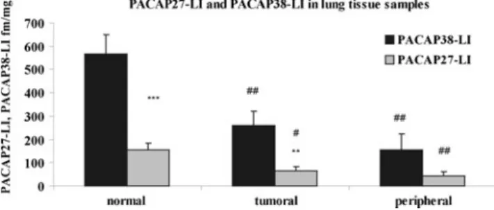
In the first part of the study, we investigated the level of PACAP38-LI and PACAP27-LI in normal, tumoral, and peritumoral (peripheral) lung tissue samples. Our results showed significantly lower level of PACAP38-LI and PACAP27-LI in tumoral and peripheral samples compared
with normal healthy tissue. Significantly higher PACAP38- LI was found in normal and tumoral tissue samples com- pared with PACAP27-LI (Fig.1).
The RIA examination of colon tissue samples showed similar results: both PACAP38-LI and PACAP27-LI were significantly lower in tumoral and peripheral samples com- pared with normal healthy tissue, and PACAP38-LI was sig- nificantly higher in all samples compared with PACAP27-LI in the same tissue (Fig.2).
In the second part of the study, we collected samples from the heart during cardiac surgery. We compared the PACAP38-LI and PACAP27-LI in heart tissue samples from ischemic heart disorder and valvular abnormalities (aorta stenosis or mitral insufficiency). PACAP38-LI was signifi- cantly higher in all samples compared with PACAP27-LI.
PACAP38-LI and PACAP27-LI were significantly higher in patients, where cardiac surgery was indicated for ischemic heart disease compared with those who were suffering from valvular dysfunction (Fig.3).
In the lung, we have a rich capillary meshwork around the alveoli, therefore, the relatively high amount of blood in the lung samples can lead to higher PACAP38-LI and PACAP27-LI in the lung tissue compared with colon and heart samples (Figs.1,2, and3). The baseline quantitative data are therefore not compared between samples originated from different tissues.
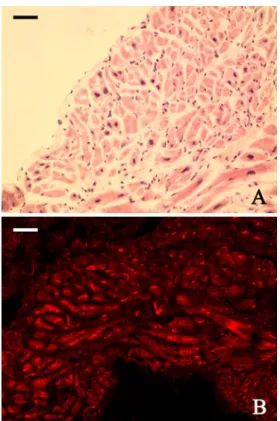
In order to investigate the expression of PAC1 receptors in the heart muscle, we obtained heart muscle samples during cardiac surgery. The samples were excised from the heart muscle wall during the insertion of cannula of extra- corporeal circulating system to the right atrium of patients with ischemic cardiac disease. We used hematoxilin–eosin staining to orientate the heart muscle cells in cross- and longitudinal sections. In parallel slides, PAC1 receptor im- munohistochemistry showed receptor expression in the heart muscle cells. The endocardial connective tissue remained PAC1-receptor-negative, but the non-specific binding of the red blood cells was detectable as an auto- fluorescence between the heart muscle cells (Fig.4).
Fig. 1 PACAP38-like immunoreactivity (LI) and PACAP27-LI deter- mined by RIA from normal, tumoral, and peripheral lung tissue sam- ples. Data are means±SEM. **p<0.01; ***p<0.001 vs. PACAP38-LI;
#p<0.05; ##p<0.01 vs. normal tissue samples
Discussion
In the first part of our experiment, we examined PACAP38- LI and PACAP27-LI in human samples from normal and cancerous lung tissues. We found significantly lower levels of PACAP27-LI and PACAP38-LI in tumoral and peripheral tissues compared with normal samples. In humans, PACAP- positive nerve fibers are found in the tracheal and bronchial wall, situated among smooth muscle bundles, and around glands and small blood vessels (Luts et al.1993). Numerous experiments investigated the presence of different PACAP receptors in normal and tumoral human lung tissue samples using different methods (Reubi 2000; Reubi et al. 2000;
Busto et al.1999,2000,2003). PACAP and PACAP recep- tors have been described both in small cell lung cancer (SCLC) and NSCLC lines (Moody et al. 1993,2000; Zia et al.1995). PACAP affects the growth and proliferation of lung cancer cells; for example, the protooncogene c-fos plays a role in cell proliferation and differentiation, and PACAP is able to increase c-fos mRNA in SCLC cells (Draoui et al. 1996). The PACAP antagonist PACAP6-38 reverses the increase in c-fos mRNA and inhibits SCLC growth in vitro and in vivo (Draoui et al.1996) and NSCLC growth in vivo (Zia et al.1995). PACAP also stimulates the MAPK and EGF receptor tyrosine phosphorylation and vascular endothelial cell growth factor mRNA in lung
cancer cell lines (Moody et al. 2002,2012). As PACAP is an autocrine growth factor for lung cancer, the relatively lower amount of PACAP38-LI and PACAP27-LI in tumoral and peritumoral tissues may suggest the overuse of the peptide by tumoral cells. On the other hand, the significantly lower immunoreactivity in the tumor might be caused by the degeneration of the structures containing high amount of PACAP such as smooth muscle and endocrine cells.
Similarly to the lung samples, we also showed significant difference between the normal and tumoral colon tissue samples, where PACAP38-LI and PACAP27-LI were also significantly lower in tumoral and peripheral tissue samples compared with normal tissue. The presence of PACAP in the gastrointestinal tract is well-known (Portbury et al.
1995; Hannibal et al. 1998), where it regulates secretion and motility (Lauff et al. 1999). Similarly to lung tissue, Reubi and coworkers have shown the predominance of VPAC1 receptors in healthy gastrointestinal mucosa and colorectal carcinoma with specific autoradiography (Reubi 2000; Reubi et al.2000). PACAP and PAC1 receptor immu- noreactivities have been shown in nerve fibers of the muco- sa, muscles, and myenteric plexus of the stomach and distal colon of the rat where intensive staining has been detected in the ganglionic nerve cells (Sundler et al.1992; Portbury et al.
1995; Hannibal et al.1998; Miampamba et al.2002). PACAP and PAC1 receptor expression have also been detected in Fig. 2 PACAP38-like immunoreactivity (LI) and PACAP27-LI deter-
mined by RIA from normal, tumoral, and peripheral colon tissue samples. Data are means±SEM. *p<0.05; **p<0.01 vs. PACAP38- LI; #p<0.05; ##p<0.01 vs. normal tissue samples
Fig. 3 PACAP38-like immunoreactivity (LI) and PACAP27-LI deter- mined by RIA from heart samples. Data are means±SEM. **p<0.01;
***p<0.001 vs. PACAP38-LI; #p<0.05; ##p<0.01 vs. normal tissue samples
Fig. 4 Representative pictures of human heart muscle stained by hematoxilin–eosin (a) and PAC1 receptor immunohistochemistry (b).
Scalebar, 20μm
HCT8 human colon tumor cells. PACAP38 increases the number of viable HCT8 cells via the regulation of Fas-R expression in human colon cancer cell line, indicating possible role of PACAP in the regulation of colon cancer growth (Le et al. 2002). PACAP plays a suppressive role in the development of inflammation-associated colon cancer, because PACAP-deficient mice showed more aggressive colo- rectal tumor growth upon induction of colitis (Nemetz et al.
2008). Similarly to our results, PACAPergic nerve fibers in the submucosal and myenteric plexus decreased significantly in pathological sections from sigmoid and rectum cancer com- pared with sections from uninvolved intestinal wall (Godlewski and Lakomy2010). The significant decrease in the PACAP immunoreactivity of the tumoral samples may be caused by the degeneration of the myenteric plexus and the dysfunc- tion in the innervation of colon in tumoral and peritumoral part.
In the second part of the study, we investigated cardiac muscle samples collected from patients with ischemic heart disease, mitral valve insufficiency, or aortic stenosis during heart surgery. We demonstrated that PACAP38-LI and PACAP27-LI were significantly higher in heart muscle of patients with ischemic heart disorder compared with valvu- lar abnormalities. The occurrence of PACAP has been dem- onstrated in the cardiac muscle as well as in intracardial plexus and cardiac ganglia of different species (Schoenfeld et al.2000; Chang et al.2005). PACAP is suggested to act as a cardioprotective factor (Dvorakova 2005). Our research group has shown that PACAP is able to protect the cardio- myocytes against oxidative stress-, ischemia/reperfusion- and doxorubicin-induced apoptosis involving different apo- ptotic pathways (Gasz et al. 2006a, b; Racz et al. 2008, 2010; Roth et al. 2009). Sano and coworkers (2002) have found that PACAP reduces cardiac fibrosis, and the cardio- protective effect of PACAP is also supported by the ob- served pulmonary hypertension and heart failure in PAC1- receptor-deficient mice (Otto et al.2004). Recently, Mori and coworkers (2010) have shown that PACAP-deficient mice display an increased vulnerability to doxorubicin- induced cardiomyopathy in an in vivo experiment. In the present study, we found significantly higher level of PACAP38-LI and PACAP27-LI in the ischemic heart tissue, suggesting its possible protective function during ischemic disorders. The protective effect of PACAP against different toxic agents can be attenuated by PACAP6-38, indicating that the cardioprotective effect of PACAP is mostly mediat- ed by PAC1 receptors. Dominant expression of PAC1 recep- tors is well known in cultured cardiomyocytes; furthermore, human heart tissue expresses RNA transcripts for all three PACAP receptors (Wei and Mojsov1996). In our study, we showed PAC1 receptors in human heart muscle from patients with ischemic heart disease with immunohisto- chemical examination.
It is well known that PACAP38 is the dominant form in the mammalian tissue (Vaudry et al. 2009). In the present study, we also detected higher level of PACAP38-LI com- pared with PACAP27-LI in all of the clinical samples in different disorders.
Significantly higher PACAP38-LI and PACAP27-LI were measured in the healthy lung and colon tissues com- pared with the respective tumor samples, but further inves- tigations are necessary to describe the exact function of PACAP in oncogenesis. It is known that VIP antagonist are potent inhibitor lung and colon cancer lines (Levy et al.2002), and PACAP6-38 also inhibits the effect of PACAP on different cancer lines, suggesting potential therapeutic effect in the clinical practice (Le et al. 2002). We showed that PACAP38-LI and PACAP27-LI are significantly higher in ischemic heart disorders compared with valvular abnor- malities, suggesting that PACAP might play a role in ische- mic heart disorders.
Acknowledgments This study was supported by the following grants: OTKA (K72592, 75965, CNK78480), TAMOP (4.2.1.B-10/2/
KONV-2010-002, 4.2.2.B-10/1-2010-0029), Bolyai Scholarship, Richter Foundation, PTE AOK Research Grant KA-4039/10-26, PTE-MTA “Lendulet” Program. The authors also thank all the volunteers.
References
Atlasz T, Szabadfi K, Kiss P et al (2010) Pituitary adenylate cyclase activating polypeptide in the retina: focus on the retinoprotective effects. Ann N Y Acad Sci 1200:128–139
Borzsei R, Mark L, Tamas A et al (2009) Presence of pituitary adeny- late cyclase activating polypeptide-38 in human plasma and milk.
Eur J Endocrinol 160:561–565
Botia B, Basille M, Allais A et al (2007) Neurotrophic effects of PACAP in the cerebellar cortex. Peptides 28:1746–1752 Bourgault S, Chatenet D, Wurtz O et al (2011) Strategies to convert
PACAP from a hypophysiotropic neurohormone into a neuro- protective drug. Curr Pharm Des 17:1002–10024
Brubel R, Boronkai A, Reglodi D et al (2010) Changes in the expres- sion of pituitary adenylate cyclase-activating polypeptide in the human placenta during pregnancy and its effects on the survival of JAR choriocarcinoma cells. J Mol Neurosci 42:450–458 Brubel R, Reglodi D, Jambor E et al (2011) Investigation of pituitary
adenylate cyclase activating polypeptide in human gynecological and other biological fluids by using MALDI TOF mass spectrom- etry. J Mass Spectrom 46:189–194
Busto R, Carrero I, Guijarro LG et al (1999) Expression, pharmaco- logical, and functional evidence for PACAP/VIP receptors in human lung. Am J Physiol 277(1 Pt 1):L42–L48
Busto R, Prieto JC, Bodega G, Zapatero J, Carrero I (2000) Immuno- histochemical localization and distribution of VIP/PACAP recep- tors in human lung. Peptides 21:265–269
Busto R, Prieto JC, Bodega G, Zapatero J, Fogue L, Carrero I (2003) VIP and PACAP receptors coupled to adenylyl cyclase in human lung cancer: a study in biopsy specimens. Peptides 24:429–436 Calupca MA, Vizzard MA, Parsons RL (2000) Origin of pituitary adeny-
late cyclase-activating polypeptide (PACAP)-immunoreactive fibers
innervating guinea pig parasympathetic cardiac ganglia. J Comp Neurol 423:26–39
Chang Y, Lawson LJ, Hancock JC, Hoover DB (2005) Pituitary adenylate cyclase-activating polypeptide: localization and differ- ential influence on isolated hearts from rats and guinea pigs.
Regul Pept 129:139–146
DeHaven WI, Cuevas J (2002) Heterogeneity of pituitary adenylate cyclase-activating polypeptide and vasoactive intestinal polypep- tide receptors in rat intrinsic cardiac neurons. Neurosci Lett 328:45–49
Dejda A, Jolivel V, Bourgault S et al (2008) Inhibitory effect of PACAP on caspase activity in neuronal apoptosis: a better under- standing towards therapeutic applications in neurodegenerative diseases. J Mol Neurosci 36:26–37
Delgado M, Ganea D (2001) Vasoactive intestinal peptide and pituitary adenylate cyclase activating polypeptide inhibit expression of Fas ligand in activated T lymphocytes by regulating c-Myc, NF-κB, NF-AT, and early growth factors 2/3. J Immunol 166:1028–1040 Draoui M, Hida T, Jakowlew S, Birrer M, Zia F, Moody TW (1996) PACAP stimulates c-fos mRNAs in small cell lung cancer cells.
Life Sci 59:307–313
Dvorakova MC (2005) Cardioprotective role of the VIP signaling system. Drug News Perspect 18:387–391
Gasz B, Racz B, Roth E et al (2006a) PACAP inhibits oxidative stress- induced activation of MAP kinase dependent apoptotic pathway in cultured cardiomyocytes. Ann N Y Acad Sci 1070:293–297 Gasz B, Racz B, Roth E et al (2006b) Pituitary adenylate cyclase
activating polypeptide protects cardiomyocytes against oxidative stress-induced apoptosis. Peptides 27:87–94
Godlewski J, Lakomy IM (2010) Changes in vasoactive intestinal peptide, pituitary adenylate cyclase-activating polypeptide and neuropeptide Y-ergic structures of the enteric nervous system in the carcinoma of the human large intestine. Folia Histochem Cytobiol 48:208–216
Hannibal J, Ekblad E, Mulder H, Sundler F, Fahrenkrug J (1998) Pituitary adenylate cyclase activating polypeptide (PACAP) in the gastrointestinal tract of the rat: distribution and effects of capsaicin or denervation. Cell Tissue Res 291:65–79
Hardwick JC, Tompkins JD, Locknar SA, Merriam LA, Young BA, Parsons RL (2006) Calcium influx through channels other than voltage-dependent calcium channels is critical to the pituitary adenylate cyclase-activating polypeptide-induced increase in ex- citability in guinea pig cardiac neurons. Ann N Y Acad Sci 1070:317–321
Hirose M, Leatmanoratn Z, Laurita KR, Carlson MD (2001) Effects of pituitary adenylate cyclase-activating polypeptide on canine atrial electrophysiology. Am J Physiol Heart Circ Physiol 281:H1667– H1674
Jakab B, Reglodi D, Jozsa R et al (2004) Distribution of PACAP-38 in the central nervous system of various species determined by a novel radioimmunoassay. J Biochem Biophys Methods 61:189–198 Koppan M, Varnagy A, Reglodi D et al (2012) Correlation between oocyte number and follicular fluid concentration of pituitary adenylate cyclase-activating polypeptide (PACAP) in women after superovulation treatment. J Mol Neurosci (in press) doi:10.1007/s12031-012-9743-3
Lauff JM, Modlin IM, Tang LH (1999) Biological relevance of pitui- tary adenylate cyclase-activating polypeptide (PACAP) in the gastrointestinal tract. Regul Pept 84:1–12
Le SV, Yamaguchi DJ, McArdle CA, Tachiki K, Pisegna JR, Germano P (2002) PAC1 and PACAP expression, signaling, and effect on the growth of HCT8, human colonic tumor cells. Regul Pept 109:115–125
Levy A, Gal R, Granoth R, Dreznik Z, Fridkin M, Gozes I (2002) In vitro and in vivo treatment of colon cancer by VIP antagonists.
Regul Pept 109:127–133
Luts A, Uddman R, Alm P, Basterra J, Sundler F (1993) Peptide- containing nerve fibres in human airways: distribution and coex- istence pattern. Int Arch Allergy Immunol 101:52–60
Miampamba M, Germano PM, Arli S et al (2002) Expression of pituitary adenylate cyclase-activating polypeptide and PACAP type 1 receptor in the rat gastric and colonic myenteric neurons.
Regul Pept 105:145–154
Moody TW, Zia F, Makheja A (1993) Pituitary adenylate cyclase activating polypeptide receptors are present on small cell lung cancer cells. Peptides 14:241–246
Moody TW, Walters J, Casibang M, Zia F, Gozes Y (2000) VPAC1 receptors and lung cancer. Ann N Y Acad Sci 921:26–32 Moody TW, Leyton J, Casibang M, Pisegna J, Jensen RT (2002)
PACAP-27 tyrosine phosphorylates mitogen activated protein kinase and increases VEGF mRNAs in human lung cancer cells.
Regul Pept 109:135–140
Moody TW, Osefo N, Nuche-Berenguer B, Ridnour L, Wink D, Jensen RT (2012) Pituitary adenylate cyclase activating polypeptide causes tyrosine phosphorylation on the EGF receptor in lung cancer cells. J Pharmacol Exp Ther 341:873–881
Mori H, Nakamachi T, Ohtaki H et al (2010) Cardioprotective effect of endogenous pituitary adenylate cyclase-activating polypeptide on doxorubicin-induced cardiomyopathy in mice. Circ J 74:1183–
1190
Naruse S, Suzuki T, Ozaki T, Nokihara K (1993) Vasodilator effect of pituitary adenylate cyclase activating polypeptide (PACAP) on femoral blood flow in dogs. Peptides 14:505–510
Nemetz N, Abad C, Lawson G et al (2008) Induction of colitis and rapid development of colorectal tumors in mice deficient in the neuropeptide PACAP. Int J Cancer 122:1803–1809
Otto C, Hein L, Brede M et al (2004) Pulmonary hypertension and right heart failure in pituitary adenylate cyclase-activating poly- peptide type I receptor-deficient mice. Circulation 110:3245–3251 Parsons RL, Rossignol TM, Calupca MA, Hardwick JC, Brass KM (2000) PACAP peptides modulate guinea pig cardiac neuron membrane excitability and neuropeptide expression. Ann N Y Acad Sci 921:202–210
Portbury AL, McConalogue K, Furness JB, Young HM (1995) Distribu- tion of pituitary adenylyl cyclase activating peptide (PACAP) im- munoreactivity in neurons of the guinea-pig digestive tract and their projections in the ileum and colon. Cell Tissue Res 279:385–392 Racz B, Gasz B, Borsiczky B et al (2007) Protective effects of pituitary
adenylate cyclase activating polypeptide in endothelial cells against oxidative stress-induced apoptosis. Gen Comp Endocrinol 153:115–123
Racz B, Gasz B, Gallyas F Jr et al (2008) PKA-Bad-14-3-3 and Akt- Bad-14-3-3 signaling pathways are involved in the protective effects of PACAP against ischemia/reperfusion-induced cardio- myocyte apoptosis. Regul Pept 145:105–115
Racz B, Reglodi D, Horvath G et al (2010) Protective effect of PACAP against doxorubicin-induced cell death in cardiomyocyte culture.
J Mol Neurosci 42:419–427
Reglodi D, Gyarmati J, Ertl T et al (2010) Alterations of pituitary adenylate cyclase-activating polypeptide-like immunoreactivity in the human plasma during pregnancy and after birth. J Endo- crinol Invest 33:443–445
Reglodi D, Kiss P, Lubics A, Tamas A (2011) Review on the protective effects of PACAP in models of neurodegenerative diseases in vitro and in vivo. Curr Pharm Des 17:962–972
Reubi JC (2000) In vitro evaluation of VIP/PACAP receptors in healthy and diseased human tissues. Clinical implications. Ann N Y Acad Sci 921:1–25
Reubi JC, Laderach U, Waser B, Gebbers JO, Robberecht P, Laissue JA (2000) Vasoactive intestinal peptide/pituitary adenylate cyclase-activating peptide receptor subtypes in human tumors and their tissues of origin. Cancer Res 60:3105–3112
Roth E, Weber G, Kiss P et al (2009) Effects of PACAP and precondi- tioning against ischemia/reperfusion-induced cardiomyocyte apo- ptosis in vitro. Ann N Y Acad Sci 1163:512–516
Sano H, Miyata A, Horio T, Nishikimi T, Matsuo H, Kangawa K (2002) The effect of pituitary adenylate cyclase activating poly- peptide on cultured rat cardiocytes as a cardioprotective factor.
Regul Pept 109:107–113
Schoenfeld LK, Souder JA, Hardwick JC (2000) Pituitary adenylate cyclase-activating polypeptide innervation of the mudpuppy car- diac ganglion. Brain Res 882:180–190
Seaborn T, Masmoudi-Kouli O, Fournier A, Vaudry H, Vaudry D (2011) Protective effects of pituitary adenylate cyclase-activating polypep- tide (PACAP) against apoptosis. Curr Pharm Des 17:204–214 Somogyvari-Vigh A, Reglodi D (2004) Pituitary adenylate cyclase
activating polypeptide: a potential neuroprotective peptide. Curr Pharm Des 10:2861–2889
Sundler F, Ekblad E, Absood A, Hakanson R, Koves K, Arimura A (1992) Pituitary adenylate cyclase activating peptide: a novel
vasoactive intestinal peptide-like neuropeptide in the gut. Neuro- science 46:439–454
Szakaly P, Kiss P, Lubics A et al (2008) Effects of PACAP on survival and renal morphology in rats subjected to renal ischemia–reper- fusion. J Mol Neurosci 36:89–96
Ushiyama M, Sugawara H, Inoue K, Kangawa K, Yamada K, Miyata A (2006) Characterization of the PAC1 variants expressed in the mouse heart. Ann N Y Acad Sci 1070:586–590
Vaudry D, Falluel-Morel A, Bourgault S et al (2009) Pituitary adeny- late cyclase-activating polypeptide and its receptors: 20 years after the discovery. Pharmacol Rev 61:283–357
Wei Y, Mojsov S (1996) Multiple human receptors for pituitary adenylyl cyclase-activating polypeptide and vasoactive intestinal peptide are expressed in a tissue-specific manner. Ann N Y Acad Sci 805:624–627
Zia F, Fagarasan M, Bitar K et al (1995) Pituitary adenylate cyclase activating peptide receptors regulate the growth of non-small cell lung cancer cells. Cancer Res 55:4886–4891