Description of myxosporeans (Cnidaria: Myxozoa) infecting the popular food fi sh Notopterus notopterus (Pisces: Notopteridae) in Malaysia and India
Muhammad Ha fi z Borkhanuddin
a,1, Urvashi Goswami
b,1, Gábor Cech
b, Kálmán Molnár
b, Stephen D. Atkinson
c, Csaba Székely
b,⁎
aFaculty of Science & Marine Environment, Universiti Malaysia Terengganu, 21030 Kuala Nerus, Malaysia
bInstitute for Veterinary Medical Research, Centre for Agricultural Research, POB 18, H-1581 Budapest, Hungary
cDepartment of Microbiology, Oregon State University, Corvallis, OR 97330, USA
a r t i c l e i n f o a b s t r a c t
Article history:
Received 9 May 2020
Received in revised form 12 August 2020 Accepted 19 August 2020
This study was a co-operative investigation of myxosporean infections ofNotopterus notopterus, the bronze featherback, which is a popular foodfish in the South Asian region. We examined fish from Lake Kenyir, Malaysia and the River Ganga, Hastinapur, Uttar Pradesh, India, and ob- served infections with two myxosporeans: Myxidium cf. notopterum (Myxidiidae) and Henneguya ganapatiae (Myxobolidae), respectively. These species were identified by myxospore morphology, morphometry and host tissue affinity, and the original descriptions supplemented with small-subunit ribosomal DNA sequences and phylogenetic analysis. Free myxospores ofM.cf.notopterumwere found in the gallbladder, and measured 14.7 ± 0.6μm long and 6.3 ± 0.6μm wide; host, tissue and myxospore dimensions overlapped with the type, but differed in morphological details (spore shape, valve cell ridges) and locality (Malaysia versus India). Plasmodia and spores ofH. ganapatiaewere observed in gills, and myxospores had a spore body 9.7 ± 0.4μm long, 4.5 ± 0.5μm wide; sample locality, host, tis- sue, spore morphology and morphometry matched the original description. Small-subunit ribo- somal DNA sequences were deposited in GenBank (M.cf.notopterumMT365527,H. ganapatiae MT365528) and both differed by >7% from congeneric species. Although the pathogenicity and clinical manifestation of myxozoan in humans are poorly understood, consumption of rawfish meat with myxozoan infection was reported to be associated with diarrhea. Identification of current parasite fauna fromN. notopterusis an essentialfirst step in assessing pathogen risks to stocks of this important foodfish.
© 2020 Published by Elsevier Inc. on behalf of International Association of Food and Waterborne Parasitology. This is an open access article under the CC BY-NC-ND license (http://
creativecommons.org/licenses/by-nc-nd/4.0/).
Keywords:
Myxidium Henneguya Bronze featherback Asia
Tropical Parasite Myxospore
1. Introduction
The bronze featherback,Notopterus notopterusPallas, 1769 (Osteoglossiformes) is a member of a group commonly called“knife fishes”, which are distributed widely in Africa, South and Southeast Asia (Talwar and Jhingran, 1991). Knifefish have commercial value for recreational anglers, and species ofNotopterus,Chitala,PapyrocranusandXenomystushave been categorized as a com-
⁎ Corresponding author.
E-mail address:szekely.csaba@atk.hu. (C. Székely).
1 MHB and UG contributed equally to this work.
https://doi.org/10.1016/j.fawpar.2020.e00092
2405-6766/© 2020 Published by Elsevier Inc. on behalf of International Association of Food and Waterborne Parasitology. This is an open access article under the CC BY- NC-ND license (http://creativecommons.org/licenses/by-nc-nd/4.0/).
Contents lists available atScienceDirect
Food and Waterborne Parasitology
j o u r n a l h o m e p a g e :w w w . e l s e v i e r . c o m / l o c a t e / f a w p a r
mercially importantfish by the Food and Agriculture Organization of the United Nations for food and the ornamental trade (Casavas et al., 1996).Notopterus notopterusis a particularly important foodfish, with high commercial value. For example, in Cambodia this species has had the highest export value of anyfish (Mille et al., 2016). Thefish is also valued as a decorative spe- cies in the aquarium trade (Mohanty and Samanta, 2016).
Wild populations of bronze featherback are declining, and it is regarded as a threatened species (CAMP, 1998). Natural popula- tions are impacted by excessive harvesting, and pollution from industrial, domestic and agricultural sources, which has led to in- creased concentrations of heavy metals infish tissue (Ngor et al., 2003;Shah et al., 2009;Mohanty and Samanta, 2016). Initial attempts have been made to culture thefish (Rahmatullah et al., 2009;Mohanty and Samanta, 2018). Identification of parasites that infectN. notopterusis a fundamental part of assessing disease risks to both wild and cultured stocks of this important foodfish.
The parasite fauna of bronze featherback is poorly studied. The ciliophoran protozoa,Trichodina monopteriwas described by Mitra and Haldar (2004).Two cystidicolid nematodes,Pseudoproleptus notopteriandSpinitectus notopteriwere described by Karve and Naik (1951)and redescribed byMoravec et al. (2016). One isopod,Alitropus typusEdwards, 1840, was recorded from the bronze featherback byAhmad et al. (2016)in the Lake Chasma, Pakistan. The myxosporean fauna of thisfish is relatively better studied, asSarkar (1996)describedMyxobolus meglitschusfrom the gills andMyxidium notopterumfrom the liver. Three Henneguyaspecies are known from India:Quadri (1965, 1970)describedH. notopteraeandH. ganapatiae, whileLalita Kumari (1969)describedH. singhi. Myxozoa is one of the economically most important groups of microscopic metazoan parasites, as they infectfishes from both food and pet trades. Stocks of foodfish species impacted by myxozoan infections includefinfish in Mediterranean aquaculture, byEnteromyxumspp. (Palenzuela et al., 2002), catfish byHenneguya ictaluri(Pote et al., 2000), and salmonids byMyxobolus cerebralisandCeratonova shasta(AFS-FHS, 2014). In addition to reducing foodfish stocks, myxozoans can affect humans directly. Several species ofKudoaare known to degradefillet quality after catch (Henning et al., 2012;
Langdon, 1991) and anotherKudoa,K. septempunctata, can cause diarrhea and emesis after consumption of raw infected olive flounder (Harada et al., 2012). The pathogenicity ofK. septempunctatawas demonstrated in an in vitro experiment on human in- testinal cells, which were rapidly invaded by sporoplasms (Ohnishi et al., 2013). OtherKudoaspecies evoke allergic reactions in consumers (Martínez de Velasco et al., 2008). Importantly, as both diversity and pathogenic effects of myxozoans are still being revealed, surveillance and detection of novel species are important for assessing risks to and from foodfish.
Myxidiumis one of the largest myxozoan taxa (Class Myxosporea) with over 232 nominal species (Eiras et al., 2011). It is a polyphyletic genus of typically coelozoic (rarely histozoic) parasites, with most species described from the gall bladder, with some from the kidneys or urinary bladder. HistozoicMyxidiumspecies are known from the gills and skin (Eiras et al., 2011;
Heiniger and Adlard, 2014). GenusHenneguyaThélohan, 1892 has 189 species (Eiras, 2002;Eiras and Adriano, 2012) and is the second largest group within the Myxozoa.Henneguyaspp. are common parasites of marine and fresh-waterfish and are typ- ically histozoic in different organs and tissues, particularly the gills, skin, kidney, musculoskeletal system or gastrointestinal tract (Kent et al., 2001;Eiras, 2002;Bahri and Marques, 2008). In India,Kalavati and Nandi (2007)report 24Henneguyaspecies, the majority of which are from West Bengal, with three species (H. ganapatiae, H. notopterae, H. singhi) described from N. notopterus. However, like most myxozoans, manyHenneguyaspecies have been described on the basis of morphological and morphometric characters only, and presently lack corresponding DNA sequence data, which makes accurate re-identification chal- lenging. The importance of species from this genus as pathogens of freshwaterfish has been described by several authors (Dyková and Lom, 1978;Kalavati and Narasimhamurti, 1985;Lom and Dyková, 1995;Martins and Souza, 1997;Martins et al., 1999). With several species causing economic impacts onfish farm activities (Feist and Longshaw, 2006).
In this present study, we describe myxozoans fromN. notopterusfrom India and Malaysia, using morphology and small- subunit ribosomal DNA (ssrDNA) sequencing. We found aMyxidiumspecies,M. cf.notopterum, from the gall bladder offish from Lake Kenyir, Malaysia, and we re-describedHenneguya ganapatiae, from gills ofN. notopterusfrom River Ganga, Hastinapur, Uttar Pradesh, India.
2. Materials & methods
2.1. Collection and morphological examination of bronze featherback in Malaysia
Notopterus notopteruswere collected with gill nets in the Tasik Kenyir Water Reservoir (4°48′33.45″N, 102°47′10.45″E) in May 2011. Fish (N = 13; length 21–24 cm; weight 1.0–1.5 kg) were transported live to the Institute of Tropical Aquaculture (AKUATROP), University Malaysia Terengganu (UMT), and maintained in an aerated aquarium. Within 2 days after capture,fish were pithed, dissected and examined for the presence of myxosporeans using a stereomicroscope and a compound microscope.
Emphasis was placed on examining organs typically associated with myxozoan development: gills,fins, muscle, kidneys, gall blad- der, and intestine. Bile was smeared on a slide, and examined wet by microscopy. When suspected myxospores were found, they were studied with an advanced light microscope (Nikon Eclipse 80i). Thirty fresh spores from a single host were measured and characterized according to the guidelines ofLom and Arthur (1989); with the exception that we use the more structurally accu- rate term“polar tubule”instead of“polarfilament”. Spores were preserved in 80% ethanol for subsequent molecular analysis.
2.2. Collection and morphological examination of bronze featherback in India
Fish were purchased at afish market, but purportedly were caught in the River Ganga, in Hastinapur (29°01′N, 77°45′E), Meerut, Uttar Pradesh, India in February 2018. Fish (N = 20; body length 20–25 cm) and kept on ice until necropsy. Eachfish
was examined for myxozoan parasites with special attention to infections of the gill. Due to degradation of the gill tissue, location and structure of plasmodia could not be characterized in detail. Spores were collected from damaged plasmodia by scraping gill filaments, then preserved in 4% formalin for morphological studies, and in 90% ethanol for molecular analysis. Twenty twofixed spores from a singlefish were measured and described forMyxidiumspecies using Nomarski differential interference contrast and photographed. All measurements are expressed in micrometers (Tables 2 and 3).
2.3. DNA extraction, amplification and sequencing
Total genomic DNA was extracted from spores preserved in ethanol. The spores were centrifuged at 9600 ×gfor 10 min and the supernatant removed. For theMyxidiumspores, total DNA was extracted from the spore pellet using a DNeasy Blood & Tissue Kit (Qiagen, Hilden, Germany), while forHenneguya, total DNA was extracted using a Genaid Tissue Genomic DNA Mini Kit (New Taipei City, Taiwan), following the manufacturers' instructions, with a 100μlfinal elution step. ssrDNA was amplified using a nested PCR described in detail byCech et al. (2015). Universal eukaryotic primers ERIB1 and ERIB10 (Barta et al., 1997) were used in the first round PCR. Myxozoan specific primers Myx1F (Hallett and Diamant, 2001) and SphR (Eszterbauer and Székely, 2004) were used in the second round PCR. The primer sequences are listed inTable 1. Amplicons were analysed by elec- trophoresis in a 1% agarose gel. The PCR products were excised from the gel, purified with the Gel/PCR DNA Fragments Extraction Kit (Geneaid, New Taipei City, Taiwan) and sequenced directly using the BigDye Terminator v3.1 Cycle Sequencing Kit (Life Tech- nologies) with an ABI PRISM 3100 Genetic Analyser (Life Technologies), using the amplification and inner primers.
2.4. Phylogenetic analysis
The sequence fragments were assembled using MEGA 6 (Tamura et al., 2013) and ambiguous bases were clarified by visual examination of the corresponding ABI chromatograms. Sequences of the discovered myxozoans were aligned with reference se- quences from the NCBI GenBank database (based on a BLAST similarity >88% and coverage >75% forH. ganapatiae, and similarity
>89% and coverage >77% forM.cf.notopterum) with CLUSTAL W (Thompson et al., 1994). Final alignments of 24 sequences for M.cf.notopterumand 34 sequences forH. ganapatiaewere tested using MEGA 6 for the nucleotide substitution model of bestfit as indicated by the Akaike Information Criterion (AIC). Phylogenetic relationships forM.cf.notopterumandH. ganapatiaewere in- ferred using the maximum likelihood (ML) method with the G + I and GTR + G + I substitution models respectively, and bootstrapped with 1000 replicates. Initial trees for the heuristic search were obtained by applying the Neighbor-Joining method to a matrix of pairwise distances estimated using the Nearest Neighbor Interchange (NNI) approach forM.cf.notopterumand Maximum Composite Likelihood (MCL) approach forH. ganapatiae. A discrete Gamma distribution was used to model evolution- ary rate differences among sites (5 categories; +G, parameter = 0.3388). The rate variation model allowed for some sites to be evolutionarily invariable ([+I]; 0.0000% sites). All alignments with <75% site coverage were eliminated.Chloromyxum cristatum andCeratonova shastawere chosen as the outgroups forH. ganapatiaeandM.cf.notopterumanalyses, respectively.
3. Results
3.1. Myxidium cf. notopterum
Bronze featherback (N = 13; 21–24 cm total length) were examined from the Tasik Kenyir water reservoir in Malaysia. Free- floating matureMyxidiumspores were found in the gall bladders of 2fish (15.4%). Although the myxospore morphometry most closely resembledM. notopterumSarkar, 1996, from the same type host in India, they differed somewhat in morphology (described below). No ssrDNA sequence was available from the type species to compare with the novel sequence data we provide here.
Table 1
Primers used in PCRs and sequencing.
Primer name Sequence (5′-3′) Used to Application Reference
ERIB1 A`CCTGGTTGATCCTGCA Both 1st round PCR Barta et al., 1997
ERIB10 CTTCCGCAGGTTCACCTACGG Both 1st round PCR Barta et al., 1997
Myx1F GTG AGA CTG CGG ACG GCT CAG Both 2nd round PCR Hallett and Diamant, 2001
SphR GTT ACC ATT GTA GCG CGC GT Both 2nd round PCR & sequencing forHenneguya Eszterbauer and Székely, 2004
ACT1FR TTG GGT AAT TTG CGC GCC TGC Both Sequencing Hallett and Diamant, 2001
CR1 R GAT YAG ATA CCG TCS TAGT Henneguya Sequencing Székely et al., 2015
CR1 F CGA AGA CGA TCA GAT ACC GTC CTA Henneguya Sequencing Székely et al., 2015
ACT3F CAT GGA ACG AAC AAT Henneguya Sequencing Hallett and Diamant, 2001
Myxgen4R ACC TGT TAT TGC CAC GCT Myxidium Sequencing Kent et al., 2000
Myxgen3F GGA CTA ACR AAT GCG AAG GCA Myxidium Sequencing Kent et al., 2000
MTseg2F GCA AGA GGT GAA ATT CTT G Myxidium Sequencing Kent et al., 2000
MB5r ACC GCT CCT GTT AAT CAT CAC C Myxidium Sequencing Eszterbauer, 2004
MC5 CCTGAGAAACGGCTACCACAT Henneguya Sequencing Molnár et al., 2002
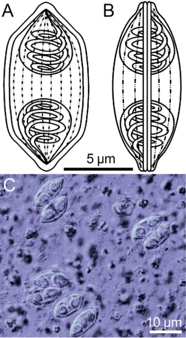
Description of spores: In valvular view spores fusiform with acuminated tips, in sutural view sigmoid, with twist on longitudinal axis giving reniform appearance. Longitudinal suture line thin (Fig. 1A, B, C). Spore valve with 8–10 longitudinal surface ridges.
Length 14.7 ± 0.6 (13.8–16.0μm), width 6.3 ± 0.6 (5.5–7.7μm). Polar capsules pyriform, at either end of spore, having equal dimensions: length 5.7 ± 0.5 (4.6–6.4μm), width 4.7 ± 0.4 (3.6–5.3μm) (Table 2). Polar tubules with 3–4 coils perpendicular to the long axis of each polar capsule (Fig. 1A, B, C).
Type host: Bronze featherback, local name“belida”,Notopterus notopterus(Pallas) (Notopteridae).
Site of infection: Gall bladder Prevalence of infection: 15.4% (2/13)
Reference materials: Digitized photos of syntype spores were deposited in the collection of Fish Pathology and Parasitology Group, Centre for Agricultural Research, Institute for Veterinary Medical Research, Budapest, Hungary.
Molecular data: Small subunit rDNA sequence data of 1667 bp from a single hostfish, has been deposited in NCBI GenBank (accession no. MT365527). Sequences with highest similarity wereMyxosporeagen. sp. PBS-2015 (93.0%; KP030767) and Myxidium cuneiforme(90.6%, DQ377709).
Fig. 1.Myxospores ofMyxidiumcf.notopterum. A–B: Line drawings of mature myxospores in frontal and valvular view showing polar capsules with coiled polar tubules and the longitudinal grooves. C: Fresh, unstained myxospores in frontal view showing the two pyriform polar capsules.
Table 2
Comparison of hosts, infection site, myxospore dimensions and localities ofMyxidiumcf.notopterumand other closely related congeners. All measurements are in mi- crometer (μm).
Species Host Infection site Spore body Polar capsule No. of polar
tubule coils
Locality Reference
Length Width Length Width
Myxidiumcf.
notopterum
Notopterus notopterus
Gall bladder 13.8–16.0 (14.7 ± 0.6)
5.5–7.7 (6.3 ± 0.6)
4.6–6.4 (5.7 ± 0.5)
3.6–5.3 (4.7 ± 0.4)
3–5 Malaysia Present study
M.
incomptaverni
Diplectanocotyla gracilis
Parenchymal tissue
11.3–11.8 (11.6)
4.2–5.6 (4.9)
2.4–3.3 (2.9)
1.8–2.0 (1.9)
2–3 Malaysia Freeman and
Shinn, 2011 M. notopterum Notopterus
notopterus
Liver 13.5–16.0
(15.37)
7.0–9.0 (8.3)
4.5–6.0 (5.65)
4.5–5.5 (5.05)
3–4 India Eiras et al.,
2011 M. cuneiforme Cyprinus carpio Gall Bladder 12.0–13.1
(12.5 ± 0.3) 4.8–6.1 (5.4 ± 0.3)
4.0–4.6 (4.3 ± 0.2)
2.6–3.4 (3.1 ± 0.2)
5–6 China Li et al., 2016
M. amazonense Corydoras melini Gall bladder 16.1–17.9 (17.0 ± 0.9)
3.0–4.4 (3.7 ± 0.7)
4.9–5.9 (5.4 ± 0.5)
2.8–4.0 (3.4 ± 0.6)
4–5 Brazil Mathews et al.,
2015 M. scripta Trachemys
scripta elegans
Gall bladder 16.6–20.4 (18.8)
4.6–5.9 (5.1)
5.1–7.8 (6.6)
2.6–4.1 (3.8)
6–8 USA Roberts et al.,
2008
M. truttae Salmo trutta fario Gall bladder 11–12 7.0–7.3 3.7 diameter NA France Eiras et al.,
2011
Fig. 2.Phylogenetic tree generated by maximum likelihood analysis of ssrDNA sequences ofMyxidiumcf.notopterum, and other closely-related myxosporean species; GenBank accession numbers shown after the species name. Novel data are in bold. Numbers at nodes indicate the bootstrap confidence values >50 (ML).Ceratonova shastawas used as an outgroup.
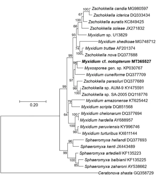
Phylogenetic analysis: Maximum likelihood (ML) analysis (Fig. 2), with 26 species in the ingroup andCeratonova shasta (GQ358729) as an outgroup, showed seven clades of Myxidiidae (Myxidium and Zschokkella), and Sphaeromyxidae (Sphaeromyxa), all of which are coelozoic species infecting the gall bladder or the bile duct of theirfish hosts. Specifically, M.cf.notopterumclustered withMyxidiumspecies that parasitize the gall bladder of fresh waterfish hosts.
Remarks: Myxospores ofM.cf.notopterumwere morphometrically similar toMyxidium notopterum(Sarkar, 1996) described from the samefish in West Bengal, India. Spores of our species have a slightly sigmoidal shape, whereasM. notopterumspores are cylindrical with rounded ends. Polar capsules ofM.cf.notopterumare more elongated than those ofM. notopterum. Polar tubules have 3–4 turns inM. notopterumcompared with 4–5 forM.cf.notopterum(Table 2). No ridges are reported on the spore surface ofM. notopterum, whereasM.cf.notopterumhas 8–10 striations.
3.2. Redescription of Henneguya ganapatiaeQuadri, 1970
Bronze featherback (N = 20; 20–25 cm total length) were collected from River Ganga, Hastinapur, Uttar Pradesh, India. Ma- ture myxospores ofH. ganapatiaewere observed in gills of 15/20 (75%)fish.
Fig. 3.Myxospores ofHenneguya ganapatiae. A–B: Line drawings of mature myxospores in frontal and valvular view showing polar capsules and caudal processes.
C: Fresh, unstained myxospore in frontal view showing the two pyriform polar capsules and caudal processes.
Description of spores: Spore body ellipsoidal in both frontal and sutural views, with two slightly curved caudal processes. Spore valves have thin walls, surface smooth, without ridges (Fig. 3A, B, C). Spore body length 9.7 ± 0.4 (9.3–10.0μm), width 4.5 ± 0.5 (4.0–4.8μm). Two polar capsules, pyriform, approximately equal size, length 3.3 ± 0.2 (2.6–3.2μm) and width 1.6 ± 0.1 (1.4–1.8μm). Polar tubules not observed. Length of caudal processes 23.7 ± 1.4 (22.0–25.0μm) (Table 3).
Type host: Bronze featherback, local name“patra”,Notopterus notopterus(Pallas, 1769) (Notopteridae).
Site of infection: Gillfilaments
Reference materials: Digitized photos of syntype spores retained in the collection of Fish Pathology and Parasitology Group, Centre for Agricultural Research, Institute for Veterinary Medical Research, Budapest, Hungary.
Prevalence: 75% (15/20)
Molecular data: Sequence data of the ssrDNA ofH. ganapatiae(1660 bp) from a single hostfish, was deposited in NCBI GenBank (accession number MT365528). Pairwise comparisons revealed that the most similar myxozoans were H. chaudharyi (89.4%; from spotted snakehead fish Channa punctata; KT279402), andH. setiuensis(90.7%; MH743111), H. calcariferi (90.8%; MH743109) andH. voronini (90.4%; MH743110) described from barramundi Lates calcariferfrom Malaysia.
Phylogenetic analysis:Henneguya ganapatiaeclustered with otherHenneguyaspecies that parasitize fresh and brackish water fish hosts (Fig. 4).
Remarks: The parasite species observed in bronze featherback had morphology and measurements that correspond to Henneguya ganapatiaeQuadri (1970).
4. Discussion
The parasite fauna offishes belonging to Notopteridae is poorly studied, with data available only for the bronze featherback, N. notopterus. Our cooperative work onfinding myxosporean infections in this host from Malaysia and India resulted in detecting two species. We used morphology, morphometry and ssrDNA sequencing, to identify these taxa asM. cf.notopterumand H. ganapatiae, respectively.
Myxidium notopterumSarkar, 1996was described from the liver ofN. notopterusfrom India. We identified morphometrically very similar myxospores from the gall bladder of the same host, from Malaysia. We regard the tissue difference as minor, as spores in the gall bladder probably originated in the liver, and no developmental stages were observed in the gall bladder itself.
Morphological features differed from the type species (overall sigmoid versus cylindrical shape, more elongate polar capsules, more tubule turns, and valve cell ridges), however no molecular data are available from the type species for comparison. Thus we regard the currently available data as insufficient for identifyingM. notopterum, or describing a new species. We recommend that the parasite be collected and re-described from the original biotope in India, and a molecular comparison made. As for the other congeners, morphometry, host and geographic origin differentiateM.cf.notopterumfromM. cuneiforme,M. amazonense, M. scripta, andM. truttae. Nevertheless, some measurements of spore features overlap among species, especially polar capsule di- mensions and the count of polar tubule turns (Eiras et al., 2011;Li et al., 2016;Mathews et al., 2015;Roberts et al., 2008). This is Table 3
Comparison of hosts, infection site, myxospore dimensions and localities ofHenneguya ganapatiaeand other closely relatedHenneguyacongeners. All measurements are in micrometer (μm).
Species Host Infection site Spore body Polar capsule Locality Reference
Length Width Length Width
Henneguya ganapatiae
Notopterus notopterus
Gills 9.7 ± 0.4
(9.3–10.0)
4.5 ± 0.5 (4.0–4.8)
3.3 ± 0.2 (3.2–2.6)
1.6 ± 0.1 (1.4–1.8)
Uttar Pradesh, India
Present study
H. ganapatiae Notopterus notopterus
Gill contents 9.9–10.0 4.0–4.4 3.21–2.6 1.4–1.8 Andra Pradesh, India
Quadri, 1970
H. bicaudi Cirrihinus mrigala
Gillfilaments 21.0 6.2 3.1 2.0 Harike wetlands, Punjab, India
Kaur and Attri, 2015
H.
ophiocephali
ophiocephalus punctatus
Gills and muscles
41.5–52.5 6.2–7.2 6.2 9.3 West Bengal, India Chakravarty, 1939
H. chudharyi Channa punctatus
Gill lamellae 10.5–13.2 (11.6 ± 1.1)
3.6–4.2 (3.8 ± 0.2)
5.5–7.2 (6.5 ± 0.6)
1.0–1.3 (1.1 ± 0.1)
Uttar Pradesh, India
Bajpai and Haldar, 1982 H. voronini Lates calcarifer Base of the gill
filament
9.9 ± 0.3 (9.5–10.3)
5.9 ± 0.3 (5.8–6.0)
3.7 ± 0.2 (3.5–4.0)
2.1 ± 0.1 (2.0–2.2)
Malaysia Borkhanuddin et al., 2020
H. setiuensis Lates calcarifer Within gill lamellae
8.9 ± 0.4 (8.3–9.5)
5.9 ± 0.3 (5.8–6.0)
3.3 ± 0.2 (3.1–3.5)
2.1 ± 0.1 (2.0–2.2)
Malaysia Borkhanuddin et al., 2020
H. calcarifer Lates calcarifer Skeletal muscle
9.9 ± 0.3 (9.5–10.3)
5.9 ± 0.3 (5.8–6.0)
3.7 ± 0.2 (3.5–4.0)
2.1 ± 0.1 (2.0–2.2)
Malaysia Borkhanuddin et al., 2020
H. bicornuata Ophiocephalus punctatus
Branchial epithelium
26.4–29.1 2.8–3.8 3.3–3.6 1.1–1.4 West Bengal, India Ray-Chaudhuri and Chakravarty, 1970 H. zahoori Ophiocephalus
punctatus
Gillfilaments 20.0–30.0 2.1–3.0 4.9–6.7 0.7–1.1 Uttar Pradesh, India
Bhatt and Siddiqui, 1964
thefirst report ofMyxidiumspecies from a Malaysian freshwaterfish, and only one other congener is known from this region, the marineMyxidium incomptaverni, which is a hyperparasite ofDiplectanocotyla gracilis(Monogenea) (Freeman and Shinn, 2011).
Generally, myxospore morphology is a lesser important correlate in phylogenetic relationships, with myxozoans from verte- brate hosts yielding the strongest evolutionary signals, followed by aquatic hosts and tissue tropism (Carriero et al., 2013;
Rocha et al., 2018;Kent et al., 2001;Eszterbauer, 2004;Holzer et al., 2004;Fiala, 2006). Due to their paraphyletic and polyphyletic nature, similar morphological features, and closely-related genetic identity, suggestions have been made to merge the genera Myxidium and Zschokella (Fiala, 2006;Li et al., 2016). Phylogenetic analysis from this study produced results that are congruent with previous observations of polyphyly of these two genera, withM.cf.notopterumin a sister clade to severalZschokkellaspecies.
The phylogeny supported tissue tropism as an evolutionary signal forM.cf.notopterum, as this species clustered with other gall bladder/bile duct infecting myxosporeans. We found only weak correlation of vertebrate host with myxozoan species in this group, as multiple host taxa/groups (e.g. Siluriformes; Cypriniformes) were represented by myxozoans that clustered withM.
cf. notopterum (host = Osteoglossiformes). Although we have added sequence data from the first Myxidium from an Fig. 4.Phylogenetic tree generated by maximum likelihood analysis of ssrDNA sequences ofHenneguya ganapatiaeand other closely-related myxosporean species;
GenBank accession numbers shown after the species name. Novel data are in bold. Numbers at nodes indicate the bootstrap confidence values >50 (ML).
Chloromyxum cristatumwas used as an outgroup.
osteoglossiform, further clarity of phylogenetic relationships should be gleaned from future sequencing of related myxozoans, par- ticularly from South Asianfishes.
Henneguyaspp. are important disease agents in both wild and farmed foodfish, with a considerable increase in the number of novelHenneguyaspecies identified recently (Eiras and Adriano, 2012).Henneguyais within the family Myxobolidae, and is distin- guished on the basis of myxospores having two caudal processes. Five myxobolid myxozoans are known from the host,Notopterus spp.: threeHenneguyaspp. (H. ganapatiae,H. notopterae,H. singhi) and twoMyxobolus(M. meglitschusandM. notopterum). As we only encountered one of theHenneguyaspp. recorded from gills of the bronze featherback, we were unable to test the validity of the other two species with either a redescription of morphology, or addition of molecular data. We confirmed that the species we found,H. ganapatiae, shows morphometric differences toH. notopteraeandH. singhiin polar capsules and spore body. However, as with theMyxidiumspecies we found, no sequence data were available fromHenneguyaspp. fromN. notopterusin India, for com- parison. From available data, our phylogenetic analysis showed thatH. ganapatiaewas most similar (90.7%) to threeHenneguya species from gillfilaments, gill lamellae and muscles, respectively, ofLates calcariferfrom Malaysia, andHenneguya chaudharyi (89.4%) from gillfilaments ofChanna punctatusfrom India.
Due to difficulties of obtaining live infectedN. notopterus, we were unable to prepare histological sections and thus could not study developmental stages or the specific site preference of H. ganapatiae on the gills; both of which are important non-molecular characters for species descriptions. We identified the parasite found asH. ganapatiaeQuadri, 1970on the basis of host, geographic locality, tissue tropism and similarity of myxospore morphology and morphometry. We supplement the type description with molecular data and analysis of the phylogenetic position of this species. Additional myxozoan surveys from South Asianfishes are needed to improve both taxonomy and broader context of known and novel myxozoan parasites from this region. Identification and surveillance for species that parasitizeN. notopterusis important for assessing risks to both wild and cultured stocks of this important foodfish species.
Declaration of competing interest
The authors certify that they have no affiliations with or involvement in any organization or entity with anyfinancial interest (such as honoraria; educational grants; participation in speakers' bureaus; membership, employment, consultancies, stock owner- ship, or other equity interest; and expert testimony or patent-licensing arrangements), or non-financial interest (such as personal or professional relationships, affiliations, knowledge or beliefs) in the subject matter or materials discussed in this manuscript.
Acknowledgements
Hungarian-Indian Research and Development Cooperation Tender: 2017-2.3.7-TÉT-IN-2017-00003 and Ministry of Higher Educa- tion, Malaysia (MOHE) for the Niche Research Grant Scheme NRGS/2015/53131/33. MHB and UG contributed equally to this work.
References
AFS-FHS (American Fisheries Society-Fish Health Section), 2014. FHS Blue Book: Suggested Procedures for the Detection and Identification of Certain Finfish and Shell- fish Pathogens. 2014 ed. Accessible at.http://afs-fhs.org/bluebook/bluebook-index.php.
Ahmad, I., Afshan, K., Ramzan, M., Hayat, S., Rizvi, S.S.R., Qayyum, M., 2016.Effect of water quality parameters on isopod parasiteAlitropustypus(Aegidae) of ecto- therms in Chashma Lake, Pakistan. Pak. J. Zool. 48 (3), 769–779.
Bahri, S., Marques, A., 2008.Gill infection ofSymphodus tincabyHenneguyasp. (Myxozoa, Myxobolidae) in Kerkennah Islands, Tunisia. Bull. Eur. Assoc. Fish Pathol. 28, 42–45.
Bajpai, R.N., Haldar, D.P., 1982.A new myxosporidian,Unicauda chaudhuryin. sp., (Myxozoa: Myxosporea) from the fish,Ophiocephalus punctataBloch. Riv. Parasitol.
43, 147–152.
Barta, J.R., Martin, D.S., Liberator, P.A., Dashkevicz, M., Anderson, J.W., Feighner, S.D., Elbrecht, A., Perkins-Barrow, A., Jenkins, M.C., Danforth, H.D., Ruff, M.D., Profous- Juchelka, H., 1997.Phylogenetic relationships among eightEimeriaspecies infecting domestic fowl inferred using complete small subunit ribosomal DNA se- quences. J. Parasitol. 83, 262–271.
Bhatt, V.S., Siddiqui, W.A., 1964.Four new species of myxosporidia from the Indian freshwater fish,Ophiocephalus punctataBloch. J. Protozool 11, 314–316.
Borkhanuddin, M.H., Cech, G., Molnár, K., Shaharom-Harrison, F., Khoa, T.N.D., Samshuri, M.A., Mazelan, S., Atkinson, S.D., Székely, C., 2020.Henneguya(Cnidaria:
Myxosporea: Myxobolidae) infections of cultured barramundi,Lates calcarifer(Perciformes, Latidae) in an estuarine wetlands system of Malaysia: description ofHenneguya setiuensisn. sp.,Henneguya voroninin. sp. andHenneguya calcarifern. sp. Parasitol. Res. 119, 85–96.
CAMP, 1998.Conservation Assessment and Management Plan for Freshwater Fishes of India. Zoo outreach organization and NBFGR, Lucknow, p. 156.
Carriero, M.M., Adriano, E.A., Silva, M.R., Ceccarelli, P.S., Maia, A.A., 2013.Molecular phylogeny of theMyxobolusandHenneguyagenera with several new South American species. PLoS One 8, e73713.
Casavas, I., Doulman, D.J., Petr, T.O., Padro, J., Debas, L., 1996.Cambodia—Rehabilitation and Development Needs of the Fishery Sector. FAO, Rome.
Cech, G., Borzák, R., Molnár, K., Székely, C., 2015.Three new species ofMyxobolusBütschli, 1882 (Myxozoa: Myxobolidae) infecting the common naseChondrostoma nasus(L.) in the River Danube. Syst. Parasitol. 92, 101–111.
Chakravarty, M.M., 1939.Studies on myxosporidia from fishes of Bengal, with a note on myxosporidian infection in aquaria fishes. Arch. Protistenknd. 92, 169–178.
Dyková, I., Lom, J., 1978.Histopathological changes in fish gills infected with myxosporidian parasites of the genusHenneguya. J. Fish Biol. 12, 197–202.
Eiras, J.C., 2002.Synopsis of the species of the genusHenneguyaThélohan, 1892 (Myxozoa: Myxosporea: Myxobolidae). Syst. Parasitol. 52, 43–54.
Eiras, J.C., Adriano, E.A., 2012.A checklist of new species ofHenneguyaThélohan, 1892 (Myxozoa: Myxosporea, Myxobolidae) described between 2002 and 2012. Syst.
Parasitol. 83, 95–104.
Eiras, J.C., Saraiva, A., Cruz, C.F., Santos, M..J., Fiala, I., 2011.Synopsis of the species ofMyxidiumBütschli, 1882 (Myxozoa: Myxosporea: Bivalvulida). Syst. Parasitol. 80, 81–116.
Eszterbauer, E., 2004.Genetic relationship among gill-infectingMyxobolusspecies (Myxosporea) of cyprinids: molecular evidence of importance of tissue-specificity.
Dis. Aquat. Org. 58, 35–40.
Eszterbauer, E., Székely, C., 2004.Molecular phylogeny of the kidney-parasiticSphaerospora renicolafrom common carp (Cyprinus carpio) andSphaerosporasp. from goldfish (Carassius auratus auratus). Acta Vet. Hung. 52, 469–478.
Feist, W.S., Longshaw, M., 2006.Phylum Myxozoa. In: Woo, P.T.K. (Ed.), Fish Diseases and Disorders, Vol. 1. Protozoan and Metazoan Infections, 2nd ed. CAB Interna- tional, UK, pp. 230–296.
Fiala, I., 2006.The phylogeny of Myxosporea (Myxozoa) based on small subunit ribosomal RNA gene analysis. Int. J. Parasitol. 36, 1521–1534.
Freeman, M.A., Shinn, A.P., 2011.Myxosporean hyperparasites of gill monogeneans are basal to the Multivalvulida. Parasit. Vectors 4, 220.
Hallett, S.L., Diamant, A., 2001.Ultrastructure and small-subunit ribosomal DNA sequence ofHenneguya lesterin. sp. (Myxosporea), a parasite of sand whitingSillago analis(Sillaginidae) from the coast of Queensland, Australia. Dis. Aquat. Org. 46, 197–212.
Harada, T., Kawai, T., Jinnai, M., Ohnishi, T., Sugita-Konishi, Y., Kumeda, Y., 2012.Detection ofKudoa septempunctata18S ribosomal DNA in patient fecal samples from novel food-borne outbreaks caused by consumption of raw olive flounder (Paralichthys olivaceus). J. Clin. Micro. 50, 2964–2968.
Heiniger, H., Adlard, R.D., 2014.Relatedness of novel species ofMyxidiumBütschli, 1882,ZschokkellaAuerbach, 1910 andEllipsomyxaKøie, 2003 (Myxosporea:
Bivalvulida) from the gall bladders of marine fishes (Teleostei) from Australian waters. Syst. Parasitol. 87, 47–72.
Henning, S., Louwrens, C., Hoffman, M.M., 2012.A review ofKudoa-induced myoliquefaction of marine fish species in South Africa and other countries. South Afr. J. Sci.
109, 1–5.
Holzer, A.S., Sommerville, C., Wootten, R., 2004.Molecular relationships and phylogeny in a community of myxosporeans and actinosporeans based on their 18S rDNA sequences. Int. J. Parasitol. 34, 1099–1111.
Kalavati, C., Nandi, N.C., 2007.Handbook on Myxosporean Parasites of Indian Fishes. Zoological Survey of India. Kolkata, New Delhi, p. 293.
Kalavati, C., Narasimhamurti, C.C., 1985.Histopathological changes in the gills ofChanna punctatusBL. infected withHenneguya waltairensis. Arch. Protistenkd. 129, 199–202.
Karve, J.N., Naik, G.G., 1951.Some parasitic nematodes of fishes–II. J. Univ. Bombay, Section B 19, 1–37.
Kaur, H., Attri, R., 2015.Morphological and molecular characterization ofHenneguya bicaudin. sp. (Myxosporea: Myxobolidae) infecting gills ofCirrhinus mrigala (Ham.) in Harike Wetland, Punjab (India). Parasitol. Res. 114 (11), 4161–4167.
Kent, M.L., Khattra, J., Hedrick, R.P., Devlin, R.H., 2000.Tetracapsula renicolan. sp. (Myxozoa:Saccosporidae); the PKX myxozoan—the cause of proliferative kidney dis- ease of salmonis fishes. J. Parasitol. 86 (1), 103–111.
Kent, M.L., Andree, K.B., Bartholomew, J.L., El-Matbouli, M., Desser, S.S., Devlin, R.H., et al., 2001.Recent advances in our knowledge of the Myxozoa. J. Euk. Microbiol.
48, 395–413.
Lalita Kumari, P.S., 1969.Studies on parasitic protozoa (Myxosporidia) of freshwater fishes of Andhra Pradesh. India. Riv. Parasitol. 30, 153–156.
Langdon, J.S., 1991.Myoliquefaction post-morten (“milky flesh”) due toKudoa thyrsites(Gilchrist) (Myxosporea. Multivalvulida) in mahi mahi,Coryphaena hippurusL.
J. Fish Dis. 14, 45–54.
Li, C., Suo, D., Yang, C., Zhao, Y., 2016.Redescription ofMyxidium cuneiformeFujita, 1924 (Myxosporea: Bivalvulida) and molecular phylogeny with its relative species.
in Chinese. Sichuan J. Zool. 35, 384–390.
Lom, J., Arthur, J.R., 1989.A guideline for preparation of species description in Myxosporea. J. Fish Dis. 12, 151–156.
Lom, J., Dyková, L., 1995.Myxosporea (Phylum Myxozoa). In: Woo, P.T.K. (Ed.), Fish Diseases and Disorders - Protozoan and Metazoan Infections. vol. 1. Cab Interna- tional, pp. 87–147.
Martínez de Velasco, G., Rodero, M., Cuéllar, C., Chivato, T., Mateos, J.M., Laguna, R., 2008.Skin prick test ofKudoasp. antigens in patients with gastrointestinal and/or allergic symptoms related to fish ingestion. Parasitol. Res. 103, 713–715.
Martins, M.L., Souza, V.N., 1997.Henneguya piaractusn. sp. (Myxozoa: Myxobolidae), a gill parasite ofPiaractus mesopotamicusHolmberg, 1887 (Osteichthyes:
Characidae), in Brazil. Rev. Bras. Biol. 57, 239–245.
Martins, M.L., Souza, V.N., Moraes, J.R.E., Moraes, F.R., Costa, A.J., 1999.Comparative evaluation of the susceptibility of cultivated fishes to the natural infection with myxosporean parasites and tissue changes in the host. Rev. Bras. Biol. 59, 263–269.
Mathews, P.D., Silva, M.R.M., Maia, A.A.M., Adriano, E.A., 2015.Ultrastructure and ssrRNA sequencing ofMyxidium amazonensen.sp. a myxosporean parasite of Corydoras melinifrom the Rio Negro river, Amazonas state, Brazil. Parasitol. Res. 114, 4675–4683.
Mille, G., Hap, N., Loeng, N., 2016.Economic Value of Fish in Cambodia and Value Added Along the Trade Chain. Inland Fisheries Research and Development Institute (Fisheries Administration) and WorldFish, p. 62.
Mitra, A.K., Haldar, D.P., 2004.First record ofTrichodinella epizootica(Raabe.1950) Sramek-Husek, 1953, with description ofTrichodina notopteridaesp. n. (Ciliphora:
Peritrichida) from freshwater fishes of India. Acta Protozool. 43, 269–274.
Mohanty, D., Samanta, L., 2016.Multivariate analysis of potential biomarkers of oxidative stress inNotopterus notopterustissues from Mahanadi River as a function of concentration of heavy metals. Chemosphere 155, 28–38.
Mohanty, D., Samanta, L., 2018.Dietry supplementation ofSpirulina amelioratesiron-induced oxidative stress in Indian knife fishNotopterus notopterus. Environm.
Toxicol. Pharmacol. 61, 71–78.
Molnár, K., Eszterbauer, E., Székely, C., Dan, A., Harrach, B., 2002.Morphological and molecular biological studies on intramuscularMyxobolusspp. of cyprinid fish.
J. Fish Dis. 25, 643–652.
Moravec, F., Pachanawan, A., Kamchoo, K., 2016.Redescription of two species of cystidicolid nematodes (Spirurina: Cystidicolidae) fromNotopterus notopterus (Osteichthyes) in Thailand. Acta Parasitol. 61, 278–290.
Ngor, S., Aun, L.D., Hortle, K.G., 2003.The Dai Trey Linh fishery on the Tonle Touch (Touch River), southeast Cambodia. Proceedings of the 6th Technical Symposium on Mekong Fisheries.
Ohnishi, T., Furusawa, H., Yoshinari, T., Yamazaki, A., Horikawa, K., Kamata, Y., Sugita-Konishi, Y., 2013.Electron microscopic study of Kudoa septempunctata infecting Paralichthys olivaceus (Olive Flounder). Jpn. J. Infect. Dis. 66, 348–350.
Palenzuela, O., Redondo, M.J., Alvarez-Pellitero, P., 2002.Description ofEnteromyxum scophthalmigen. nov., sp. nov. (Myxozoa), an intestinal parasite of turbot (Scophthalmus maximusL.) using morphological and ribosomal RNA sequence data. Parasitol 124 (4), 369–379.
Pote, L.M., Hanson, L.A., Shivaji, R., 2000.Small subunit ribosomal RNA sequences link the cause of proliferative gill disease in channel catfish toHenneguyan. sp.
(Myxozoa: Myxosporea). J. Aquat. Anim. Health 12 (3), 26–34.
Quadri, S.S., 1965.Study on a new myxosporean parasite from the freshwater fish,Notopterus notopterus. Zool. Anz. 175, 225–228.
Quadri, S.S., 1970.On a new parasite,Henneguya ganapatiaen. sp. from freshwater fish of Hyderabad,Notopterus notopterus. Prof. Ganabati Shas. Comm 1–5.
Rahmatullah, M., Das, N.K., Rahman, M.A., Sultana, T., Jahan, R., 2009.A preliminary study on co-cultivation of Mozambique tilapia (Oreochromis mossambicus) with bronze featherback (Notopterus notopterus) in shallow homestead ponds. Indian J. Fisher. 56, 43–45.
Ray-Chaudhuri, S., Chakravarty, M.M., 1970.Studies on Myxosporidia (Protozoa: Sporozoa) from the food fishes of Bengal. 1. Three new species fromOphiocephalus punctatusBloch. Acta Protozool. 8, 167–174.
Roberts, J.F., Whipps, C.M., Bartholomew, J.L., Schneider, L., Jacobson, E.R., 2008.Myxidium scriptan. sp. identified in urinary and biliary tract of Louisiana farmed red eared slider turtlesTrachemys scriptaelegans. Dis. Aquat. Org. 80, 199–209.
Rocha, S., Azevedo, C., Oliveira, E., Alves, Â., Antunes, C., Rodrigues, P., Casal, G., 2018.Phylogeny and comprehensive revision of mugiliform-infecting myxobolids (Myxozoa, Myxobolidae), with the morphological and molecular redescription of the cryptic speciesMyxobolus exiguus. Parasitol. 1–18.
Sarkar, N.K., 1996.On two new myxosporidian parasites (Myxozoa, Myxosporea) of a freshwater teleost,Notopterus notopterus(Pallas) of West Bengal, India. Proc.
Zool. Soc., Calcutta. 49, 5–10.
Shah, A.Q., Kazi, T.G., Arain, M.B., Baig, J.A., Afridi, H.I., Kandhro, G.A., Khan, S., Jamali, M.K., 2009.Hazardous impact of arsenic on tissues of same fish species collected from two ecosystem. J. Haz. Mat. 167, 511–515.
Székely, C., Cech, G., Chaudhary, A., Borzák, R., Singh, H.S., Molnár, K., 2015.Myxozoan infections of the three Indian major carps in fish ponds around Meerut, UP, India, with descriptions of three new species,Myxobolus basuhaldarisp. n.,M. kalavatiaesp. n. andM. meerutensissp.n. and the redescription ofM. catlaeand M. bhadrensis. Parasitol. Res. 114, 1301–1311.
Talwar, P.K., Jhingran, A.G., 1991.Inland Fishes of India and Adjacent Countries. 1st ed. Oxford and IBH Publishing Pvt, New Delhi.
Tamura, K., Stecher, G., Peterson, D., Filipski, A., Kumar, S., 2013.MEGA6: molecular evolutionary genetics analysis version 6.0. Mol. Biol. Evol. 30, 2725–2729.