Interaction of zearalenone-14-sulfate with cyclodextrins and the removal of the modi fi ed mycotoxin from aqueous solution by beta-cyclodextrin bead polymer
Zelma Faisal
a,b, Eszter Fliszár-Nyúl
a,b, Luca Della fi ora
c, Gianni Galaverna
c, Chiara Dall'Asta
c, Beáta Lemli
b,d, Sándor Kunsági-Máté
b,d, Lajos Szente
e, Miklós Poór
a,b,⁎
aDepartment of Pharmacology, Faculty of Pharmacy, University of Pécs, Szigeti út 12, H-7624 Pécs, Hungary
bJános Szentágothai Research Centre, University of Pécs, Ifjúság útja 20, H-7624 Pécs, Hungary
cDepartment of Food and Drug, University of Parma, Via G.P. 7 Usberti 17/A, 43124 Parma, Italy
dInstitute of Organic and Medicinal Chemistry, Medical School, University of Pécs, Szigeti út 12, H-7624 Pécs, Hungary
eCycloLab Cyclodextrin Research & Development Laboratory, Ltd., Illatos út 7, H-1097 Budapest, Hungary
a b s t r a c t a r t i c l e i n f o
Article history:
Received 25 February 2020
Received in revised form 23 April 2020 Accepted 25 April 2020
Available online 28 April 2020 Keywords:
Zearalenone-14-sulfate Modified mycotoxin Cyclodextrins Host-guest interaction Fluorescence spectroscopy Mycotoxin binder
The xenoestrogenic mycotoxin zearalenone is a common food contaminant produced byFusariumstrains. The modified mycotoxin zearalenone-14-sulfate (Z14S) is formed in both fungi and mammals during the biotransfor- mation of zearalenone. Cyclodextrins (CD) are cyclic oligosaccharides which can form host-guest type complexes with some mycotoxins, including zearalenone, zearalenols, and zearalenone-14-glucoside. As a result of the com- plex formation, thefluorescence signal of these mycotoxins strongly increases. Furthermore, CD polymers seem to be suitable for the extraction of some mycotoxins from aqueous solutions and beverages. In this study, the in- teraction of Z14S with CDs and soluble CD polymers was examined withfluorescence spectroscopy and molec- ular modeling. Furthermore, the removal of Z14S from aqueous solution byβ-CD bead polymer (BBP) was also tested. Our results demonstrate the formation of stable Z14S-CD complexes (K= 0.1 to 5.0 × 10⁴L/mol).
Dimethyl-β-CD (DIMEB) produced the most stable complexes with Z14S at pH 5.0 and 7.4. At pH 10.0, the bind- ing constant of Z14S-DIMEB complex decreased and quaternary ammonium-β-CD showed similar affinity to- ward the mycotoxin than DIMEB. In addition, Z14S was successfully removed from aqueous solutions by BBP.
Considering the above-listed observations, besides the parent mycotoxins, some of their modified/masked deriv- atives can also interact with CDs.
© 2020 The Authors. Published by Elsevier B.V. This is an open access article under the CC BY license (http://
creativecommons.org/licenses/by/4.0/).
1. Introduction
Zearalenone is a toxic secondary metabolite ofFusariumspecies, which frequently contaminate grains (especially maize) and other commodities (e.g., fruits, meat, pastry, and beverages) [1,2]. The con- sumption of zearalenone-contaminated food and feed can result in the development of reproductive disorders in animals and humans, due to the xenoestrogenic effect of the mycotoxin and its metabolites [2–4]. Zearalenone is highly biotransformed by mammals, plants, and fungi, during which reduced (zearalenols, zearalanone, and zearalanols) and conjugated metabolites are formed [4,5].
Mycotoxin conjugates produced by plants are designated as masked mycotoxins, while other compounds formed by structural modifica- tion of the parent mycotoxins are referred to modified mycotoxins [5,6]. Zearalenone-14-sulfate (Z14S; also termed as zearalenone-4- sulfate;Fig. 1) is produced by different strains (e.g., Aspergillus oryzae,FusariumandRhizopusspecies) as a fungal metabolite or by sulfotransferase (SULT) enzyme during the phase II metabolism of zearalenone in mammals [4,5]. Generally, the sulfate conjugation re- sults in less toxic derivatives, due to the increased hydrophilicity of the metabolite formed. However, Z14S can be degraded partly to the more toxic parent compound by the human intestinal microbiome [7]. Therefore, we need to consider Z14S as a similarly toxic compound to zearalenone, and should take into consideration during the health risk assessment of the exposure [5].
Cyclodextrins (CDs) are cyclic glucose oligomers joined through 1–4 glycosidic linkage. CDs possess a lipophilic internal cavity and a hydro- philic external part, the latter provides them high aqueous solubility [8,9]. Since CDs can entrap nonpolar molecules in their internal cavity,
⁎ Corresponding author at: Department of Pharmacology, Faculty of Pharmacy, University of Pécs, Szigeti út 12, H-7624 Pécs, Hungary.
E-mail addresses:faisal.zelma@gytk.pte.hu(Z. Faisal),eszter.nyul@aok.pte.hu (E. Fliszár-Nyúl),luca.dellafiora@unipr.it(L. Dellafiora),gianni.galaverna@unipr.it (G. Galaverna),chiara.dallasta@unipr.it(C. Dall'Asta),beata.lemli@aok.pte.hu(B. Lemli), kunsagi-mate.sandor@gytk.pte.hu(S. Kunsági-Máté),szente@cylolab.hu(L. Szente), poor.miklos@pte.hu(M. Poór).
https://doi.org/10.1016/j.molliq.2020.113236
0167-7322/© 2020 The Authors. Published by Elsevier B.V. This is an open access article under the CC BY license (http://creativecommons.org/licenses/by/4.0/).
Contents lists available atScienceDirect
Journal of Molecular Liquids
j o u r n a l h o m e p a g e :w w w . e l s e v i e r . c o m / l o c a t e / m o l l i q
they are commonly utilized by pharmaceutical, cosmetic, and food in- dustries. The three most frequently applied CDs areα-,β-, andγ-CDs, containing six, seven, and eightD-glucopyranose units, respectively [9]. The complexation of guest molecules by CDs is strongly influenced by the chemical modification of the host [9,10]. As it has been reported, CDs can interact with some mycotoxins (and their derivatives), includ- ing aflatoxins, citrinin, dihydrocitrinone, ochratoxin A, zearalenone, zearalenols, and zearalenon-14-glucoside [11–17]. Typically, the com- plex formation offluorescent mycotoxins with CDs increases theirfluo- rescence signal, which may be beneficial from the analytical point of view [18,19]. Furthermore, based on previous studies, CD polymers seem to be suitable for the removal of certain mycotoxins (e.g., patulin, ochratoxin A, alternariol, zearalenone, and zearalenols) and masked mycotoxins (e.g., zearalenone-14-glucoside) from aqueous solutions, including beverages (such as fruit juices, wine, or beer) [17,20–23].
Despite the fact that modified mycotoxins commonly appear in grains and in other commodities, we have only limited information re- garding their interactions with CDs. Since the masked mycotoxin zearalenone-14-glucoside forms complexes with CDs [17], it is reason- able to hypothesize that other modified mycotoxins may also be entrapped by CDs. Therefore, in this study, the complex formation of Z14S with native and chemically-modified CDs and soluble CD polymers (pH 5.0–10.0) was investigated employingfluorescence spectroscopy and molecular modeling. Furthermore, the removal of Z14S from aque- ous solution by insolubleβ-cyclodextrin bead polymer (BBP) was also investigated. Our results demonstrate that, similarly to zearalenone, Z14S also formed stable complexes with native and chemically- modified CDs and BBP successfully removed the modified mycotoxin from aqueous solution.
2. Materials and methods 2.1. Reagents
Zearalenone-14-sulfate ammonium salt (Z14S) was obtained from ASCA GmBh (Berlin, Germany). Z14S stock solution (5000μM) was pre- pared in spectroscopic grade ethanol (96 v/v%; Reanal, Budapest, Hungary) and stored at−20 °C. Cyclodextrins, includingβ-CD (BCD), γ-CD (GCD), randomly methylatedβ-CD (RAMEB), heptakis-2,6-di-O- methyl-β-cyclodextrin (DIMEB), 6-monodeoxy-6-monoamino-β- cyclodextrin (MABCD), (2-hydroxy-3-N,N,N-trimethylamino)propyl- β-cyclodextrin (QABCD), (2-hydroxypropyl)-β-CD (HPBCD), sulfobutyl-β-cyclodextrin (SBCD), epichlorohydrin cross-linked soluble BCD polymer (BCD content: 70 m/m%), epichlorohydrin cross-linked soluble QABCD polymer (QABCD content: 60 m/m%), and insoluble (water-swellable)β-cyclodextrin bead polymer (BBP; cyclodextrin- epichlorohydrin cross-linked bead polymer; BCD content: 50 m/m%) were provided by CycloLab Cyclodextrin Research and Development Laboratory, Ltd. (Budapest, Hungary).
2.2. Spectroscopic measurements
Fluorescence spectra were recorded at 25 °C in the presence of air, using a Hitachi F-4500fluorimeter (Tokyo, Japan). In spectroscopic studies, sodium acetate (0.05 M, pH 5.0), sodium phosphate (0.05 M, pH 7.4), and sodium borate (0.05 M, pH 10.0) buffers were applied.
Fluorescence emission spectra of Z14S were examined in the presence of increasing CD concentrations, applying 320 nm excitation wavelength.
Binding constants (K; unit: L/mol) of Z14S-CD complexes were de- termined based on the graphical application of Benesi-Hildebrand equa- tion [13]:
I0
I−I0
ð Þ¼1
Aþ 1
A K CD½ n ð1Þ
whereI0andIare thefluorescence emission intensity of Z14S in the ab- sence and presence of CDs, respectively.Ais a constant,nis the number of binding sites, and [CD] is the concentration (unit: L/mol) of the host molecule.
2.3. Molecular modeling studies
The molecular modeling approach relied on a combination of pharmacophoric analysis of CD cavity integrated with docking studies to provide a plausible architecture of binding. The 3D structures ofβ- CD,γ-CD, and DIMEB derived from the crystallographic structures re- corded in the Cambridge Crystallographic Data Center (CCDC) database (https://www.ccdc.cam.ac.uk/structures) having accession code WEWTOJ, LAJLALO2, and ZULQAY, respectively. The 3D coordinates of Z14S were retrieved from PubChem (https://pubchem.ncbi.nlm.nih.
gov; CID = 45359500, accessed on November 21, 2019). The consis- tency of atom and bond types assignment of Z14S and CDs were checked with the Sybyl software (version 8.1;www.certara.com), as previously reported [24]. Z14S was computed in the deprotonated form and the 3D structure was energetically minimized using Powel al- gorithm (with a coverage gradient of≤0.05 kcal/(mol × Å) and a maxi- mum of 250 iterations) before the analysis, as it has been reported [17].
The description of CD sites was carried out using the Flapsite tool of the FLAP software (Fingerprint for Ligand And Protein;https://www.
moldiscovery.com) while the GRID algorithm was used to investigate the corresponding pharmacophoric space [25,26] in agreement with previous studies [27]. The GOLD software [28] was used to perform all the docking simulations as it previously proved to be reliable in predicting the binding architectures of host-guest type complexes [17,29,30]. GOLD setting reported by Dellafiora and co-workers was used [29]. As exception, ten poses were generated for each compound in each CD and the best-scored pose according to the GOLDScore scoring function was considered to represent the most probable architecture of binding, in agreement with previous works [17,31–33].
2.4. Extraction of Z14S from aqueous buffer by BBP
To investigate the removal of Z14S by BBP, the mycotoxin (2μM in 1.5 mL volume) was incubated with increasing amounts of the bead polymer (0.0, 1.0, 2.5, 5.0, 10.0, and 20.0 mg/1.5 mL) in sodium acetate buffer (0.05 M, pH 5.0; 40 min, 1000 rpm, 25 °C). Then BBP was sedimented by pulse centrifugation (4000g, 6 s, room temperature) and the mycotoxin content of the supernatant was determined by high-performance liquid chromatography (HPLC; see inSection 2.5).
Using the same experimental conditions, increasing mycotoxin con- centrations (0.2, 2.5, 5.0, 7.5, and 10.0μM in 1.5 mL volume) were added to standard amount of BBP (2.0 mg/1.5 mL), after which the Z14S con- tent of the supernatants were quantified. Data were evaluated applying the Langmuir and Freundlich sorption isotherms [21,23]. The Langmuir Fig. 1.Chemical structure of zearalenone-14-sulfate (Z14S).
equation was described as:
qe¼Q0KLCe
1þKLCe ð2Þ
whereqeis the bound amount of Z14S (mg/g BBP),Q0represents the maximum amount of Z14S bound (mg/g BBP),Cedenotes the free amount of Z14S (mg) in the solution at equilibrium, andKLis the Lang- muir equilibrium constant (L/mg). The Freundlich equation was de- scribed as:
qe¼KFC1e=n ð3Þ
wherenandKFare the heterogeneity index and the Freundlich constant, respectively.
2.5. HPLC analysis
Z14S concentrations in the supernatants were quantified by an inte- grated HPLC system (Jasco, Tokyo, Japan), which includes an autosampler (AS-4050), a binary pump (PU-4180), and afluorescence detector (FP-920). A 20-μL volume of samples was driven through a Phenomenex Security Guard™(C18, 4.0 × 3.0 mm) guard column joined to a Kinetex (C18, 250 × 4.6 mm, 5μm) analytical column. The isocratic elution was performed with 1.0 mL/minflow rate at room tem- perature. The mobile phase contained methanol, acetonitrile, and dis- tilled water (6:35:59 v/v%). Z14S was detected at 465 nm (λex= 330 nm), and the chromatograms were evaluated with ChromNAV software.
2.6. Statistics
Statistical analyses were performed with one-way ANOVA test (IBM SPSS, New York, NY, USA). The level of significance was set topb0.01.
3. Results and discussion
3.1. Fluorescence excitation and emission spectra of zearalenone-14-sulfate
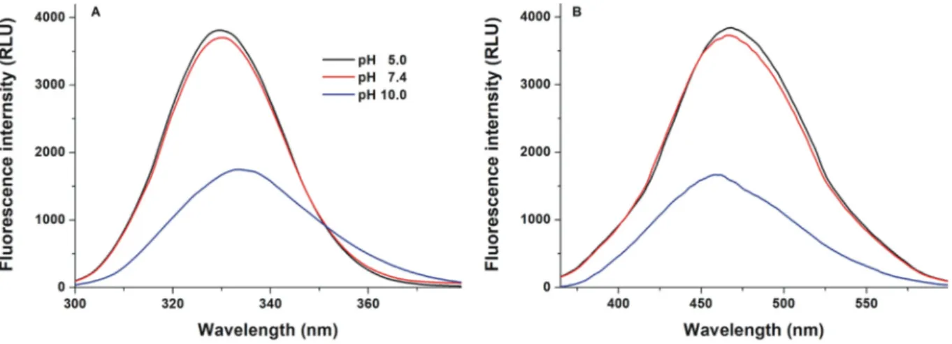
First, thefluorescence excitation and emission spectra of Z14S were recorded in different buffers (pH 5.0, 7.4, and 10.0;Fig. 2). The excitation wavelength maximum of Z14S was noticed around 330 nm under weakly acidic (pH 5.0) and physiological (pH 7.4) circumstances. How- ever, at pH 10.0, a slight red shift of the maximum (330 nm→334 nm) and a significant decrease in the fluorescence signal were noticed (Fig. 2A). Furthermore, at pH 5.0 and 7.4, the emission spectra of the
mycotoxin were similar, while a blue shift of the wavelength maximum (465 nm→458 nm) and the decreased emission signal were observed at pH 10.0 (Fig. 2B). Since the protonation state can influence the ab- sorption andfluorescence spectra of thefluorophores [34–36], these spectral changes are likely resulted from the deprotonation of the myco- toxin at the highest pH tested (similarly to zearalenone, zearalenols, and zearalenone-14-glucoside [17,37,38]): The phenolic hydroxyl group of Z14S on C16 may lose its proton under alkaline conditions.
3.2. Effects of cyclodextrins on the fluorescence emission signal of zearalenone-14-sulfate
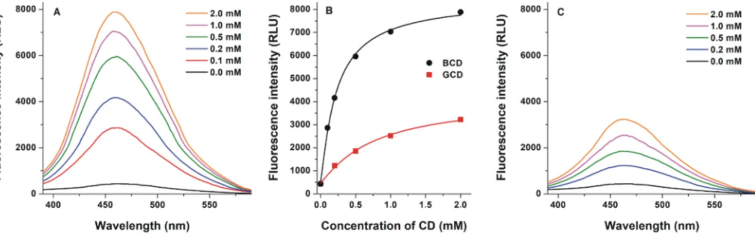
Fluorescence spectra of Z14S were also examined in the presence of nativeβ- andγ-CDs at pH 5.0. CDs induced a blue shift in the excitation wavelength maximum of Z14S (data not shown); therefore, emission spectra were recorded using 320 nm excitation wavelength. In the pres- ence of CDs, thefluorescence emission signal of the mycotoxin was con- siderably increased and a slight blue shift of the emission wavelength maximum (465 nm→460 nm) was observed (Fig. 3). Typically, the in- clusion of organicfluorophores byβ-CDs results in a blue shift in their fluorescence emission wavelength maxima [34,35]. This fact and the ob- servation that CDs alone did not exert significantfluorescence under the applied conditions suggest the formation of host-guest type Z14S-CD complexes. Thefluorescence enhancement observed can be explained by the decreased quenching effect of water molecules due to the inclu- sion of Z14S in the nonpolar CD cavity [39,40]. BCD induced higherfluo- rescence enhancement of the mycotoxin (Fig. 3B) and formed more stable (approximately three-fold; see details later inSection 3.3) com- plexes with Z14S than GCD. Thus, Z14S favors the smaller cavity of BCD vs. GCD, similarly to zearalenone [37]. Interestingly, the masked mycotoxin zearalenone-14-glucoside showed the opposite [17]. In the following experiments, BCD and its chemically modified derivatives were studied further.
3.3. Interaction of zearalenone-14-sulfate with native and chemically mod- ified cyclodextrins in different buffers
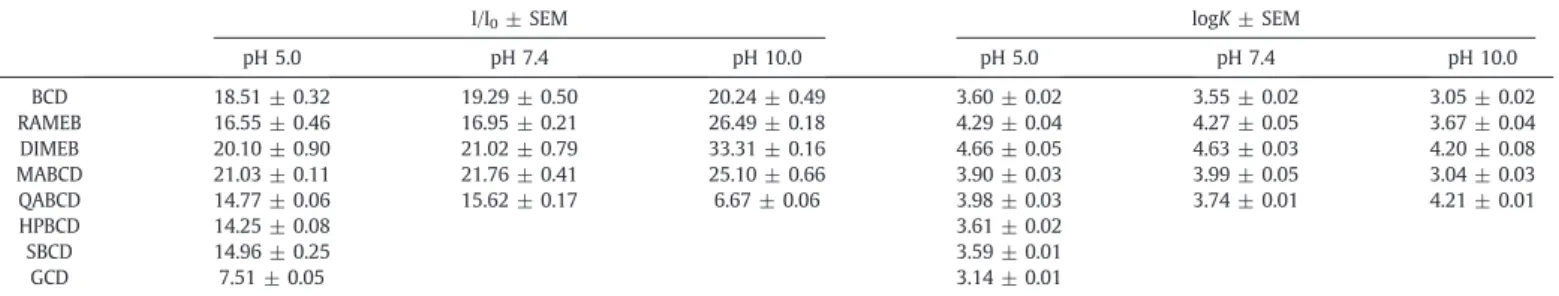
Emission spectrum of Z14S was recorded in the presence of increas- ing CD concentrations in different buffers. At pH 5.0,β-CDs induced 14- to 21-fold increase in the emission signal of the mycotoxin at 460 nm (Fig. 4 andTable 1). Monoamino- (MABCD) and dimethyl-β-CDs (DIMEB) caused stronger while other derivatives resulted in weaker fluorescence enhancement than the native BCD (Fig. 4). Binding con- stants of Z14S-CD complexes were determined using the Benesi- Hildebrand equation (Eq.(1)). Benesi-Hildebrand plots showed excel- lentfitting (R2= 0.990–0.999) with the 1:1 stoichiometry model
Fig. 2.Fluorescence spectra of Z14S (25μM). Excitation (A; pH 5.0 and pH 7.4:λem= 465 nm; pH 10.0:λem= 458 nm) and emission (B; pH 5.0 and pH 7.4:λex= 330 nm; pH 10.0:λex= 334 nm) spectra of the mycotoxin in different buffers (0.05 M sodium acetate, pH 5.0; 0.05 M sodium phosphate, pH 7.4; 0.05 M sodium borate, pH 10.0). (For interpretation of the references to colour in thisfigure legend, the reader is referred to the web version of this article.)
(Fig. S1). Theβ-CDs examined, formed stable complexes with Z14S (logK= 3.6 to 4.7) at pH 5.0 (Table 1). Hydroxypropyl- (HPBCD) and sulfobutyl-substitution (SBCD) did not affect the affinity ofβ-CD toward Z14S; therefore, these derivatives were not tested further. MABCD and QABCD complexes showed approximately two-fold higher binding con- stants vs. Z14S-BCD. Furthermore, the methylatedβ-CDs, namely DIMEB and RAMEB formed the most stable complexes with the myco- toxin (approximately 11- and 5-fold compared to BCD, respectively) (Table 1). As it has been reported, the masked mycotoxin zearalenone-14-glucoside formed less stable complexes with BCD (logK= 2.8) than Z14S [17]; however, the binding constants of Z14S- BCD (logK= 3.6) and zearalenone-BCD (logK= 4.0) [37] complexes were similar at pH 5.0. Furthermore, the stability of Z14S complexes with RAMEB and DIMEB is practically identical to zearalenone com- plexes; however, SBCD is less while QABCD is more suitable for the en- trapment of Z14S compared to zearalenone [37].
The interaction of Z14S with BCD, RAMEB, DIMEB, MABCD, and QABCD were also investigated at pH 7.4 and 10.0. The absolutefluores- cence intensities of Z14S-CD complexes were the highest at pH 5.0 (see inFig. 4). Furthermore, the relative enhancement in thefluorescence of Z14S by CDs is demonstrated inTable 1. At pH 7.4, the CD-induced rel- ative increase influorescence was similar to observed at pH 5.0. Despite the fact that the absolute intensities of Z14S-CD complexes were the
lowest at pH 10.0 (see representative spectra in Fig. S2), the relative en- hancement was significantly higher under these conditions (Table 1).
The only exception was Z14S-QABCD complex, which showed poor emission signal at pH 10.0 (Fig. S2, bottom right). Under alkaline cir- cumstances, the phenolic hydroxyl group of Z14S likely becomes deprotonated which results in a blue shift in its emission maximum and the significant decrease in its emission signal (see inFig. 2). CDs tested (except QABCD) behaved similarly and likely favor the proton- ated form of the mycotoxin. Because the same emission wavelength maximum (approximately at 460 nm) was observed at each pH exam- ined, it is reasonable to hypothesize that most of the CDs formed stable complexes with the same (protonated) form of Z14S. This hypothesis is supported by the following observations: (1) the relative increase in the fluorescence is larger at pH 10.0 due to the higherfluorescence signal of protonated Z14S (since CDs produce more stable complexes with this form, the equilibrium changes in favor of the protonated Z14S);
(2) the higher relativefluorescence enhancement accompanied with significantly lower binding constant at pH 10.0, because CDs form com- plexes with the protonated mycotoxin which is presumably not the dominant form under alkaline conditions. On the other hand, QABCD seems to favor the deprotonated form based on the following observa- tions (Fig. S2): (1) At pH 10.0, the emission wavelength maximum of Z14S-QABCD (447 nm) is different from the other CD complexes tested;
(2) Z14S-QABCD complex has much lower emission signal than other complexes at pH 10.0, despite the fact that it was an effective enhancer at pH 5.0 and 7.4; (3) Z14S-QABCD shows the highest stability at pH 10.0. Since QABCD contains cationic substituents (tetraalkylammonium moieties), it is reasonable to hypothesize that its ionic interaction with the deprotonated mycotoxin may stabilize the formed complex.
3.4. Molecular modeling studies
Z14S showed a similar mode of binding regardingβ- andγ-CD (Fig. 5), even though the capability to get sunk within the diverse cavi- ties was found dependent on the internal radius of CDs. Specifically, the interaction with either BCD or DIMEB was more superficial, while it was deeper within the GCD (whose internal radius is higher in comparison to that of BCD and DIMEB). The analysis of the binding poses showed that Z14S arranged the aliphatic ring within the hydrophobic core of CD cavity, while it used the sulfate and keto groups to engage the large border of the CDs with polar contacts. The binding poses calcu- lated in the light of the affinity to CDs observed experimentally (DIMEB≫BCDNGCD) suggested the stark importance of hydrophobic interactions to stabilize the Z14S-CD complexes. Indeed, the 2-O- methylation regarding DIMEB was thought providing an additional Fig. 3.Fluorescence emission spectra of Z14S (1μM) in the presence of increasing BCD (A) and GCD (C) concentrations (0.0–2.0 mM) in 0.05 M sodium acetate buffer (pH 5.0;λex= 320 nm). (B) BCD- and GCD-induced increase in thefluorescence of Z14S (λex= 320 nm,λem= 460 nm).
Fig. 4.Fluorescence emission intensities of Z14S (1μM) in the presence of increasing CD concentrations (0–2 mM) in 0.05 M sodium acetate buffer (pH 5.0;λex= 320 nm;
λem= 460 nm).
hydrophobic patch that could better stabilize the interaction with the aromatic ring of Z14S in comparison to either BCD or GCD. Concerning the diverse affinity between BCD and GCD observed experimentally, it could be partially explained by the differential sinking of Z14S within the two hydrophobic cavities. In particular, keeping in mind that keto group in position #7 and the 2-oxabicyclo group represent hydrophilic patches in the aliphatic ring of Z14S, the more superficial interaction with BCD is likely more favored than that within GCD, wherein both groups are deeper included within the hydrophobic core of GCD's cavity.
3.5. Interaction of zearalenone-14-sulfate with solubleβ-cyclodextrin polymers
We also tested the interactions of the mycotoxin with soluble (epi- chlorohydrin cross-linked) BCD and QABCD polymers at pH 5.0 and 10.0 (the soluble polymer of DIMEB is not available). Thefluorescence emission spectrum of Z14S (1μM) was recorded in the presence of in- creasing concentrations of soluble polymers. In order to apply compara- ble CD concentrations with the previous investigations, the polymer was normalized to its CD“monomer”content (0.0, 0.05, 0.1, 0.2, 0.5,
1.0, and 2.0 mM). Sometimes the CD polymers have higher affinity to- ward guest molecules due to the cooperativity of the monomers [22,41,42]. Therefore, the binding constants of the formed complexes were calculated based on the CD content of polymers applied (Eq.(1)), as it has been reported previously [22]. The complexes of Z14S with soluble BCD polymer showed 3.65 (±0.04) and 2.74 (±
0.06) logKvalues at pH 5.0 and 10.0, respectively. These data are in good agreement with our previous results with the BCD monomer (Table 1). The soluble QABCD polymer formed slightly less stable com- plexes with Z14S at pH 10.0 (logK= 3.96 ± 0.03) than the QABCD monomer. However, approximately three-fold higher affinity of the polymer (logK = 4.48 ± 0.01) vs. the monomer was noticed at pH 5.0. These observations suggest that soluble QABCD polymer is suit- able to form highly stable complexes with Z14S at both pH 5.0 and 10.0, in agreement with the data presented inTable 1.
3.6. Extraction of zearalenone-14-sulfate from aqueous solution by insolu- ble (water-swellable)β-cyclodextrin bead polymer
As it has been demonstrated, insoluble CD polymers are suitable for mycotoxin removal from aqueous solutions, including certain beverages Table 1
CD-induced relative increase in thefluorescence emission signal of Z14S (I/I0; 1μM Z14S + 2.0 mM CD) in different buffers (λex= 320 nm,λem= 460 nm). Decimal logarithmic values of binding constants (K; unit: L/mol) of Z14S-CD complexes.
I/I0± SEM logK± SEM
pH 5.0 pH 7.4 pH 10.0 pH 5.0 pH 7.4 pH 10.0
BCD 18.51 ± 0.32 19.29 ± 0.50 20.24 ± 0.49 3.60 ± 0.02 3.55 ± 0.02 3.05 ± 0.02
RAMEB 16.55 ± 0.46 16.95 ± 0.21 26.49 ± 0.18 4.29 ± 0.04 4.27 ± 0.05 3.67 ± 0.04
DIMEB 20.10 ± 0.90 21.02 ± 0.79 33.31 ± 0.16 4.66 ± 0.05 4.63 ± 0.03 4.20 ± 0.08
MABCD 21.03 ± 0.11 21.76 ± 0.41 25.10 ± 0.66 3.90 ± 0.03 3.99 ± 0.05 3.04 ± 0.03
QABCD 14.77 ± 0.06 15.62 ± 0.17 6.67 ± 0.06 3.98 ± 0.03 3.74 ± 0.01 4.21 ± 0.01
HPBCD 14.25 ± 0.08 3.61 ± 0.02
SBCD 14.96 ± 0.25 3.59 ± 0.01
GCD 7.51 ± 0.05 3.14 ± 0.01
Fig. 5.Interactions of Z14S with BCD, GCD, and DIMEB. (A) Representation of the calculated complexes in cut surfaces (CDs) and sticks (Z14S). The dashed line serves as geometrical reference point and it indicates the centroid of hexose sugars of each CD: the inclusion of Z14S is deeper within GCD than within either BCD or DIMEB. (B) Representation of the calculated complexes in sticks. Yellow dotted lines indicate the formation of polar contacts while the grey, blue, and red mesh indicates regions sterically and energetically suitable to receive hydrophobic, hydrogen bond donor and hydrogen bond acceptor groups, respectively. The red ring indicates the position of the 2-O-methylation of DIMEB, which is thought favoring the interaction with the aromatic ring of Z14S. (For interpretation of the references to colour in thisfigure legend, the reader is referred to the web version of this article.)
[21,23]. Zearalenone was successfully extracted from spiked corn beer [23] and the zearalenone-14-glucoside content of aqueous solution was also effectively decreased by BBP [17]. Some chemically-modified CDs have higher affinity toward Z14S than BCD; however, only the BCD polymer is available in insoluble (water-swellable) form. There- fore, the removal of Z14S by BBP was tested in 0.05 M sodium acetate buffer (pH 5.0). In a concentration-dependent fashion, BBP considerably decreased the Z14S content of the solution, resulting in 95% reduction of the mycotoxin concentration in the presence of 20.0 mg/1.5 mL BBP (vs.
1μM Z14S) (Fig. 6A). Based on these results, Z14S can be extracted by BBP with similar efficacy from aqueous solution than zearalenone [23].
In order to describe quantitatively the interaction of Z14S with BBP, increasing Z14S concentrations were added to standard amount of BBP (see details inSection 2.4) in sodium acetate buffer (0.05 M, pH 5.0). The binding ability of BBP was evaluated using the Langmuir (Eq.(2)) and Freundlich (Eq.(3)) isotherms (Fig. 6B). The Langmuir affinity constant (KL) was 0.096 ± 0.051 L/mg and theQ0value was 9.53 ± 4.49 mg/g.
The Freundlich model showed a 0.827 ± 0.019 (mg/g) × (L/mg)1/n Freundlich constant (KF) and a 0.895 ± 0.045 1/nvalue.KLandKFvalues of Z14S-BBP interaction were lower compared to zearalenone-BBP (KL= 0.60 L/mg,KF= 1.16 (mg/g) × (L/mg)1/n) [23], suggesting the slightly lower adsorptive capacity of BBP regarding Z14S vs. the parent mycotoxin. Thenvalue suggests the relatively homogenous sorption of Z14S to BBP.
4. Conclusions
In summary, the interaction of the modified mycotoxin Z14S with native and chemically-modified CDs as well as with soluble CD poly- mers was investigated. Furthermore, the extraction of Z14S from aque- ous solution by insolubleβ-CD bead polymer was examined. Z14S form similarly stable complexes with CDs than zearalenone, and also favors β- vs.γ-CDs. CDs induced strong increase in thefluorescence signal of Z14S: MABCD, DIMEB, and BCD proved to be the most effective en- hancers. The methyl derivatives (DIMEB and RAMEB) formed the most stable complexes with Z14S at pH 5.0 and 7.4. Furthermore, at pH 10.0, Z14S-QABCD showed similarly high stability to the Z14S- DIMEB complex. Soluble BCD and QABCD polymers demonstrated com- parable binding ability regarding Z14S than CD monomers. BBP success- fully decreased the Z14S content of aqueous solution, suggesting its possible suitability as mycotoxin binder. Based on the above-listed ob- servations, CD technology seems to be a promising tool to make more sensitive thefluorescence detection of Z14S as well as to develop
mycotoxin binders which can entrap modified mycotoxins besides the parent compound.
Funding
The project was supported by the Hungarian National Research, De- velopment and Innovation Office (FK125166; M.P., Z.F., and E.F.-N.), and by the GINOP-2.3.2-15-2016-00049 grant (S.K.-M.).
CRediT authorship contribution statement
Zelma Faisal:Methodology, Investigation, Writing - original draft, Writing - review & editing.Eszter Fliszár-Nyúl:Methodology, Investi- gation.Luca Dellafiora:Methodology, Investigation, Writing - original draft, Writing - review & editing.Gianni Galaverna:Methodology, In- vestigation. Chiara Dall'Asta: Methodology, Investigation. Beáta Lemli:Methodology, Formal analysis, Investigation.Sándor Kunsági- Máté:Methodology, Formal analysis, Investigation, Funding acquisition.
Lajos Szente:Conceptualization, Resources.Miklós Poór:Conceptuali- zation, Methodology, Writing - original draft, Writing - review & editing, Supervision, Funding acquisition.
Declaration of competing interest
The authors declare that they have no known competingfinancial interests or personal relationships that could have appeared to influ- ence the work reported in this paper.
Acknowledgements
The authors thank to Katalin Fábián for her excellent assistance in the experimental work. This project was supported by the János Bolyai Research Scholarship of the Hungarian Academy of Sciences (M.P.). In addition, the authors would like to acknowledge Prof. Gabriele Cruciani for the courtesy of FLAP software (www.moldiscovery.com).
Appendix A. Supplementary data
Supplementary data to this article can be found online athttps://doi.
org/10.1016/j.molliq.2020.113236.
Fig. 6.(A) Removal of Z14S (1μM in 1.5 mL volume) from aqueous solution (0.05 M sodium acetate buffer; pH 5.0) byβ-cyclodextrin bead polymer (BBP; incubation: 40 min, 1000 rpm, 25 °C; *pb0.01). (B) Langmuir (solid line) and Freundlich (dashed line) isotherms for the Z14S-BBP interaction.
References
[1] J.I. Pitt, Mycotoxins, in: J.G. Morris Jr., M.E. Potter (Eds.), Foodborne Infections and Intoxications, 4th ed., Volume 30, Academic Press, Cambridge, MA, USA 2013, pp. 409–418.
[2] S. Yazar, G.Z. Omurtag, Fumonisins, trichothecenes and zearalenone in cereals, Int. J.
Mol. Sci. 9 (2008) 2062–2090.
[3] C.M. Maragos, Zearalenone occurrence and human exposure, World Mycotoxin J. 3 (2010) 369–383.
[4] European Food Safety Authority (EFSA), Risks for animal health related to the pres- ence of zearalenone and its modified forms in feed, EFSA J. 15 (2017) 4851.
[5] European Food Safety Authority (EFSA), EFSA Panel on Contaminants in the Food Chain, Scientific opinion on the risks for human and animal health related to the presence of modified forms of certain mycotoxins in food and feed, EFSA J. 12 (2014) 3916.
[6] M. Rychlik, H.U. Humpf, D. Marko, S. Dänicke, A. Mally, F. Berthiller, H. Klaffke, N.
Lorenz, Proposal of a comprehensive definition of modified and other forms of my- cotoxins including“masked”mycotoxins, Mycotoxin Res. 30 (2014) 197–205.
[7] A. Dall’Erta, M. Cirlini, M. Dall’Asta, D. Del Rio, G. Galaverna, C. Dall’Asta, Masked my- cotoxins are efficiently hydrolyzed by human colonic microbiota releasing their aglycones, Chem. Res. Toxicol. 26 (2013) 305–312.
[8] L. Szente, Highly soluble cyclodextrin derivatives: chemistry, properties, and trends in development, Adv. Drug Deliv. Rev. 36 (1999) 17–28.
[9] G. Crini, Review: a history of cyclodextrins, Chem. Rev. 114 (2014) 10940–10975.
[10] L. Szente, J. Szemán, Cyclodextrins in analytical chemistry: host–guest type molecu- lar recognition, Anal. Chem. 85 (2013) 8024–8030.
[11] M. Poór, S. Kunsági-Máté, L. Szente, G. Matisz, G. Secenji, Z. Czibulya, T. Kőszegi, In- teraction of ochratoxin A with quaternary ammonium beta-cyclodextrin, Food Chem. 172 (2015) 143–149.
[12]Q. Wu, J. Xu, H. Xu, Interactions of aflatoxin B1 and related secondary metabolites with native cyclodextrins and their potential utilization, Food Control 94 (2018) 102–107.
[13] Y. Zhou, J. Chen, L. Dong, L. Lu, F. Chen, D. Hu, X. Wang, A study offluorescence prop- erties of citrinin inβ-cyclodextrin aqueous solution and different solvents, J. Lumin.
132 (2012) 1437–1445.
[14] Z. Faisal, S. Kunsági-Máté, B. Lemli, L. Szente, D. Bergmann, H.U. Humpf, M. Poór, In- teraction of dihydrocitrinone with native and chemically modified cyclodextrins, Molecules 24 (2019) E1328.
[15] C. Dall’Asta, A. Faccini, G. Galaverna, R. Corradini, A. Dossena, R. Marchelli, Complex- ation of the mycotoxin zearalenone withβ-cyclodextrin: study of the interaction andfirst promising applications, Mycotoxin Res. 24 (2008) 14–18.
[16] C. Dall’Asta, A. Faccini, G. Galaverna, R. Corradini, A. Dossena, R. Marchelli, Complex- ation of zearalenone and zearalenols with native and modifiedβ-cyclodextrins, J.
Incl. Phenom. Macrocycl. Chem. 64 (2009) 331–340.
[17] Z. Faisal, E. Fliszár-Nyúl, L. Dellafiora, G. Galaverna, C. Dall’Asta, B. Lemli, S. Kunsági- Máté, L. Szente, M. Poór, Cyclodextrins can entrap zearalenone-14-glucoside: inter- action of the masked mycotoxin with cyclodextrins and cyclodextrin bead polymer, Biomolecules 9 (2019) 354.
[18] C.M. Maragos, M. Appell, V. Lippolis, A. Visconti, L. Catucci, M. Pascale, Use of cyclo- dextrins as modifiers offluorescence in the detection of mycotoxins, Food Addit.
Contam. A 25 (2008) 164–171.
[19] D.A. Larionova, I.Y. Goryacheva, C. Van Peteghem, S. De Saeger, Thin-layer chroma- tography of aflatoxins and zearalenones withβ-cyclodextrins as mobile phase addi- tives, World Mycotoxin J. 4 (2011) 113–117.
[20]M. Appell, M.A. Jackson, Synthesis and evaluation of cyclodextrin-based polymers for patulin extraction from aqueous solutions, J. Incl. Phenom. Macrocycl. Chem.
68 (2010) 117–122.
[21]M. Appell, M.A. Jackson, Sorption of ochratoxin A from aqueous solutions usingβ- cyclodextrin polyurethane polymer, Toxins 4 (2012) 98–109.
[22]E. Fliszár-Nyúl, B. Lemli, S. Kunsági-Máté, L. Szente, M. Poór, Interactions of myco- toxin alternariol with cyclodextrins and its removal from aqueous solution by beta-cyclodextrin bead polymer, Biomolecules 9 (2019) 428.
[23] M. Poór, Z. Faisal, A. Zand, T. Bencsik, B. Lemli, S. Kunsági-Máté, L. Szente, Removal of zearalenone and zearalenols from aqueous solutions using insoluble beta- cyclodextrin bead polymer, Toxins 10 (2018) 216.
[24] L. Dellafiora, R. Ruotolo, A. Perotti, M. Cirlini, G. Galaverna, P. Cozzini, A. Buschini, C.
Dall’Asta, Molecular insights on xenoestrogenic potential of zearalenone-14- glucoside through a mixed in vitro/in silico approach, Food Chem. Toxicol. 108 (2017) 257–266.
[25] M. Baroni, G. Cruciani, S. Sciabola, F. Perruccio, J.S. Mason, A common reference framework for analyzing/comparing proteins and ligands. Fingerprints for Ligands and Proteins (FLAP): theory and application, J. Chem. Inf. Model. 47 (2007) 279–294.
[26]E. Carosati, S. Sciabola, G. Cruciani, Hydrogen bonding interactions of covalently bondedfluorine atoms: from crystallographic data to a new angular function in the GRID forcefield, J. Med. Chem. 47 (2004) 5114–5125.
[27] L. Dellafiora, B. Warth, V. Schmidt, G. Del Favero, H. Mikula, J. Fröhlich, D. Marko, An integrated in silico/in vitro approach to assess the xenoestrogenic potential of Alternaria mycotoxins and metabolites, Food Chem. 248 (2018) 253–261.
[28] G. Jones, P. Willett, R.C. Glen, A.R. Leach, R. Taylor, Development and validation of a genetic algorithm forflexible docking, J. Mol. Biol. 267 (1997) 727–748.
[29] L. Dellafiora, M. Marchetti, F. Spyrakis, V. Orlandi, B. Campanini, G. Cruciani, P.
Cozzini, A. Mozzarelli, Expanding the chemical space of human serine racemase in- hibitors, Bioorg. Med. Chem. Lett. 25 (2015) 4297–4303.
[30]J.M. Rollinger, D. Schuster, E. Baier, E.P. Ellmerer, T. Langer, H. Stuppner, Taspine:
bioactivity-guided isolation and molecular ligand-target insight of a potent acetyl- cholinesterase inhibitor from Magnolia x soulangiana, J. Nat. Prod. 69 (2006) 1341–1346.
[31] A. Amadasi, C. Dall’asta, G. Ingletto, R. Pela, R. Marchelli, P. Cozzini, Explaining cyclodextrin-mycotoxin interactions using a‘natural’forcefield, Bioorg. Med.
Chem. 15 (2007) 4585–4594.
[32]P. Cozzini, G. Ingletto, R. Singh, C. Dall’Asta, Mycotoxin detection plays“cops and robbers”: cyclodextrin chemosensors as specialized police? Int. J. Mol. Sci. 9 (2008) 2474–2494.
[33] P. Cozzini, M. Fornabaio, A. Marabotti, D.J. Abraham, G.E. Kellogg, A. Mozzarelli, Sim- ple, intuitive calculations of free energy of binding for protein-ligand complexes. 1.
Models without explicit constrained water, J. Med. Chem. 45 (2002) 2469–2483.
[34]M.V. Enoch, R. Rajamohan, M. Swaminathan, Fluorimetric and prototropic studies on the inclusion complexation of 3,3′-diaminodiphenylsulphone with beta- cyclodextrin and its unusual behavior, Spectrochim. Acta A Mol. Biomol. Spectrosc.
77 (2010) 473–477.
[35] I.V.M.V. Enoch, S. Yousuf,β-Cyclodextrin inclusion complexes of 2-hydroxyfluorene and 2-hydroxy-9-fluorenone: differences in stoichiometry and excited state prototropic equilibrium, J. Solut. Chem. 42 (2013) 470–484.
[36] S. Yousuf, R. Alex, P.M. Selvakumar, I.V.M.V. Enoch, P.S. Subramanian, Y. Sun, Picking out logic operations in a naphthalene b-diketone derivative by using molecular en- capsulation, controlled protonation, and DNA binding, ChemistryOpen 4 (2015) 497–508.
[37] M. Poór, S. Kunsági-Máté, N. Sali, T. Kőszegi, L. Szente, B. Peles-Lemli, Interactions of zearalenone with native and chemically modified cyclodextrins and their potential utilization, J. Photochem. Photobiol. B Biol. 151 (2015) 63–68.
[38] M. Poór, A. Zand, L. Szente, B. Lemli, S. Kunsági-Máté, Interaction ofα- andβ- zearalenols withβ-cyclodextrins, Molecules 22 (2017) 1910.
[39] G. Ramirez-Galicia, R. Garduno-Juarez, M.G. Vargas, Effect of water molecules on the fluorescence enhancement of Aflatoxin B1 mediated by Aflatoxin B1:β-cyclodextrin complexes. A theoretical study, Photochem. Photobiol. Sci. 6 (2007) 110–118.
[40] M. Aghamohammadi, N. Alizadeh, Fluorescence enhancement of the aflatoxin B1 by forming inclusion complexes with some cyclodextrins and molecular modeling study, J. Lumin. 127 (2007) 575–582.
[41] A. Harada, M. Furue, S. Nozakura, Cyclodextrin-containing polymers. 2. Cooperative effects in catalysis and binding, Macromolecules 9 (1976) 705–710.
[42]W. Saenger, Cyclodextrin inclusion compounds in research and industry, Angew.
Chem. Int. Ed. Eng. 19 (1980) 344.