COMPARISON OF POLYMERASE CHAIN REACTION-RESTRICTION ENZYME ANALYSIS
METHOD AND DNA SEQUENCE ANALYSIS RESULTS IN THE IDENTIFICATION OF NON-
TUBERCULOUS MYCOBACTERIA
ÖZGÜRAPPAK1*, SELÇUKTÜRKEL2, NURANESEN1and AY ¸SEAYDANÖZKÜTÜK1
1Department of Medical Microbiology, Dokuz Eylul University,İzmir, Turkey
2Department of Medical Microbiology, Training and Research Hospital, Aksaray University, Aksaray, Turkey
(Received: 9 February 2018; accepted: 3 April 2018)
The typing of non-tuberculous mycobacteria (NTM) is important from a clinical and epidemiological perspective. The polymerase chain reaction-restriction enzyme analysis (PRA) method and DNA sequence analysis method were utilized to target a gene region that codes the 65-kDa heat-shock protein for typing 150 suspected NTM samples isolated from the respiratory tract.Mycobacterium abscessus, Mycobacterium xenopi, Mycobacterium fortuitum, and Mycobacterium peregrinum were most fre- quently found by both methods. Six isolates that could not be defined by the PRA method were defined asNocardia cyriacigeorgica, Nocardia abscessus, andMyco- bacterium intracellulareby DNA sequence analysis. Discordance between the results of the two methods was observed for only one isolate. The isolate that was defined as Mycobacterium gordonaetype 6 by the PRA method was defined asMycobacterium senegalenseby sequence analysis. The PRA method is simple and gives rapid results.
Compared with DNA sequence analysis, it gives consistent and reliable results up to a ratio of 90%. DNA sequence analysis is the gold standard method in which all strains can be defined. However, given our laboratory conditions, its disadvantage is that it takes longer to reach a diagnosis than through the PRA method.
Keywords:non-tuberculous mycobacteria, DNA sequence analysis, restriction enzyme analysis, heat-shock protein
Introduction
Non-tuberculous mycobacteria (NTM) represents a large number of non- pathogenic mycobacterial species other than the Mycobacterium tuberculosis
*Corresponding author; E-mail:ozgurappak@yahoo.com.tr
First published online July 14, 2017
complex (MTC) (Mycobacterium bovis, M. bovis Bacille Calmette–Guérin, Mycobacterium africanum, Mycobacterium canetti, Mycobacterium microti, Mycobacterium pinnipedii, and Mycobacterium caprae) and Mycobacterium leprae[1].
Today, more than 170 types of NTM are found intensely in nature, especially in soil and water sources, including drinking water systems (http://
www.bacterio.net/mycobacterium.html). These may lead to diseases especially in people who undergo immune-suppressive treatment or who suffer from immune deficiency, but NTM infections can also be observed in people with normal immune systems. The NTM can infect many systems, primarily respiratory, and can lead to various clinical pictures [2]. The prevalence of pulmonary NTMs varies among countries, but it is increasing worldwide and becoming an important public health problem, especially for elderly people. Although the exact reason for this increase is not known, it can be explained by the increase in the patient population sensitive to NTM, the use of advanced molecular-based tests, the rise in environmental mycobacterial infection sources, and more awareness among clinicians against NTMs. Because NTMs can be found everywhere as saprophytes, their isolation from the respiratory tract does not show that they are always causative agents. Therefore, the diagnosis should be verified clinically, microbi- ologically, and radiologically [3–5].
NTMs should be defined at a species level, because they are resistant to classic antituberculosis drugs and the different types have different susceptibilities to antibiotics. Additionally, the definition is necessary to enable the collection of epidemiological data. In this study, the polymerase chain reaction (PCR)-restriction enzyme analysis (PRA) method and DNA sequence analysis method were used to define samples isolated from the respiratory tract that were not MTC and were suspected of being NTM. The objective was to compare the results of the two methods and evaluate the epidemiological data collected in our hospital over a specific period.
Materials and Methods Study group
We studied 185 NTM suspect samples that were detected not to be MTC from respiratory tract samples, between 2004 and 2010, at the Dokuz Eylul University Hospital’s Mycobacteriology Laboratory. The study was funded by the university and confirmed by its ethics committee. Isolates from 150 patients were included in the study, after excluding the isolates of repeated samples and those
that were not reproduced after passage. The following were used as quality control strains: Mycobacterium intracellulare type 1 (ATCC 13950), Mycobacterium abscessustip 1 (ATCC 19977),Mycobacterium fortuitumtype 1 (ATCC 6841), Mycobacterium gordonae type 1 (ATCC 14470), Mycobacterium scrofulaceum type 1 (ATCC 19981),Mycobacterium szulgai(ATCC 35799), Mycobacterium kansasii type 1 (ATCC 12478), andM. tuberculosisH37Rv.
Identification of NTM
The MTC and NTM distinction was made by p-nitro-α-acetylamino-β- hydroxypropiophenone (NAP) and p-nitro benzoic acid. Phenotypic and conven- tional methods were used for those that could not be differentiated. An amount of 0.5 ml was taken from BACTEC 12B and MGIT 960 bottles that belonged to NTM-suspected samples, their fresh passage was made to the Löwenstein–Jensen (LJ) medium and their growth feature was controlled.
DNA extraction
Colonies from the LJ mediums, where the growth was detected, were added to a 1-ml physiological saline solution and centrifuged at 11,000×g for 1 min.
A 250-μl 1× TE tampon (10 mM Tris, 1 mM EDTA, pH 8.0) was added to the precipitate and mixed using vortex agitation. Then, it was again centrifuged at 11,000×gfor 1 min and a 250-μl 1×TE tampon was added to the precipitate and incubated at 95 °C for 10 min. The centrifuge processing was repeated and the supernatant portion was maintained at−20 °C as a DNA extract until inclusion in the study.
Polymerase chain reaction (PCR)
The heat-shock protein 65 (hsp65) gene region of NTM was reproduced by the PCR reaction. The mixture, whose total volume was 50μl, included the isolated DNA sample, dH2O, MgCl2(25 mM), dNTP (10 mM), 10×Buffer, Primer 1 (Tb11;
5′-ACCAACGATGGTGTGTCCAT-3′), Primer 2 (Tb12; 5′–CTTGTCGAACCG- CATACCCT-3′), and hot start taq DNA polymerase. This mixture was placed in a“thermal cycler”device (Techne TC-412, Bioblock, Illkirch, France) and subjected to initial denaturation at 95 °C for 5 min. Then, 40 cycles were applied: 94 °C for 1 min, 60 °C for 1 min, 72 °C for 1 min, and again for 10 min. The reaction was stopped at 4 °C and the amplification products were shown at gel electrophoresis [6].
A 50-base pair (bp) DNA reagent was loaded on thefirst and the last wells as an indicator of molecular magnitude and PCR was loaded on the other wells. After applying electrophoresis at 130 V for 45 min, the gel was viewed on a UV transilluminator.
Restriction enzyme analysis
For both enzymes (BstEII andHaeIII), a 10-μl PCR product was added to the mixtures that contained 12.0μl dH2O, 2.5μl 5×buffer, and 0.5μl enzyme. The mixture that containedBstEII was maintained at 60 °C for 1 h and the mixture that containedHae III was maintained at 37 °C for 1 h. After the cutoff period, the patterns were viewed at 4% metaphor gel. The results are available inhttp://app.
chuv.ch/prasite/index.html.
DNA sequence analysis
A 40-μl portion of the PCR products equaling 441 bp in length at agarose gel electrophoresis, at a suitable clearness and viewed as pure band, were saved at 2–8 °C and sent to Korea for purification and double-sided sequencing (Macrogen Inc., Korea). The Tb11 and Tb12 primers used at PCR reaction were used for the sequencing process. The results were e-mailed to us as chromotogram files in ab1 file format and converted to fasta format by the BioEdit (Biological sequence alignment editor; Ibis Biosciences, Inc., Canada) program and recorded. These sequences were compared with the reference mycobacteria hsp65 gene region downloaded from Genbank (National Center for Biotechnology Information, National Library of Medicine, USA) and were aligned using the BioEdit program. The DNA sequences in alignedfastaformat were analyzed using the Basic Local Alignment Search Tool (National Library of Medicine, USA) program, and 150 isolates were defined at the species level.
Results
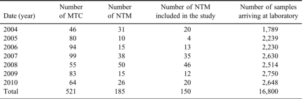
TableIshows a distribution by years of 150 patients’samples, which were not MTC, but were detected to have NTM growth. About 42.7% (64 out of 150) of the isolates belonged to female patients. The sampling distribution was 64 (42.7%) sputum, 73 (48.6%) bronchial lavage, 11 (7.3%) bronchoalveolar lavage, and 2 (1.3%) tracheal secret.
When the sequences obtained by the DNA sequence analysis were com- pared with the standard sequences at the GenBank, 51 of the isolates showed
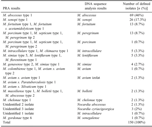
100% compatibility, 94 showed 99%, 1 showed 98%, 3 showed 97%, and 1 showed 95%. Out of 14 different microorganisms that were detected,we had 12 different NTM types and 2 different Nocardia types. The most isolated NTM type was M. abscessus with 69 (46%), whereas the least-defined NTM types were Mycobacterium porcinum, Mycobacterium avium, and Mycobacterium senega- lensewith one isolate each (0.7%).
The PRA method could not define 6 of the 150 isolates.M. abscessustype 1 was the most isolated NTM with 69 isolates (46%) (Figures 1 and2). The six isolates that could not be defined by PRA were defined by the DNA sequence analysis as Nocardia cyriacigeorgica (3/6), Nocardia abscessus (2/6), and M. intracellulare (1/6). The isolate defined asM. gordonaetype 6 by PRA was defined as M. senegalenseby sequence analysis (Table II). The distribution by years of the defined isolates can be seen in TableIII.
Table I.Distribution by years of isolates isolated from clinical samples defined as MTC and NTM
Date (year)
Number of MTC
Number of NTM
Number of NTM included in the study
Number of samples arriving at laboratory
2004 46 31 20 1,789
2005 80 10 4 2,239
2006 94 15 13 2,230
2007 99 38 35 2,630
2008 55 50 46 2,514
2009 83 15 12 2,750
2010 64 26 20 2,648
Total 521 185 150 16,800
Note:MTC:Mycobacterium tuberculosiscomplex; NTM: non-tuberculous mycobacteria.
Figure 1.PCR bands views obtained from NTMs. M: marker (50 bp), 1-12 wells; study samples (expected band size: 441 bp)
Figure 2.PCR-restriction enzyme analysis cutting patterns. M: marker (50 bp); B: band patterns of BstE IIenzyme cutting products; H: band patterns ofHae IIIenzyme cutting products; BI and H1:
KK2 [Mycobacterium abscessus type 1(ATCC 19977)] cutting patterns, for BstE II enzyme 235-201 bp, forHae III enzyme 145-70-60 bp. Other wells; study samples
Table II.Comparison of results of PRA and DNA sequence analysis
PRA results
DNA sequence analysis results
Number of defined isolates [n(%)]
M. abscessustype 1 M. abscessus 69 (46%)
M. xenopitype 1 M. xenopi 26 (17.3%)
M. fortuitumtype 1,M. fortuitum s. acetamidolyticumtype 1
M. fortuitum 13 (8.7%)
M. porcinumtype 1,M. septicumtype 1, M. peregrinumtip 2
M. peregrinum 13 (8.7%)
M. porcinumtype 1,M. septicumtype 1, M. peregrinumtype 2
M. porcinum 1 (0.7%)
M. intracellularetype 1,M. chimaeratype 1 M. intracellulare 5 (3.3%) M. simiaetype 5,M. lentiflavumtype 1,
M.florentinumtype 1
M. lentiflavum 5 (3.3%)
M. genavensetype 2,M. simiaetype 1 M. simiae 4 (2.7%)
M. colombiensetype 1,M. avium s. avium type 2
M. avium 1 (0.7%)
M. avium s. aviumtype 1 M. aviumizolat 2 (1.3%)
M. avium s. Paratuberculosistype 1 M. avium s. Silvaticumtype 1
M. massiliensetype 1,M. bolletiitype 1, M. abscessustype 2
M. bolletii 2 (1.3%)
M. chelonaetype 1 M. chelonaetype 2 (1.3%)
Unidentified 2 isolate Nocardia abscessus 2 (1.3%)
Unidentified 3 isolate Nocardia cyriacigeorgica 3 (2%)
Unidentified 1 isolate M. intracellulare 1 (0.7%)
M. gordonaetype 6 M. senegalense 1 (0.7%)
Total 150 (100%)
Note:PRA: PCR-restriction enzyme analysis.
Discussion
The biochemical tests are not suitable for routine usage in the definition of NTM because they are not practical, require a long time, can only distinguish certain types from each other, and are inadequate in a definition of new mycobacteria types. Nowadays, in-house or commercial molecular-based tests are frequently used in defining types of mycobacteria. The cutting patterns obtained by theBstEII andHaeIII enzymes are type specific and allow for the definition of many mycobacteria that are clinically important. The PRA method is used by many laboratories for being easy, fast, and repeatable [6, 7]. Some commercial systems that use DNA probe technology, such as the AccuProbe system (Hologic GenProbe, San Diego, CA), the INNO-LiPA Mycobacteria system (Fujirebio Europe, Ghent, Belgium), and the GenoType Mycobacterium system (Hain Lifescience, Nehren, Germany), are used in the definition of NTMs.
However, some probes used in the commercial systems are not 100% specific and in addition to what is declared by producers, some cross-reactions can be observed [8,9]. Sequence analysis is the reference method in the definition of mycobacteria and, for this purpose, the 16S rRNA, hsp65, ITS, gyrB, recA, and rpoB gene regions are targeted. Common properties of these gene regions are that they exist in all mycobacteria and include sequences that are well protected from mutation and are variable specific to each type. 16S rRNA and hsp65 are the most important and most frequently used gene regions [10,11]. In this study, we compared the
Table III.Distribution by years of the defined isolates
Identified isolates 2004 2005 2006 2007 2008 2009 2010 Total
M. abscessus 6 3 12 16 26 5 1 69
M. xenopi 1 – – 8 10 5 2 26
M. peregrinum – – – – 2 1 10 13
M. fortuitum 6 1 – 3 3 – – 13
M. intracellulare 1 – – 5 – – – 6
M. simiae – – – 1 2 – 1 4
M. lentiflavum 2 – – – 2 – 1 5
M. avium 1 – – – – 1 1 3
N. cyriacigeorgica 3 – – – – – – 3
N. abscessus – – – 2 – – – 2
M. chelonae – – – – – – 2 2
M. bolletii – – – – – – 2 2
M. senegalense – – – – 1 – – 1
M. porcinum – – 1 – – – – 1
Total 20 4 13 35 46 12 20 150
results obtained by the hsp65 PRA method with the results of the reference method, DNA sequence analysis.
Our DNA sequence analysis results with the PRA method were compati- ble with 95.3% (143/150) and incompatible with 0.6% (1/150). The study by Chimara et al. [12] indicated 90.3% compatibility between the two methods, and the PRA method defined only 1.2% of the samples incorrectly. Sajduda et al.
evaluated 38 reference strains by the PRA method and observed that the dimensions of the cutting patterns were 7 bp smaller than the sequence analysis.
However, they observed that the repeatability of PRA patterns and the accuracy level of the results were high. In the same study, they blindly applied the 64 strains to the traditional biochemical tests, the commercial INNO-LiPA method, and DNA sequence analysis. In the biochemical tests, they found that 14 of the 64 isolates were defined incorrectly, but that correct results were yielded with 78%. Despite that, they observed that the results obtained by the PRA method were compatible with INNO-LiPA by 89% and by DNA sequence analysis by 93.3% [13]. Gürtler et al. [14] identified mycobacteria species with 16S-23S rDNA (ISR)-restriction fragment site polymorphisms rapidly between 2 days and 4 weeks and all identifications were found to be compatible with DNA sequencing. Important advantages of the PRA method are that it can be applied easily, gives results quickly, and obtains over 90% conformity in the results.
However, a big disadvantage of the PRA method is that the band length is evaluated visually, which makes standardization and evaluation of small band patterns (especially cut byHaeIII) difficult. Furthermore, a single base change can lead to the appearance or disappearance of a new cutting area [15]. For instance, a point mutation in one of the cutting pattern regions can shift the typical PRA model ofM. malmoenseto aM. marinum-specific PRA model [16].
The parasite website used for interpreting the PRA cutting patterns is helpful.
However, it does not use the real dimension of manual cutting patterns, but instead uses the patterns reported by publications, which may lead to problems in evaluation. Although the PRA method is useful for many types, it cannot be used for the taxonomy of new types [17]. The restriction enzymes used in this study caused the production of identical cutting patterns for different mycobacteria types (for instance, M. porcinum type 1, M. septicum type 1, M. peregrinum type 2,BstEII 235–210 bp, andHaeIII 140–125–100 bp). Therefore, changing these restriction enzymes with more distinctive enzymes seems necessary for a definite distinction.
Özçolpan et al. [18] defined ten different types in their sequence analysis study that targeted hsp65 and 16S rRNA in the definition of 101 NTM isolates.
M. porcinum(39.60%), Mycobacterium lentiflavum(35.65%), andM. abscessus (5.64%) were the most isolated types. A sequence analysis carried out in four
centers in Turkey defined 17 different types in 90 NTM isolates;M. gordonae (23%),M. abscessus(14%), andM. lentiflavum(10%) were determined to be the most frequently isolated types [19]. Babalık et al. [20] examined 75 NTM cases, 43% of which were defined by the hsp65 PRA method, andM. abscessus(28%), M. avium complex(25%), andM. kansasii(16%) were the most frequently seen NTMs. Velayeti et al. analyzed 96 articles that mentioned 1,751 NTM isolates in the Middle Eastern countries and found thatM. fortuitum was among the slow- growing NTMs andM. avium complexwas among the fast-growing NTMs. In the same study, they analyzed 21 articles published in Turkey between 1993 and 2013 and the frequency order of 280 NTM isolates were defined as follows:M. avium complex (54/280),M. gordonae(53/280),M. fortuitum(48/280), andM. abscessus (45/280) [21]. The NTMs predominantly exist in soil and water, environments that are basic sources of infection. Therefore, the reason for the differences of dominant types between laboratories is considered to be a result of different environmental conditions [22].
In this study, six isolates considered to be NTM could not be defined by the PRA method. These samples for during the period 2004–2007 were routinely processed by the LJ and BACTEC 460 TB systems, and the NAP test was used to make the MTC and NTM distinction. Five isolates suspected of being NTM, which were proved not to be MTC by the NAP test, did not present any band pattern with the BstE II (440-0-0) and Hae III (0-0-0-0) enzymes used in the PRA. The bacteriological and clinical properties of Nocardias are similar to those of myco- bacteria and should additionally be considered for a differential diagnosis. The mycobacteria can be distinguished from each other easily and quickly byBstEII cutting patterns with the PRA, but some publications indicate that other actino- mycetes that also contain Nocardias do not haveBstEII cutting patterns. It has been reported that this property can be used to define nocardias. However, Brunello et al.
[23] have shown that theBstEII (440-0-0) cutting patterns of some mycobacteria (Mycobacterium confluentis, Mycobacterium gilvum, Mycobacterium tusciae, Mycobacterium brumae, Mycobacterium duvalii, M. Szulgai, andMycobacterium gadium) did not exist, just as they did not exist in Nocardia. Besides, revealing that Nocardia ignorataandNocardia cerradoensishaveBstEII cutting patterns makes one think that the estimated definition can be inconvenient. In addition, the fact that Hae III patterns are different than the algorithm defined by Chimara et al. [12]
explains why Nocardias are not defined due to the enzymes used in the PRA method. At 235-120-100 bp, cutting patterns were observed in the other isolate undefined by the PRA method, the BstE II enzyme, but could not be defined because theHaeIII and band patterns were not observed. The sequence analysis defined it as M. intracellulare. Sequence analysis is superior to the PRA in its ability to define these types.
The isolate defined asM. gordonaetype 6 by the PRA method was defined asM. senegalense (99% compatible with the reference sequence) by sequence analysis. The internet site prasite defines four types of M. senegalense.While BstEII cutting patterns ofM. senegalensetype 2 andM. senegalensetype 6 are the same (235/130/85 bp), theirHaeIII cutting patterns are different (140/125/
60/50 bp and 140/120/95 bp, respectively).M. gordonaecan easily be obtained from fresh water, water installations, and laboratory taps. A slow-growing mycobacteria is generally considered a contaminant [5].M. senegalense, how- ever, is a fast-growing mycobacteria and generally leads to infections affecting cattle in East Africa. It is reported to have caused catheter-related bacteremia for a patient with immune deficiency and soft tissue infection related to an aquarium glass cut in a normal healthy child [24, 25]. Although we do not have the patient’s clinical data, defining it asM. senegalenseor wrongly asM. gordonae type 6 would not create a problem for the patient as these do not cause any infection in the respiratory tract.
It is important to know the culture positivity ratios in mycobacteriology laboratories. If the ratios are higher than expected, the laboratory supervisor should take note. Successive positivity in the samples coming from the bron- choscopy unit, with the same factor isolated, could signal a problem in the sterilization of endoscopic material [26]. In addition, the NTMs may lead to pseudo-positivity in the cultures by contamination of the tampons and water used in laboratories. If these pseudo-infections can be detected, people can be prevented from unnecessary treatments.
The lack of evidence related to person-to-person infection and the fact that people generally get infected by NTMs from the environment presents an important problem concerning the studies about the epidemiology of NTMs.
People become exposed to NTMs by drinking tap water, swimming, or inhaling aerosols while taking a shower. To understand whether the NTMs detected in this study are causative agents or contaminants, and their epidemiological meanings, more comprehensive clinical work is needed.
In summary, the PRA method is an easily applicable method that gives results quickly. Compared with DNA sequence analysis, it gives over 90%
compatible and reliable results. For NTM-suspected samples undefined by the PRA, other microorganisms should be considered. DNA sequence analysis is the gold standard method and has defined all our strains. However, as it is not a method we apply directly in our laboratory, a disadvantage is that it takes longer to reach a diagnosis than through the PRA method. For patient management and follow-up treatment, the NTM results should be considered with the clinical findings.
Conflict of Interest The authors declare no conflict of interest.
References
1. Cousins, D. V., Bastida, R., Cataldi, A., Quse, V., Redrobe, S., Dow, S., Duignan, P., Murray, A., Dupont, C., Ahmed, N., Collins, D. M., Butler, W. R., Dawson, D., Rodrı´guez, D., Loureiro, J., Romano, M. I., Alito, A., Zumarraga, M., Bernardelli, A.:
Tuberculosis in seals caused by a novel member of the Mycobacterium tuberculosis complex:Mycobacterium pinnipediisp. nov. Int J Syst Evol Microbiol53, 1305–1314 (2003).
2. Guglielmetti, L., Mougari, F., Lopes, A., Raskine, L., Cambau, E.: Human infections due to nontuberculous mycobacteria: The infectious diseases and clinical microbiology specia- lists’point of view. Future Microbiol10, 1467–1483 (2015).
3. Shah, N. M., Davidson, J. A., Anderson, L. F., Lalor, M. K., Kim, J., Thomas, H. L., Lipman, M., Abubakar, I.: PulmonaryMycobacterium avium-intracellulareis the main driver of the rise in non-tuberculous mycobacteria incidence in England, Wales and Northern Ireland, 2007–2012. BMC Infect Dis16, 195 (2016).
4. Piersimoni, C., Scarparo, C.: Extrapulmonary infections associated with nontuberculous mycobacteria in immunocompetent persons. Emerg Infect Dis15, 1351–1358 (2009).
5. Griffith, D. E., Aksamit, T., Brown-Elliott, B. A., Catanzaro, A., Daley, C., Gordin, F., Holland, S. M., Horsburgh, R., Huitt, G., Lademarco, M. F., Iseman, M., Oliver, K., Ruoss, S., von Reyn, C. F., Wallace, R. J., Winthrop, K.: An official ATS/IDSA statement:
Diagnosis, treatment, and prevention of nontuberculous mycobacterial diseases. Am J Respir Crit Care Med175, 367–416 (2007).
6. Telenti, A., Marchesi, F., Balz, M., Bally, F., Bottger, E. C., Bodmer, T.: Rapid identification of mycobacteria to the species level by polymerase chain reaction and restriction enzyme analysis. J Clin Microbiol31, 175–178 (1993).
7. Verma, A. K., Kumar, G., Arora, J., Singh, P., Arora, V. K., Myneedu, V. P., Sarin, R.:
Identification of mycobacterial species by PCR restriction enzyme analysis of the hsp65 gene an Indian experience. Can J Microbiol61, 293–296 (2015).
8. Tortoli, E.: Microbiological features and clinical relevance of new species of the genus Mycobacterium. Clin Microbiol Rev27, 727–752 (2014).
9. Tortoli, E., Pecorari, M., Fabio, G., Messinò, M., Fabio, A.: Commercial DNA-probes for mycobacteria incorrectly identify a number of less frequently encountered species. J Clin Microbiol48, 307–310 (2010).
10. Kim, H., Kim, S. H., Shim, T. S., Kim, M. N., Chae, G. T., Bai, G. H., Park, Y. G., Lee, S. H., Cha, C. Y., Kook, Y. H., Kim, B. J.: Differentiation ofMycobacterium species by analysis of the heat-shock protein 65 gene (hsp65). Int J Syst Evol Microbiol 55, 1649–1656 (2005).
11. McNabb, A., Eisler, D., Adie, K., Amos, M., Rodrigues, M., Stephens, G., Black, W. A., Isaac-Renton, J.: Assessment of partial sequencing of the 65-kilodalton heat shock protein
gene (hsp65) for routine identification of Mycobacteriumspecies isolated from clinical sources. J Clin Microbiol42, 3000–3011 (2004).
12. Chimara, E., Ferrazoli, L., Ueky, S. Y., Martins, M. C., Durham, A. M., Arbeit, R. D., Leao, S. C.: Reliable identification of mycobacterial species by PCR-restriction enzyme analysis (PRA)-hsp65 in a reference laboratory and elaboration of a sequence-based extended algorithm of PRA-hsp65 patterns. BMC Microbiol8, 48–54 (2008).
13. Sajduda, A., Martin, A., Portaels, F., Palomino, J. C.: hsp65 PCR-restriction analysis (PRA) with capillary electrophoresis in comparison to three other methods for identification of Mycobacteriumspecies. J Microbiol Methods80, 190–197 (2010).
14. Gürtler, V., Harford, C., Bywater, J., Mayall, B. C.: Direct identification of slowly growing Mycobacteriumspecies by analysis of the intergenic 16S-23S rDNA spacer region (ISR) using a GelCompar II database containing sequence based optimization for restriction fragment site polymorphisms (RFLPs) for 12 enzymes. J Microbiol Methods64, 185–99 (2006).
15. Ringuet, H., koua-Koffi, C., Honore, S., Varnerot, A., Vincent, V., Berche, P., Gaillard, J. L., Pierre-Audigier, C.: hsp65 sequencing for identification of rapidly growing myco- bacteria. J Clin Microbiol37, 852–857 (1999).
16. Nolte, O., Haag, H., Hafner, B.: A mutation in the 65, 000 Dalton heat shock proteingene, commonly used for molecular identification of non-tuberculous mycobacteria, leads to the misidentification of Mycobacterium malmoense as Mycobacterium marinum. Mol Cell Probes19, 275–277 (2005).
17. Pourahmad, F., Thompson, K. D., Adams, A., Richards, R. H.: Comparative evaluation of polymerase chain reaction–restriction enzyme analysis (PRA) and sequencing of heat shock protein 65 (hsp65) gene for identification of aquatic mycobacteria. J Microbiol Methods76, 128–135 (2009).
18. Özçolpan, O. O., Sürücüoglu, S., Özkütük, N., Çavu¸so˘ glu, C.: Distribution of nontuber-˘ culous mycobacteria isolated from clinical specimens and identified with DNA sequence analysis. Mikrobiyol Bul49, 484–493 (2015).
19. Günaydin, M., Yanik, K., Eroglu, C., Sanic, A., Ceyhan, I., Erturan, Z., Durmaz, R.:
Distribution of nontuberculous mycobacteria. Ann Clin Microbiol Antimicrob 12, 33 (2013).
20. Babalık, A., Kuyucu, T., Ordu, E. N., Ernam, D., Partal, M., Köksalan, K.: Non-tuberculous mycobacteria infection: 75 cases. Tuberk Toraks60, 20–31 (2012).
21. Velayati, A. A., Rahideh, S., Nezhad, Z. D., Farnia, P., Mirsaeidi, M.: Nontuberculous mycobacteria in Middle East: Current situation and future challenges. Int J Mycobacteriol 4, 7–17 (2015).
22. Cook, J. L.: Nontuberculous mycobacteria: Opportunistic environmental pathogens for predisposed hosts. Br Med Bull96, 45–59 (2010).
23. Brunello, F., Ligozzi, M., Cristelli, E., Bonora, S., Tortoli, E., Fontana, R.: Identification of 54 mycobacterial species by PCR-restriction fragment length polymorphism analysis of the hsp65 gene. J Clin Microbiol39, 2799–2806 (2001).
24. Oh, W. S., Ko, K. S., Song, J. H., Lee, M. Y., Ryu, S. Y., Taek, S., Kwon, K. T., Lee, J. H., Peck, K. R., Lee, N. Y.: Catheter associated bacteremia byMycobacterium senegalensein Korea. BMC Infect Dis5, 107 (2005).
25. Talavlikar, R., Carson, J., Meatherill, B., Desai, S., Sharma, M., Shandro, C., Tyrrell, G. J., Kuhn, S.:Mycobacterium senegalensetissue infection in a child afterfish tank exposure.
Can J Infect Dis Med Microbiol22, 101–103 (2011).
26. Middleton, A. M., Chadwick, M. V., Gaya, H.: Disinfection of bronchoscopes, contami- nated in vitro with Mycobacterium tuberculosis, Mycobacterium and Mycobacterium chelonaein sputum, using buffered peracetic (“Nu-Cidex”) acid solution. J Hosp Infect 37, 137–143 (1997).