The management of symptomatic simple renal cysts in children
C. Koh
a, T. Cserni
a, R. Hawkes
b, A.P. Dickson
a, S. Hennayake
a, D.J.B. Keene
a,*aDepartment of Paediatric Urology, Royal Manchester Children’s Hospital, Manchester, Oxford Road, M13 9WL, United Kingdom
bDepartment of Paediatric Radiology, Royal Manchester Children’s Hospital, Manchester, Oxford Road, M13 9WL, United Kingdom
a r t i c l e i n f o
Article history:
Received 28 July 2017 Received in revised form 30 August 2017
Accepted 2 September 2017 Available online 5 September 2017
Keywords:
Simple renal cyst Sclerotherapy Symptomatic Management Children
a b s t r a c t
Renal cysts are being increasingly diagnosed in children due to the increasing use of ultrasound scanning (USS). Most are asymptomatic, but they may present withflank pain, haematuria, palpable mass or even urinary tract infections or urosepsis. There is a wide range of management options for symptomatic cysts, the choice of which depends on patient factors as well as surgeons’preference. In this case series, we present our experience in the management offive cases of symptomatic renal cysts which have been treated in varying ways, ranging from minimally invasive procedures to drain and sclerose the cysts, to more invasive procedures to excise and deroof the cysts.
Ó2017 The Authors. Published by Elsevier Inc. This is an open access article under the CC BY-NC-ND license (http://creativecommons.org/licenses/by-nc-nd/4.0/).
A review of patients with symptomatic simple renal cysts was undertaken retrospectively from the authors’prospectively main- tained database. Five patients were identified (three female, two male).Table 1summarises the features of thefive patients identi- fied, and their differing management plans.
1. Case reports 1.1. Patient 1
An 11 year old female presented to an adult centre with recur- rent left loin pain and suprapubic pain especially during micturi- tion. Initial ultrasound scan (USS) showed several upper pole cysts on the left, the largest measuring 1.6 cm in diameter, with normal right kidney and bladder (images unavailable). Urodynamic study was normal and showed no vesicoureteric reflux. Dimercapto- succinic acid radionuclide (DMSA) scan showed bilateral upper pole photopaenia which was more marked on the side of the cyst with a split uptake of 48% and 52% in the left and right kidneys respectively.
At 15 years of age, symptoms of persistent recurrent left loin pain led to the decision to proceed with ultrasound guided drainage of the cyst, during which 2 ml of clear serousfluid was aspirated.
This resulted in improvement in loin pain but not complete resolution.
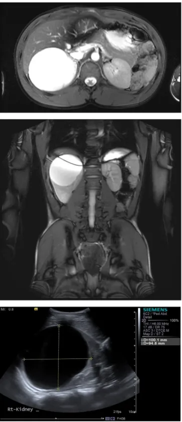
Further magnetic resonance (MR) imaging raised the possibility of a right sidedpara-calyceal cyst (5 mls in volume), along with the left sided cortical cyst which had re-accumulated (9 mls in volume) (Fig. 1). The patient’s symptoms were much improved, and there- fore no further intervention was performed.
1.2. Patient 2
Afive year old female presented to our paediatric urology clinic with a history of lower and upper urinary tract infections, along with backache over the previous year. Upon further questioning a history of daytime urgency and occasional nocturnal enuresis was revealed. She was commenced on prophylactic antibiotics and given lifestyle advice regarding overactive bladder symptoms.
*Corresponding author.
E-mail address:dkeene@nhs.net(D.J.B. Keene).
Contents lists available atScienceDirect
Journal of Pediatric Surgery CASE REPORTS
j o u r n a l h o me p a g e : w w w . j p s c a s e r e p o r t s . c o m
2213-5766/Ó2017 The Authors. Published by Elsevier Inc. This is an open access article under the CC BY-NC-ND license (http://creativecommons.org/licenses/by-nc-nd/4.0/).
https://doi.org/10.1016/j.epsc.2017.09.002
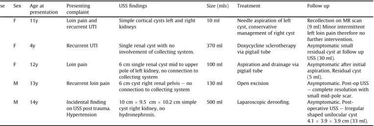
Initial USS performed revealed a large cystic structure measuring 9.78.08.2 cm (370 ml) centrally within the left kidney (Fig. 2) with a markedly dilated central collecting system.
Functional imaging showed a split function of 42% on the left side and 58% on the right side with very good emptying on both sides without any obstruction (Fig. 3).
The scans showed that the collecting system was non-dilated and non-obstructed. Regular monitoring over the subsequent 12 months showed afluctuating but steady increase in size of the cyst, up to 10.410.38.5 cm (Fig. 4). This increase in dimensions was reflected in reported worsening of loin pain, and increase in fre- quency of urinary tract infections.
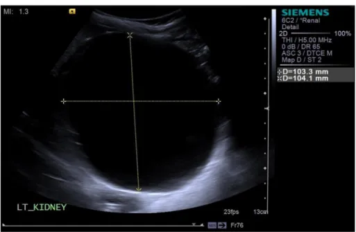
The decision to attempt drainage of the cyst, and delineate the relationship to the collecting system was made. A 6F pigtail tube was inserted under ultrasound guidance and the fluid drained from the cyst. Contrast was then infused under radiological control and no connection with the collecting system was seen (Fig. 5).
A novel use of doxycycline was proposed to sclerose the renal cyst. Current practice within our hospital uses doxycycline as a sclerosing agent for cervico-facial lymphatic malformations in paediatric cases. Using these guidelines, 200 mg (10 mg/ml) of Table 1
Summary of patients’demographic details, diagnosis and management.
Case Sex Age at presentation
Presenting complaint
USSfindings Size (mls) Treatment Follow up
1 F 11y Loin pain and
recurrent UTI
Simple cortical cysts left and right kidneys
10 ml Needle aspiration of left cyst, conservative management of right cyst
Recollection on MR scan (9 ml) Minor intermittent left loin pain therefore no further intervention.
2 F 4y Recurrent UTI Single renal cyst with no
involvement of collecting system.
370 ml Doxycycline sclerotherapy via pigtail tube
Asymptomatic small residual cyst at follow up USS (30 ml).
3 F 12y Loin pain 6 cm single renal cyst mid to upper
pole of left kidney, no connection to collecting system
100 ml Aspiration and drainage via pigtail tube
Asymptomatic after initial aspiration. Residual cyst (5 ml).
4 M 13y Recurrent loin pain 6 cm cyst right renal pelviseno connection to collecting system
130 ml Open excision Asymptomatic. Post-op USS ecomplete resolution with small mid-pole scar.
5 M 14y Incidentalfinding
on USS post trauma.
Hypertension
10 cm9.5 cm10.2 cm simple cyst right kidney, no
hydronephrosis.
500 ml Laparoscopic deroofing. Asymptomatic. Post- operative USSeIrregular shaped unilocular cyst 4.13.93.9 cm (33 ml).
Fig. 1.MR abdomen, showing possible right sidedpara-calyceal cyst, and re-accu- mulated left sided cortical cyst.
Fig. 2.USS showing large 9.78.08.2 cm (370 ml) cystic structure within the left kidney.
C. Koh et al. / J Ped Surg Case Reports 28 (2018) 21e29 22
Fig. 3.Functional imaging showing split function of 42% on the left and 58% on the right. There was good emptying bilaterally without any obstruction.
Fig. 4.Surveillance USS showed that the cyst had increased in size to 10.310.48.5 cm.
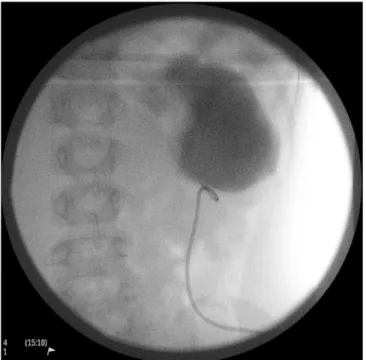
doxycycline was administered via the pigtail tube. The tube was then clamped for 24 h and left to drain for a further 7 days. Repeat USS 7 days later (15 days post-operatively) confirmed that there was a residual cyst (Fig. 6) and therefore further sclerotherapy was performed with the 200 mg of doxycycline used previously. The tube was then removed.
Repeat USS at three and six months following the sclero- therapy showed a small residual cyst which measuring 30 mls in
volume, which was asymptomatic and showed no increase in size. At 24 months post-op, the cyst measured 2.6 ml in volume (Fig. 7).
1.3. Patient 3
A 12 year old female presented with left loin pain and sepsis.
CT imaging confirmed the presence of a large 6 cm cyst in the mid to upper portion of the left kidney (images unavailable).
There was no suspicion on connection to the collecting system.
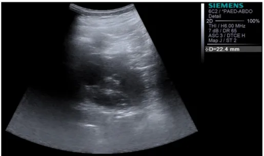
The patient underwent ultrasound guided insertion of an 8F pigtail drain into the renal cyst drainage under general anaes- thetic. A small amount of pus was drained, followed by 100 mls of serousfluid. A pigtail tube was left in place to drain the cyst to completion. Plans were made for alcohol sclerotherapy however unfortunately the drain fell out in the interim. Subsequent scans showed a 2.22.41.9 cm cyst in the left upper pole (Fig. 8).
She remained asymptomatic and did not require further inter- vention to the small residual cyst.
1.4. Patient 4
A 13 year old male presented with a four tofive year history of recurrent generalised abdominal pain which occurred every couple of months and lasting for a two to three days at a time.
This was associated with significant nausea but no vomiting.
Besides this, he did not suffer from any bowel symptoms or abdominal distention, fever or urinary tract infection associated with the pain.
An ultrasound scan performed in the first instance showed a 6.3 cm cyst in the right renal pelvis which was felt to be parapelvic in origin. CT scan confirmed that the cyst was not connected to the calyceal system, but demonstrated calcification on the wall of the cyst (images unavailable). Therefore the decision for complete excision was made.
At age 14 the patient underwent an open complete cyst excision.
Intra-operatively several paracystic calyces were damaged and Fig. 5.Contrast being infused into the cyst, showing no connection with the collecting
system.
Fig. 6.USS at 15 days post op showing the residual cyst, drain in situ.
C. Koh et al. / J Ped Surg Case Reports 28 (2018) 21e29 24
Fig. 7.USS at 3 (1/5/15etop), 6 (4/8/15 - middle) and 24 months (bottom) post op.
repaired. A double J stent was inserted to facilitate drainage of urine. Post operative scan showed no evidence of residual cyst formation, urinoma or upper pole dilatation. The stents were sub- sequently removed.
Histology confirmed a solitary renal cyst with calcification within the wall. The patient had an uneventful recovery with
complete resolution of symptoms. Follow up USS showed a smaller irregular right kidney with a small scar in the mid zone consistent with the surgery. DMSA scan demonstrated that the left kidney contributed 56% function and the right 55%.
Fig. 8.USS showing residual cyst. The patient remained asymptomatic and therefore did not require any further intervention.
Fig. 9.USS showing 7.47.3 cm right upper pole renal cyst.
C. Koh et al. / J Ped Surg Case Reports 28 (2018) 21e29 26
1.5. Patient 5
A 14 year old male was seen in the general paediatric clinic at a district general hospital for investigation of hypertension. This was identified incidentally after his involvement in a road traffic accident. He was otherwise completely asymptomatic and had no history of recurrent urinary tract infections. An initial USS showed a 7.47.36.7 cm renal cyst at the right upper pole (Fig. 9), with no recognisable renal cortex and a bipolar length of 11.8 cm. The left renal bipolar length was 9.3 cm. A DMSA scan showed a normally functioning left kidney with an uptake of 44%, whilst the right kidney had dilated upper moiety with reduction in uptake of 8%, and a non-dilated lower moiety with an uptake of 48% (Fig. 10). This was initially thought to be a duplex system, but further imaging in the form of MR Urogram (Fig. 11) revealed a well circumscribed 8.49.6 9.2 cm cyst with no communication with the renal calyceal system and no solid components or associated vascularity.
He was trialled on Enalapril for approximately 12 months with variable adherence with some improvement in his hypertension.
The decision was therefore made to perform laparoscopic
deroofing of the right upper pole renal cyst, and clearfluid with no debris was drained from the cyst. He was discharged on post- operative day 2 after an uncomplicated recovery. Cytology revealed straw coloured fluids with low cellularity including a few macrophages and some possibly urothelial cells. Histology showed cyst wall lining of a transitional epithelial type, which in places appeared to be attenuated and with atrophic and scarred renal parenchymal tissue present and no evidence of a neoplastic process.
A follow up renal US showed a unilocular cyst measuring 4.13.93.9 cm (Fig. 12). He is asymptomatic and remains under follow up.
2. Discussion
Increasing use of ultrasound scanning has resulted in simple renal cysts being diagnosed with increasing prevalence in children [1]. They are usually unilateral and solitary lesions which are defined as one with normal surrounding renal parenchyma, normal renal function with no associated systemic illness or disorders[2].
Marietti et al. examined the incidence of urological anomalies Fig. 10.Pre-operative DMSA scan showing left kidney contributing to 44% of renal function, whilst right kidney was divided into upper moiety contributing 8% and lower moiety contributing 48% to renal function.
found in emergency paediatric CT scans performed for other causes [3]. Of these 69 of 213 patients were found to have renal cysts. The traditional classification of cysts using computer tomography (CT) findings, known as Bosniak classification [4,5], is rarely used in children due to the high radiation exposure with CT scanning. The vast majority (95%) of children remain asymptomatic with simple renal cysts[6]therefore the management is largely conservative. In children, simple renal cysts tend to slowly increase in size[6]and the frequency of complications are higher in large cysts and young patients.
Symptomatic renal cysts present with discomfort,flank pain, haematuria or a palpable mass related directly to the cyst[6,7].
The surgical strategy in symptomatic patients varies from a minimally invasive procedure, that is a percutaneous puncture with or without sclerotherapy to either marsupialization, total cyst resection, or nephrectomy by laparoscopy or laparotomy[1].
Bayram et al. retrospectively examined 45 children with simple renal cysts[6]. Of these 49% increased in size of, on average, 1% a year. Two of the patient group underwent intervention for serious complications secondary to the cyst; thefirst had a ne- phrectomy for rapid size increase, and the second underwent percutaneous aspiration.
In our series offive patients this trend in surgical strategy can also be seen, with most patients receiving cyst aspiration, lapa- roscopic deroofing or open surgery and one patient receiving percutaneous sclerotherapy. Simple aspiration resulted in recur- rence of cysts in both patients it was used in. Open cyst excision proved a major undertaking with a risk of nephrectomy. The
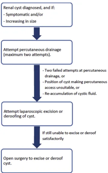
flowchart in Fig. 13 shows the decision
making algorithm for the management of renal cysts in our department.
Different sclerosing agents have been described in the pae- diatric literature, including tetracycline [8], 99% ethanol[9]and hypertonic saline [10]. Paananen treated 32 adults with simple renal cysts with ethanol sclerotherapy and monitored the pa- tients for serum alcohol concentrations, serial cyst measurements and presence of pain [11]. A single patient underwent sclero- therapy of a 6 cm diameter cyst with hypertonic saline [10].
Seven patients (22%) had complete resolution of cyst, where the mean cyst diameter decreased from 7.8 cm to 1.7 cm for the whole group. The use of doxycycline sclerotherapy in cystic lymphatic malformations has been used for several years with good efficacy[12]. Sclerotherapy with tetracycline 150 mg was described in a series of 4 patients with simple renal cysts but none had a cyst as large as the patient described above [8].
The authors describe the use of doxycycline in a child with a large simple renal cyst, avoiding the risks ethanol poses as a sclerosant in terms of systemic absorption[11], calcification[9]
and increase in pain post instillation [11]. The strategy of inserting a nephrostomy tube for delivery, allowed repeated in- stillations (three doses) of doxycycline, seven days apart, until the cyst was seen to shrink, and it avoided further general anaesthetic after the initial nephrostomy placement. The neph- rostomy tube was clamped between instillations allowing it to be concealed easily under clothing and reducing the risk of acci- dental displacement.
Fig. 11.Pre-operative imaging for patient 5 - MR Urogram and USS showing 8.49.69.2 cm cyst with no communication with the renal calyceal system and no solid components or associated vascularity.
C. Koh et al. / J Ped Surg Case Reports 28 (2018) 21e29 28
Conflicts of interest
None. This research did not receive any specific grant from funding agencies in the public, commercial, or not-for-profit sectors.
References
[1] Koutlidis N, Joyeux L, Méjean N, Sapin E. Management of simple renal cyst in children: French multicenter experience of 36 cases and review of the liter- ature. J Pediatr Urol 2015;11(3):113e7.https://doi.org/10.1016/j.jpurol.2015.
03.003.
[2] Bisceglia M, Galliani CA, Senger C, Stallone C, Sessa A. Renal cystic diseases: a review. Adv Anat Pathol 2006;13(1):26e56.https://doi.org/10.1097/01.pap.
0000201831.77472.d3.
[3] Marietti S, Woldrich J, Durbin J, Sparks S, Kaplan G, Chiang G. Urologicfindings on computed tomography of the abdomen and pelvis in a pediatric popula- tion. J Pediatr Urol 2013;9(5):609e12.https://doi.org/10.1016/j.jpurol.2012.
05.014.
[4] Bosniak MA. The Bosniak renal cyst classification: 25 years later. Radiol 2012;
262(3):781e5.https://doi.org/10.1148/radiol.11111595.
[5] Bosniak MA. The current radiological approach to renal cysts. Radiol 1986;
158(1):1e10.https://doi.org/10.1148/radiology.158.1.3510019.
[6] Bayram MT, Alaygut D, Soylu A, Serdaroglu E, CakmakçıH, Kavukçu S. Clinical and radiological course of simple renal cysts in children. Urol 2014;83(2):
433e7.https://doi.org/10.1016/j.urology.2013.08.055.
[7] Canning DA. Re: clinical and radiological course of simple renal cysts in chil- dren. J Urol 2015;193(5):1646e7.https://doi.org/10.1016/j.juro.2015.02.024.
[8] Reiner I, Donnell S, Jones M, Carty HLM, Richwood AMK. Percutaneous sclerotherapy for simple renal cysts in children. Br J Radiol 1992;65(771):
281e2.https://doi.org/10.1259/0007-1285-65-771-281.
[9] Akinci D, Gumus B, Ozkan O, Ozmen M, Akhan O. Single-session percutaneous ethanol sclerotherapy in simple renal cysts in children: long-term follow-up.
Pediatr Radiol 2005;35(2):155e8.https://doi.org/10.1007/s00247-004-1337- [10] Kabaalioy. glu A1, Apaydin A, Ozkaynak C, Melikoglu M, Sindel T, Lüleci E.
Percutaneous sclerotherapy of a symptomatic simple renal cyst in a child:
observation of membrane detachment sign. Eur Radiol 1996;6(6):872e4.
https://doi.org/10.1007/BF00240694.
[11] Paananen I, Hellström P, Leinonen S, Merikanto J, Perälä J, Päivänsalo M, et al.
Treatment of renal cysts with single-session percutaneous drainage and ethanol sclerotherapy: long-term outcome. Urol 2001;57(1):30e3.https://doi.
org/10.1016/S0090-4295(00)00889-X.
[12] Shergill A, John P, Amaral JG. Doxycycline sclerotherapy in children with lymphatic malformations: outcomes, complications and clinical efficacy.
Pediatr Radiol 2012;42(9):1080e8. https://doi.org/10.1007/s00247-012- 2406-2.
Fig. 12.Post-operative USS showing small residual cyst 4.13.93.9 cm.
Fig. 13.Algorithm for the management of symptomatic renal cysts at our institution.