Novel innervation pathways in the regulation of basal forebrain neurons
Ph.D. Thesis
Zsuzsanna Bardóczi
Semmelweis University
János Szentágothai Ph.D. School of Neuroscience
Supervisor: Imre Kalló, M.D., Ph.D.
Official reviewers: István Ábrahám, Ph.D., D.Sc., Habil Zsuzsanna Várnainé Tóth, Ph.D.
Head of the Final Examination Committee: András Csillag, M.D., Ph.D., D.Sc.
Members of the Final Examination Committee: Ádám Dénes, M.D., Ph.D.
Dobolyi Árpád, Ph.D., D.Sc., Habil
Budapest
2018
2 1. Introduction
The basal forebrain (BF) areas (i.e., medial septum (MS), ventral pallidum (VP), diagonal band nuclei (DBN), substantia innominata (Si)/extended amygdala (EA), nucleus basalis of Meynert (NBM) and peripallidal regions) contain a heterogeneous mixture of cell types:
neuropeptide containing neurons such as Gonadotropin-releasing hormone (GnRH)- synthesizing neurons, a heterogeneous collection of cholinergic, GABAergic, glutamatergic projection neurons, and various interneurons.
In the first part of my Ph.D. thesis, we describe our experiments, which examined the potential glycinergic input of GnRH and cholinergic neurons and led to the discovery of morphological and functional connection of cholinergic neurons with glycine transporter- positive cells of the brain stem and the BF.
According to Mesulam and his colleagues, four major subgroups of cholinergic neurons can be distiguished in the BF. The cholinergic neurons, which are located in the MS (Ch1) and vertical limb of the diagonal band of Broca (VDB) (Ch2) project mainly to the the hippocampus. The medial septal cholinergic neurons send axons also to the entorhinal cortex.
The output of cholinergic neurons in the horizontal limb of the diagonal band of Broca (HDB)/magnocellular preoptic nucleus (MCPO) (Ch3) targets mainly the olfactory bulb, where they regulate the activity of various cells and synapses. The Ch3 cholinergic neurons send axons also to the piriform, and entorhinal cortices. The cholinergic neurons, which are distributed in the VP, NBM, SI/EA, globus pallidus, internal capsule (Ch4) project to the basolateral amygdala and innervate the entire neocortex. Furthermore, the cholinergic neurons, which are located in the NBM and DBN, project to the prefrontal cortex (PFC). The BF cholinergic neurons have been implicated in attention, arousal, sleep-wake cycle, learning, memory and olfactory-function.
The GnRH neurons are scattered along the MS, rostral preoptic area (rPOA), ventral anterior hypothalamic area (AHA) and mediobasal hypothalamus (MBH). Most of the GnRH processes terminate in the external zone of the median eminence, where GnRH is secreted in the hypothalamo-hypophysial portal bloodstream, which carries it to gonadotrope cells in the anterior pituitary gland. Under the control of GnRH, luteinizing hormone (LH) and follicles stimulating hormone (FSH) are released from the gonadotropes in a pulsatile manner and they act on the gonads to stimulate gametogenesis and secretion of gonadal steroids. The gonadal steroids exert negative (in females and males) and positive (only in females) feedback effects on the hypothalamo-pituitary unit. In females, most of the estrus cycle estradiol has a suppressive, negative feedback effect on gonadotropin secretion. However, this negative feedback effect switches to a positive feedback and the pattern of LH secretions turns into a
3
non-pulsatile surge. Estrogens’ feedback actions are mediated predominantly via Estrogen Recetor α (ERα), however ERα is absent from the GnRH neurons. Thus, estrogen’s feedback action is mediated to GnRH neurons via ERα-expressing afferents, such as the Kisspeptin (KP) neurons. The hypothalamic KP system appears to be involved in both the pulsatile and surge modes of GnRH release. It consists of two distinct cell populations located within the rostral periventricular area of the third ventricle (RP3V), involving the anteroventral periventricular nucleus (AVPV) and the periventricular preoptic nucleus (Pe), and the arcuate nucleus (Arc). Both of these KP cell populations express ERα and send projections that innervate GnRH-IR neurones; thus, they have the capacity to convey critically important estrogen-dependent signals to GnRH neurons.
According to the classical concept of the the ascending arousal system, a major branch of the complex pathways from the rostral pons and caudal midbrain reaches the hypothalamus and the BF and mediates activating function for these brain regions. Based on preliminary studies showing immunoreactive cell processes for glycine plasma membrane transporter type 1 (GLYT1) and type 2 (GLYT2) and glycine receptor (GlyR) alpha 1 subunit expression in GnRH neurons, our laboratory has postulated the possible existence of an ascending inhibitory (glycinergic) pathway to BF neurons and local action of glycine on BF cholinergic and GnRH neurons. Thus, one of the major aims of my PhD work was to reveal whether GnRH and BF cholinergic neurons are targets of glycine signaling in the BF.
In the second part of my Ph.D. thesis, we demonstrate the experiments, which aimed to investigate the hypothalamic neuronal network regulating reproduction, especially the neuronal projections of GnRH neurons to hypothalamic target cells. Thus, we investigated the ultrastructure of the processes of GnRH neurons and the synaptic connections established in regions, such as the RP3V and/or Arc, which play fundamental roles in the regulation of GnRH secretion. Furthermore, we examined putative changes of the identified neuronal connections under changing hormonal (i.e. lactation) and circadian circumstances. Finally, we examined the existance of KP-KP neuronal interaction in mice and its relevance in human.
The GnRH neurons have long processes (over 1000 µm), which mostly project to the median eminence. These processes have axon- and dendrite-like properties, thus nowadays they are termed as „dendron” by Herde and his colleagues. However, previous anatomical studies provided evidence for axon-like branches of the GnRH neurons in the RP3V and Arc regions, which form synapses on unidentified neuronal populations. We hypothesized that one of the target could be the KP neurons, which populate both the RP3V and Arc and establish a major afferentation to GnRH neurons. The second target was thought to be the hypothalamic dopamine (tyrosine-hydroxylase (TH)-synthesizing) neuronal population, which reside also in
4
the RP3V and Arc. The dopaminergic neurons maintain an inhibitory control on prolactin- secreting cells in the hypophysis; this tonic effect is important for the pulsatile secretion of GnRH, since a reduction of dopamine secretion during lactation results in an increased prolactin level, and consequently, suspension of the pulsatile secretion of GnRH. Variations in the circadian time-points (ZT4–5 and ZT11–12) and lactation may critically influence not only the function of the GnRH neurons, but also their neuronal connections. Thus, we examined potential interaction of GnRH-IR axons with KP and/or TH-IR neurons, and analyzed the identified GnRH appositions onto KP- and/or TH-IR neurons in various hormonal status.
The KP neurons in the Arc play an important role in the regulation of pulsatile release of GnRH. The KP neurons in the Arc, coexpress Neurokinin B (NKB) and Dynorphin Dyn, thus they are termed as KNDy neurons. Several studies in rodents have showed that they are interconnected, and may function synchronously to control the pulsatile secretion of GnRH.
Thus, we have carried out comparative ultrastructural studies on KP neurons to find analogue for interconnection in the human infundibular nucleus (INF).
2. Aims
2.1. Investigating potential target cells of glycine in the BF
2.1.1. GlyR in GnRH and cholinergic neurons
2.1.2. GLYT2-IR afferents to GnRH and cholinergic neurons 2.1.3. Source of glycinergic fibers in the BF
2.1.4. GLYT1-IR astrocytic processes in the vicinity of GnRH and cholinergic neurons.
2.1.5. Membran potential properties of GnRH and cholinergic neurons in the presence of glycine
2.2. Characterization of GnRH efferents and their target cells in mice and humans 2.2.1. Ultrastructural features of GnRH processes in mice
2.2.2. KP-KP contacts in mice and human
2.2.2. Effects of circadian and lactation-related changes on the GnRH input to KP- and TH-IR neurons in mice
5 3. Material and Methods
3.1. Mouse samples
3.1.1. Brain tissue collected for immunohistochemical processing
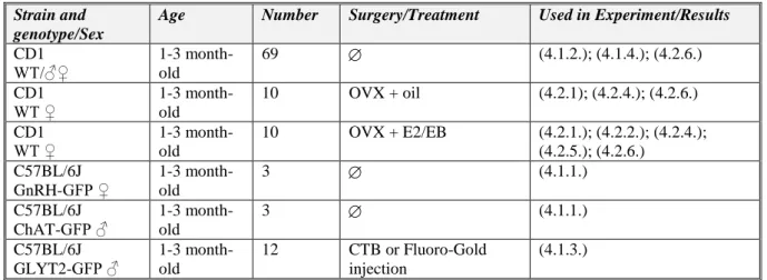
Wild-type (CD1, Charles River) and transgenic mice (1–3 month-old, 25–30 g body weight) were housed under controlled lighting (12 h light/dark cycle; lights on at 7:00 A.M.), and temperature (22°C) conditions with access to food and water ad libitum. The list of animal models used in the different experiments is summarized in Table I. below. A group of animals were ovariectomised (OVX, day 0) and 7 days later (day 7) implanted subcutaneously with a capsule (ID 1.57 mm, OD 3.18 mm) containing either 17β-estradiol (0.625 μg in 20 μl sunflower oil; OVX+E2) or vehicle (OVX+Oil). Three days after implantation (day 10), mice were colchicine-treated (intracerebroventricularly 40 μg in 4 μl 0.9% saline) and 24 h later (day 11) they were sacrificed at either ZT4–5 or ZT11–12. Surgery was performed on animals under deep anesthesia induced by an intraperitoneally injected cocktail of ketamine (25 mg/kg body weight), Xylavet (5 mg/kg body weight), and Pipolphen (2.5 mg/kg body weight) in saline. All studies were performed with permission from the Animal Welfare Committee of the Institute of Experimental Medicine (No. 2285/003), the Debrecen University (No.
6/2011/DE MÁB and 5/2015/DEMÁB), the Eötvös Loránd University (PEI/001/37-4/2015) and in accordance with legal requirements of the European Community (Decree 86/609/EEC).
Table I. Mouse models used in the different experiments.
Strain and genotype/Sex
Age Number Surgery/Treatment Used in Experiment/Results
CD1 WT/♂♀
1-3 month- old
69 (4.1.2.); (4.1.4.); (4.2.6.)
CD1 WT ♀
1-3 month- old
10 OVX + oil (4.2.1); (4.2.4.); (4.2.6.) CD1
WT ♀
1-3 month- old
10 OVX + E2/EB (4.2.1.); (4.2.2.); (4.2.4.);
(4.2.5.); (4.2.6.) C57BL/6J
GnRH-GFP ♀
1-3 month- old
3 (4.1.1.)
C57BL/6J ChAT-GFP ♂
1-3 month- old
3 (4.1.1.)
C57BL/6J GLYT2-GFP ♂
1-3 month- old
12 CTB or Fluoro-Gold injection
(4.1.3.)
Table II. Acute slices collected for electrophysiological examination.
Strain Genotype/Sex
Age Number Surgery/Treatment Used in Experiment/Results
C57BL/6J GnRH-GFP proestrus ♀
1-3 month- old
3 (4.1.5.)
C57BL/6J ChAT-GFP ♂
1-3 month- old
26 (4.1.5.)
6 3.2. Human samples
Human hypothalami were collected at autopsy from the 1st Department of Pathology and Experimental Cancer Research, Semmelweis University, Budapest, Hungary, and the Department of Pathology, Saint Borbála Hospital, Tatabánya. Three-four hours after death, the brains were removed from the skull of a 77-year-old (SKO5) and a 71-year-old (SKO8) female subject and a 55-year-old male individual (SKO7). Ethic permissions were obtained from the Hungarian Medical Research Council /ETT TUKEB 33268-1/2015/ EKU (0248/15) and 31443/2011/EKU (518/PI/11)/ and from the Regional and Institutional Committee of Science and Research Ethics of Semmelweis University (SE-TUKEB 251/2016), in accordance with the Hungarian Law (1997 CLIV and 18/1998/XII.27. EÜM Decree/).
3.3. Preparation of mouse and human sections for light microscopic studies
The mice were perfused transcardially, first with phosphate buffered saline (PBS) solution (10 ml 0.1M PBS; pH 7.4) and then with PBS containing 4% paraformaldehyde (PFA) (100 ml 4% PFA in 0.1M PBS). The brains were post-fixed in 2% PFA/PBS solution for 24h at 4 °C, cryoprotected overnight in 25% sucrose. Serial 30-μm thick coronal sections were cut with a Leica SM 2000R freezing microtome. Human hypothalamic tissue blocks were dissected from the brain of three postmenopausal women (aged 53-88) within 24 h after death. The tissues were rinsed briefly with running tap water and then, immersionfixed with 4% PFA in 0.1 M PBS for 10 days. The tissue blocks were infiltrated with 20% sucrose and cut serially either at 30 or at 100 μm thickness with a freezing microtome. After the endogenous peroxidase activity had been quenched with 0.5% H2O2 (10 min), sections were permeabilized with 0.5%
Triton X-100. Finally, 2% normal horse serum (NHS) was applied (for 20 min) to reduce nonspecific antibody binding. Subsequent treatments and interim rinses in PBS (3X for 5 min) were performed at room temperature, except for incubation in the primary antibody or fluorochrome conjugate, which took place at 4°C.
3.4. Preparation of mouse and human sections for electron microscopic studies Mice were perfused first with PBS (10 ml, 0.1M; ph=7.4), then a mixture of 2% PFA and 4%
acrolein. Brains were postfixed in 2% PFA/PBS solution for 24h at 4 °C. 30-μm thick coronal sections were cut with Leica VTS-1000 Vibratome and treated with 1% sodium borohydride (30 min), 0.5% H2O2 (15 min) and permeabilised with three freeze-thaw cycles, as described previously. Human tissue samples were used from elderly subjects previously shown to contain high levels of KP immunoreactivities. The internal carotid and vertebral arteries were cannulated, and the brains were perfused first with physiological saline (1.5 L for 30 min) containing 5 mL Na-heparin (5000 U/mL), followed by a fixative solution (3–4 L for 2.0–2.5
7
h) containing 4% PFA, 0.05% glutaraldehyde, and 0.2% picric acid in 0.1 M PB. The hypothalami were dissected out and postfixed overnight in 4% PFA without glutaraldehyde.
Fifty-micrometer-thick coronal sections were prepared from the hypothalami with a Leica VTS-1000 Vibratome. 2% normal horse serum (NHS) was applied (for 20 min) to reduce nonspecific antibody binding. Subsequent treatments and interim rinses in PBS (3X for 5 min) were performed at room temperature, except for incubation in the primary antibody which took place at 4°C. After immunohistochemical detection of tissue antigens, the labelled mice and human sections were treated with 1% osmium tetroxide (1 h) and 1% uranyl acetate (in 70% ethanol; 40 min), dehydrated in an ascending series of ethanol and acetonitrile, and flat- embedded in TAAB 812 medium epoxy resin between glass microscope slides pre-coated with a liquid release agent. The resin was allowed to polymerize at 56 °C for 2 days.
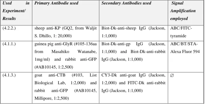
3.5. Single and Multiple labeling of tissue antigens for confocal analyses
Sections of mice or humans were incubated for 72 h in a single primary antibody or a cocktail of two or three primary antibodies (KP, GlyR, GFP, CTB, GnRH, TH). The antigen-antibody complexes were detected by incubating the sections in corresponding FITC-, CY3-, or CY5- conjugated secondary antibodies (12h). In the case of GlyR, the signal was amplified by using biotinylated tyramide (BT) and Alexa Fluor 594 fluorochrome bound to streptavidin. For studying KP contacts in the human INF, FITC-tyramide was employed to amplify the signal.
The immunofluorescent sections were mounted onto glass slides from 0.1 M Tris–HCl buffer (pH 7.6) and coverslipped with an aqueous mounting media (Mowiol).
Table III. Details of single- and multiple labeling for confocal microscopic analysis.
Used in
Experiment/
Results
Primary Antibodie used Secondary Antibodies used Signal Amplification employed
(4.2.2.) sheep anti-KP (GQ2, from Waljit S. Dhillo,1: 20,000)
Biot-Dk-anti-sheep IgG (Jackson, 1:1,000)
ABC/FITC- tyramide (4.1.1.) guinea pig anti-GlyR (#105-136aa
from Masahiko Watanabe, 1mg/ml) and rabbit anti-GFP (#AB10145, 1:2,500)
Biot-Dk-anti-gp IgG (Jackson, 1:1,000) and Biot-Dk-anti-rabbit IgG (Jackson, 1:1,000)
ABC/BT/STA- Alexa Fluor 594
(4.1.3.) goat anti-CTB (#103, List Biological Lab, 1:2,000) and rabbit anti-GFP (#AB10145, Millipore, 1:2,500)
CY3-Dk anti-goat IgG (Jackson, 1:2,000) and FITC-Dk anti-rabbit IgG (Jackson, 1:1,000)
8
(4.2.6.) guinea-pig anti-GnRH (#1018, from Erik Hrabovszky, 1:600,000), rabbit anti-KP (#566, from Alan Caraty, 1:50,000) and chicken anti-TH (#TYH, Aves Laboratories Inc., 1:1,000)
FITC-Dk anti-guinea pig IgG (Jackson, 1:1,000), CY3-Dk anti- rabbit IgG (Jackson, 1, 2,000) and CY5-Dk anti-chicken IgG (Jackson, 1:1,000)
3.6. Single- and dual labelling for light- and electron microscopy
Sections of mice or humans were single or double labelled for light- and electron microscopic examinations. The sections were incubated (72 hours) concurrently in the primary antibodies.
This was followed by visualization of the KP-, GlyR-, GnRH-, GLYT2-, GLYT1-IR structures by incubating the sections sequentially in appropriate biotinylated secondary antibody (12 h) and then Vectastain ABC Elite solution (1: 1,000, 1.5 h). The peroxidase reaction was carried out in the presence of 0.006% H2O2 and nickel-diaminobenzidine (NiDAB), and post-intensified with silver-gold (SGI-NiDAB). After this reaction, the KP-, TH-, GnRH-, ChAT-IR structures were detected by incubating sections sequentially in the appropriate biotinylated secondary antibody (1 day) and then Vectastain ABC Elite solution (1: 1,000; 1.5 h). The peroxidase reaction was carried out in the presence of 0.006 % H2O2 and (diaminobenzidine) DAB alone. For light-microscopic examinations, the sections were mounted onto glass slides and cover slipped with the mounting media (Depex). For electron microscopic examinations, the sections were dehydrated and embedded in epoxy resin (see above).
Table IV. Details of single- and dual labeling for light- and electron microscopic analysis.
Used in Experiment/
Results
Primary Antiserum used Secondary Antibodies used Visualization of antigen-antibody complexes (4.2.2.) sheep anti-KP (GQ2, from Waljit
S. Dhillo, 1: 20,000)
Biot-Dk-anti-sheep IgG (Jackson, 1:1,000)
SGI-NiDAB
(4.1.1.) guinea-pig anti-GlyR (#105- 136aa,from Masahiko Watanabe, 1 mg/ml)
Biot-Dk-anti-gp IgG (Jackson, 1:1,000)
SGI-NiDAB
(4.2.5.) guinea-pig anti-GnRH (#1018, from Erik Hrabovszky, 1:600,000) and rabbit anti-KP (#566, from Alan Caraty, 1:100,000)
Biot-Dk-anti-gp IgG (Jackson, 1:1,000) and Biot-Dk-anti-rabbit IgG (Jackson, 1:1,000)
SGI-NiDAB and DAB
9
(4.2.5.) guinea-pig anti-GnRH (#1018, from Erik Hrabovszky, 1:600,000) and chicken anti-TH (#TYH, Aves Laboratories Inc., 1:1,000)
Biot-Dk-anti-gp IgG (Jackson, 1:1,000) and Biot-Dk-anti-chicken IgG (Jackson, 1:1,000)
SGI-NiDAB and DAB
(4.1.2.) rabbit anti-GLYT2 (#N30aa,from Masahiko Watanabe, 1 mg/ml) and guinea-pig anti-GnRH (#1018, from Erik Hrabovszky, 1:600,000)
Biot-Dk-anti-rabbit IgG (Jackson, 1:1,000) and Biot-Dk-anti-gp IgG (Jackson, 1:1,000)
SGI-NiDAB and DAB
(4.1.2.) rabbit anti-GLYT2 (#N30aa,from Masahiko Watanabe, 1 mg/ml) and goat anti-ChAT (#AB144P, Millipore, 1:1,1500)
Biot-Dk-anti-rabbit IgG (Jackson, 1:1,000) and Biot-Dk-anti-sheep IgG (Jackson, 1:1,000)
SGI-NiDAB and DAB
(4.1.4.) goat anti-GLYT1 (#AB1770, Millipore, 1:10,000) and guinea- pig anti-GnRH (#1018, from Erik Hrabovszky, 1:600,000)
Biot-Dk-anti-sheep IgG (Jackson, 1:1,000) and Biot-Dk-anti-gp IgG (Jackson, 1:1,000)
SGI-NiDAB and DAB
(4.1.4.) goat anti-GLYT1 (#AB1770, Millipore, 1:10,000) and goat anti-ChAT (#AB144P, Millipore, 1:1,1500)
Biot-Dk-anti-sheep IgG (Jackson, 1:1,000) and Biot-Dk-anti-sheep IgG (Jackson, 1:1,000)
SGI-NiDAB and DAB
3.7. Tract tracing to identify glycinergic afferents to BF neurons Table V. Summary of tract tracing experiments.
Tracer used
Injection parameters Visualization of
antigen-antibody complexes/Dilution CTB
(0,5%
solution) Fluoro- Gold (2.5–
5.0%
solution)
The following stereotaxic coordinates were used, respectively, with reference to the bregma (B) planes: MS: anteroposterior, +0.61 mm;
mediolateral, +0.0 mm; dorsoventral, -4 mm; HDB: anteroposterior, +0.37 or +0.02 mm; mediolateral, +0.80 or +1.40 mm; dorsoventral, - 4.90 or -5.00 mm (Paxinos, 2013); and VP/SI: anteroposterior, +0.13 mm; mediolateral, +1.20 mm; dorsoventral, -4.25 mm. CTB and Fluoro-Gold were injected via unilateral iontophoresis (5 µA, 7 s on- off) into the BF for 20 min.
In the case of CTB:
FITC-conjugated donkey anti-rabbit IgG (Jackson, 1:1,1000).
Fluro-Gold was not amplified.
10 3.8. Microscopy and data analysis
3.8.1. Correlated Light- and Electron Microscopy
The flat-embedded sections were initially investigated by light microscopy at 60x magnification. The black color of SGI/Ni-DAB-labeled structures were easily distinguishable from the brown, DAB-labeled structures and made the selection of potential contact sites possible in flat-embedded sections. The areas exhibiting appositions of GLYT1-, GLYT2- or GnRH-IR processes on the somatodendritic region of ChAT-, GnRH-, KP- or TH-IR neurons were further processed. Semithin (1 µm) and ultrathin (50–60 nm) sections were cut with a Leica Microsystems ultracut UCT ultramicrotome. The ultrathin sections were collected in ribbons onto Formvar-coated single slot grids, contrasted with 2% lead citrate and examined with a Jeol 100 C Transmission Electron Microscope. The contact sites were identified in serial ultrathin sections.
3.8.2. Confocal laser microscopy
Regions of interest (ROI; 50589 µm2) containing TH- and/or KP-IR neurons in the periventricular regions of the POA (3-6 ROI/Bregma levels to cover the entire area) and Arc (3-7 ROI/Bregma levels) were scanned (to a depth of 19-20 µm) in one side of the selected three sections of the POA or Arc regions. Each perikarya showing TH- and/or KP- immunoreactivity and receiving GnRH afferent(s) has been recorded. Appositions (defined by the absence of any visible gap between the juxtaposed profiles in at least one optical slice) and immunoreactive perikarya were numbered. Both perikaryal and dendritic appositions were counted; dendrites were considered only if their connections to the perikaryon was traceable. To avoid double counting of perikarya or appositions, immunoreactive profiles appearing repeatedly in the overlapping parts of neighboring Z-stacks or neighboring optical slices of the Z-stacks were identified and encoded with the same number.
3.8.3. Mapping and quantification
KP-immunopositive or KP-immunonegative TH-IR cell populations, and TH- immunonegative KP-IR neurons were distinguished in the POA and named as KP-/TH+, KP+/TH+, KP+/TH-, respectively. In accordance with previous studies, the TH-IR neurons in the Arc were immunonegative for KP. The percentage of neurons immunoreactive either for TH or KP, or for both, and the percentage of GnRH appositions onto each of these neurons in the POA were determined and comparisons made for the different models by means of one- way ANOVA, with the post hoc Tukey HSD test. The mean number of GnRH appositions on the different phenotype of cells were also determined. This approach made comparisons
11
between the preoptic and arcuate TH-IR cell populations possible by using two-way ANOVA, with the post hoc Tukey HSD. Statistical significance was defined at p<0.05.
To identify the source of glycinergic input to the BF, the retrograde tracers CTB or Fluoro- Gold were injected into the MS, HDB, VP and SI of mice expressing GFP under the control of GLYT2 promoter. Although the distribution of retrogradely labeled GFP-positive neurons varied from brain to brain depending on the exact location and size of injection sites, there were brainstem areas and nuclei commonly labeled for the tracer.
4. Results
4.1. Examination of glycinergic input of BF neurons
4.1.1. Subnucleus and cell-specific appearance of GlyRs in the BF
Using a panα-GlyR antibody, immunoreactive puncta were detected in all subdivisions of BF and septal-preoptic area. Although many neurons in the septal-preoptic area were immunopositive for GlyR subunits, double-labeling revealed no clear evidence for the presence of this receptor in GnRH neurons. Contrasting the GnRH neurons, GlyR-IR sites were identified in strong association with most cholinergic neurons, suggesting that glycine can directly influence BF cholinergic functions.
4.1.2. Distribution of glycinergic (GLYT2-IR) afferents in the BF and their appositions to GnRH and cholinergic neurons
By immunohistochemical labeling of GLYT2, we detected a high density of glycinergic (GLYT2-IR) axons in the mouse BF and septo-preoptico-hypothalamic region, including the areas where GnRH- and ChAT-IR neurons are distributed. Light microscopy analyses of GLYT2-IR fibers and GnRH- or ChAT-IR neurons in the BF areas demonstrated several axo- somatic and axo-dendritic contacts between them. Analyses of high-power light microscopic images often revealed GLYT2-IR fiber varicosities, with a central non-labeled area indicating embedded processes of target cells and concave joined surfaces indicating axo-spinous connections. These contacts were further investigated at the ultrastructural level. In the case of GnRH neurons, in spite of the relatively frequent appositions seen at light microscopy levels, EM analyses of serial ultrathin sections failed to detect synapses at the contact sites (n=54 investigated). In contrast to the GnRH neurons, 32 synapses were identified with cholinergic profiles at the ultrastructural level. GLYT2-IR axon terminals were found to surround smaller-diameter as well as larger-diameter ChAT-IR dendrites and form symmetric synapses with dendritic shafts (n=20) and perikarya (n=1). In addition, the correlated bright-field and electron microscopic approach and analysis of serial ultrathin sections also revealed axo- spinous connections on somatic (n=2) and dendritic spines (n= 9).
12
4.1.3. Localization of glycinergic neurons projecting to the BF
To identify the source of glycinergic input to the BF, the retrograde tracers CTB or Fluoro- Gold were injected into the MS, HDB, VP, and SI of mice expressing GFP under the control of GLYT2 promoter. We did not inject tracer into the MPA region because of the missing evidence for connection between glycinergic fibers and GnRH neurons. The highest number of double-labeled neurons were in the raphe magnus (25±7.4% of all GFP neurons, n=6).
Retrogradely labeled GFP-positive neurons were also commonly present in the different parts of the pontine reticular nucleus and the gigantocellular reticular nucleus. A few cells could also be detected in the periaqueductal gray.
4.1.4. Glycine transporter 1 immunoreactive astroglial processes in the vicinity of GnRH and cholinergic neurons
Contrasting the dorsal forebrain areas, GLYT1 immunoreactivity in the BF is very strong, especially in the major regions populated by GnRH neurons and/or cholinergic neurons.
GLYT1 immunoreactivity appears as confluent patches. Light microscopy studies revealed that the GnRH and cholinergic neurons were embedded in GLYT1-IR astrocytic microenvironment of the BF. At the ultrastructural level, the presence of GLYT1 immunoreactivity in thin glial processes was confirmed, often adjacent to axon terminals establishing asymmetric or symmetric synapse with the GnRH and cholinergic neurons.
4.1.5. Collaborative studies on membrane events of GnRH and cholinergic neurons related to glycine’s effect
GnRH and cholinergic neurons were tested for direct effects of glycine in the BF by using electrophysiological approaches. In the case of GnRH neurons, whole cell recordings revealed that glycine (4 µM) had no significant effect on the frequency of action potential firing on GnRH neurons of proestrous mice. In contrast to GnRH neurons, electrophysiological recordings support a substantial glycinergic input to cholinergic neurons in all subdivisions of the BF. Approximately 80% of the recorded neurons, selected randomly from the BF displayed bicuculline-resistant, strychnine-sensitive spontaneous IPSCs.
4.2. Characterizing GnRH efferents and their target cells in mice and humans 4.2.1. Ultrastructure of GnRH-IR processes in mice
We identified GnRH-IR axon terminals forming asymmetric synaptic contacts on unlabeled dendrites in the RP3V and Arc. The axon terminals contained small clear vesicles and immunolabeled dense core vesicles and the diameters of axon terminals were about 0.5 µm. In accordance with the recently published paper by Herbison’s lab, we also detected GnRH dendrons in the Arc with the following diameters i.e. 0.712±0.211 µm and 1.62±0.748 µm.
13
Similarly to the axon varicosities and terminals, they contain mitochondria, small clear and dense core vesicles, but were in post-synaptic position by receiving synaptic inputs from unidentified axon terminals.
4.2.2. Analysis of KP contacts in mice
We observed a dense plexus of KP-IR processes within the POA and Arc by confocal microscopy. The KP-IR varicosities were in close contact with other KP-IR somata both in the POA and Arc. Ultrastructural analysis identified KP-IR axon terminals containing large dense core granules and small clear vesicles in the POA and Arc. KP-IR terminals (n=5 in the POA; n=3 in the Arc) were found to form symmetric, axo-somatic synapses with other KP-IR soma.
4.2.3. Analysis of KP contacts in human
Confocal microscopic study of human INF revealed that most of the KP-positive cells were localized to the caudal INF and fewer to the infundibular stalk (InfS). Analysis of serial ultrathin sections identified classical synapses between KP-IR axons and non-labeled cell bodies or dendrites. In these regions, the KP-IR axons often formed contacts also with other KP-IR neurons. These involved axo-dendritic and axo-somatic synapses, as well as axo- axonic apposition.
4.2.4. Confocal microscopic analysis of GnRH-IR processes forming appositions on KP- and/or TH-IR neurons in the RP3V and Arc
GnRH-IR cell bodies were found in the septo-preoptico-hypothalamic continuum, while their processes were seen to course through and form varicosities in the RP3V and MBH two regions which contain KP- and/or TH-IR neurones. Studies using triple-label immunofluorescence identified KP- and/or TH-IR neurones in the RP3V within a plexus of GnRH-IR processes and in the Arc, KP- and TH-IR neurones were found in the vicinity of GnRH-IR processes. Confocal microscopic analysis of the labelled cellular profiles identified GnRH-IR axon varicosities juxtaposed to all three types of labeled neurones (i.e. KP-, TH-, or KP/TH-IR) in the RP3V and to both phenotypes (i.e. TH-IR or KP-IR neurones) in the Arc.
4.2.5. Identifying synaptic targets of GnRH axons
The immunofluorescent identification of appositions between GnRH-IR varicosities and KP- and/or TH-IR neurones in the RP3V and Arc prompted an initial investigation for classical synapses at the ultrastructural level. Analysis of the selected areas in serial ultrathin sections identified 13 cases of either axo-somatic or axo-dendritic synapses between GnRH-IR terminals and KP-IR neurones. In the case of TH-IR neurons, the presence of synaptic specialization has been proved for 12 appositions. The GnRH-IR presynaptic profiles
14
contained abundant small round clear vesicles and a few dense core granules that were heavily labelled with silver grains. All of the 25 synapses detected in the RP3V (n = 8) and Arc (n = 17) were found to be asymmetric.
4.2.6. Hormonal and lactation-related effects on the GnRH input of KP- and TH-IR neurons
4.2.6.1 Circadian effect on GnRH input to KP neurons
We tested whether diurnal changes can influence the quantity of GnRH inputs to KP neurons in the RP3V and Arc. In the animal models used for quantifying the innervation in the RP3V (colchicine-treated OVX+E2 mice) and in the Arc (colchicine-treated OVX+Oil mice), GnRH-IR axon varicosities were seen to contact approximately 25% of the RP3V KP-IR neurones [23.79 ± 6.93% at ZT4–5 (n = 5) and 28.19 ± 4.41% at ZT11–12 (n = 7)] and approximately 50% of the Arc KP-IR neurones [45.99 ± 3.02% at ZT4–5 (n = 5) and 55.13 ± 4.62% at ZT11–12 (n = 5)]; the percentage was unaffected by the different ZT investigated.
4.2.6.2. Effect of Lactation on GnRH input to KP- and/or TH-IR neurons To test for potential plastic changes of the GnRH input to dopaminergic neurons in lactating mothers (postpartum day 11) and in mothers deprived from pups for 24 hours, immunohistochemical triple labeling (for GnRH, TH and KP) and confocal microscopic analyses were conducted. The numbers of GnRH-IR fiber appositions to KP positive as well as KP negative TH-IR neurons (KP+/TH+-IR and KP-/TH+-IR) were analyzed in lactating mothers and compared to the values shown by non-lactating (OVX+E2) mice, and mothers deprived of their pups for 24h.
Lower ratio of TH-IR neurones immunoreactive for KP in the POA of lactating mothers A relatively high percentage of TH neurons was found to be immunoreactive for KP in OVX- E2 mice (57.8±4.3%). This ratio was significantly lower in lactating mice (16.1±5% of all IR cells counted, F= 28.069, p< 0.001; one-way ANOVA, post hoc Tukey). The percentage of neurons single-labeled either for TH (KP-/TH+-IR) or KP (KP+/TH--IR) was in turn significantly elevated in lactating mice (for TH; 39.8±3.7% vs. 72.4±2.9% for all IR cells counted, F=30.986, p< 0.001; one-way ANO-VA, post hoc Tukey, for KP; 2.4±0.8% vs.
15.4±4.1% for all IR cells counted, F=5.056, p= 0.03; one-way ANOVA, post hoc Tukey).
Removing the pups from the litter for 24 hours did not change the co-localization percentages in the mothers (17.3±4.6% of all IR cells counted).
15
Effect of Lactation or Pup-deprivation on the Number of GnRH-IR Fiber Appositions onto TH-IR Neurons
Using confocal microscopic analyses, varicose GnRH-IR fibers were observed in apposition to all the three phenotypes of labeled neurons (i.e. KP+/TH+, KP-/TH+, or KP+/TH-) in the POA and to the TH-IR neurons (i.e. KP-/TH+) in the Arc. Mothers showed a significantly elevated percentage of GnRH-IR appositions to KP-/TH+ neurons in the POA (68.5±4.3% for lactating mice, 72.3±6% for pup-deprived mothers vs. 48.2±3.2% for the OVX+E2 mice, F=
8.834, p=0.006; one-way ANOVA, post hoc Tukey). In contrast, KP+/TH+ neurons received a significantly reduced GnRH innervation in the same groups of animals (13.1±3.8% and 12.4±3.7% vs. 49.3±3.4%, respectively, F=36.225, p< 0.001; one-way ANOVA, post hoc Tukey). The net result was a small reduction in the percentage of GnRH appositions on the entire population of preoptic TH-IR neurons of lactating female mice compared to non- lactating animals (F=4.946, p< 0.032; one-way ANOVA, post hoc Tukey). Removing the pups from the litter caused no significant changes in the number of contacts. The percentages of GnRH appositions targeting the TH-IR neuron population (KP+ and KP-) were high (96.9±1.5%, 81.6 ±4.1%, 84.8±5.9% in the POA of OVX+E2 mice, lactating or pup-deprived mothers, respectively). The mean number of GnRH appositions per TH-IR neuron did not differ among the experimental groups, but its value was significantly lower in the POA than in the Arc for each experimental group (F=34.043, p< 0.001; two-way ANOVA, post hoc Tukey).
5. Conclusion
In the first part of my PhD work, we examined the potential target cells of glycine signalling in the BF. Thus, we performed morphological and functional examinations to identify the target cells and areas in the BF.
We investigated the putative presence of GlyR in GnRH and cholinergic neurons. We found that all of the BF regions contain GlyRs, including the areas where the GnRH and cholinergic neurons are located. Immunofluorescence labeling revealed no clear evidence for the presence of GlyR in GnRH neurons, whereas the cholinergic neurons were positive for GlyR in all BF regions. Using double immunostaining, we were not able to confirm the presence of synaptic specializations between GLYT2-IR axon terminals and GnRH neurons. In contrast to GnRH neurons, cholinergic neurons received GLYT2-IR axon terminals with both axo-somatic and axo-dendritic arrangement. These synapses belonged to the symmetric category. We found the synapses frequently on more distal branches, indicating less powerful but still significant inhibitory influence on target cells, involving plasticity. To identify the source of glycinergic
16
afferents to BF, we used tract tracing examinations. We injected CTB or Fluoro-Gold into different BF regions and analyzed the distribution of double labelled cells in the brainstem of transgenic mice expressing GFP in GLYT2-eypressing cells (i.e. glycinergic neurons). We found that the glycinergic cell bodies located mainly in the Raphe magnus, Gigantocellular formation of the brain stem and Periaquaductal grey. We tested also the distribution of the GLYT1-IR astrocytic processes in the vicinity of GnRH and cholinergic neurons. Double immunostaining confirmed that the GnRH and cholinergic neurons were embedded into the rich network of GLYT1-IR glial processes. At the ultrastructural level, these glial processes were in the vicinity of asymmetric and symmetric synapses onto GnRH or cholinergic neurons, suggesting that the glycine concentration is highly controlled in the extracellular space at these synapses. Finally, we tested the potential effects of glycine on the membrane properties of the GnRH and cholinergic neurons in collaboration with other labs. At whole cell patch clamp recording conditions, we could not detect effects of glycine on the firing of the GnRH neurons. In contrast to GnRH neurons, the cholinergic neurons were inhibited by glycine.
In the second part of my PhD studies, we characterized the GnRH efferents and their target cells in mice and humans. At first, we have studied the ultrastructure of the GnRH-IR processes in the MBH and in the Arc. GnRH-IR processes could be identified with varying diameters (0.712±0.211 µm-1.62±0.748 µm); these processes often established synaptic specializations with axon terminals, as well as the soma and dendritic processes. Thus, some of those clearly function in a postsynaptic arrangement, while others fulfill a pre-synaptic position in the neuronal connections established. The KP neurons play an important role in the regulation of GnRH neurons in both mice and human. We investigated the KP-KP interactions at ultrastructural level in the mouse RP3V and Arc and in the human INF. In mice, we found axo-somatic synapses between KP neurons in both regions. These interactions seem to be phylogenetically conserved, since studies support their existence in rats, sheep, and also in humans. Our observations revealed axo-somatic and axo-dendritic synapses, as well as axo-axonic connections between KP neurons. Our findings indicate that the operation of the KP neurons could be highly synchronized also in humans. However, the neurotransmitters/neuromodulators used by these connections might show certain species- difference, e.g. dynorphin immunoreactivity in the human KP neurons is fairly low. Besides forming the final common pathway of the reproduction and releasing into the hypothalamo- hypophyseal circulation, GnRH neurons innervate hypothalamic areas including the RP3V and Arc. In my PhD work, we examined putative synaptic targets of the GnRH neurons in the RP3V and Arc. We demonstrated the first time synaptic contacts between GnRH and KP- or
17
TH-IR neurons by electron microscopy. Furthermore, we also investigated a potential effect of circadian and lactation on the GnRH afferents to KP- and TH-IR neurons. Using triple labelling immunohistochemistry, we quantified the GnRH appositions on the KP-, TH- and KP/TH IR neurons at different circadian time points and during lactation and lactation but pup-removed conditions in both areas. KP and TH co-localization levels were influenced significantly by the altered hormonal conditions. We observed lower ratio of TH-IR neurons immunoreactive for KP in the POA of lactating mothers. However, the circadian and hormonal changes did not influence the interaction between the GnRH neurons and KP or TH-IR neurons at the studied intercellular connection, time points and subcellular levels.
Taken together, the number of appositions were maintained but one cannot exclude the possibility that plastic changes may occur at other post partum time points, ultrastructural and/or molecular levels.
18 6. List of publications
6.1. List of publications underlying the thesis
1. Szabolcs Takács1 , Zsuzsanna Bardóczi1 , Katalin Skrapits , Balázs Göcz , Viktória Váczi , Zsófia Maglóczky , Iván Szűcs , Gergely Rácz , András Matolcsy , Waljit S Dhillo , Masahiko Watanabe , Andrea Kádár , Csaba Fekete , Imre Kalló , Erik Hrabovszky Post mortem single-cell labeling with DiI and immunoelectron microscopy unveil the fine structure of kisspeptin neurons in humans BRAIN STRUCTURE & FUNCTION 223:(5) pp. 2143-2156. (2018)
1Authors have contributed equally to this work.
2. Zsuzsanna Bardóczi , Tamás Wilheim , Katalin Skrapits , Erik Hrabovszky , Gergely Rácz , András Matolcsy , Zsolt Liposits , Joanna H. Sliwowska , Árpád Dobolyi and Imre Kalló GnRH neurons provide direct input to hypothalamic tyrosine hydroxylase immunoreactive neurons which is maintained during lactation FRONTIERS IN ENDOCRINOLOGY DOI: 10.3389/fendo.2018.00685 11 p. (2018)
3. Bardóczi Z , Pál B , Kőszeghy Á , Wilheim T , Watanabe M , Záborszky L , Liposits Z , Kalló I Glycinergic input to the mouse basal forebrain cholinergic neurons JOURNAL OF NEUROSCIENCE 37:(39) pp. 9534-9549. (2017)
4. Kallo I , Vida B , Bardoczi Z , Szilvasy-Szabo A , Rabi F , Molnar T , Farkas I , Caraty A , Mikkelsen J , Coen CW , Hrabovszky E , Liposits Z Gonadotropin- Releasing Hormone Neurones Innervate Kisspeptin Neurones in the Female Mouse Brain. NEUROENDOCRINOLOGY 98:(4) pp. 281-289. (2013)
6.2. List of other publications
1. Hollo K , Ducza L , Hegyi Z , Docs K , Hegedus K , Bakk E , Papp I , Kis G , Meszar Z , Bardoczi Z , Antal M Interleukin-1 receptor type 1 is overexpressed in neurons but not in glial cells within the rat superficial spinal dorsal horn in complete Freund
adjuvant-induced inflammatory pain. JOURNAL OF
NEUROINFLAMMATION 14: Paper 125. (2017)
2. Jo S , Kallo I , Bardoczi Z , Arrejo e Drigo R , Zeold A , Liposits Z , Oliva A , Lemmon VP , Bixby JL , Gereben B , Bianco AC Neuronal Hypoxia Induces Hsp40- Mediated Nuclear Import of Type 3 Deiodinase As an Adaptive Mechanism to Reduce Cellular Metabolism JOURNAL OF NEUROSCIENCE 32:(25) pp. 8491-8500.
(2012)
3. Kallo I , Mohacsik P , Vida B , Zeold A , Bardoczi Z , Zavacki AM , Farkas E , Kadar A , Hrabovszky E , Arrojo e Drigo R , Dong L , Barna L , Palkovits M , Borsay BA , Herczeg L , Lechan RM , Bianco AC , Liposits Z , Fekete C , Gereben B A Novel Pathway Regulates Thyroid Hormone Availability in Rat and Human Hypothalamic Neurosecretory Neurons PLOS ONE 7:(6) Paper e37860. 16 p. (2012)