Reproduction of Cell Type
IRWIN R. KÖNIGSBERG AND STEPHEN D. HAUSCHKA
Department of Embryology, Carnegie Institution of Washington, Baltimore, Maryland
Introduction
The phrase "reproduction of cell type" may, to some students of de- velopment, seem to contain an internal contradiction. We classify cells on the basis of the specialized cytoarchitecture, biochemistry, and physiol- ogy of the state we define, somewhat arbitrarily, as fully differentiated. Yet cells which have reached this stage rarely divide. T o be sure, under the special circumstances which initiate regeneration, many types of "fully"
differentiated cells do participate in the initial proliferative stages. In so doing, however, they rapidly lose at least those cytological features by which they can be recognized as definitive types of cells.
Therefore, in discussing the manner in which populations of a particu- lar type of cell are augmented, we are in essence treating two distinctly different processes. We must first consider the replication of some progeni- tor cell and second, the processes by which these progenitor cells become specialized.
During vertebrate embryogenesis, and indeed in the immature animal as well, both processes—cell division and cytodifferentiation—occur con- comitantly within the same tissue or organ. It has been assumed that these two processes do not occur in the same cell, but not until relatively recently has it been possible to examine this assumption critically.
Using radioautographic techniques, several investigators have been able to identify the proliferative members of such developing populations (Stockdale and Holtzer, 1961; Wessells, 1964a; Makela and Nossal, 1962;
Young, 1962; Leblond, 1964; Kitiyakara and Angevine, 1963). The con- sensus has been that DNA synthesis occurs only in the progenitor cells:
those cells in which the specialized products specific to the cell type cannot be detected (but see Leblond, 1964).
243
T o date, the most extensive investigations into the proliferative phases associated with the production of differentiated cell types are those of Leblond and his colleagues (recently reviewed in Leblond, 1964). This group has been engaged in examining the growth of a variety of organs in the postnatal rat using a combination of biochemical, radioauto- graphic, and cytological techniques. On the basis of such studies, Leblond (1964) classifies cell populations within the organism as static, expanding, or renewing. Static populations are homogeneous groups of cells in which no mitotic activity can be detected and in which the DNA content re- mains constant. Although neurons fall into this category, other neural elements (e.g., spongioblasts) proliferate, but the frequency of mitosis declines with time. Expanding populations are those in which scattered mitoses can be detected in quantities which account for the increase in total DNA content. In such populations (kidney, pancreas, and adrenal) DNA accumulation is quite rapid immediately after birth but gradually approaches constancy. In these organs, mitosis is not restricted solely to connective tissue cells. Paired labeled nuclei are found in adjacent differ- entiated parenchymal cells (e.g., pancreatic acinar cells, proximal con- voluted tubule cells of the kidney, and follicular cells of the thyroid).
In the last category, the renewing population, an intensive cell prolifera- tion occurs, providing cells in numbers far exceeding those required to account for any increase in the DNA content. In such populations this high rate of cell production is balanced by cell loss. Cell loss may occur either by emigration (e.g., hematopoietic tissue, reticuloendothelial system, and seminiferous tubules) or by cell attrition (e.g., digestive tract, pleural epithelium, and epidermis).
Those tissues in which provision is made for continuous replacement are generally those which suffer the greatest attrition in the organism.
Continuous renewal of these tissues is an adaptation which extends longevity by preventing the accumulation of defects. In the renewing populations mitotic activity is restricted to a stem cell line frequently set apart anatomically from the specialized cells of the tissue. In the epi- dermis, for example, the basal layer of cells consists of actively proliferat- ing cells which are cytologically and biochemically unspecialized. Indi- vidual cells of this basal layer lose their attachment to the underlying basement membrane, presumably because they are forced up into the more superficial layers of the epidermis. Only after losing their associa- tion with the basement membrane do these cells initiate keratin synthesis.
Thus, during embryogenesis and continuing through postnatal life into adulthood, the reproduction of cell type occurs through an inter- mediary—the progenitor cell. We know that these progenitor cells do
not contain detectable quantities of the specific products characteristic of their ultimate fate. We do not know, however, the extent to which their repertoire of developmental responses may have been restricted. From a variety of experimental situations, it is clear that the basal cell of the epidermis and of specialized epidermal structures such as sebaceous glands and hair can undergo specializations characteristic of other epidermal regions (Montagna, 1956). T h e well-known transformation of keratinizing epidermis into mucoid-secreting epithelium by vitamin A is only one ex- ample (reviewed by Fell, 1961).
Whether the basal cells of an adult epidermis can be channeled into specializations other than those in the epidermal repertoire is unknown.
Since these particular transformations are reversible in the tissue as a whole, a distinction is often drawn between the responses of basal cells and true "differentiation." Some authors (cf. Mercer, 1961) prefer to consider them "modulations" (see Weiss, 1950), that is, different physiological states of a single type of cell. The confusion perhaps results from the fact that no distinction is made between the basal cell population and the indi- vidual differentiated descendants of this population. In fact, a basal cell is always channeled into one epidermal specialization or another; when the environment is changed, it is not the specialized cell that reverts. On the contrary, it is another cell, still in the basal layers, still unspecialized, which is influenced by the new conditions (Fell and Mellanby, 1953). In no sense does the same cell modulate between two different specialized states (see Weiss and James, 1955, p. 392).
Regulation of Stem Cell Differentiation
Stem cell populations such as those represented by the basal layers of stratified squamous epithelium are in a steady state. T h e production of new cells balances the continuous loss of cells from the upper layers of the epithelium. By removing one superficial layer at a time, Pinkus (1951, 1952) was able to demonstrate that the removal of as few as four layers of the stratum corneum of epidermis invoked a significant increase in mitotic activity. T o maintain a steady state, loss of cells from the basal layer must occur at a rate comparable to the rate at which new cells are produced.
After a cell moves out of the basal layer it no longer divides, but pre- sumably as a response to its changed location, initiates the synthesis of proteins characteristic of cornified epithelial cells.
An older view held that the shift to synthetic activities of specialization was due to a differential division of a stem cell. This view has been ex- amined by Leblond and his associates (1964) and found to be untenable.
According to the hypothesis, the division of a basal cell would be in some manner unequal. One daughter cell would remain in the basal layer and continue to serve as a stem cell while the other daughter cell would emigrate to progressively more superficial layers. In the process this cell would become progressively more keratinized, die, and be sloughed off.
Such a mechanism, however, is not compatible with the distribution pat- tern of labeled nuclei in radioautographs of esophageal epithelium at progressively longer intervals after the administration of tritiated thymi- dine. During the first 12 hours, all the labeled cells complete mitosis, yet an insignificant number of labeled cells is found in the spinous layer.
After 12 hours, the number of labeled cells in the spinous layer gradually rises. By using criteria such as the proximity of labeled cells and rela- tive grain density of cells, Leblond and his collaborators were able to recognize the daughter cells of a single mitotic division. T h e migration of one or both daughter cells of a pair occurs at random. There is thus no evidence that one cell invariably remains behind and that the other differentiates.
Differentiation does not appear to be related to some special feature of the preceding mitosis. Leblond suggests that migration is initiated simply by the random displacement of a basal cell resulting from population pressures. We would suspect that the decisive cue which triggers differ- entiation is a function of new environmental relationships. It is not known whether loss of contact with the basement membrane or the influ- ences emanating from surrounding spinous cells is the critical factor.
In the differentiation of an epithelial stem cell there is a suggestion that differentiation is directed by the cellular environment, as in embryo- genesis. Although less dramatic than the classic examples of embryonic induction, interactions of a similar nature may continue to play a role in late embryonic, postnatal, and adult life (see Auerbach, 1964). Such interactions include any influence exerted locally by a dissimilar cell which alters and stabilizes biosynthetic activities and eventually the cyto- logical appearance of the responding cell. Although tissue interactions between cells of dissimilar type are more usual, it is not unreasonable to assume that a more differentiated cell might influence the development of a less differentiated cell of the same series. For example, a spinous cell might influence a displaced basal cell.
The Cellular Environment and Stem Cell Differentiation
T h e persistence of an inductive interaction into late embryonic and perhaps early postembryonic life is best exemplified by recent investiga-
tions into the growth and development of the lens. T h e lens contains both a population of differentiating cells and a segregated germinal stem cell line. In this respect, the lens is analogous to the epidermis with which it shares a common embryonic origin. Although differentiation leads ulti- mately to death of the cell in both tissues, differentiated cells in the lens form the definitive lens and are retained. T h e cuboidal germinal cells form an epithelium over the anterior surface of the lens. As individual germinal cells pass the margin of the lens, they start to elongate and to synthesize a (third) specific lens protein, gamma crystalline (Papaconstan-
tinou, 1964, 1965; Takata et al, 1965). Two lines of evidence indicate that as these cells pass the margin they are subjected to influences emanating from the posterior tissue of the eye.
If the lens of a chick embryo is removed and rotated through 180 de- grees before it is replaced in the eye, it will still regulate in time to form a more or less normal lens even if the operation is performed at a rela- tively late stage (J. L. Coulombre and Coulombre, 1963). The germinal epithelial cells, most of which now face the back of the eye, stop dividing and start to elongate; only those epithelial cells at the lens margin con- tinue to proliferate. These cells now migrate both anteriorly and pos- teriorly from this proliferative marginal zone. T h e former re-establish a new lens epithelium while the latter elongate and contribute in normal fashion to the growing lens. T h e interpretation of these results is that the germinal cells, exposed by the operation to the posterior chamber of the eye, respond to influences they would not normally encounter until they had passed the equator of the lens.
An even more direct demonstration of the influence of posterior tissues of the eye upon the developing lens has been obtained using culture tech- niques. Epithelium from the lens of 13-day-old mouse embryos fails to differentiate further when isolated in culture. When the epithelium is combined with isolated neural retina, however, either directly or with an ultrathin Millipore filter interposed, a complete new lens with oriented fibers is formed (Auerbach, 1964; Muthukkaryppan, 1964). Philpott and Coulombre (1965) found that isolated lens epithelium in culture will palisade to a limited extent, but only if the medium contained a protein supplement (serum or ascitic fluid). Such cultured epithelium failed to differentiate further when grafted into the coelomic cavity of 5-day-old chick embryos. These epithelial cells, when transplanted back into a lentectomized eye, continued to elongate and differentiate into lens fibers of more normal appearance.
The neural retina, derived from that part of the optic vesicle which
induces the primary lens placode, apparently continues to influence lens development in later stages. Earlier investigators had suggested that lens induction is a protracted event (reviewed by A. J. Coulombre, 1965a,b).
Stone (1954) has in fact shown that the neural retina influences the forma- tion of a lens from the dorsal iris in Wolffian regeneration in the adult urodele. T h e extent to which these influences exerted at later stages are analogous to the primary inductive event is problematical but not untestable.
Many of our concepts of embryonic induction have changed with the passage of time and with the availability of new analytical approaches.
We know now, for example, that cellular contact is not an indispensable requisite of all inductive events (Grobstein, 1953). It would seem that another time-honored criterion of embryonic induction, namely, that the effect persists in the absence of the inducer, needs re-evaluation. This may be true of the original cells which are induced, but in the further growth of the induced structure differentiation of additional primordial cells may be dependent upon a persistent source of directive influences.
Such influences may be provided by derivatives of the original inductive tissue as seems to be the case in the lens-neural retina dependency dis- cussed above. It is also possible that such influences are exerted by the induced cells themselves. It may be recalled that Mangold and Spemann (1927) demonstrated that medullary plate could, in turn, induce medul- lary plate in gastrula ectoderm. Moreover, specific areas of the medullary plate impose regional specificity on the responding gastrula ectoderm.
E x p e r i m e n t a l A p p r o a c h e s to E m b r y o n i c I n d u c t i o n
The problem of embryonic induction or tissue interaction has been most profitably examined over the past 12 years by an approach which greatly simplifies the experimental design of earlier studies. Moreover, it permits manipulations which were previously either impossible or ex- tremely difficult. These techniques, introduced and developed by Grob stein (1953, 1954), have been applied to a group of inductive events which occur between epithelial primordia of a variety of organs and their sub jacent mesenchymal components. Briefly, the two components are sepa- rated with trypsin and fixed in apposition on either side of an ultrathin
(20 μ) filter with pores of microscopic dimensions. Culture in this fashion permits the normal interaction to occur under conditions which lend them- selves to a variety of analytical approaches.
These studies have fundamentally altered our concept of embryonic
induction. By far the most important contribution has been a demon- stration that inductive effects can be transmitted across short distances.
This means that in certain instances there is a substance that can be iso- lated, purified, and identified. If this can be accomplished, the mechanism of action can be studied.
Grobstein's studies demonstrate that some macromolecular material is entrapped in the filter (Grobstein, 1954, 1963). This material is sensitive to proteolytic enzymes and exhibits some of the staining properties char- acteristic of the general class of mucopolysaccharides. Radioautographic studies indicate that this macromolecular material crosses the membrane (Kallman and Grobstein, 1965); one of these macromolecules has been identified (Kallman and Grobstein, 1964, 1965). None of the materials has as yet been identified with the active factor or factors of the mesen- chymal cell. One encouraging finding recently reported is that a high con- centration of embryo juice or of a particulate fraction of this extract replaces the effects exerted by mesenchymal cells when present in the medium (Rutter et al., 1964). T h e embryo contains a high proportion of mesenchymal cells and it is entirely reasonable that the extract should be active. This finding is encouraging because it indicates that the factor (or factors) is relatively stable. If it were highly unstable, the prospects of its eventual purification would be reduced. In yet another inductive sys- tem—the spinal cord-chondrogenic interaction—an active extract has been prepared from pooled embryonic spinal cords which can replace the living effector tissue (Hommes et al., 1962; Strudel, 1962; Lash et al.,
1962; Lash, 1963). T h e identity of the active component is in doubt presumably because of the difficulty of purifying the trace quantities which are available.
Cytodifferentiation in Cell Culture
Our own inclination has been to study differentiation in still simpler systems. All the single events which occur during the development of an organism are simple and comprehensible. T h e complexity that exists, exists only in the mind of the investigator. T h e value of a simple system is that we can more readily ask a clear and simple question.
In developing a test system, we are fully aware that natural limits exist.
The number of components could be reduced to a point at which the system no longer retains those properties we are interested in studying.
Cytodifferentiation, as Grobstein (1962a) has cautioned, occurs in vivo
"in the context of a higher level of integration and interaction between
diverse tissues." At what time in its history and to what extent can a cell be isolated from the integrative influences which might regulate its differ- entiation in the organism? It was to this question that we initially addressed our experimental approach.
We did not anticipate that the initial question would lead ultimately, as it has, to a consideration of cell and tissue interactions. Our major con- cern was at first with the more crucial issue of whether a single isolated cell could survive, divide, and still retain its capacity for differentiation in culture. The cumulative experiences of many investigators suggested that this was not possible. The many theoretical arguments based on this experience, however, were often constructed without regard for the em- pirical nature of culture approaches. T h e issue involving stability of differentiative function in culture has been discussed at length elsewhere (Königsberg, 1963); additional positive evidence for stability has been dis- cussed more recently (Eagle, 1965). At the present time, it seems of little value to ruminate over issues which are rapidly losing their controversial content.
The choice of tissue, skeletal muscle, was in retrospect not an ideal one because unlike the epitheliomesenchymally derived organs used in the transfilter approach we had no clear insight into the roles of the inter- acting cell types. It did have the advantage, however, that the major cell type of the tissue, the muscle "fiber" was easily characterized on the cyto- logical as well as molecular level.
The first phases of the investigation dealt with the behavior in culture of relatively large numbers of single cells liberated by trypsinization from embryonic chick leg muscle (Königsberg et al., 1960; Königsberg, 1960, 1961a). Although the tissue was taken from advanced embryos, it still con- tained large numbers of mononucleated cells which were, by this criterion, relatively undifferendated (Fig. 1); these mononucleated cells are the predominant type in cell suspensions prepared from embryonic muscle tissue. Whether such disorganized cells, dispersed on a glass surface in a liquid culture medium, continue to differentiate and form typical skeletal muscle fibers was the initial question; we found that they did.
Hence, tissue organization per se is not indispensable to the normal differ- entiation of skeletal muscle cells.
Studying the characteristics of these cultures, we can recognize two distinct phases. During the first period, the cells attach and spread on the surface of the petri dish, and then multiply rapidly, forming a con- fluent sheet of cells by the third or fourth day (Königsberg, 1961a). By gross inspection at least, the cells are devoid of any indication of frank
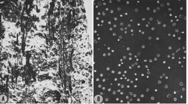
FIG. 1. A. Sagittal section through a thigh muscle from a 12-day-old chick embryo. Fixed in Allen's B-17;
stained in iron hematoxylin. Elongated multinucleate myotubes are clearly in evidence. In addition, large num- bers of mononucleated cells, presumably both myoblasts and fibroblasts, are also present. B. Cell suspension pre- pared by trypsinization (10 minutes, at 37°C in 0.05% 1-300 trypsin) of a mince of leg muscle from a 12-day-old chick embryo. Photographed in phase contrast.
r
>
σ
C M
S H M
>
H
S
muscle differentiation during most of this period. As connuency is ap- proached, however, long, multinucleated, ribbon-like cells resembling myotubes rapidly appear (see Konisberg, 1965b). These are the primitive muscle fibers observed in vivo. Subsequently, the multinucleated cells can be positively identified by virtue of their spontaneous contractility and the presence of cross-striated myofibrils. Concomitant with the attain- ment of connuency, a pronounced transition from exponential growth to a very slow linear increase in cell number (DNA per culture) was observed (Königsberg, 1961a).
P o p u l a t i o n D e n s i t y a n d C y t o d i f f e r e n t i a t i o n
These three phenomena—confluency, cessation of growth, and the ap- pearance of multinucleated myotubes—seemed to be closely correlated.
Moreover, the time of appearance of all three could be shifted simul- taneously by varying the size of the inoculum.
This correlation did not prove, of course, that causal relationship ex- isted between the attainment of confluency and the formation of multi- nucleated cells. If such a relationship did exist, however, there seemed to be two sound and testable alternative mechanisms by which it might operate. T h e attainment of confluency might promote myotube forma- tion simply by reducing intercellular distance, thus increasing the fre- quency of random cell-to-cell collision. This seemed a perfectly plausible explanation since the best available evidence at the time suggested that multinuclearity occurred by a process of successive cell fusions (reviewed in Königsberg, 1965b). Alternately, as confluency was approached, the increased cell density might produce appreciable alteration of the medium as a result of the metabolic activities of the cultured cells. The possibility of such a mechanism had in fact been demonstrated by the work of Earle and his associates (Sanford et al., 1948) who used "conditioned" medium, and by Puck and his collaborators (Puck and Marcus, 1955) who employed
"feeder" layers.
T o discriminate between these alternatives "parabiotic" cultures were set up in which the same medium circulated over both a sparsely seeded and a densely seeded culture. Myotubes formed in both cultures at ap- proximately the same time, and earlier than the event occurred in sparsely seeded controls. T h e next question was obvious. Were alterations in the medium of a stable nature or was the continued presence of a large number of cells required? Not only was the question obvious, but the experimental design was clear.
Replicate cultures of the same cell suspension were subsequently fed either with freshly prepared medium or conditioned medium recovered from older cultures which had already attained confluency. Under the conditions of these experiments, myotube formation occurred 24 hours earlier in conditioned medium (Königsberg, 1962).
Conditioned Medium and the Development of Muscle Clones
We had learned that the normal tissue architecture was not indispen- sable to the subsequent differentiation of myoblasts in vitro. We also knew that differentiation was promoted by some influence of a high popu- lation density and that this influence was transmitted via the medium.
Consideration of whether this population density-dependent influence was at all related to the course of normal myogenesis in vivo was for the moment postponed. Whether physiological or not, was it sufficient to permit the growth and differentiation of single isolated myoblasts?
An elegant series of techniques for cloning animal cells in culture had been devised by Puck's group (Puck et al., 1956). With relatively minor variations, these were applied to freshly isolated embryonic muscle cells.
After a rather tedious empirical examination of the parameters of the procedure for preparing conditioned medium, we devised a schedule which yielded consistent results (Königsberg, 1963). It was abundantly clear that properly conditioned medium was indeed adequate to support the growth and differentiation of colonies of cells descended from single isolated myoblasts.
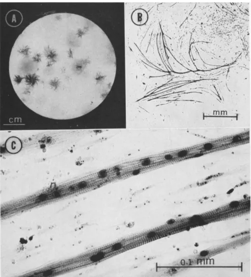
Small numbers of dissociated cells from embryonic muscle were inocu- lated into petri plates containing conditioned medium and cultured for approximately 2 weeks. At the end of this period, a gross examination of fixed and stained cultures revealed the presence of two distinctly different colonial forms (Fig. 2). By microscopic examination, those colonies which have a fibrous appearance can be identified as muscle colonies using the same criteria by which skeletal muscle cells are recognized in vivo (Fig. 3).
That these colonies can indeed originate from a single cell was investi- gated both by following the entire process and by physically confining single cells within small glass cylinders (Königsberg, 1963). Such isolated colonies contain large numbers of the same multinuclear, cross-striated muscle fibers observed earlier in mass cultures and in embryonic muscle fixed and sectioned at the appropriate stage. In addition, we also observed mononucleated cells within the same colony. These mononucleated cells,
|ΙΙΙΙ|ΙΙΙΙ|ΙΙΙΙ|ΙΙΙΙ|ΙΙΙΙ|ΙΙΙΙ|ΙΙΙΙ|ΙΙΗ|ΙΙΙΙ|ΙΙΙΙ|
CENTIMETERS
FIG. 2. Two petri plate cultures seeded with 200 cells each. Fixed in Bouin's and stained in Meyers' hematoxylin. A. Fixed after 6 days of culture. By gross inspection alone muscle colonies cannot be distinguished from fibroblast colonies at this time.
B. Fixed after 13 days of culture. Two distinctly different types of colonies can be observed. The fibrous colonies with irregular edges can be identified as colonies con- taining differentiated skeletal muscle cells (Fig. 3). The regular discoid colonies are composed of fibroblastic cells.
if selected (by the use of antimycin A which specifically destroys the differentiated muscle fibers) and transferred by trypsinization to petri plates containing conditioned medium, can recapitulate the entire process of growth and differentiation and form a macroscopic muscle colony (Königsberg, 1965a).
T h e Conditioning Process: Possible Mechanisms
Satisfied that conditioned medium was adequate to support the growth and differentiation of isolated myoblasts, we turned to a consideration of the kind of influence densely populated "farm" cultures could be exerting on the conditioned medium. The two most likely alterations of the medium are either detoxification or the relief of specific nutritional defi- ciencies. Fisher and Puck (1956) have demonstrated that feeder layers can perform both functions. Recently, Eagle and his collaborators (Eagle and Piez, 1962; Eagle, 1963) have emphasized the relationship between cell density and the nutritional requirements of cultured cells. They found that since cells are usually cultured under conditions in which the extra- cellular space (the culture medium) is disproportionately larger than it is
FIG. 3. At progressively higher magnification the distinctive fibrous-appearing colo- nies can be identified as colonies of skeletal muscle cells by the same criteria used to identify skeletal muscle in vivo. A. Petri plate culture fixed after 16 days in Zenkers' and stained in iron hematoxylin. B. At higher magnification, the network of myotubes of a small muscle colony can be distinguished. T h e colony figured here is approximately 5 mm from the center, at 10 o'clock, in the petri plate in A. C. An area of the myotube network of B at still higher magnification. These cells, like skeletal muscle cells in vivo, are multinucleate. T h e cross-striated pattern, typical of voluntary muscle, can also be observed in these cells.
in vivo, losses of cell metabolites to the extracellular space severely limit survival and growth. Examining several amino acid requirements of cul- tured cells, they found that even in cases in which the cell could synthesize a specific amino acid, it frequently required an exogenous supplement of that amino acid. At sufficiently high cell densities, however, such supple- mentation becomes unnecessary. At these high levels, the particular amino acid accumulates so rapidly that it reaches concentrations sufficient to maintain the necessary intracellular concentration. In addition to fairly ubiquitous metabolites such as amino acids, we could not ignore the possi- bility of similar phenomena regulating the availability of cell products more specific to particular cell types. Although Eagle's group employed pure cell strains in their studies, our farm cultures contained mixed popu- lations of at least two types, presumptive fibroblasts as well as myoblasts.
We could not dismiss the possibility of cross feeding among different types of cells.
There was certainly no dearth of possible mechanisms or of molecular species which could conceivably play a role in the effects exerted by condi- tioned medium. T h e only clue we had was that if it were simply the supplementation of one or more factors, these would have to be reasonably stable. T o be sure, one possible approach was a methodical investigation of the levels of all known metabolites both before and after conditioning the media. Eventually, we might even hope to be able to reproduce con- ditioned medium synthetically. However, the life-span of man is still roughly "three score years and ten" (Ps XC:10). Furthermore, the prospect of engaging in such research was not a palatable one even if success were guaranteed. Instead, we have attempted to define the biological param- eters of the effects of conditioned medium.
Biological Parameters of Conditioning
During the past year, we have attempted to reduce the formidability of the problem by asking well-defined questions of more limited scope. One question with which we have been concerned was whether conditioned medium represented an absolute requirement of the isolated myoblast.
We had known for some time that even in unconditioned medium a small variable number of aberrant muscle colonies developed (Königsberg,
1961b, 1963). These, of course, might have been formed by a small number of "variant" myoblasts which were not dependent upon conditioned medium (Figs. 4 and 5). It was equally possible, however, that during the cloning procedure the colonies themselves were to some degree condition- ing the medium in which they were growing.
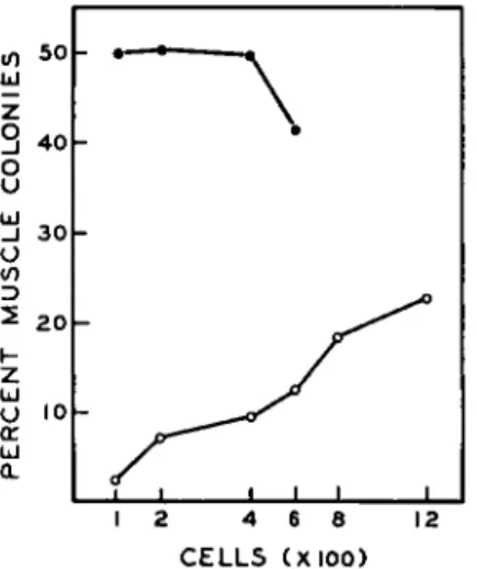
We reasoned that if cross feeding did occur between colonies, crowding the colonies closer together should enhance the exchange. Series of cul- tures were set up in which the number of inoculated cells varied between 100 and 1200 cells. Parallel series were run in conditioned as well as unconditioned medium. Plotting the number of muscle colonies against the total number of colonies after a 2-week growth period (Fig. 6) indi- cated that crowding did not increase the yield of muscle colonies cultured in conditioned medium. By progressively crowding the colonies closer together in unconditioned medium, however, progressively higher per-
|lill|IIIIJIIII|llll|llll|llll|llll|llll|llll|llll|
CENTIMETERS
FIG. 4. A comparison of colony types which develop in unconditioned medium (A;
and in conditioned medium (B). Both plates were fixed after 13 days of culture. The circles and checks on the petri plate in A are an aid in scoring colony types. The circles mark the locations of colonies containing identifiable myotubes. Compare the minute size of these colonies to the muscle colonies on plate B.
centages of muscle colonies were obtained. We concluded that some interaction between adjacent colonies occurs in unconditioned medium leading to the development of muscle colonies which either would not have formed at all or would have been scored as fibroblastic. Since the same effect is not observed in conditioned medium, it may mean that with conditioning already optimal, the maximum number of muscle colony progenitor cells have expressed themselves in this medium.
These observations at least established the plausibility of the thesis that the few aberrant muscle colonies which develop in unconditioned medium are also dependent upon conditioning. Another line of investiga-
tion suggests that the development of muscle clones is entirely dependent upon the use of conditioned medium.
In a series of experiments designed to test another question, we pre- pared cell suspensions by trypsinization of single colonies of either muscle or hbroblastic type. These suspensions were transferred to individual petri plates containing conditioned medium and incubated for the standard 2-week period. Such second generation colonies breed true to the parental type (Figs. 7 and 8).
We now took cell suspensions prepared from single muscle colonies and pipetted half the cells into a plate containing conditioned medium and half into unconditioned medium. The transfer cultures in unconditioned medium were usually completely devoid of colonies or, on occasion, con- tained a few minute colonies which could not be identified as muscle. In contrast, the plates with conditioned medium contained large numbers of muscle colonies which frequently were extremely crowded. Although both plates had received heavy inocula of muscle cells, only in conditioned medium are these cells competent to form colonies. Some fibroblastic colonies at least will clone in either type of medium (Fig. 9). T h a t all fibroblastic colonies do not behave in a uniform fashion may indicate, as suggested earlier (Königsberg, 1963), that these colonies do not represent a homogeneous population.
From these experiments we may conclude that some colonies of fibro- blastic cells either do not require conditioned medium or can effectively condition the medium themselves. None of the cells obtained from muscle colonies will form clones unless conditioned medium is provided. Ap- parently they cannot condition the medium sufficiently to establish them- selves. This suggests that conditioning is primarily a function of the fibro- blasts.
We knew, in fact, that conditioned medium could be prepared by using farm cultures consisting almost exclusively of fibroblastic cells. Medium so prepared was as effective as the medium conditioned by mixed popula- tions of muscle cells and fibroblasts.
FIG. 5. Microscopic examination of one of the minute muscle colonies which develop in unconditioned medium. A. Culture fixed and stained after 10 days. T h e outlined area is magnified in B. B. Arrow indicates the small muscle colony visible within the area outlined in A. Note that this colony is in close proximity to a colony of fibroblastic
cells. This is frequently although not invariably true of muscle colonies in uncondi- tioned medium. C. and D. At higher magnifications the multinucleate myotubes can be seen. T h e pattern of cross striation can be observed in unstained preparations of such cells examined in polarized light (Königsberg, 1961b).
ι-Λ·. ^>*ν ^^^^^é^SS^^''
gift, ^ ;V-* : ^ ^ i | w ;
1*3 .... i^llL
Such fibroblast farm cultures are seeded with cell suspensions prepared by trypsinizing primary cultures which have grown to confluency. In these confluent cultures most, if not all, of the myoblasts have fused to form multinucleated fibers. These differentiated fibers fragment during trypsinization, but most of them are removed by passing the suspensions through bolting silk. It is problematical whether these fragments are viable. In any event, secondary cultures prepared in this manner con- tain relatively few myotubes. According to our knowledge of the optimal
<n so
Id
z 3 40 u o
Lu _J 30 υ </)
Ώ 2 2 0 I-z
id u io o:
Id Q_
1 2 4 6 8 12 CELLS (XIOO)
FIG. 6. Variation of the percentage of muscle colonies (ordinate) as a function of inoculum size (abcissa). Cultures were inoculated with freshly suspended cells within the range indicated. They were cultured for 13 days with a change of medium every 3 days (no fourth feeding was given). The medium was either conditioned (φ φ) or unconditioned (Q O)· Throughout this range of inoculum sizes the plating efficiency either remained unchanged or declined slightly with increasing inoculum.
Although the percentage muscle colonies in unconditioned medium increased with increasing inoculum this was not true of the percentage muscle in conditioned medium (see text). The range of inocula used with conditioned medium could not be extended beyond 600 cells because of the difficulties in counting such crowded cultures accurately.
cell numbers required to condition medium effectively, it is highly unlikely that these few myotubes could be responsible for conditioning.
These observations added support to the premise that conditioning is accomplished principally by the fibroblasts.
T h e Critical Period
We next turned to the question of whether there was a specific time during the development of a muscle clone during which conditioned
j i i i I L
medium was required. Replicate cultures were set up in conditioned medium with small numbers of freshly isolated muscle cells. At 3-day in- tervals the medium was replaced, and at each interval a group of cultures which had been fed up to that time with conditioned medium was now
|IIII|IIIIJIIII|IIII|IIII|IIII|IIII|IIII|IIII|II^
CENTIMETERS
FIG. 7. Second generation subclones of muscle and fibroblastic colonies in conditioned medium. A. Sister culture of the primary plates used as the source of the colonies transferred. This culture was fixed on the same day (day 12) that the transfers were made. The petri plate in B was inoculated with cells suspended by trypsinization of a single muscle colony. C. Subclones of a single colony of fibroblastic cells.
switched to unconditioned medium. These "switched" plates were then carried in unconditioned medium for the remainder of the standard 2-week incubation period. An examination of the plates clearly indicated that col- onies of muscle cells cultured in conditioned medium for 3 days were in- distinguishable from those cultivated for the entire 2-week period in the
same medium. Moreover, with some batches of conditioned medium an interval as short as the first 24 hours in culture was sufficient.
In the cultures initiated in conditioned medium and transferred to unconditioned medium, both the plating efficiency and the percentage of muscle colonies were comparable to those of full-term conditioned medium plates. Even more striking, the muscle colonies on the short-term
"switched" plates were identical to the large sworling muscle colonies observed in our standard conditioned medium plates, in sharp contrast to the small aberrant muscle colonies observed in unconditioned medium (Figs. 4 and 5).
It was quite clear that there was indeed a specific period during which conditioned medium was required. This period, the 24-hour interval during which the cells settle to the floor of the petri plate where they attach and stretch, proved to be surprisingly short.
Conditioning the Petri Plate Surface
The system with which we have been working consists essentially of three components: cells, petri plate, and conditioned medium. Ap- parently, conditioned medium is effective even if present for only the short period during which the cultures are initiated. It seemed logical to inquire whether the first interaction occurs between cells and medium or between medium and the surface of the petri plate. We wanted to deter- mine whether the brevity of the period during which the cells required conditioned medium might indicate that the petri plate surface was altered in some way by exposure to this medium and whether, once this new surface was established, conditioned medium was dispensable. (It seems pertinent to point out that in our laboratory it is standard proce- dure to fill the petri plates with medium and store them in the incubator for an hour or two before use. This practice was adopted originally to reduce the time the cells need be exposed to normal atmosphere and room temperature.)
T o test this premise it would be necessary to treat petri plates with conditioned medium, in the absence of cells, and then substitute un-
FIG. 8. Higher magnification photomicrographs of areas of the second generation subclones in Fig. 7. B. Second generation colonies subcloned from a single muscle colony. C. Daughter colonies of a primary colony of fibroblastic cells. Sworls seen in the center of some of these colonies (upper right) are composed of mononucleated cells. They are due, most probably, to multilayering in colony centers.
conditioned medium before inoculating the cells. Therefore we pre- incubated plates with conditioned medium for a period of 3 days at 36.5°C in an atmosphere of 5% C 02, aspirated off the conditioned medium and replaced it with 2 ml of unconditioned medium. These
!hll|lillJlill|llll|llll|llll|llll|llll|IIH
CENTIMETERS
FIG. 9. Second generation fibroblastic clones grown in both conditioned and uncon- ditioned medium. Plates A and B received equal numbers of cells from the same primary colony of fibroblastic cells. Plate A contained conditioned medium and plate B, unconditioned. Approximately the same number of colonies are present in both plates.
The same protocol was used for cultures C and D (C, conditioned medium; D, uncon- ditioned). In this case plating was inferior in unconditioned medium.
plates were now inoculated with small numbers of freshly dissociated muscle cells, cultured for a 2-week period, and then examined. Qualita- tively, the muscle colonies grown in unconditioned medium but on pre- treated petri plates were indistinguishable from colonies grown on con- ditioned medium (Fig. 10). Pre treatment with unconditioned medium,
however, is completely ineffective. Colonies grown on control plates pre- treated with unconditioned medium are in no way different from those cultured in unconditioned medium.
FIG. 10. The effect of pretreating petri plate surfaces with conditioned medium before use. A. Control plate cultured in unconditioned medium. B. Control cultured in conditioned medium. C and D. Cultures grown in unconditioned medium but on pre- treated surfaces. The surface of the petri plate in C was treated with unconditioned medium; plate D was treated with conditioned medium. The protocol of pretreatment was as follows: Petri plates were filled with 2 ml of medium and incubated at 36.5°C in a humidified, gassed incubator for 3 days. At the end of this incubation period the medium was removed by aspiration. All of the pretreated plates were then filled with 2 ml of unconditioned medium, inoculated with cells, and cultured in the usual manner.
There are three possible explanations for the results obtained by pre- treating petri plates with conditioned medium: (1) T h e petri plate surface is altered by the removal of material; (2) alteration of the surface is
effected by the deposition of material; (3) small traces of soluble material might be left behind on the plates after pretreatment. These small traces may be sufficient to supplement, in the usual nutritional sense, the un- conditioned medium with which the plates are subsequently filled.
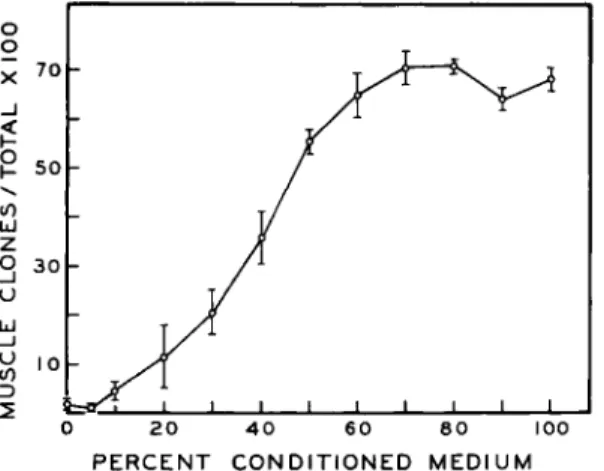
In all the experiments, the medium was removed by careful and thorough aspiration. Although it seemed unlikely that a significant amount of soluble material was left behind, this possibility was easily tested. If we assumed that the pretreatment results were due to a simple dilution phenomenon, they should be reproduced in conditioned medium diluted with unconditioned medium. In this manner we should be able to determine the minimal amount of conditioned medium required to supplement the volume of unconditioned medium used to test the pre- treated plates. Mixtures of both types of medium were prepared contain- ing progressively smaller percentages of conditioned medium. Equal inocula of cells were cultured in these mixtures, fixed and stained after 2 weeks, and scored. In Fig. 11 we have plotted the percentage of muscle
o o x ™
_i
£ 50 u z
O 30 U Id D
0 20 4 0 60 80 100 PERCENT CONDITIONED MEDIUM
FIG. 11. T h e percentage of muscle colonies as a function of the percentage of con- ditioned medium in dilutions of conditioned medium with unconditioned. T h e standard error of each plotted mean is indicated by the vertical bar through each point.
colonies for each mixture. The percentages of muscle colonies obtained on pretreated plates are included in Table I (see also Table II). Comparing these figures to the values plotted in Fig. 11 we note that the percentages of muscle colonies obtained on pretreated plates correspond to values obtained with mixtures containing 40-50% conditioned medium. Since we pretreat with 2 ml of conditioned medium which is subsequently re- placed with 2 ml of unconditioned medium, we would have had to leave
behind almost all the conditioned medium. Such gross carelessness is highly unlikely.
An even more compelling argument than that based on the conditioned medium dilution curve, is the fact that the effect of preincubation persists even after the treated petri plate is rinsed thoroughly with distilled water.
Petri plates were again filled with 2 ml of conditioned medium and placed in the incubator for a period of 3 days after which the conditioned medium was removed by aspiration. This time, however, some of the
TABLE I
T H E EFFECT OF PRETREATMENT OF PETRI PLATES WITH CONDITIONED MEDIUM UPON THE SUBSEQUENT DEVELOPMENT OF MUSCLE CLONES
Number of
petri plates Muscle Fibroblastic Per cent
in each clones clones muscle Treatment« group (mean ± S.E.) (mean ± S.E.) (mean ± S.E.)&
Unconditioned
medium controls 4 0.8 ± 0.5 41.0 ± 3.5 2.0 ± 1.6 Conditioned
medium controls 4 28.5 ± 2.1 24.0 ± 1.5 54.3 ± 2.1 Pretreatment (con-
ditioned medium) grown in uncon-
ditioned medium 4 18.0 ± 0 . 7 21.8 ± 2 . 3 45.8 ± 4.0
» Pretreatment as described in text.
ö Per cent muscle values [(muscle colonies/total colonies) 100] are presented since they normalize the data with respect to intergroup differences in plating efficiency.
Tests of the significance of the difference between the means ("Students" t) for per cent muscle indicate that the conditioned medium controls and the pretreated groups are both significantly different from the unconditioned medium controls (p < 0.001 in both cases). T h e mean per cent muscle of the conditioned medium group is not sig- nificantly different from the pretreated group (/?>0.3). T h e mean number of muscle clones per plate, however, is significantly different in these two groups (p < 0.02).
plates were rinsed either once or three times with 2 ml of sterile distilled water. They were then filled with 2 ml of unconditioned medium, inocu- lated with a small number of cells, and cultured for the usual 2-week period. Without question, rinsing with distilled water as many as three times gave results indistinguishable from results with preincubated plates which had not been rinsed at all (Fig. 12). In either case the cultures on pretreated petri plates were not significantly different from those obtained on standard plates with conditioned medium. If a film is deposited or adsorbed on the bottom of the petri plate during pretreatment, it must
TABLE II
T H E EFFECT OF DISTILLED WATER WASHES UPON THE SURFACE OF PRETREATED PETRI PLATES
Number of petri
plates in each Group Treatment« group
( Pretreat:uncond. medium A J Wash :none
Grown runcond. medium I Pretreat:cond. medium B J Wash :none
I Grown :cond. medium ( Pretreat:cond. medium Q J Wash :none
Grown runcond. medium
Î
Pretreaf.cond. medium Wash :1Grown runcond. medium f Pretreatrcond. medium
E J Wash :3
Grown runcond. medium
« Pretreatment as described in text.
& Per cent muscle values [(muscle colonies/total colonies) 100] are presented since they normalize the data with respect to intergroup differences in plating efficiency.
Tests of the significance of the difference between the means ("Student's" t) for per cent muscle indicate no significant difference between the conditioned medium control (B) and any of the pretreated groups (C, D, and E) (p> Λ to p > .8). The mean number of muscle clones on pretreated plates which were not washed (C) and similar plates washed three times (E) are also not significantly different (p>0.1).
be composed of materials insoluble in distilled water. At present we favor this hypothesis, if for no other reason than that it can be most readily tested. Assuming that some film is deposited on the petri plate surface—
what sort of material might it be?
Collagen Synthesis during Conditioning
In reference to our speculations that the fibroblasts were principally responsible for conditioning the medium, one piece of information now assumed importance.
During the period when we were examining alternative protocols for preparing conditioned medium we found that the most effective prepara- tions were those collected from crowded confluent monolayers. Accord- ingly, farm cultures are grown for 5 days until they reach confluency. At
Muscle Fibroblastic Per cent
clones clones muscle (mean ± S.E.) (mean ± S.E.) (mean ± S.E.)&
3 0.0 ± 0.0 38.7 ± 3.3 0.0 ± 0.0 3 32.3 ± 3 . 5 22.0 ±2.7 59.6 ± 2.5 3 25.7 ± 2 . 2 11.7 ± 1 . 6 68.8 ± 2.6 3 18.3 ± 1.0 12.0 ± 1.0 60.6 ± 1.7 3 20.4 ± 2 . 0 12.0 ± 1 . 5 63.8 ± 1.2
this time they are refed, and the medium is later collected, filtered, and used. This same protocol is also used when we prepare conditioned medium with farms consisting of second passage cells (predominantly
1 ' |l ' 2 1 3l ' 4 1 ' 51 CENTIMETERS
FIG. 12. T h e effect of washing petri plate surfaces after pretreatment with condi- tioned medium. T h e pretreatment schedule is given in the legend of Fig. 10. Cells were cultured on these pretreated plates in unconditioned medium. A. Control: no washes after pretreatment. B. Washed once with 2 ml of sterile distilled water before use. C.
Washed with three changes of distilled water, 2 ml each, before use. Each volume of distilled water was left in the petri plate for five minutes and then removed by aspira- tion. After the last wash the plate was filled with unconditioned medium and inoculated.
fibroblasts). Measurements of the growth of such farm cultures indicate that the cells are in the stationary phase during the conditioning period (Fig. 13).
One striking change in the biosynthetic pattern of fibroblasts which
occurs in the stationary phase has been clearly demonstrated (Green and Goldberg, 1963, 1964; Goldberg and Green, 1964). Using a cell line derived from embryonic mouse cells these investigators have been able to demon- strate both the synthesis and deposition of collagen, but only in stationary nondividing cultures. Using isotopically labeled proline they were unable to demonstrate labeled protein-bound hydroxyproline in either cells or medium from exponentially growing cultures. If our cultures were com- parable to those studied by Green and Goldberg, the period during which effective conditioned medium can be collected corresponds to the time during which the cultures should be synthesizing collagen.
100
I 2 3 4 5 6 7 8 DAYS IN C U L T U R E
FIG. 13. A semilog plot of the growth, in terms of DNA per culture, of conditioning farms of fibroblastic cells. DNA was determined by Burton's modification of the diphenylamine reaction. T h e arrows indicate replacement of the medium. Farms were inoculated with 106 cells from a suspension of primary cultures.
In order to test the validity of such a comparison, we have examined the cell monolayers in our fibroblast farm cultures using electron micro- scopic techniques. Cell monolayers were fixed and embedded in situ after the conditioned medium was harvested. The results are as yet preliminary, but they indicate that fibrous material is deposited extracellularly be- tween adjacent cells of the monolayer (Fig. 14). This material can be resolved as extremely broad fibers with a definite major period but with no detectable fine periodicity (Fig. 15). Measurements of the interperiod indicate that the periodicity is 850 Â, unlike either native collagen or any of the reconstituted fibers which have as yet been precipitated from solu- tions of tropocollagen. Such fibers have been observed by Goldberg and Green (1964) in stationary cultures of a mouse fibroblast line. Similar
CELL AND TISSUE INTERACTIONS 271
o Z
*4S .
fibers have also been described by Jakus (1961, 1962) in the limbic region of the cornea and most frequently in Descemet's membrane. Jakus (1961) suggests that although no direct evidence exists, the wide variety of fibers observed in the region of the cornea may indicate the existence of an environment in which collagen may assume many forms. She suggests that mucopolysaccharides may be involved in producing these different
FIG. 15. Fibers similar to those seen in Fig. 14. The periodicity is 850 Â.
forms, and has, in fact, demonstrated such long-spacing fibers precipitating from collagen solutions extracted from the cornea under conditions which give the native spacing with collagens extracted from other tissues (Jakus, 1964). If these fibers are repeatedly dissolved in a citric acid buffer and reprecipitated, eventually only fibers exhibiting the native periodicity are formed (Jakus, 1964). One possible explanation of these observations
is that recycling these fibers removes a contaminant (i.e., mucopolysac- charide) which maintains the long-spaced structure. Our tentative con- clusion is that the fibers we have observed in our conditioning farm cul- tures are an atypical form of collagen.
A Hypothetical Role of Conditioned Medium
The new information which we now had—the facts, as well as the reasonable assumptions can be summarized as follows: (1) Fibroblasts can effectively condition the medium, and perhaps may be solely responsible for conditioning; (2) pretreatment of the petri plates with conditioned medium alters the surface of the plate in some manner so that it will now support the growth and differentiation of muscle clones even when the liquid overlay is unconditioned medium; (3) this alteration of the petri plate surface may be due to the deposition of material on the surface of the plate; (4) in our fibroblast farm cultures, what appears to be an atypical collagen fiber is laid down and we assume that collagen is being synthesized.
Reviewing these points suggests the following hypothesis: Collagen in some form is present in the conditioned medium; it is deposited on the petri plate surface during incubation and there in some manner permits the development of muscle clones from single myoblasts.
If this hypothesis is true, we should be able to extract collagen from a recognized source of the protein and coat the surface of the petri plate with collagen; this tactic should replace the requirement for conditioned medium.
This, in fact, is what we have done. Acid extracts of rat tail tendon were prepared and thin films spread over the surface of the petri plate (Ehrmann and Gey, 1956). Collagen was precipitated either by exposure of the film to ammonia vapor or by adding sodium chloride to 1 % of the final volume before spreading the acid collagen film. In either case, after the films had set, they were washed with sterile distilled water and equilibrated against unconditioned medium. Petri plates containing a liquid overlay of unconditioned medium over a surface film of precipi- tated collagen were inoculated with a small number of single cells. These cultures were subsequently handled in a manner identical to companion controls in either conditioned or unconditioned medium but on conven- tional polystyrene, tissue-culture petri plates (Falcon).
The results of these experiments were unequivocal. Single cells cultured on collagen films in unconditioned medium gave rise to cultures com-
parable in every respect to controls cultured in conditioned medium. In Table III, the results of six separate trials are tabulated. By quantita- tive criteria, that is, the percentage of muscle colonies per culture, a collagen substratum completely replaces the necessity for conditioned medium (see Hauschka and Königsberg, 1966).
In our experience, however, the more reliable and striking characteristic of muscle colonies which develop in conditioned medium is the size and morphology of such colonies. The large sworling muscle colonies, im- mediately identifiable by their fibrous appearance even to the naked eye, were never before observed in unconditioned medium unless the plates had been pretreated with conditioned medium. They are very much in evidence, however, when single cells are cultured on a collagen film despite the fact that the medium used has not been conditioned (Fig. 16).
Certain experimental facts and reasonable assumptions have led us to construct a hypothetical explanation of the role played by conditioned medium in the development of muscle clones. These same facts and assumptions suggested a simple test of the hypothesis and the results of this test have been overwhelmingly positive. This does not mean that each assumption we have made has been verified. It does suggest, however, that the hypothesis, as a working hypothesis, has merit.
Our thesis is that the development of muscle clones is influenced by the metabolic activity of fibroblasts and that the influence is exerted by collagen or materials associated with collagen. T o examine this hy- pothesis critically, we first need to know if material is indeed deposited on the petri plate during pretreatment with conditioned medium. If this proves to be the case, the identity of these materials will need to be estab- lished. Since the amino acid composition and the solubility properties of collagen are so unique (Harrington and von Hippel, 1961), we would anticipate no difficulty in at least establishing whether collagen is or is not one of these materials.
We also need to examine the effects of more highly purified collagens to resolve the question of whether the phenomenon we have observed is due to collagen or to those contaminants usually associated with collagen (mucopolysaccharides, for example). Also of relevance is the question of whether molecules structurally related to collagen would be efficacious.
When we have these answers, we will be in a better position to specu- late on mechanisms. We may at least know whether we need consider the uptake of materials into the myoblasts or an effect exerted at the cell surface.
T H E E F F E C T O F A C O L L A G E N SUBSTRATUM U P O N T H E D E V E L O P M E N T O F M U S C L E C L O N E S «
Experiment number
1 2 3 4 5 6
Number of petri
plates of each
type 6 8 6 6 5 6 Average
Conditioned med (group A) Mean no.
of muscle clones 38.5 ± 1.1 20.8 ± 1.3 28.8 ± 2.3 20.8 ± 1.9 21.6 ±2.0 29.0 ± 2.2
Mean no.
of fibro.
clones 32.2 ± 2.3 28.5 ±1.4 32.7 ± 2.0 9.2 ± 0.8 10.2 ± 0.4 26.2 ±1.1
urn Mean per cent
muscle 54.8 ±1.6 42.0 ± 0.7 48.4 ±4.0 69.4 ± 2.5 67.1 ± 2.0 52.5 ± 2.0
55.7
Unconditioned medium Untreated surface
(group B) Mean no.
of muscle clones 1.2 ±0.4 0.13 ± 0.05 0.2 ± 0.2 0.0 ±0.0 0.0 ± 0.0 1.5 ±0.5
Mean no.
of fibro.
clones 42.2 ±3.3 34.4 ± 1.2 31.5 ±2.4 35.3 ± 2.3 27.2 ± 6.7 24.2 ± 1.5
Mean per cent
muscle 2.6 ± 0.9 0.4 ± 0.4 0.6 ±1.8 0.0 ± 0.0 0.0 ± 0.0 6.4 ±1.9
1.7
Collagen surface (group C) Mean no.
of muscle clones 31.0 ±1.1 2.19 -h 1.4 19.0 ± 0.9
20.8 ± 1.3 16.6 ± 1.1 21.7 ± 0.7
Mean no.
of iîbro.
clones 15.2 ± 0.8 14.8 ± 1.4 17.0 ± 1.9 12.7 ± 0.8 17.6 ± 1.5 10.3 ± 0.9
Mean per cent
muscle 67.3 ± 2.5 59.9 ±3.9 53.6 ± 0.9 62.2 ± 2.3 47.8 ± 5.5 67.7 ± 2.3
59.8
"Values are expressed as means per petri plate ± standard error. Mean per cent muscle values [(muscle colonies/total colonies) 100] are presented since they normalize the data with respect to fluctuations in plating efficiency from experiment to experiment. Tests of the significance of the differences between means for per cent muscle indicate that in every experiment the values for both group A and group C are significantly different from the value of group B (p <C 0.001 in all cases). Differences between the means of groups A and C were not significant in three experiments and significant or of marginal significance in the other three experiments.
n w r r
>
z Ό
C m z H M
>
Ci H δ z
Macromolecules and Cell Culture
T h e classic approach to the study of the properties of embryonic cells upon which much of modern developmental biology is grounded, in-
I ' il ' 2 ! 3I ' 4| ' si CENTIMETERS
FIG. 16. T h e development of muscle clones on a substratum of reconstituted collagen in unconditioned medium. A. Control culture: untreated surface; unconditioned medium. B. Control culture: untreated surface; conditioned medium. C. Colonies which have developed on a film of reconstituted collagen with a liquid overlay of un- conditioned medium. Compare plates A and C. Both of these cultures have been carried on unconditioned medium. However, note the number of large muscle clones which have developed on the collagen film (C).
volved testing the differentiative expression of a particular group of cells in a variety of ectopic locations in the embryo. Cell and tissue-culture techniques merely extend the range of such tests. In fact, the technique of in vitro culture was devised for just this purpose (Harrison, 1907).