Signal Transduction
Manifestation of Novel Social Challenges of the European Union in the Teaching Material of
Medical Biotechnology Master’s rogrammes at the University of Pécs and at the University of DebrecenP
Tímea Berki MD, PhD – Ferenc Boldizsár MD, PhD – Mariann Szabó MD – Gergő Talabér, MD, PhD – Zoltán Varecza PhD
Signal Transduction (Medical Biotechnology)
Tímea Berki MD, PhD – Ferenc Boldizsár MD, PhD – Mariann Szabó MD – Gergő Talabér MD, PhD – Zoltán Varecza MsC, PhD
“Manifestation of Novel Social Challenges of the European Union
in the Teaching Material of
Medical Biotechnology Master’s Programmes at the University of Pécs and at the University of Debrecen”
Identification number:TÁMOP-4.1.2-08/1/A-2009-0011
University of Pécs – Pécs, 2011
© Tímea Berki MD, PhD; Ferenc Boldizsár MD, PhD;
Mariann Szabó MD; Gergő Talabér MD, PhD; Zoltán Varecza MsC, PhD, 2011 The project is funded by the European Union and
co-financed by the European Social Fund.
Editor in charge: University of Pécs
Editor in charge: Tímea Berki MD, PhD and Ferenc Boldizsár MD, PhD, Rita Bognár Technical editor: Zsolt Bencze, Veronika Csöngei and Szilvia Czulák
Lector: Dr. György Miskei Length: 155 pages
Identification number:
TÁMOP-4.1.2-08/1/A-2009-0011
3
Content
LIST OF FIGURES ... 7
LIST OF TABLES ... 11
LEGEND ... 13
I GENERAL SIGNAL TRANSDUCTION ... 15
I.1 INTRODUCTION, OVERVIEW OF EXTRACELLULAR SIGNALING ... 15
I.2 FAMILIES OF EXTRACELLULAR RECEPTORS ... 20
I.2.1 Ion-channel receptors ... 21
I.2.2 7-transmembrane-spanning receptors (7-TM) ... 23
I.2.3 Enzyme-linked receptors ... 27
I.3 INTRACELLULAR RECEPTORS ... 39
I.4 INTRACELLULAR SIGNAL TRANSMITTING MOLECULES ... 40
I.4.1 G-proteins ... 40
I.4.2 Second messengers ... 42
I.4.3 The Ca2+-signal ... 46
I.4.4 Transcription factors ... 52
I.5 OVERVIEW OF MAJOR SIGNALING PATHWAYS ... 59
I.5.1 cAMP-PKA pathway ... 59
I.5.2 PLCγ-DAG-PKC ... 59
I.5.3 MAPK-pathway ... 60
I.5.4 PI3-kinase-PKB (Akt) ... 60
I.5.5 JAK-STAT ... 60
4 The project is funded by the European Union and co-financed by the European SocialFund.
II DETAILED (SYSTEMATIC) SIGNAL TRANSDUCTION ... 61
II.1 SIGNALING IN THE IMMUNE SYSTEM ... 61
II.1.1 Signaling in the specific immune system 1: B cell signaling ... 61
II.1.2 Signaling in the specific immune system 2: T cell activation and signaling ... 65
II.1.3 Fcγ Receptor signaling ... 70
II.1.4 Fcε Receptor signaling ... 73
II.1.5 Cytokine signaling ... 78
II.1.6 Chemokine signaling ... 87
II.1.7 Signaling in the innate immune system, PRR signaling ... 90
II.2 HORMONE AND GROWTH FACTOR SIGNALING ... 97
II.2.1 Tyrosine kinase-linked receptors ... 97
II.2.2 G-protein-linked receptors (epinephrine, serotonin, glucagon) ... 103
II.2.3 Intracellular/nuclear receptor signaling (steroid hormones and thyroxin) ... 106
II.2.4 Non-genomic steroid hormone signaling pathways ... 112
II.3 SIGNALING IN TUMOR CELLS (EGF-R, HER-2R, ADHESIONMOLECULES) ... 118
II.4 APOPTOSIS SIGNALING ... 123
II.5 RECEPTOR INTERACTIONS, SIGNALING “CROSS-TALK” ... 129
II.6 WNT RECEPTOR SIGNALING ... 133
Content
Identification number:
TÁMOP-4.1.2-08/1/A-2009-0011
5
II.7 SIGNALING IN THE NERVOUS SYSTEM ... 140
II.7.1 Acetylcholine (Ach) ... 141
II.7.2 Noradrenalin (NA) ... 144
II.7.3 Dopamine (D) ... 144
II.7.4 Serotonin (5-HT) ... 144
II.7.5 GABA ... 144
II.7.6 Glutamate ... 145
II.7.7 Glycine ... 145
II.7.8 ATP... 145
II.8 PHARMACOLOGICAL INFLUENCE OF THE SIGNALING ... 146
FURTHER READING ... 155
Identification number:
TÁMOP-4.1.2-08/1/A-2009-0011
7
List of figures
Figure I.1-1: Main types of receptors ... 19
Figure I.1-2: Intracellular receptor signaling ... 19
Figure I.2-1: Extracellular receptor types ... 21
Figure I.2-2: Cys-loop ion-channel receptors ... 22
Figure I.2-3: Synapse between two neurons - neurotransmission ... 23
Figure I.2-4: 7-transmembrane-spanning receptors (7-TM) ... 25
Figure I.2-5: Structure of 7-TM receptors ... 26
Figure I.2-6: Receptor desensitization ... 27
Figure I.2-7: Kinase-phosphatase balance ... 28
Figure I.2-8: Receptor- and cytoplasmic PTPs ... 29
Figure I.2-9: Natriuretic peptide signaling ... 29
Figure I.2-10: Receptor tyrosine kinase family ... 31
Figure I.2-11: Receptor tyrosine kinase (RTK) signaling ... 32
Figure I.2-12: Members of the signaling complex ... 33
Figure I.2-13: Dimerization of GF receptors ... 33
Figure I.2-14: GF receptor signaling pathways ... 34
Figure I.2-15: MAPK/ERK in growth and differentation ... 37
Figure I.4-1: Activation of G-protein-coupled receptors (GPCR) ... 41
Figure I.4-2: G-proteins ... 41
Figure I.4-3: cAMP-PKA pathway ... 43
Figure I.4-4: More receptors using the same second messenger system ... 44
Figure I.4-5: IP3 receptor pathway ... 44
Figure I.4-6: Several pathways use the Ca2+ signal ... 48
8 The project is funded by the European Union and co-financed by the European SocialFund.
Figure I.4-7: Intra/extracellular compartments of Ca2+-signaling, Ca2+-channels ... 49
Figure I.4-8: Effector mechanisms of Ca2+-signaling ... 51
Figure I.4-9: Regulation of transcription ... 52
Figure I.4-10: Functional domains of transcription factors ... 54
Figure I.4-11: Structural groups of transcription factors ... 55
Figure I.4-12: Role of transcription factors in thymocyte development ... 56
Figure I.4-13: Th - Tc cell decision ... 57
Figure I.4-14: Th differentiation ... 57
Figure II.1-1: Overview of BcR signaling ... 63
Figure II.1-2: Short/long term BcR stimulation ... 64
Figure II.1-3: Co-stimularory pathways of BcR signaling ... 65
Figure II.1-4: Molecules of the “immunological synapse” ... 66
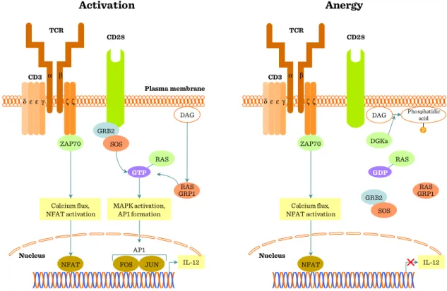
Figure II.1-5: Overview of TcR/CD3 signaling pathway ... 67
Figure II.1-6: T cell activation pathways ... 68
Figure II.1-7: Co-stimulatory pathways regulate the TcR signal ... 70
Figure II.1-8: Types of Fcgamma receptors ... 71
Figure II.1-9: Activator and inhibitory Fcγ receptor signaling ... 72
Figure II.1-10: Overview of Fcγ receptor signaling ... 73
Figure II.1-11: IgE bound FcεR I ... 74
Figure II.1-12: IgE bound FcεR II ... 75
Figure II.1-13: Biological effects of FcεR signaling ... 77
Figure II.1-14: FcεRI mediated signaling ... 77
Figure II.1-15: Similarities in TcR and FcεR signaling ... 78
Figure II.1-16: Cytokine receptors ... 80
Figure II.1-17: Characteristics of multichain cytokine receptors ... 81
List of figures
Identification number:
TÁMOP-4.1.2-08/1/A-2009-0011
9
Figure II.1-18: The structure of JAK and STAT proteins ... 82
Figure II.1-19: Overview of cytokine signalling ... 83
Figure II.1-20: TNF receptor mediated apoptosis I ... 85
Figure II.1-21: TNF receptor mediated apoptosis II ... 86
Figure II.1-22: Chemokine signal through receptors coupled with G-proteins ... 89
Figure II.1-23: Chemokine signaling pathways ... 89
Figure II.1-24: Toll-like receptors-pattern recognition ... 92
Figure II.1-25: Overview of complement receptor (CR) and Toll-like receptor signaling ... 93
Figure II.1-26: Toll-like receptor inhibitors ... 95
Figure II.1-27: Complement receptors ... 96
Figure II.2-1: Growth factor (GF) receptors ... 98
Figure II.2-2: Autophosphorylation of RTKs ... 99
Figure II.2-3: Overview of EGF signaling ... 100
Figure II.2-4: General characteristics of GF signaling ... 100
Figure II.2-5: GF receptors as therapeutic targets ... 101
Figure II.2-6: Adrenergic receptors ... 104
Figure II.2-7: Nuclear receptor superfamily ... 108
Figure II.2-8: Functional domains of transcription factors ... 109
Figure II.2-9: Mechanism of steroid receptor action ... 110
Figure II.2-10: Genomic steroid actions ... 111
Figure II.2-11: Genomic and non-genomic GC effects ... 113
Figure II.2-12: Summary of genomic and non-genomic glucocorticoid effects ... 114
Figure II.3-1: Immune selection in the development of cancer: no two tumors are alike ... 119
10 The project is funded by the European Union and co-financed by the European SocialFund.
Figure II.3-2: Tumor and activated T cells ... 119
Figure II.3-3: TGF-β signaling in tumor signaling and cancer progression ... 120
Figure II.3-4: What happens when Fas-stimulated immune cells resist to die? ... 120
Figure II.4-1: Apoptosis pathways ... 124
Figure II.4-2: Mitochondrial apoptosis pathway ... 125
Figure II.4-3: Bcl-family ... 126
Figure II.4-4: Apoptosome ... 126
Figure II.4-5: Apoptosis signaling intervention ... 128
Figure II.5-1: Growth factor receptor – integrin signaling interaction ... 131
Figure II.5-2: Convergence of signaling pathways ... 132
Figure II.6-1: β-catenin in cellular adhesion ... 134
Figure II.6-2: Alzheimer’s disease I ... 135
Figure II.6-3: Wnt signaling pathways ... 135
Figure II.6-4: Alzheimer’s disease II ... 136
Figure II.6-5: Canonical Wnt pathway ... 138
Figure II.7-1: Neurotransmission ... 141
Figure II.7-2: Acetylcholine ... 142
Figure II.7-3: Acetylcholine receptors ... 142
Figure II.8-1: Potential drug targets in signaling pathways ... 146
Figure II.8-2: Various levels of intervention ... 147
Figure II.8-3: ERB signaling intervention ... 148
Figure II.8-4: Calcineurin and rapamycin ... 150
Figure II.8-5: Rapamycin ... 151
Figure II.8-6: Proteosome inhibitors-Bortezomib ... 152
Figure II.8-7: HSP-90 inhibitors ... 153
Identification number:
TÁMOP-4.1.2-08/1/A-2009-0011
11
List of tables
Table I.2-1: Receptor types ... 24
Table I.4-1: Some important transcription factors ... 53
Table II.2-1: Receptor classes ... 98
Table II.2-2: Intracellular receptor families ... 107
Table II.6-1: Wnt signaling ... 133
Table II.8-1: Selected kinase inhibitors in clinical development ... 147
Identification number:
TÁMOP-4.1.2-08/1/A-2009-0011
13
Legend
Kinase
Phosphatase Enzyme
Cyclin, pro-apoptotic Pro-survival
GTP-ase GAP/GEF Caspase
Transcription factor
Identification number:
TÁMOP-4.1.2-08/1/A-2009-0011
15
I General signal transduction
I.1 Introduction, overview of extracellular signaling
Soluble mediators transmit information through the extracellular space over various distances in cell-to cell communication. In local (short distance) cell signaling, some cells may be in direct contact with each other in order to communicate. Cell-to-cell signaling means that mediators can pass from one cell to another cell through cell junctions, which are found in both animals and plants. Long distance signaling is mediated by hormones between animal cells (endocrine signaling), or growth factors between plant cells. Another general form of long distance signaling is synaptic signaling used mainly in the nervous system. In plants and animals extracellular signaling molecules control metabolic processes, growth and differentiation of tissues, synthesis and secretion of proteins, and the composition of intracellular and extracellular fluids.
Communication by extracellular signals usually involves six steps
(1) Synthesis and release of the extracellular mediator molecule by the signaling cell;
(2) Transport of the mediator to the target cell;
(3) Reception: detection of the signal by a specific receptor protein;
(4) Transduction: binding of the extracellular mediator molecule to a specific receptor on the target cell, and this signal is interpreted by a series of subcellular reactions called signal transduction events.
16 The project is funded by the European Union and co-financed by the European SocialFund.
(5) Response: The signal triggers the desired reaction within the cell, for example a change in cellular metabolism, function, or development triggered by the receptor-signal complex.
(6) Termination: removal of the signal, which often terminates the cellular response.
Signaling molecules operate over various distances
Based on the distance over which extracellular, secreted molecules transmit the signal, cell-to-cell communication can be classified into three types: endocrine (long distance between the source of the mediator and the target cell – the mediator is transported by the circulation, sometimes bound to transport proteins), paracrine (the source of the mediator and the target cell are relatively close to each other – the mediator is transported by simple diffusion), or autocrine (in this case the mediator-producing- and the target cell is the same). In addition, certain membrane-bound proteins on a cell can directly transmit a signal to adjacent cells.
Receptor proteins exhibit ligand-binding and effector specificity
The cellular response to a particular extracellular signaling molecule depends on its binding to a specific receptor protein located on the surface or in the nucleus or cytosol of a target cell. The signaling molecule (a hormone, pheromone, or neurotransmitter) acts as a ligand, which binds to, or “fits” into a site on the receptor. Binding of a ligand to its receptor causes a conformational change in the receptor that initiates a sequence of reactions leading to a specific cellular response.
The response of a cell or tissue to specific hormones is determined by the particular hormone receptors it possesses and by the intracellular reactions initiated by
Introduction, overview of extracellular signaling
Identification number:
TÁMOP-4.1.2-08/1/A-2009-0011
17 the binding of any one hormone to its receptor. Different cell types may have different sets of receptors for the same ligand, each inducing a different response. Or the same receptor may appear on various cell types, and binding of the same ligand may trigger a different response in each type of cell (e.g. acetylcholine). Clearly, different cells respond in a variety of ways to the same ligand. On the other hand, different receptor- ligand complexes can induce the same cellular response in some cell types (e.g.
glucagons and epinephrine).
Thus, a receptor protein is characterized by binding specificity for a particular ligand, and the resulting hormone-ligand complex exhibits effector specificity (i.e., mediates a specific cellular response).
Hormones can be classified based on their solubility and receptor location
Most hormones fall into three major categories: (1) small lipophilic molecules that diffuse across the plasma membrane and interact with intracellular receptors; and (2) hydrophilic or (3) lipophilic molecules that bind to cell-surface receptors (Figure I.1-1).
(1) Lipophilic hormones with intracellular receptors: many lipid-soluble hormones diffuse across the plasma membrane and interact with receptors in the cytosol or nucleus. The resulting hormone-receptor complexes bind to transcription-control regions of the DNA thereby affecting expression of specific genes. Hormones of this type include the steroids (e.g., cortisol, progesterone, estradiol, and testosterone), thyroxine, and retinoic acid (Figure I.1-1 and Figure I.1-2).
(2) Water-soluble hormones with cell surface receptors: As water-soluble signaling molecules cannot diffuse across the plasma membrane, they all bind to cell-surface receptors. This large class of compounds is composed of two
18 The project is funded by the European Union and co-financed by the European SocialFund.
groups: (a) peptide hormones, such as insulin, growth factors, and glucagon, which range in size from a few amino acids to protein-size compounds, and (b) small charged molecules, such as epinephrine and histamine, that are derived from amino acids and function as hormones or neurotransmitters.
Many water-soluble hormones induce a modification in the activity of one or more enzymes already present in the target cell. In this case, the effects of the surface-bound hormone are usually nearly immediate, but persist for a short period only. These signals also can give rise to changes in gene expression that may persist for hours or days. In yet other cases water-soluble signals may lead to irreversible changes, such as cellular differentiation.
(3) Lipophilic hormones with cell-surface receptors: The primary lipid-soluble hormones that bind to cell-surface receptors are the prostaglandins. There are at least 16 different prostaglandins in nine different chemical classes, designated PGA – PGI. Prostaglandins are part of an even larger family of hormones containing 20 carbon atoms called eicosanoid hormones. In addition to prostaglandins, they include prostacyclins, thromboxanes, and leukotrienes. Eicosonoid hormones are synthesized from a common precursor, arachidonic acid. Arachidonic acid is generated from phospholipids and diacylglycerol.
Introduction, overview of extracellular signaling
Identification number:
TÁMOP-4.1.2-08/1/A-2009-0011
19 Figure I.1-1: Main types of receptors
Figure I.1-2: Intracellular receptor signaling
Cytoplasm Outside of cell
Apolar signal
Receptor
Polar signal
Membrane bound receptor Cell membrane
Inside of cell
Plasma membrane
Nucleus Receptor
Cytoplasm
Signal
Chaperone protein Outside of cell
Inside of cell
20 The project is funded by the European Union and co-financed by the European SocialFund.
I.2 Families of extracellular receptors
Introduction
Ligands act on extra- or intracellular receptors according to their hydrophilic or hydrophobic nature, respectively (Figure I.1-1). Hydrophobic/lipophilic molecules (e.g.
steroid hormones, thyroid hormone, vitamin D) can diffuse through the plasma membrane lipid layer, thus reaching intracellular receptors (Figure I.1-2) (for details see Chapter II.2.3, page 106). Hydrophylic/water soluble ligands (e.g. peptide hormones, cytokines, chemokines, neurotransmitters), on the other hand, are unable to penetrate the lipid rich outer barrier of the cells; therefore, they need receptors protruding from the outer surface of the cell membrane.
Extracellular receptor groups
In case of extracellular receptors, the effect of ligand binding has to be transmitted into the cell. A number of signal transduction pathways have evolved to serve this purpose.
Extracellular receptors belong to 3 major categories (Figure I.2-1):
(1) Ion-channel receptors
(2) 7-transmembrane-spanning receptors (7-TM), also called G-protein-coupled receptors (GPCR)
(3) Enzyme-linked receptors
Families of extracellular receptors
Identification number:
TÁMOP-4.1.2-08/1/A-2009-0011
21 Figure I.2-1: Extracellular receptor types
I.2.1 Ion-channel receptors
Ligand-operated receptors in the plasma membrane (e.g. GABAA, GABAC, iGlu, Glycine, Serotonin, nicotinic Ach, P2X receptors) belong to this group. They are quite abundant in the nervous system and on contractile cells (smooth/striated/heart muscle).
Their function is relatively simple: upon activation they open and ion currents occur as a consequence of the concentration gradient between the extra- and intracellular environment. The transient ion concentration changes lead to the contraction or depolarization of the target cells. There are 3 groups of ion-channel receptors:
(1) Cys-loop receptors: have pentameric structure with 4 trans-membrane (TM) regions in each subunit (e.g. Acetylcholin (Ach) Nicotinic Receptor – Na+ channel; GABAA, GABAC, Glycine – Cl- channels [inhibitory role in CNS]) (Figure I.2-2 and Figure I.2-3).
ENZYME-LINKED RECEPTORS G-PROTEIN-LINKED RECEPTORS ION-CHANNEL-LINKED RECEPTORS
Ions
Signal molecule
Cytoplasm Plasma membrane
GDP β γ α
GTP β γ α
β γ
Enzyme Enzyme Enzyme
GTP α Signal molecule
G-protein Activated G-protein Activated enzyme
Dimer of signal molecule
Inactive catalytic domain
Enzyme Active catalytic
domain
Signal molecule
Activated enzyme
22 The project is funded by the European Union and co-financed by the European SocialFund.
(2) Glutamate-activated cationic channels: have tetrameric structure with 3 TM regions in each subunit (e.g. iGlu) [excitatory role in CNS]
(3) ATP-gated channels: have three homologous subunits with two TM regions in each subunit (e.g. P2X purinoreceptor)
Figure I.2-2: Cys-loop ion-channel receptors
α β γ
α β p1
p2 Receptor type
Subunit diversity α1−6,β1−3, γ1−3, δ,ε,κ, andθ p1-3 α1−4, β
GABAA GABAC Glycine
TM 1
TM 2
TM 3
TM 4 N
C
N C
N C C N N C
Pore
Families of extracellular receptors
Identification number:
TÁMOP-4.1.2-08/1/A-2009-0011
23 Figure I.2-3: Synapse between two neurons - neurotransmission
I.2.2 7-transmembrane-spanning receptors (7-TM)
Groups of the 7-transmembrane (7-TM) spanning receptors
The 7-TM receptor family (Table I.2-1) is one of the largest gene families in vertebrates comprising more than 700 members. They fall into:
(1) Class A: Rhodopsin-like (e.g. prostaglandins, thromboxanes, serotonine, dopamine, histamine, catecholamines, Ach (M), rhodopsin, melatonin, chemokines, bradykinin, somatostatin, opioid, vasopressin receptors)
(2) Class B: Secretin family (e.g. glucagon, GnRH, PTH, CRH receptors)
(3) Class C: Glutamate and GABA (metabotropic) (Glutamate, GABA, sweet taste, secretin receptors)
(4) Frizzled (e.g. Wnt, Hedgehog, bitter taste receptors)
Presynaptic neuron (axon terminal)
Postsynaptic neuron
Neurotransmitter molecule
NT transporter
Synaptic vesicles
Voltage-gated sodium channel
GPCR (modulatory)
Ligand-gated ion channel (direct excitation
or inhibition) +
+
24 The project is funded by the European Union and co-financed by the European SocialFund.
(5) Adhesion family (e.g. chondroitin sulfate receptors) groups.
Despite the complexity of the already identified ligands, it has to be noted, that there are still more than 200 “orphan” receptors (= no identified ligand yet) in the 7-TM family.
Table I.2-1: Receptor types
Structure of the 7-transmembrane (7-TM) spanning receptors
As implicated by their name, the polypeptide chain of these receptors crosses the plasma membrane 7 times (Figure I.2-4); the N-terminus is extracellular, while the C- terminus is intracellular. The transmembrane (TM) α-helical domains are separated from each other by extracellular- and intracellular loops (EL and IL). These domains take on a “barrel-like” conformation in the membrane, with the ligand-binding site in the middle. While the extracellular ligand-binding region of the receptors show variation,, the transmembrane and intracellular parts, on the other hand are more conserved. Palmitoylation of cysteine residues at the C terminus establish the
Receptor properties Ligands Ligand binds in the core region of the
7 transmembrane helices
11-cis-retinal (in rhodopsin) acetylcholine
catecholamines
biogenic amines (histamine, serotomine, etc.) nucleosides and nucleotides
leukotrienes, prostaglandins, prostacyclins, thromboxanes Short peptide ligands bind partially in
the core region and to the external loops
peptide hormones (ACTH, glucagon, growth hormone) parathyroid hormone, calcitonin
Ligands make several contacts with the N-terminal segment and the external loops
hypothalamic glycoprotein releasing factors (TRH, GnRH)
Induce an extensive reorganization of an extended N-terminal segment
metabotropic receptors for neurotransmitters (such as GABA and glutamate)
Ca2+-sensing receptors, for example on parathyroid cells, thyroidal C-cells (which secrete calcitonin) and on the renal juxtaglomerular apparatus
Proteinase activated receptors receptors for thrombin amd thrypsin
Families of extracellular receptors
Identification number:
TÁMOP-4.1.2-08/1/A-2009-0011
25 connection of the 7-TM receptors to cholesterol and sphingolipid rich membrane microdomains (“rafts”). IL2 and IL3 are important for the association with G-proteins (Figure I.2-5) (see next chapter).
Figure I.2-4: 7-transmembrane-spanning receptors (7-TM)
Ligand-binding GαC-terminal tail Other Gαsurfaces Helix 8 (Gβγ-binding)
Gα-binding
Interaction surface
IL1 IL2 IL3
EL1 EL2 EL3 Extracellular loops (EL1-3)
Intracellular loops (IL1-3)
N
C
GRK phosphorylation (Desensitization) PKC phosphorylation
(Desensitization)
PKC phosphorylation (Desensitization) Palmitoylation (Lipid raft localization) N-Glycosylation
(Receptor folding, trafficking)
E/DRY Motif (Receptor activity and protein-protein interactions)
Plasma membrane
TM 1
TM 2
TM 3
TM 4
TM 5
TM 6
TM 7
Transmembrane helix (TM1-7) TM
4
26 The project is funded by the European Union and co-financed by the European SocialFund.
Figure I.2-5: Structure of 7-TM receptors
Regulation of 7-TM receptors (GPCR)
(1) 7-TM activation is regulated by the phosphorylation of the C terminus of the receptors by PKA (feedback phosphorylation) or G-protein receptor kinases (GRK1-7).
(2) Translocation: the active receptor with the surrounding membrane is internalized – dephosphorylated in acidic vesicles and recycled to the surface.
(3) Arrestin linking: binding of arrestin molecules inhibit the binding of Gs proteins to the receptors (e.g. rhodopsin in retina); + activation of alternative pathways: MAPK, PI3-K, PKB/Akt, Src (Figure I.2-6).
Side perspective
Intracellular persective
TM 1
TM 4
TM 5 TM
6 TM
7
IL 1
IL2 IL3 EL1
EL2 N EL 3
C TM2
TM 3
Intracellular loops
Extracellular loops
TM 1
TM 2
TM
3 TM
4 TM
5 TM
6 TM
7
IL1
IL3
EL1 EL2
EL3 N
C
Gα-binding surface Non-covalent
bond
IL2
TM 1
TM
2 TM
4 IL1
IL2 IL3
EL1
EL2 N EL3
C
TM 7
TM 6
TM 3
TM 5 Gα
TM 1
TM 5 TM TM 6
7 N
C
TM 4 TM
2 Gα
TM 3
Gα C-terminal
tail of Gα
Agonist
Active GPCR Inactive GPCR
Families of extracellular receptors
Identification number:
TÁMOP-4.1.2-08/1/A-2009-0011
27 Figure I.2-6: Receptor desensitization
I.2.3 Enzyme-linked receptors
Enzyme-linked receptors are a group of multi-subunit transmembrane proteins that possess either intrinsic enzymatic activity in their intracellular domain or associate directly with an intracellular enzyme (Figure I.2-1, page 21). Generally, upon ligand binding a conformational change is transmitted via a transmembrane helix, which activates enzymatic activity initiating signaling cascades.
Groups of receptors that have intrinsic enzymatic activities include:
(1) Receptor Tyrosine Kinases (RTK) (e.g. PDGF, insulin, EGF, VEGF and FGF receptors) (Figure I.2-7);
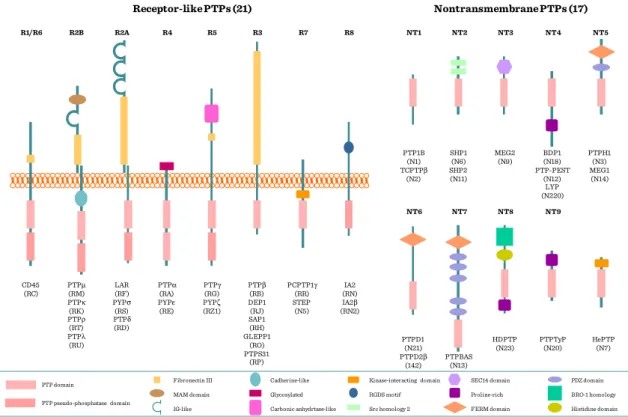
(2) Receptor Tyrosine Phosphatases (e.g. CD45 [cluster determinant-45] protein of T cells and macrophages) (Figure I.2-7 and Figure I.2-8);
(3) Receptor Guanylate Cyclases (e.g. natriuretic peptide receptors) (Figure I.2-9);
(4) Receptor Serine/Threonine Kinases (e.g. activin and TGF-β receptors).
(5) Tyrosine-Kinase Associated Receptors: Receptors that associate with proteins that have tyrosine kinase activity (Cytokine Receptors, T- and B cell receptors, Fc receptors)
GRK
ATP ADP
Arrestin P P P P P
P
G-protein linked receptor kinase
Activated receptor Desensitized receptor
28 The project is funded by the European Union and co-financed by the European SocialFund.
Receptors with intrinsic tyrosine kinase activity are capable of autophosphorylation as well as phosphorylation of other substrates. Additionally, several families of receptors lack intrinsic enzyme activity, yet are coupled to intracellular tyrosine kinases by direct protein-protein interactions (see below).
Figure I.2-7: Kinase-phosphatase balance
Phosphorylase kinase (ser/thr kinase)
PP1c (ser/thr phosphatase) Phosphorylase b
Phosphorylase b
Phosphorylase a P Phosphorylase a P
Inactive Active
P
ATP ADP
CD45 (tyr phosphatase)
Csk (tyr kinase)
ADP ATP
Inactivep56Lck
P Y505
Y394
Primedp56Lck
Y505
Y394
Activep56Lck
P Y394 P
Families of extracellular receptors
Identification number:
TÁMOP-4.1.2-08/1/A-2009-0011
29 Figure I.2-8: Receptor- and cytoplasmic PTPs
Figure I.2-9: Natriuretic peptide signaling
Receptor-like PTPs (21) Nontransmembrane PTPs (17)
CD45 (RC) R1/R6
PTPμ (RM) PTPκ (RK) PTPρ (RT) PTPλ (RU) R2B
LAR (RF) PYPσ (RS) PTPδ (RD) R2A
PTPα (RA) PYPε (RE) R4
PTPγ (RG) PYPζ (RZ1) R5
PTPβ (RB) DEP1 (RJ) SAP1 (RH) GLEPP1
(RO) PTPS31
(RP) R3
PCPTP1γ (RR) STEP (N5) R7
IA2 (RN) IA2β (RN2) R8
HDPTP (N23) NT8 MEG2 (N9) NT3
HePTP (N7) PTPH1 (N3) MEG1 (N14) NT5
SHP1 (N6) SHP2 (N11) NT2
PTPBAS (N13)
NT7
PTPD1 (N21) PTPD2β
(142) NT6 PTP1B (N1) TCPTPβ
(N2) NT1
BDP1 (N18) PTP-PEST
(N12) LYP (N220)
NT4
PTPTyP (N20)
NT9
MAM domain Glycosylated RGDS motif Proline-rich BRO-1 homology
Fibronectin III Cadherine-like Kinase-interacting domain SEC14 domain PDZ domain
IG-like Carbonic anhydrtase-like Src homology 2 FERM domain Histidine domain
PTP domain
PTP pseudo-phosphatase domain
↑NP degradation
↓cAMP?
↑IP3?
↑Vasorelaxation
↑Diuresis, natriuresis
↓Renin, aldosterone
↓Cell proliferation
↓Cardiac fibrosis
↑Vasorelaxation
↓Cell proliferation
↑Long bone gowth Kinase homology domain
Plasma membrane Ligand binding domain Receptor
Hinge region
Guanylyl cyclase domain
Physiologic response Natriuretic peptide
NPR-C NPR-A
(GC-A)
NPR-B (GC-B)
ANP BNP CNP
cGMP
GTP GTP cGMP
PP PPPP
PPPPP PPP PP PPPPP
P
Natriuretic peptide
Hormone bound
Active Desensitised
Kinase
Phosphatase ATP
ATP
cGMP GTP
P P
P
P P
P P Basal
ATP
30 The project is funded by the European Union and co-financed by the European SocialFund.
I.2.3.1 Receptor tyrosine kinases
Introduction, definitions
Tyrosine kinases are signaling proteins with catalytic activity to phosphorylate tyrosine residues. Tyrosine-phosphorylation, in turn, is a ubiquitous signaling event in several pathways. There are two major groups of tyrosine kinases:
(1) “Complete” Receptor tyrosine kinases (RTK) are cell surface receptors with intrinsic kinase activity (i.e. own intracellular kinase domain) [e.g. growth factor receptors].
(2) “Incomplete” or Non-receptor tyrosine kinases (nRTK) are cytosolic or membrane-anchored kinases associated with different cell surface receptors and transmit their signal towards the intracellular signaling networks [e.g. Src family kinases, Syk family kinases].
Families and structure of receptor tyrosine kinases
There are 90 unique Tyr kinases in the human genome, 58 are RTKs, most of them are growth factor-, cytokine- and hormone receptors (Figure I.2-10). Classes: I – EGFR family (ErbB); II – Insulin rec. family; III – PDGF family; IV – FGF family; V – VEGF family; VI – HGF family (c-Met); VII – Trk family; VIII – Eph family; IX – AXL family; X – LTK family; XI – TIE family; XII – ROR family; XIII – DDR family;
XIV – RET family; XV – KLG family; XVI – RYK family; XVII – MuSK family.
Some of those are expressed ubiquitously (EGFR), while others are tissue-specific (NGFR).
All receptor tyrosine kinases have extracellular ligand binding domain(s), a trans- membrane domain and intracellular kinase domain(s). The structure of the ligand
Families of extracellular receptors
Identification number:
TÁMOP-4.1.2-08/1/A-2009-0011
31 binding domains is highly variable, they are built from fibronectin III-, cysteine rich-, factor VIII like-, Ig like-, leucin rich-, EGF like-, kringle-, C1r like-, glycine rich-, cadherin or acid box domains, whereas the transmembrane- and kinase domains are similar (Figure I.2-10).
Figure I.2-10: Receptor tyrosine kinase family
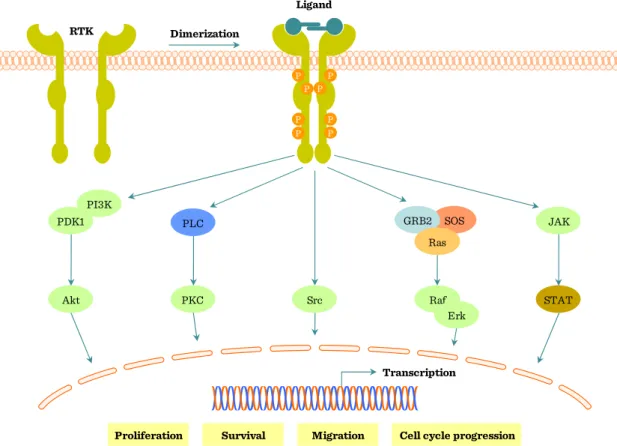
Signaling through receptor tyrosine kinases
Main steps of RTK signaling (Figure I.2-11, Figure I.2-12 and Figure I.2-13):
(1) Ligand binding
(2) Dimerization (except the insulin receptor, which has a tetrameric structure) (3) Autophosphorylation
(4) Signal complex (adapter proteins, kinases etc.)
Fibronectin III Leucine-rich Cysteine-rich
Acid-box Kinase
IG-like
VEGFR1 VEGFR2 VEGFR3 PDGFRα PDGFRβ CSF1R Kit Kit2
Ryk Torso
EGFR ErbB2 ErbB3 ErbB4
Met Ron Sea
TrkA TrkB TrkC INSR
IGF1R IRR
Axl Mer Sky
Eph Eck Eek Erk Elk Ehk1 Ehk2 Sek Hek Hek11 Cek-9 Myk-1 Myk-2
Ros FGFR1
FGFR2 FGFR3 FGFR4
Tie Tie2
DDR Ret Ror1 Torpedo
Ror2 Ltk Alk
EGF-like Cadherin
Factor VIII-like
Glicyne-rich Kringle C1r-like
32 The project is funded by the European Union and co-financed by the European SocialFund.
Figure I.2-11: Receptor tyrosine kinase (RTK) signaling
Proliferation Survival Migration Cell cycle progression Transcription
RTK
Ligand
P
P P P P
P P P Dimerization
Src
SOS GRB2
Ras
Raf Erk PKC
PLC
STAT JAK
Akt PI3K PDK1
Families of extracellular receptors
Identification number:
TÁMOP-4.1.2-08/1/A-2009-0011
33 Figure I.2-12: Members of the signaling complex
Figure I.2-13: Dimerization of GF receptors
SRC
PLCγ
PKC
PI3K RAS
RAF
MEK
MAPKs
Plasma membrane
SOS SHC GRB2
RSK
FAK
Differentiation
Transcriptional regulation
Differentiation/Growth Nucleus
Y Y Y
Y Y Y
Growth factor/Hormone
Receptor PTK
Cytoplasm
Plasma membrane
Cytoplasm
Juxtamembrane region Activation and catalytic loop (substrate precluding) C-terminal region Activation and catalytic loop (substrate accessible)
P P
P P
P P P
P P P
ATP ATP
Dimerization
34 The project is funded by the European Union and co-financed by the European SocialFund.
Members of this initial signal complex include (Figure I.2-14):
(1) enzymes/transcription factors e.g. Src/Syk family kinases, SHP-1, PLCγ, Sos, Vav, RasGAP, STAT1
(2) adaptors/regulators e.g. Grb2, SLP-76, SOCS1, Nck, Shc, Crk-L, p85 (3) adaptors/docking proteins e.g. FRS2, IRS1, DOK1
Figure I.2-14: GF receptor signaling pathways
Upon ligand binding dimerization of receptor tyrosine kinases occurs and receptors become autophosphorylated on tyrosine residues of the kinase domain. This leads to the buildup of the initial signal complex with the help of adaptor/docking
Targets
PIP2 PIP3
Targets Targets
Akt PDK1
PIP3
SOS Ras
Targets
Erk
Targets GRB2
GRB2 Shp2
Shp2
GRB2 GRB2
GRB2 PI3K RTK
Ligand
P P
P P P
P
P P
P
Plasma membrane P
P P P P
P P P
RTK Ligand
DAG IP3 PIP2
PKC
Ca2+
Cbl P
PLC PLC C2
PH SH2 SH2 SH3
Plasma membrane
Cytoplasm P
P P P P
P P P
Families of extracellular receptors
Identification number:
TÁMOP-4.1.2-08/1/A-2009-0011
35 proteins (Figure I.2-14). These adaptor proteins have Src-homology (SH) domains: SH2 domains associate with the autophosphorylated tyrosine residues of the receptors; while SH3 domains associate with the proline rich domains of further signaling molecules including guanine-nucleotid exchange factors (GEF; e.g. Sos, Vav) which catalyze the GDP-GTP exchange on the monomeric G-protein – Ras – which plays a key role in transducing signal from the growth factor receptors (Figure I.2-14). The GTP-bound Ras is activated and leads to activation of the mitogen-activated protein kinase pathway (MAPK-pathway). Ras proteins have only weak GTPase activity, thus, for their rapid inactivation GAP (GTPase activating protein) is also necessary.
Branching of the pathway
After RTK activation more intracellular signaling pathways are activated (Figure I.2-11, page 32):
(1) Ras – Raf – MEK – ERK (MAPK pathway) (2) PLCγ – IP3 –Ca2+ (see I.5.2, page 59) (3) PLCγ – DAG – PKC (see I.5.2, page 59)
(4) PI3 kinase (PI3K) – Protein kinase B (PKB) – Glycogen-synthase kinase (GSK)
(5) STAT activation
36 The project is funded by the European Union and co-financed by the European SocialFund.
The MAPK pathway
Ras activates Raf, a MAP3K, which is a serine/threonine protein kinase. Raf phosphorylates MEK (MAP2K), a dual specific protein kinase, capable of phosphorylating target proteins both on tyrosine and threonine residues. The substrate of MEK is ERK (MAPK), which is a proline-directed kinase that phosphorylates its target proteins on serine/threonine-proline. ERK has many target proteins and can also translocate into the nucleus thereby regulating the transcription of different genes.
MAPK-activated kinases (MK) include:
(1) Cytoplasmic Ribosomal S6 kinases (RSK) [e.g. initiation factors of translation, apoptosis machinery, oestrogen rec., Sos]. In some cases their phosphorylated form can translocate to nucleus [e.g. ATF4, c-Fos, SRF].
(2) Mitogen- and stress-activated kinases (MSK) are found in the nucleus [e.g.
CREB, histone H3, HMGN1, ATF1].
(3) MAPK-interacting kinases (MNK) are components of the translation initiation complex.
Families of extracellular receptors
Identification number:
TÁMOP-4.1.2-08/1/A-2009-0011
37 Figure I.2-15: MAPK/ERK in growth and differentation
There are parallel MAPK cascades activated by different signals. The above described “prototypic” MAPK pathway is the ERK-pathway activated by mitogen signals (Figure I.2-15). Cellular stress or cytokines activate the Jnk- or p38-pathways.
Members of the MAPK pathways form spatially organized intracellular signaling complexes regulated / held together by scaffold proteins (e.g. IMP, KSR1).
Turning-off the pathway
Regulation of the MAPK-pathway is essential to control cell growth and differentiation.
Switching-off the activation is done in part by phosphatases (e.g. PTP1B, SHP1/2, DEP1) which dephosphorylate activated members of the pathway. Phosphorylation of GEF (e.g. Sos) decreases their affinity towards the adapter (e.g. Grb2) leading to the dissociation of the initial signaling complex. GAPs inactivate Ras by changing GTP
Ca2+
FoxO3 ER
Stat1/1
CREB TIF1A
C-Myc/
N-Myc Pax6
Elk-1 C-Fos
Ets UBF ETV1
HMGN1 ATF1 Histone
H3 SRF
TIF1A ETV1
ERa C-Fos Myt1 ATF4
MITF Nur77 Mad1 C/EBPβ
Ran BP3
Erk1/2 MSK1/2
BUB1 p90RSK
P27 KIP1
Erk1 MEK1 PKC
Erk1/2
MNK1/2 MEK1/2
B-Raf c-Raf
c-Raf PKA
PAK
Src Fyn PI3K
FAK
Tpl2/
Cot1
C-TAK1
MPK-1/2 cdc25 MPK-3
PP1/
PP2At
SOS
Bim C3G
SOS
SOS Ras
Rac
Rap1 PLCγ
cPLA2
P14 MP1 IMP
PEA-15
PPARγ Spred
KSR 14-3-3
Shc FRS2
IRS GRB2
Pax CAS Tal
elF4B rpS6
Filamin A IkBa
CRK
eEF2 TSC2 Kinase DAPK
BAD GSK-3
METTL1 nNos
Ca2+
GRB2
PYK2
B-Raf
PD98059 U0126
Late endosome
Cell adhesion Transiation
control Ion channels,
receptors Heterodimer
Cytoskeletal proteins
Progression of cell cycle
Nucleus Cytoplasm
Ca2+
Ion channels RTKs
RTKs
Integrins
Spry
cAMP
p90RSK
38 The project is funded by the European Union and co-financed by the European SocialFund.
back to GDP. Finally, removal of cell surface receptors by endocytosis also contributes to the stopping of activation.
Intracellular receptors
Identification number:
TÁMOP-4.1.2-08/1/A-2009-0011
39
I.3 Intracellular receptors
See chapter II.2.3 Intracellular/nuclear receptor signaling (steroid hormones and thyroxin), page 106.
40 The project is funded by the European Union and co-financed by the European SocialFund.
I.4 Intracellular signal transmitting molecules
I.4.1 G-proteins
Trimeric G-proteins
7-TM receptors associate with G-proteins (=GTP-binding proteins); hence, they are called G-protein-coupled receptors (GPCR), too (Figure I.4-1). G-proteins bind to the intracellular IL2 and IL3 parts of the receptors. Trimeric G-proteins are a complex of α-, β- and γ subunits. In their inactive form, G-protein α subunit binds GDP; upon ligand
binding, this GDP is exchanged to GTP resulting in the active form of the Gα, which dissociates from the complex and associates to effector proteins. Finally, GTP is hydrolyzed by the Gα and the inactivated Gα re-associates with the Gβγ-7-TM receptor complex. The Gγ subunit contains C terminal isoprenyl-chains anchoring it into the plasma membrane (Figure I.4-1). Based on their function, Gα subunits have different types:
(1) Gαs: stimulation of adenylyl-cyclase leading to increase of cAMP (2) Gαi: inhibition of adenylyl-cyclase leading to decrease of cAMP (3) Gαq: activation of PLC
(4) G12: activation of RhoGEF
Activated Gβγ subunits activate K+- and Ca2+-channels and PI3-kinase isoforms (Figure I.4-2).
Intracellular signal transmitting molecules
Identification number:
TÁMOP-4.1.2-08/1/A-2009-0011
41 Figure I.4-1: Activation of G-protein-coupled receptors (GPCR)
Figure I.4-2: G-proteins
GDP β γ α
β γ GTP
α GTP
β γ α
GDP β γ α
Plasma membrane
Cytoplasm
GDP GTP G-protein coupled receptor
(GPCR)
Signal molecule
Inactive G-protein
Activated G-protein subunits GTP
β γ α
GDP β γ α
β γ
GTP Giα
G-protein coupled receptor (GPCR)
Phospholipases Ion channels
Activates Rho
Ion channels PI3K Phospholipases Adenylyl cyclases
Receptor kinases GTP
Gsα
GTP Gqα
GTP G12/13α GTP
α
Ca2+
PLC
PIP2 DAG
IP3 cAMP
Adenylyl cyclase ATP cAMP Adenylyl
cyclase ATP
Plasma membrane
Cytoplasm
42 The project is funded by the European Union and co-financed by the European SocialFund.
Monomeric G-proteins – Ras
Monomeric G proteins were first discovered as transforming oncogenes in Harvey (H- Ras) and Kirsten (K-Ras) sarcoma viruses; hence their name Ras (=Rat sarcoma). N- Ras was first found in human neuroblastoma. Ras is a 189 amino acid long polypeptide, which is anchored to the membrane through lipid chains. It is of special importance, that mutations in the Ras family (Ras, Rho, Rab, Rap, Rheb) are found in 20-30% of all human tumors. Oncogenic point mutations most frequently affect the GTP-binding region of Ras. Ras is regulated by Guanine-nucleotide exchange factors (GEFs), which catalyse the GDP-GTP exchange of Ras, leading to its activation; and GTPase activating protein (GAP), which enhances the intrinsic GTPase activity of Ras, leading to its inactivation. Ras is involved in signaling through growth factor receptors via the Ras-Raf-MEK-ERK (Mitogen-activated protein kinase=MAPK) cascade (Figure I.2-15, page 37). Increased Ras activity (=“constitutively active Ras”) promotes tumor transformation, for example increased G-nucleotide exchange due to point mutations, or decreased GTPase activity due to point mutations or the lack/inactive form of GAP.
I.4.2 Second messengers
Definition and types of second messengers
A major question of signal transduction is how the activation of different extracellular receptors by their ligands (hormones, peptides, cytokines etc.), ie. the extracellular signals, are converted, and transduced into the cells. This second layer of signaling is controlled by second messenger molecules, which are diverse in chemical nature:
(1) hydrophylic molecules e.g. cAMP, cGMP, IP3, Ca2+; (2) hydrophobic molecules (lipids) e.g. diacylglycerol (DAG), phosphatidyl-inositols; or (3) gases: NO, CO, (H2S).
Intracellular signal transmitting molecules
Identification number:
TÁMOP-4.1.2-08/1/A-2009-0011
43 3’-5’Cyclic-AMP (cAMP): the “first” second messenger
In the 1950’s E. W. Sutherland discovered that the effect of adrenaline on liver cells was mediated through cAMP, for his achievement he was awarded Nobel Prize in Physiology and Medicine in 1971. cAMP is synthesised from ATP by adenylyl-cyclase;
and is broken down by cAMP-phosphodyesterase. cAMP activates Protein Kinase A by binding to its regulatory subunits. The targets of PKA include enzymes, structural proteins, and transcription factors like CREB (cAMP-responsive element binding factor) (Figure I.4-3 and Figure I.4-4).
Figure I.4-3: cAMP-PKA pathway
PO4gated channel Gαgated
channel
cAMP gated channel
Receptor
Inactive PKA Activated
PKA GTP
β γ α
Adenylyl cyclase
R R
C C cAMP cAMP cAMP
cAMP R
R C C
C C R cAMP cAMP
cAMPR cAMP
CRE CREB
cAMP Response Element
Gene expression P
CREB P CREB ATP cAMP
GTP α
Nucleus P GTP cAMP
α
P
ADP ATP
44 The project is funded by the European Union and co-financed by the European SocialFund.
Figure I.4-4: More receptors using the same second messenger system
IP3 and DAG
Phospholipase C cleaves phophatidyl-inositol-4,5-bisphosphate (PIP2) found in the plasma membrane into the soluble inositol-trisphophate (IP3) and the membrane resident diacylglycerol (DAG). IP3 initiates a rise in intracellular Ca2+, DAG activates Protein kinase C (PKC) (Figure I.4-5).
Figure I.4-5: IP3 receptor pathway
Glucagon Secretin Adrenaline
ACTH LH FSH
Adenylyl cyclase
ATP cAMP
Hormone
Receptor
Plasma membrane
Cytoplasm
IP3R DAG
GTP
α PLC
PIP2
IP3 IP3
Ca2+
Lumen of smooth endoplasmatic reticulum
Ca2+
Ca2+
Ca2+
Ca2+
Ca2+
Ca2+
Pump Pump Ca2+
channel
+ -
GTP
β γ α G protein
Ca2+
Ca2+