Signal Transduction (Medical Biotechnology)
MD, PhD. Tímea Berki, PTE Általános Orvostudományi Kar MD, PhD. Ferenc Boldizsár, PTE Általános Orvostudományi Kar
MD. Mariann Szabó, PTE Általános Orvostudományi Kar MD, PhD. Gergő Talabér, PTE Általános Orvostudományi Kar MSc, PhD. Zoltán Varecza, PTE Általános Orvostudományi Kar Technical editor: Zsolt Bencze, Veronika Csöngei, Szilvia Czulák Editor in charge: MD, PhD. Tímea Berki, MD, PhD. Ferenc Boldizsár, Rita
Bognár
Signal Transduction (Medical Biotechnology)
by MD, PhD. Tímea Berki, MD, PhD. Ferenc Boldizsár, MD. Mariann Szabó, MD, PhD. Gergő Talabér, and MSc, PhD. Zoltán Varecza
Technical editor: Zsolt Bencze, Veronika Csöngei, Szilvia Czulák
Editor in charge: MD, PhD. Tímea Berki, MD, PhD. Ferenc Boldizsár, Rita Bognár Publication date 2011
Copyright © 2011 Pécsi Tudományegyetem
Copyright 2011, Berki Tímea MD, PhD; Boldizsár Ferenc MD, PhD; Szabó Mariann MD; Talabér Gergő MD, PhD; Varecza Zoltán MSc, PhD
Table of Contents
1. Legend ... 1
2. I General signal transduction ... 2
1. I.1 Introduction, overview of extracellular signaling ... 2
1.1. Communication by extracellular signals usually involves sixsteps ... 2
1.2. Signaling molecules operate over various distances ... 2
1.3. Receptor proteins exhibit ligand-binding and effector specificity ... 2
1.4. Hormones can be classified based on their solubility and receptor location ... 3
2. I.2 Families of extracellular receptors ... 4
2.1. Introduction ... 4
2.2. Extracellular receptor groups ... 4
2.3. I.2.1 Ion-channel receptors ... 5
2.4. I.2.2 7-transmembrane-spanning receptors (7-TM) ... 6
2.4.1. Groups of the 7-transmembrane (7-TM) spanning receptors ... 7
2.4.2. Structure of the 7-transmembrane (7-TM) spanning receptors ... 7
2.4.3. Regulation of 7-TM receptors (GPCR) ... 8
2.5. I.2.3 Enzyme-linked receptors ... 9
2.5.1. I.2.3.1 Receptor tyrosine kinases ... 11
3. I.3 Intracellular receptors ... 16
4. I.4 Intracellular signal transmitting molecules ... 17
4.1. I.4.1 G-proteins ... 17
4.1.1. Trimeric G-proteins ... 17
4.1.2. Monomeric G-proteins – Ras ... 18
4.2. I.4.2 Second messengers ... 18
4.2.1. Definition and types of second messengers ... 18
4.2.2. 3‟-5‟Cyclic-AMP (cAMP): the “first” second messenger ... 18
4.2.3. IP3 and DAG ... 19
4.2.4. Nitric-oxide (NO) and other gases ... 20
4.3. I.4.3 The Ca2+-signal ... 20
4.3.1. Physiological role ... 20
4.3.2. Measuring intracellular Ca2+ ... 21
4.3.3. Phospholipase Cγ (PLCγ) mediated Ca2+ signaling ... 21
4.3.4. Ca2+-channels in the ER (Figure I.4-7) ... 22
4.3.5. Besides IP3, “Alternative” Ca2+-releasing 2nd messengers also exist: ... 23
4.3.6. Ca2+-influx through plasma membrane channels (Figure I.4-7) ... 23
4.3.7. Store-operated Ca2+-entry (SOCE) ... 23
4.3.8. Ca2+-regulated target proteins ... 24
4.3.9. The structural basis of Ca2+-binding ... 24
4.4. I.4.4 Transcription factors ... 24
4.4.1. Definition ... 24
4.4.2. Functional groups ... 25
4.4.3. Structure ... 26
4.4.4. Transcription factors controlling T cell differentiation ... 28
4.4.5. Transcription factors in diseases ... 29
4.4.6. Studying transcription factors ... 30
5. I.5Overview of major signaling pathways ... 30
5.1. I.5.1 cAMP-PKA pathway ... 30
5.2. I.5.2 PLCγ-DAG-PKC ... 30
5.3. I.5.3 MAPK-pathway ... 31
5.4. I.5.4 PI3-kinase-PKB (Akt) ... 31
Signal Transduction (Medical Biotechnology)
1.2.1. The T cell receptor (TcR) complex ... 34
1.2.2. Activation and signaling through the TcR ... 35
1.2.3. Lipid rafts and the immunological synapse ... 36
1.3. II.1.3 Fcg Receptor signaling ... 37
1.3.1. Introduction ... 37
1.3.2. Role and expression of Fcg receptors ... 37
1.3.3. Fcg receptor ITAM/ITIM ... 38
1.3.4. Fcg receptor signal transduction pathway ... 39
1.4. II.1.4 Fce Receptor signaling ... 40
1.4.1. The structure and expression of FcεRs ... 40
1.4.2. FcεRI mediated signaling ... 41
1.4.3. FcεRII (CD23) mediated processes ... 43
1.5. II.1.5 Cytokine signaling ... 44
1.5.1. Definition ... 44
1.5.2. Division, groups ... 44
1.5.3. Receptors ... 44
1.5.4. Janus kinases (JAKs) ... 45
1.5.5. Signal Transducer and Activator of Transcription (STATs) ... 46
1.5.6. Cytokine signaling ... 46
1.5.7. Regulation of JAK/STAT signaling ... 47
1.5.8. Clinical implications: JAK inhibitors ... 47
1.5.9. TNF receptor signaling ... 47
1.6. II.1.6 Chemokine signaling ... 49
1.6.1. Definition ... 49
1.6.2. Nomenclature, groups and receptors ... 50
1.6.3. Signaling ... 50
1.6.4. Implications in diseases ... 51
1.7. II.1.7 Signaling in the innate immune system, PRR signaling ... 51
1.7.1. Endocytic Pattern-Recognition Receptors ... 51
1.7.2. Signaling of Pattern-Recognition Receptors ... 52
1.7.3. Extracellular TLRs ... 52
1.7.4. Families of TLRs ... 52
1.7.5. TLR signaling and function ... 52
1.7.6. Signaling PRRs found in the membranes of the endosomes /phagolysosomes 53 1.7.7. Signaling PRRs found in the cytoplasm: ... 53
1.7.8. Complement receptor signaling ... 54
2. II.2 Hormone and growth factor signaling ... 55
2.1. II.2.1 Tyrosine kinase-linked receptors ... 55
2.1.1. II.2.1.1 Growth-factor signaling ... 55
2.2. II.2.2 G-protein-linked receptors (epinephrine,serotonin,glucagon) ... 60
2.2.1. II.2.2.1 Epinephrine (adrenaline) ... 60
2.2.2. II.2.2.2 Glucagon ... 61
2.2.3. II.2.2.3 Serotonin ... 61
2.3. II.2.3 Intracellular/nuclear receptor signaling (steroidhormonesandthyroxin) ... 61
2.3.1. History ... 62
2.3.2. II.2.3.1 Intracellular receptor families ... 62
2.4. II.2.4 Non-genomic steroid hormone signaling pathways ... 65
2.4.1. Introduction ... 65
2.4.2. Non-genomic glucocorticoid receptor (GR) signaling pathways (Figure II.2-12) 66 2.4.3. Non-genomic effects of other steroid hormones ... 67
3. II.3 Signaling in tumor cells (EGF-R,Her-2R,adhesionmolecules) ... 68
3.1. Introduction ... 68
3.2. EGFR, HER-2 ... 69
3.3. Kidney cancer ... 70
3.4. Integrin signaling ... 70
4. II.4 Apoptosis signaling ... 70
4.1. Introduction ... 70
4.2. Initiation of the cascade ... 70
4.3. Extrinsic apoptosis pathway ... 70
4.4. Intrinsic apoptosis pathway ... 70
4.5. The mitochondrial pathway: ... 71
4.6. Caspase cascade ... 73
5. II.5 Receptor interactions, signaling “cross-talk” ... 74
5.1. Introduction ... 74
5.2. “Levels” of signal “cross-talk” ... 74
6. II.6 Wnt receptor signaling ... 76
6.1. Overview ... 76
6.2. Canonical pathway ... 79
6.3. Non-canonical pathway ... 80
6.4. The role of Wnt-s in T-cell development ... 80
7. II.7 Signaling in the nervous system ... 80
7.1. II.7.1 Acetylcholine (Ach) ... 81
7.2. II.7.2 Noradrenalin (NA) ... 83
7.3. II.7.3 Dopamine (D) ... 83
7.4. II.7.4 Serotonin (5-HT) ... 83
7.5. II.7.5 GABA ... 83
7.6. II.7.6 Glutamate ... 83
7.7. II.7.7 Glycine ... 83
7.8. II.7.8 ATP ... 84
8. II.8 Pharmacological influence of the signaling ... 84
8.1. Introduction ... 84
8.2. Growth factor receptor inhibitors ... 86
8.3. Kinase inhibitors ... 86
8.4. Calcineurin blockade ... 86
8.5. Inhibitors of mTOR ... 86
8.6. Proteosome inhibitors ... 87
8.7. Blocking Hsp-90 ... 88
4. Further reading ... 89
Chapter 1. Legend
Chapter 2. I General signal transduction
1. I.1 Introduction, overview of extracellular signaling
Soluble mediators transmit information through the extracellular space over various distances in cell-to cell communication. In local (short distance) cell signaling, some cells may be in direct contact with each other in order to communicate. Cell-to-cell signaling means that mediators can pass from one cell to another cell through cell junctions, which are found in both animals and plants. Long distance signaling is mediated by hormones between animal cells (endocrine signaling), or growth factors between plant cells. Another general form of long distance signaling is synaptic signaling used mainly in the nervous system. In plants and animals extracellular signaling molecules control metabolic processes, growth and differentiation of tissues, synthesis and secretion of proteins, and the composition of intracellular and extracellular fluids.
1.1. Communication by extracellular signals usually involves sixsteps
(1) Synthesis and release of the extracellular mediator molecule by the signaling cell;
(2) Transport of the mediator to the target cell;
(3) Reception: detection of the signal by a specific receptor protein;
(4) Transduction: binding of the extracellular mediator molecule to a specific receptor on the target cell, and this signal is interpreted by a series of subcellular reactions called signal transduction events.
(5) Response: The signal triggers the desired reaction within the cell, for example a change in cellular metabolism, function, or development triggered by the receptor-signal complex.
(6) Termination: removal of the signal, which often terminates the cellular response.
1.2. Signaling molecules operate over various distances
Based on the distance over which extracellular, secreted molecules transmit the signal, cell-to-cell communication can be classified into three types: endocrine (long distance between the source of the mediator and the target cell – the mediator is transported by the circulation, sometimes bound to transport proteins), paracrine (the source of the mediator and the target cell are relatively close to each other – the mediator is transported by simple diffusion), or autocrine (in this case the mediator-producing- and the target cell is the same). In addition, certain membrane-bound proteins on a cell can directly transmit a signal to adjacent cells.
1.3. Receptor proteins exhibit ligand-binding and effector specificity
The cellular response to a particular extracellular signaling molecule depends on its binding to a specific receptor protein located on the surface or in the nucleus or cytosol of a target cell. The signaling molecule (a hormone, pheromone, or neurotransmitter) acts as a ligand, which binds to, or “fits” into a site on the receptor.
Binding of a ligand to its receptor causes a conformational change in the receptor that initiates a sequence of reactions leading to a specific cellular response.
The response of a cell or tissue to specific hormones is determined by the particular hormone receptors it possesses and by the intracellular reactions initiated by the binding of any one hormone to its receptor. Different cell types may have different sets of receptors for the same ligand, each inducing a different response. Or the same receptor may appear on various cell types, and binding of the same ligand may trigger a different response in each type of cell (e.g. acetylcholine). Clearly, different cells respond in a variety of ways to the same ligand.
On the other hand, different receptor-ligand complexes can induce the same cellular response in some cell types (e.g. glucagons and epinephrine).
Thus, a receptor protein is characterized by binding specificity for a particular ligand, and the resulting hormone-ligand complex exhibits effector specificity (i.e., mediates a specific cellular response).
1.4. Hormones can be classified based on their solubility and receptor location
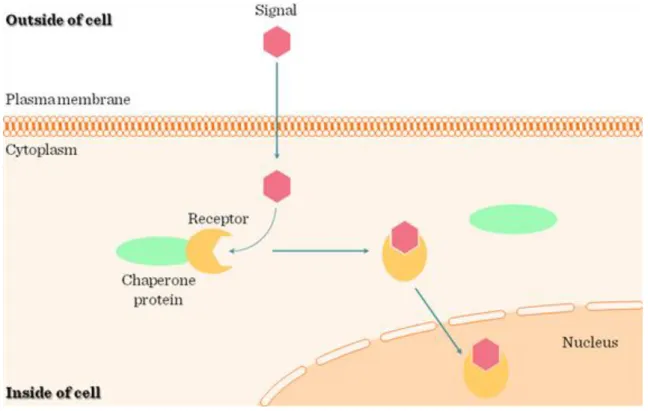
Most hormones fall into three major categories: (1) small lipophilic molecules that diffuse across the plasma membrane and interact with intracellular receptors; and (2) hydrophilic or (3) lipophilic molecules that bind to cell-surface receptors (Figure I.1-1).
(1) Lipophilic hormones with intracellular receptors: many lipid-soluble hormones diffuse across the plasma membrane and interact with receptors in the cytosol or nucleus. The resulting hormone-receptor complexes bind to transcription-control regions of the DNA thereby affecting expression of specific genes. Hormones of this type include the steroids (e.g., cortisol, progesterone, estradiol, and testosterone), thyroxine, and retinoic acid (Figure I.1-1 and Figure I.1-2).
(2) Water-soluble hormones with cell surface receptors: As water-soluble signaling molecules cannot diffuse across the plasma membrane, they all bind to cell-surface receptors. This large class of compounds is composed of two groups: (a) peptide hormones, such as insulin, growth factors, and glucagon, which range in size from a few amino acids to protein-size compounds, and (b) small charged molecules, such as epinephrine and histamine, that are derived from amino acids and function as hormones or neurotransmitters. Many water- soluble hormones induce a modification in the activity of one or more enzymes already present in the target cell.
In this case, the effects of the surface-bound hormone are usually nearly immediate, but persist for a short period only. These signals also can give rise to changes in gene expression that may persist for hours or days. In yet other cases water-soluble signals may lead to irreversible changes, such as cellular differentiation.
(3) Lipophilic hormones with cell-surface receptors: The primary lipid-soluble hormones that bind to cell- surface receptors are the prostaglandins. There are at least 16 different prostaglandins in nine different chemical classes, designated PGA – PGI. Prostaglandins are part of an even larger family of hormones containing 20 carbon atoms called eicosanoid hormones. In addition to prostaglandins, they include prostacyclins, thromboxanes, and leukotrienes. Eicosonoid hormones are synthesized from a common precursor, arachidonic acid. Arachidonic acid is generated from phospholipids and diacylglycerol.
I General signal transduction
Figure I.1-2: Intracellular receptor signaling
2. I.2 Families of extracellular receptors
2.1. Introduction
Ligands act on extra- or intracellular receptors according to their hydrophilic or hydrophobic nature, respectively (Figure I.1-1). Hydrophobic/lipophilic molecules (e.g. steroid hormones, thyroid hormone, vitamin D) can diffuse through the plasma membrane lipid layer, thus reaching intracellular receptors (Figure I.1-2) (for details see Chapter II.2.3). Hydrophylic/water soluble ligands (e.g. peptide hormones, cytokines, chemokines, neurotransmitters), on the other hand, are unable to penetrate the lipid rich outer barrier of the cells; therefore, they need receptors protruding from the outer surface of the cell membrane.
2.2. Extracellular receptor groups
In case of extracellular receptors, the effect of ligand binding has to be transmitted into the cell. A number of signal transduction pathways have evolved to serve this purpose.
Extracellular receptors belong to 3 major categories (Figure I.2-1):
(1) Ion-channel receptors
(2) 7-transmembrane-spanning receptors (7-TM), also called G-protein-coupled receptors (GPCR) (3) Enzyme-linked receptors
Figure I.2-1: Extracellular receptor types
2.3. I.2.1 Ion-channel receptors
Ligand-operated receptors in the plasma membrane (e.g. GABAA, GABAC, iGlu, Glycine, Serotonin, nicotinic Ach, P2X receptors) belong to this group. They are quite abundant in the nervous system and on contractile cells (smooth/striated/heart muscle). Their function is relatively simple: upon activation they open and ion currents occur as a consequence of the concentration gradient between the extra- and intracellular environment. The transient ion concentration changes lead to the contraction or depolarization of the target cells. There are 3 groups of ion-channel receptors:
(1) Cys-loop receptors: have pentameric structure with 4 trans-membrane (TM) regions in each subunit (e.g.
Acetylcholin (Ach) Nicotinic Receptor – Na+ channel; GABAA, GABAC, Glycine – Cl- channels [inhibitory role in CNS]) (Figure I.2-2 and Figure I.2-3).
(2) Glutamate-activated cationic channels: have tetrameric structure with 3 TM regions in each subunit (e.g.
iGlu) [excitatory role in CNS]
(3) ATP-gated channels: have three homologous subunits with two TM regions in each subunit (e.g. P2X purinoreceptor)
I General signal transduction
Figure I.2-2: Cys-loop ion-channel receptors
Figure I.2-3: Synapse between two neurons - neurotransmission
2.4. I.2.2 7-transmembrane-spanning receptors (7-TM)
2.4.1. Groups of the 7-transmembrane (7-TM) spanning receptors
The 7-TM receptor family (Table I.2-1) is one of the largest gene families in vertebrates comprising more than 700 members. They fall into:
(1) Class A: Rhodopsin-like (e.g. prostaglandins, thromboxanes, serotonine, dopamine, histamine, catecholamines, Ach (M), rhodopsin, melatonin, chemokines, bradykinin, somatostatin, opioid, vasopressin receptors)
(2) Class B: Secretin family (e.g. glucagon, GnRH, PTH, CRH receptors)
(3) Class C: Glutamate and GABA (metabotropic) (Glutamate, GABA, sweet taste, secretin receptors) (4) Frizzled (e.g. Wnt, Hedgehog, bitter taste receptors)
(5) Adhesion family (e.g. chondroitin sulfate receptors) groups.
Despite the complexity of the already identified ligands, it has to be noted, that there are still more than 200
“orphan” receptors (= no identified ligand yet) in the 7-TM family.
Table I.2-1: Receptor types
2.4.2. Structure of the 7-transmembrane (7-TM) spanning receptors
As implicated by their name, the polypeptide chain of these receptors crosses the plasma membrane 7 times (Figure I.2-4); the N-terminus is extracellular, while the C-terminus is intracellular. The transmembrane (TM) α- helical domains are separated from each other by extracellular- and intracellular loops (EL and IL). These domains take on a “barrel-like” conformation in the membrane, with the ligand-binding site in the middle.
While the extracellular ligand-binding region of the receptors show variation,, the transmembrane and intracellular parts, on the other hand are more conserved. Palmitoylation of cysteine residues at the C terminus establish the connection of the 7-TM receptors to cholesterol and sphingolipid rich membrane microdomains
I General signal transduction
Figure I.2-4: 7-transmembrane-spanning receptors (7-TM)
Figure I.2-5: Structure of 7-TM receptors
2.4.3. Regulation of 7-TM receptors (GPCR)
(1) 7-TM activation is regulated by the phosphorylation of the C terminus of the receptors by PKA (feedback phosphorylation) or G-protein receptor kinases (GRK1-7).
(2) Translocation: the active receptor with the surrounding membrane is internalized – dephosphorylated in acidic vesicles and recycled to the surface.
(3) Arrestin linking: binding of arrestin molecules inhibit the binding of Gs proteins to the receptors (e.g.
rhodopsin in retina); + activation of alternative pathways: MAPK, PI3-K, PKB/Akt, Src (Figure I.2-6).
Figure I.2-6: Receptor desensitization
2.5. I.2.3 Enzyme-linked receptors
Enzyme-linked receptors are a group of multi-subunit transmembrane proteins that possess either intrinsic enzymatic activity in their intracellular domain or associate directly with an intracellular enzyme (Figure I.2-1).
Generally, upon ligand binding a conformational change is transmitted via a transmembrane helix, which activates enzymatic activity initiating signaling cascades.
Groups of receptors that have intrinsic enzymatic activities include:
(1) Receptor Tyrosine Kinases (RTK) (e.g. PDGF, insulin, EGF, VEGF and FGF receptors) (Figure I.2-7);
(2) Receptor Tyrosine Phosphatases (e.g. CD45 [cluster determinant-45] protein of T cells and macrophages) (Figure I.2-7 and Figure I.2-8);
(3) Receptor Guanylate Cyclases (e.g. natriuretic peptide receptors) (Figure I.2-9);
(4) Receptor Serine/Threonine Kinases (e.g. activin and TGF-β receptors).
(5) Tyrosine-Kinase Associated Receptors: Receptors that associate with proteins that have tyrosine kinase activity (Cytokine Receptors, T- and B cell receptors, Fc receptors)
Receptors with intrinsic tyrosine kinase activity are capable of autophosphorylation as well as phosphorylation of other substrates. Additionally, several families of receptors lack intrinsic enzyme activity, yet are coupled to intracellular tyrosine kinases by direct protein-protein interactions (see below).
I General signal transduction
Figure I.2-7: Kinase-phosphatase balance
Figure I.2-8: Receptor- and cytoplasmic PTPs
I General signal transduction
Tyrosine kinases are signaling proteins with catalytic activity to phosphorylate tyrosine residues. Tyrosine- phosphorylation, in turn, is a ubiquitous signaling event in several pathways. There are two major groups of tyrosine kinases:
(1) “Complete” Receptor tyrosine kinases (RTK) are cell surface receptors with intrinsic kinase activity (i.e.
own intracellular kinase domain) [e.g. growth factor receptors].
(2) “Incomplete” or Non-receptor tyrosine kinases (nRTK) are cytosolic or membrane-anchored kinases associated with different cell surface receptors and transmit their signal towards the intracellular signaling networks [e.g. Src family kinases, Syk family kinases].
2.5.1.2. Families and structure of receptor tyrosine kinases
There are 90 unique Tyr kinases in the human genome, 58 are RTKs, most of them are growth factor-, cytokine- and hormone receptors (Figure I.2-10). Classes: I– EGFR family (ErbB); II– Insulin rec. family; III– PDGF family; IV– FGF family; V– VEGF family; VI– HGF family (c-Met); VII– Trk family; VIII– Eph family; IX–
AXL family; X– LTK family; XI–TIE family; XII– ROR family; XIII– DDR family; XIV– RET family; XV–
KLG family; XVI– RYK family; XVII– MuSK family. Some of those are expressed ubiquitously (EGFR), while others are tissue-specific (NGFR).
All receptor tyrosine kinases have extracellular ligand binding domain(s), a trans-membrane domain and intracellular kinase domain(s). The structure of the ligand binding domains is highly variable, they are built from fibronectin III-, cysteine rich-, factor VIII like-, Ig like-, leucin rich-, EGF like-, kringle-, C1r like-, glycine rich-, cadherin or acid box domains, whereas the transmembrane- and kinase domains are similar (Figure I.2- 10).
Figure I.2-10: Receptor tyrosine kinase family
2.5.1.3. Signaling through receptor tyrosine kinases
Main steps of RTK signaling (Figure I.2-11, Figure I.2-12 and Figure I.2-13):
(1) Ligand binding
(2) Dimerization (except the insulin receptor, which has a tetrameric structure) (3) Autophosphorylation
(4) Signal complex (adapter proteins, kinases etc.)
Figure I.2-11: Receptor tyrosine kinase (RTK) signaling
I General signal transduction
Figure I.2-12: Members of the signaling complex
Figure I.2-13: Dimerization of GF receptors
Members of this initial signal complex include (Figure I.2-14):
(1) enzymes/transcription factors e.g. Src/Syk family kinases, SHP-1, PLCg, Sos, Vav, RasGAP, STAT1 (2) adaptors/regulators e.g. Grb2, SLP-76, SOCS1, Nck, Shc, Crk-L, p85
(3) adaptors/docking proteins e.g. FRS2, IRS1, DOK1
Figure I.2-14: GF receptor signaling pathways
Upon ligand binding dimerization of receptor tyrosine kinases occurs and receptors become autophosphorylated on tyrosine residues of the kinase domain. This leads to the buildup of the initial signal complex with the help of adaptor/docking proteins (Figure I.2-14). These adaptor proteins have Src-homology (SH) domains: SH2 domains associate with the autophosphorylated tyrosine residues of the receptors; while SH3 domains associate with the proline rich domains of further signaling molecules including guanine-nucleotid exchange factors (GEF; e.g. Sos, Vav) which catalyze the GDP-GTP exchange on the monomeric G-protein – Ras – which plays a key role in transducing signal from the growth factor receptors (Figure I.2-14). The GTP-bound Ras is activated and leads to activation of the mitogen-activated protein kinase pathway (MAPK-pathway). Ras proteins have only weak GTPase activity, thus, for their rapid inactivation GAP (GTPase activating protein) is also necessary.
2.5.1.4. Branching of the pathway
After RTK activation more intracellular signaling pathways are activated (Figure I.2-11):
(1) Ras – Raf – MEK – ERK (MAPK pathway)
I General signal transduction
2.5.1.5. The MAPK pathway
Ras activates Raf, a MAP3K, which is a serine/threonine protein kinase. Raf phosphorylates MEK (MAP2K), a dual specific protein kinase, capable of phosphorylating target proteins both on tyrosine and threonine residues.
The substrate of MEK is ERK (MAPK), which is a proline-directed kinase that phosphorylates its target proteins on serine/threonine-proline. ERK has many target proteins and can also translocate into the nucleus thereby regulating the transcription of different genes. MAPK-activated kinases (MK) include:
(1) Cytoplasmic Ribosomal S6 kinases (RSK) [e.g. initiation factors of translation, apoptosis machinery, oestrogen rec., Sos]. In some cases their phosphorylated form can translocate to nucleus [e.g. ATF4, c-Fos, SRF].
(2) Mitogen- and stress-activated kinases (MSK) are found in the nucleus [e.g. CREB, histone H3, HMGN1, ATF1].
(3) MAPK-interacting kinases (MNK) are components of the translation initiation complex.
Figure I.2-15: MAPK/ERK in growth and differentation
There are parallel MAPK cascades activated by different signals. The above described “prototypic” MAPK pathway is the ERK-pathway activated by mitogen signals (Figure I.2-15). Cellular stress or cytokines activate the Jnk- or p38-pathways. Members of the MAPK pathways form spatially organized intracellular signaling complexes regulated / held together by scaffold proteins (e.g. IMP, KSR1).
2.5.1.6. Turning-off the pathway
Regulation of the MAPK-pathway is essential to control cell growth and differentiation. Switching-off the activation is done in part by phosphatases (e.g. PTP1B, SHP1/2, DEP1) which dephosphorylate activated members of the pathway. Phosphorylation of GEF (e.g. Sos) decreases their affinity towards the adapter (e.g.
Grb2) leading to the dissociation of the initial signaling complex. GAPs inactivate Ras by changing GTP back to GDP. Finally, removal of cell surface receptors by endocytosis also contributes to the stopping of activation.
3. I.3 Intracellular receptors
See chapter II.2.3 Intracellular/nuclear receptor signaling (steroidhormonesandthyroxin).
4. I.4 Intracellular signal transmitting molecules
4.1. I.4.1 G-proteins
4.1.1. Trimeric G-proteins
7-TM receptors associate with G-proteins (=GTP-binding proteins); hence, they are called G-protein-coupled receptors (GPCR), too (Figure I.4-1). G-proteins bind to the intracellular IL2 and IL3 parts of the receptors.
Trimeric G-proteins are a complex of α-, β- and γ subunits. In their inactive form, G-protein α subunit binds GDP; upon ligand binding, this GDP is exchanged to GTP resulting in the active form of the Gα, which dissociates from the complex and associates to effector proteins. Finally, GTP is hydrolyzed by the Gα and the inactivated Gα re-associates with the Gβγ-7-TM receptor complex. The Gγ subunit contains C terminal isoprenyl-chains anchoring it into the plasma membrane (Figure I.4-1). Based on their function, Gα subunits have different types:
(1) Gαs: stimulation of adenylyl-cyclase leading to increase of cAMP (2) Gαi: inhibition of adenylyl-cyclase leading to decrease of cAMP (3) Gαq: activation of PLC
(4) G12: activation of RhoGEF
Activated Gβγ subunits activate K+- and Ca2+-channels and PI3-kinase isoforms (Figure I.4-2).
Figure I.4-1: Activation of G-protein-coupled receptors (GPCR)
I General signal transduction
Figure I.4-2: G-proteins
4.1.2. Monomeric G-proteins – Ras
Monomeric G proteins were first discovered as transforming oncogenes in Harvey (H-Ras) and Kirsten (K-Ras) sarcoma viruses; hence their name Ras (=Rat sarcoma). N-Ras was first found in human neuroblastoma. Ras is a 189 amino acid long polypeptide, which is anchored to the membrane through lipid chains. It is of special importance, that mutations in the Ras family (Ras, Rho, Rab, Rap, Rheb) are found in 20-30% of all human tumors. Oncogenic point mutations most frequently affect the GTP-binding region of Ras. Ras is regulated by Guanine-nucleotide exchange factors (GEFs), which catalyse the GDP-GTP exchange of Ras, leading to its activation; and GTPase activating protein (GAP), which enhances the intrinsic GTPase activity of Ras, leading to its inactivation. Ras is involved in signaling through growth factor receptors via the Ras-Raf-MEK-ERK (Mitogen-activated protein kinase=MAPK) cascade (Figure I.2-15). Increased Ras activity (=“constitutively active Ras”) promotes tumor transformation, for example increased G-nucleotide exchange due to point mutations, or decreased GTPase activity due to point mutations or the lack/inactive form of GAP.
4.2. I.4.2 Second messengers
4.2.1. Definition and types of second messengers
A major question of signal transduction is how the activation of different extracellular receptors by their ligands (hormones, peptides, cytokines etc.), ie. the extracellular signals, are converted, and transduced into the cells.
This second layer of signaling is controlled by second messenger molecules, which are diverse in chemical nature: (1)hydrophylic molecules e.g. cAMP, cGMP, IP3, Ca2+; (2)hydrophobic molecules (lipids) e.g.
diacylglycerol (DAG), phosphatidyl-inositols; or (3)gases: NO, CO, (H2S).
4.2.2. 3’-5’Cyclic-AMP (cAMP): the “first” second messenger
In the 1950‟s E. W. Sutherland discovered that the effect of adrenaline on liver cells was mediated through cAMP, for his achievement he was awarded Nobel Prize in Physiology and Medicine in 1971. cAMP is synthesised from ATP by adenylyl-cyclase; and is broken down by cAMP-phosphodyesterase. cAMP activates Protein Kinase A by binding to its regulatory subunits. The targets of PKA include enzymes, structural proteins, and transcription factors like CREB (cAMP-responsive element binding factor) (Figure I.4-3 and Figure I.4-4).
Figure I.4-3: cAMP-PKA pathway
Figure I.4-4: More receptors using the same second messenger system
4.2.3. IP3 and DAG
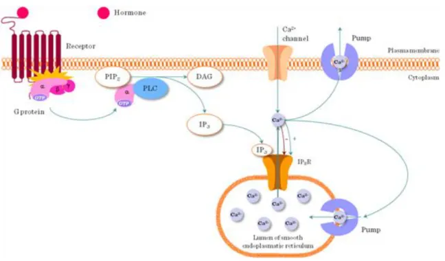
Phospholipase C cleaves phophatidyl-inositol-4,5-bisphosphate (PIP2) found in the plasma membrane into the soluble inositol-trisphophate (IP3) and the membrane resident diacylglycerol (DAG). IP3 initiates a rise in intracellular Ca2+, DAG activates Protein kinase C (PKC) (Figure I.4-5).
I General signal transduction
Figure I.4-5: IP3 receptor pathway
4.2.4. Nitric-oxide (NO) and other gases
The “quest” for “Endothelium-derived relaxation factor” (EDRF) was precipitated when multiple research groups found independently that endothelial cells could produce mediator(s) that lead to vasodilatation; this mediator turned out to be NO in 1983. The “star“ of NO rose rapidly: in 1992 NO was named “molecule of the year” by Science magazine; a “Nitric Oxide Society” was established; and in 1998 a shared Nobel Prize was awarded to F. Murad, R.F. Furchgott and L. Ignarro in Physiology or Medicine for the discovery of NO.
NO is synthesized by NO synthase, which has 3 forms: endothelial cells constitutively express eNOS; iNOS is inducible (e.g. in macrophages); and nNOS is neuronal. NOS has 2 major domains: the N-terminal Oxygenase (similar to heme-thiolate proteins), the C-terminal Reductase (similar to NADPH-cytochrome P450 reductase) and a Linker: calmodulin-binding sequence. NO is synthesized from L-arginine, in a chemical reaction where L- Arg is transformed into citrulline coupled by NO production.
NO is a highly active free radical which triggers oxidative stress. However, in smooth muscle cells NO activates guanylyl-cyclase, which produces cGMP from GTP. Protein kinase G is a Ser/Thr protein kinase activated by cGMP. PKG is expressed by vascular smooth muscle cells, platelets, endothelial cells, heart muscle, fibroblasts, renal cells, leukocytes, nervous system and regulates smooth muscle relaxation, platelet function, sperm metabolism, cell division and nucleic acid synthesis.
NO effects are utilized for various medical treatments:
(1) Vascular effects are based on vasodilatation e.g. Nitroglycerin for the treatment of coronary disease (Angina pectoris); for the treatment of erectile dysfunction to induce vasodilatation in the penis (Viagra).
(2) Heart muscle effects include decreased contractility and heart rate.
In the immune system: macrophages produce NO to kill bacteria but in severe systemic infection (sepsis) this can lead to generalized vasodilatation and shock (Septic shock).
4.3. I.4.3 The Ca2+-signal
4.3.1. Physiological role
S. Ringer found that in the presence of Ca2+ isolated frog heart maintained activity for hours, therefore Ca2+ is essential for heart function. Locke described that absence of Ca2+ inhibited neuromuscular transmission.
Kamada and Kimoshita discovered in 1943 that introduction of Ca2+ into muscle fibers caused their
contraction. Although Otto Loewi claimed “Ca2+ ist alles.” (=Ca2+ is everything), Ca2+ was identified as second messenger only after cAMP, thus became only the “second” messenger.
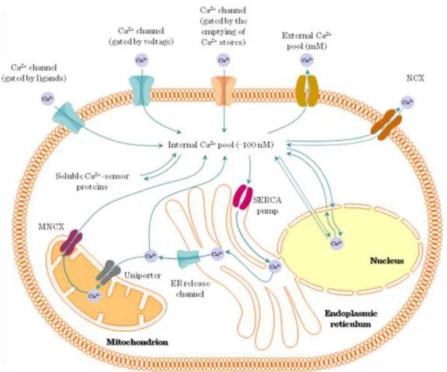
Ca2+ is found in 3 forms in the body: free, bound or trapped (hydroxiapatite in calcified tissues e.g. bones, teeth). The plasma Ca2+ level is tightly regulated: hypercalcemia leads to reduced neuromuscular transmission, myocardial dysfunction and lethargy; whereas hypocalcemia leads to increased excitability of membranes, tetany, seizures and death.
The normal range of Ca2+ in plasma or extracellular fluid is 1-2mM; 50-100nM in the intracellular space / cytoplasm; and 30-300mM in the intracellular Ca2+-stores. Cytoplasmic Ca2+ is kept low by Ca2+-ATPases in the plasma membrane and ER (SERCA), and Na+/Ca2+ exchanger in the plasma membrane. Ionophores are lipid-soluble, membrane-permeable ion-carriers e.g. A23187 (524kDa) or ionomycin (709kDa) isolated from Streptomyces.
4.3.2. Measuring intracellular Ca2+
(1) Classically, for the measurement of intracellular Ca2+ concentration changes Ca2+-sensitive photoproteins, for example Aequorin (isolated from the jelly fish Aequoria Victoria) was used, which emits blue light when bound with Ca2+. This was first microinjected into a target cell (e.g. giant squid axon) and then stimulation was applied.
(2) Fluorescent indicators, for example Quin-2, Fura-2 (UV) or Fluo-3, Fluo-4 (visible light) can be used for measuring intracellular Ca2+ level in cell suspensions using flow cytometry or spectrophotometry. Here, the signal represents the summation of individual unsynchronized contributions. Single cell measurement is possible with fluorescent/confocal microscope. On a single cell level the shape of the Ca2+ signal is usually
“spike” or “wave”.
(3) Genetically engineered indicators e.g. aequorin-transfected cells or Calmodulin-Myosin light chain Kinase- GFP construct can also be used for Ca2+ measurement.
4.3.3. Phospholipase Cγ (PLCγ) mediated Ca2+ signaling
Signals from cell surface receptors (e.g. GPCR) lead to PLCγ activation. PLCγ is a membrane proximal signaling protein which cleaves phosphatidyl-inositol-bisphosphate (PIP2) into phosphatidyl-inositol- trisphosphate (IP3) and diacyl-glycerol (DAG). IP3 releases Ca2+ from the endoplasmic reticulum, whereas DAG activates Protein kinase C (PKC). This step represents an important branching of the PLCγ pathway (Figure I.4-5). This pathway is activated by a number of different extracellular stimuli through a variety of receptors (Figure I.4-6).
I General signal transduction
Figure I.4-6: Several pathways use the Ca2+ signal
4.3.4. Ca2+-channels in the ER (Figure I.4-7)
(1) Ryanodine receptor (RyR), expressed in excitable cells (skeletal & cardiac muscle)) has four 560kDa subunits. Its modulators are Ca2+, ATP, calmodulin, FKBP12 (immunophilin).
(2) IP3 receptor (IP3R) has four 310kDa subunits.
Ca2+-induced Ca2+ release (CICR)
When cytoplasmic Ca2+ rises, neighbouring Ca2+ channels are activated progressively. Their opening leads to a Ca2+ “wave”. This is an example of positive feedback.
Figure I.4-7: Intra/extracellular compartments of Ca2+-signaling, Ca2+-channels
4.3.5. Besides IP3, “Alternative” Ca2+-releasing 2nd messengers also exist:
(1) Cyclic-ADP-ribose (cADPR) is generated by ADP-ribosyl cyclase (e.g. CD38 ectoenzyme). It participates in panceratic β-cell glucose response and TcR signaling
(2) Nicotinic acid adenine dinucleotide phosphate (NAADP) was first described in sea urchin eggs. It is a mediator of CCK effects on pancreas acinar cells and TcR signaling.
(3) Sphingosine-1-phosphate (S1P) is generated from ceramide by sphingosine-kinases upon activation by FcRs (ε, γ), GFRs (PDGF, VEGF), or cytokine rec. (IL-1, TNFα). S1P transmembrane transport is perfomed by ABCC1, cell surface receptors: S1P1, S1P5.
4.3.6. Ca2+-influx through plasma membrane channels (Figure I.4-7)
(1) Voltage-operated channels (VOCCs) are found on nerve and muscle cells. They open upon depolarization.
L, N, P/Q, R and T types
(2) Receptor-operated channels (e.g. Glutamate NMDA rec.).
(3) TRPM2 channels are activated by ADP-ribose or oxidative stress.
I General signal transduction
(1) Direct interaction between ER and plasma membrane (2) Movement of STIM1 from the ER to the plasma membrane
(3) The existence of a soluble mediator – CIF (Ca2+-influx factor) (1993.)
4.3.8. Ca2+-regulated target proteins
(1) Calmodulin-dependent (Figure I.4-8): Calmodulin phosphorylates CaM kinases, EF2 kinase, phosphorylase kinase and myosin-light chain kinase (MLCK); dephosphorylates calcineurin, which, in turn activates NFAT (Nuclear Factor of Activated T cells). Calmodulin also regulates Ca2+ transport via plasma membrane Ca2+
ATPases, cyclic nucleotide metabolism through Adenylyl-cyclase and Cyclic Nucleotide Phosphodiesterase, cytoskeleton components (e.g. MAP-2, Tau, fodrin, neuromodulin) and nitric-oxide synthase (NOS).
(2) Calmodulin-independent target proteins include:
a) Neuronal Ca2+ sensors
b) Calpain (Ca2+-activated Cys protease) c) Synaptotagmin – exocytosis
d) DAG kinase – inactivation of DAG e) Ras GEFs & GAPs
f) Cytoskeletal proteins a-actinin, gelsolin
Figure I.4-8: Effector mechanisms of Ca2+-signaling
4.3.9. The structural basis of Ca2+-binding
(1) EF-hand motifs are helix-loop-helix, the loop consists of cca.12AA-s forming the Ca2+-binding site, and they usually form pairs (=unit).
(2) C2 domains contain cca.130 AA-s, forming rigid 8-stranded antiparallel β-sheets.
4.4. I.4.4 Transcription factors
4.4.1. Definition
Transcription factors are sequence-specific DNA-binding factors that control the transmission of genetic information from DNA to mRNA. They act as activators (=promote gene expression) or repressors (=inhibit gene expression) by affecting the recruitment of RNA polymerase to the transcription initiation complex (Figure I.4-9).
Figure I.4-9: Regulation of transcription
4.4.2. Functional groups
(1) General TFs: constitutively active, present in all cells at all times, bind TATA box, form pre-initiation complex e.g. TFIIA-H.
(2) Specific transcription factors/upstream transcription factors are conditionally active (Table I.4-1).
A) Developmental (cell specific) e.g. GATA, MyoD, Hox, Winged helix B) Signal-dependent
a) extracellular ligand (e.g. nuclear receptors) b) intracellular ligand (e.g. SREBP, p53) c) cell-membrane rec. dependent d) resident nuclear CREB, AP-1, Mef-2
e) latent cytoplasmic STAT, NFAT, NFkB, Notch Table I.4-1: Some important transcription factors
I General signal transduction
Gene transcription is regulated in a complex manner: the basic transcription machinery (general transcription factors and the RNA polymerase) interacts with numerous co-regulators (specific transcription factors).
Activators bind to enhancer elements, repressors bind to silencer elements of the DNA upstream from the TATA box. However, the exact positions of such regulatory DNA elements are highly variable.
4.4.3. Structure
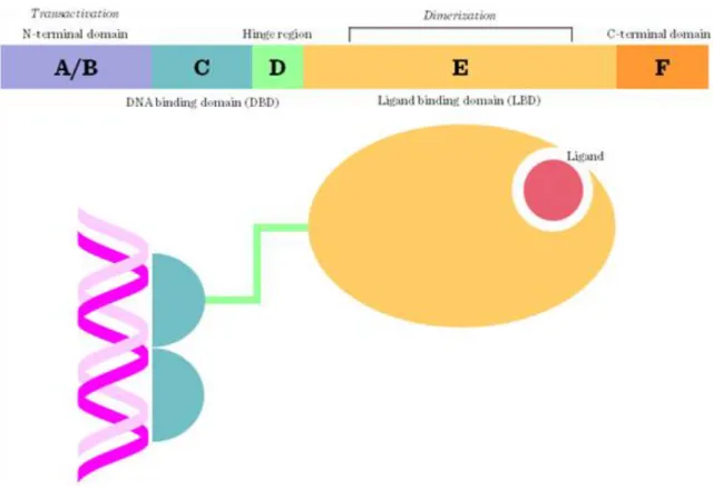
Generally, transcription factors contain (1) a DNA-binding domain (DBD) responsible for the direct interaction with DNA response elements; (2) a signal-sensing domain (SSD) responsible for the detection of extracellular signals e.g. ligand-binding; and (3) a transactivation domain (TAD) interacting with transcription co-regulators (Figure I.4-10). Most transcription factors contain helix-loop-helix, zinc-finger or leucin-zipper motifs and bind to the DNA as dimers (Figure I.4-11).
Figure I.4-10: Functional domains of transcription factors
Figure I.4-11: Structural groups of transcription factors
I General signal transduction
(3) Zinc-coordinating DNA-binding domains e.g. Zinc fingers: nuclear receptors for steroids, thyroid hormone;
GATA factors
(4) Helix-turn-helix e.g. Homeobox; Forkhead / winged helix
(5) Beta-scaffold factors with minor groove contacts e.g. NFkB. NFAT, STAT, p53 (6) Others
4.4.4. Transcription factors controlling T cell differentiation
T lymphocytes are central players of the adaptive immune mechanisms. They derive from the bone marrow common lymphoid precursor, then, the early progenitors migrate to the thymus where they undergo a series of central differentiation steps, which are tightly controlled by specific transcription factors (Figure I.4-12).
Finally, T cells leave the thymus as helper (CD4+) or cytotoxic (CD8+); this lineage decision is also under the control of transcription factors (Figure I.4-13). In the peripheral lymphatic organs, naïve CD4+ T cells reach their final differentiation stages: Th1, Th2, Th17 and Treg subpopulations, controlled by T-bet, GATA-3, RORγ and FoxP3 transcription factors, respectively (Figure I.4-14).
Figure I.4-12: Role of transcription factors in thymocyte development
Figure I.4-13: Th - Tc cell decision
Figure I.4-14: Th differentiation
I General signal transduction
(2) Rett-syndrome – MECP2 (3) Li-Fraumeni-syndrome – p53
4.4.6. Studying transcription factors
(1) Transcription factor activity might be tested by
a) Luciferase reporter assay: transfection of the target cells with a plasmid containing the luciferase gene under the control of the promoter to be studied. Upon transcription factor-binding light is emitted.
b) Chromatin immunoprecipitation (ChIP): after the biological treatment the activated transcription factors are fixed to the DNA by formaldehyde, then the genomic DNA is extracted and fragmented. The fragments are precipitated by transcription factor specific antibody(s). The precipitate is disrupted and gene-specific PCR or sequencing (ChIP-Seq) can be performed on the purified DNA. ChIP can be combined with microarray (ChIP- on-chip). Thus, high throughput screening of gene networks controlled by a transcription factor becomes possible.
c) Electrophoretic Mobility Shift Assay (EMSA) is based on the alteration in the migration speed of DNA in complex with transcription factor(s).
(2) Transcription factor interactions (physical association) can be assessed by co-immunoprecipitation.
5. I.5Overview of major signaling pathways
5.1. I.5.1 cAMP-PKA pathway
5.2. I.5.2 PLCγ-DAG-PKC
5.3. I.5.3 MAPK-pathway
5.4. I.5.4 PI3-kinase-PKB (Akt)
Chapter 3. II Detailed (systematic) signal transduction
1. II.1 Signaling in the immune system
The immune system functions as a finely regulated network of innate and adaptive mechanisms, with the ability to recognize and distinguish between self and non-self structures. Immunological steady state is continuously maintained on one hand by attacking and eliminating foreign invaders and tumor cells, and providing tolerance of important self-antigens on the other hand. Immunological recognition molecules are cell surface receptors (T cell receptor, B cell receptor, Fc receptors, Complement receptors, Toll-like receptors etc.), which, in most cases, have no intrinsic enzymatic activity, hence they use cytoplasmic non-receptor tyrosine kinases and adaptor molecules for signaling. Tyrosine-phosphorylation is a common event during immunological signaling;
thus specialized tyrosine containing signal sequence motifs have evolved: ITAM (Immunoreceptor Tyrosine- based Activation Motif) and ITIM (Immunoreceptor Tyrosine-based Inhibition Motif). ITAM is a specific sequence of amino acids (YXXL) occurring twice in close succession in the intracellular tail of a receptor, whereas ITIM sequence is as follows: I/VXXYXXL. Signals through these receptors are converted into a plethora of complex biological responses: proliferation, differentiation, phagocytosis, apoptosis or anergy.
1.1. II.1.1 Signaling in the specific immune system 1:
Bcellsignaling
1.1.1. The B cell receptor (BcR) complex
B-lymphocytes are part of the adaptive immune system, their antigen recognition molecule is the B cell receptor (BcR), which is structurally a cell surface-bound monomeric immunoglobulin molecule. Having only a short transmembrane and intracellular part, the BcR associates with the Igα/β chains, which contain ITAM motifs.
The BcR complex contains other co-stimulatory molecules as well: CD19, CD20, CD21, CD23 and CD45.
1.1.2. Activation of the BcR and signaling pathways
The BcR can be activated through cross-linking by the antigen molecule (Figure II.1-1 and Figure II.1-2).
Protein antigens (“T cell dependent antigens”) with variable epitopes can cross link only a limited number of BcRs, which alone leads to incomplete B cell activation. In this case a second simultaneous activating cytokine signal deriving from helper T cells is indispensible. Polysaccharide and lipid antigens, on the other hand, possess repeating epitopes in large number, thus, cross linking several BcRs and leading to complete B cell activation without T cells (“T cell independent antigens”).
Figure II.1-1: Overview of BcR signaling
II Detailed (systematic) signal transduction
In either case, antigen cross-linking leads to the activation of Fyn and Lyn, two Src family kinases, which phosphorylate the ITAMs of the Igα/β chains. These phosphorylated ITAMs provide docking site for the SH2 domains of Syk, which is a central non-receptor tyrosine kinase in BcR signaling. Syk activates Grb2 and PLCγ, which initiates the DAG and IP3 pathways, leading to PKC activation and the rise of the intracellular Ca2+
level, respectively. Calmodulin activates calcineurin leading to NFAT activation. Other pathways include the MAPK pathways, NFκB activation and the PI3K-Akt pathway (regulated by the CD19 co-stimulatory molecule). The non-canonical NFκB pathway is activated by BAFFR (a member of the TNF receptor family) leading to B cell survival (Figure II.1-3). Finally, on the transcription factor level, NFAT, AP-1, NFκB and ERK are activated leading to gene expression changes. The most important biological effects of the BcR signaling are the clonal proliferation and peripheral differentiation (into plasma- or memory cells) of B cells.
Figure II.1-3: Co-stimularory pathways of BcR signaling
1.2. II.1.2 Signaling in the specific immune system 2:
Tcellactivationandsignaling
1.2.1. The T cell receptor (TcR) complex
T lymphocytes perform a wide range of functions in the adaptive immune system: from the regulation of central phase of the immune response through cytokines to cytotoxic effector functions. Their antigen recognition molecule is the T cell receptor (TcR), which is a heterodimeric molecule made up from either α/β or γ/δ chains.
The TcR is complexed by the multichain signaling complex CD3 from which δ chains contain ITAM sequences (Figure II.1-4). The TcR/CD3 complex is completed by accessory (e.g. CD4, CD8, CD45 etc.) and co- stimulatory (e.g. CD28, CTLA-4, PD-1L, ICOS etc.) molecules on the cell surface.
Figure II.1-4: Molecules of the “immunological synapse”
1.2.2. Activation and signaling through the TcR
Contrasting to B cells, T cells can only be activated by processed antigen fragments (8-20 amino acid peptides) bound to MHC I or II molecules on the surface of antigen presenting cells (“MHC-restriction”). Upon close binding between the peptide-MHC complex and the TcR a sequence of signaling events follow (Figure II.1-5).
II Detailed (systematic) signal transduction
kinase), homologous to Syk in B- and mast cells, docks to the phosphorylated ITAMs on the CD3 δ chains and gets phosphorylated by Lck and itself (autophosphorylation). The activated ZAP-70 becomes a key organizer of downstream TcR signaling steps. Two important target molecules of the ZAP-70 are the adapter proteins LAT and SLP-76. Phosphorylation of these molecules leads to the formation of a multimolecular complex involving GRB2, Itk, GADS and Vav that results in activation of PLCγ1. PLCγ1, in turn, cleaves PIP2 producing two second messengers: IP3 and DAG. DAG initiates two major pathways: the Ras and PKCθ signaling. Ras triggers the MAP-kinase cascade that results in the activation of transcription factors (e.g. AP-1), while activation of PKCθ activates the NFκB pathway leading also to transcriptional regulation.
IP3 releases Ca2+ from the endoplasmic reticulum (intracellular Ca2+-store) that is followed by the opening of plasma membrane Ca2+ channels as well (capacitative influx). Elevated intracellular Ca2+ level then activates calcineurin, calmodulin and finally the transcription factor NFAT. As a consequence of all above mentioned signaling cascades a number of transcription factors are activated (AP-1, NFAT, NFκB) leading to complex gene expression changes in activated T cells (Figure II.1-6).
Figure II.1-6: T cell activation pathways
1.2.3. Lipid rafts and the immunological synapse
Recent advances in membrane cell biology have shown that the plasma membrane is not a vast “ocean” of uniform freely diffusing lipid molecules but contains important structural asymmetries. Cholesterol and sphingolipid-rich microdomains of the plasma membrane, also known as “lipid rafts” are responsible for the precise organization of the above-described signaling events. These rafts provide a platform for the molecules of the TcR signaling complex and regulate their fine molecular interactions. Importantly, lipid rafts are in close connection with the cytoskeleton network.
For a successful T cell activation the TcR signal alone is insufficient, a second, co-stimulatory signaling is also necessary (Figure II.1-7). The immunological synapse (A.Kupfer and M. Dustin) is the attachment surface between the T cell and the APC; a Supramolecular Activation Complex (SMAC) consisting of a central (c) region containing the TcR complex, CD4, CD28 and a peripheral (p) region containing adhesion molecules e.g.
LFA-1 (Figure II.1-4). Besides the binding of important ligand-receptor pairs inside the synapse, the exclusion of the CD45 phophatase is also an important factor in T cell activation. The absence or presence of the CD28 co-stimulatory signal determines whether the TcR signal causes activation or anergy (functional inactivation) of the T cell (Figure II.1-7).
Figure II.1-7: Co-stimulatory pathways regulate the TcR signal
1.3. II.1.3 Fcg Receptor signaling
1.3.1. Introduction
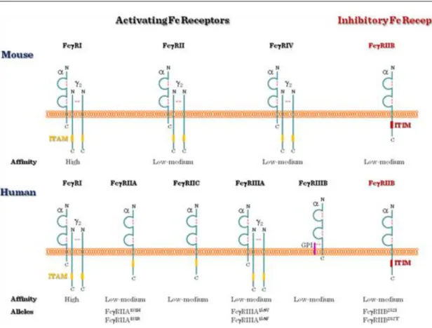
Fc-receptors (FcR) bind the constant Fc region of the immunoglobulin molecules. Based on their immunoglobulin isotype-specificity the following FcR groups can be distinguished: FcαRI (IgA), Fcα/μR, Fcγ Receptors (I, IIa/b/c, IIIa/b) (IgG), FcεRI/II (IgE), FcRH1-6, FcRX and FcRY.
1.3.2. Role and expression of Fcg receptors
Leukocyte Fc receptors promote the phagocytosis and killing of opsonized particles and deliver signals that stimulate the microbicidal activity of leukocytes. The most important Fc receptors of phagocytes are the Fcg receptors, which bind IgG immuncomplexes (Figure II.1-8). Their activity stimulates phagocytic or cytotoxic cells to destroy microbes, or infected cells by antibody-mediated phagocytosis or antibody-dependent cell- mediated cytotoxicity (ADCC).
II Detailed (systematic) signal transduction
Figure II.1-8: Types of Fcgamma receptors
1.3.3. Fcg receptor ITAM/ITIM
Similarly to the BcR and the TcR, Fcg receptors generate signals through ITAMs, too (Figure II.1-9). For example, the intracellular tail of FcγRIIA and C, and in the g chains of FcγRI and FcγRIIIA contain ITAMs.
After ligand binding, tyrosine (Y) residue of the ITAM is phosphorylated by tyrosine kinases, and a signaling cascade is generated within the cell.
FcγRIIB1 and FcγRIIB2, on the other hand, have ITIM sequences and, thus, are inhibitory Fc receptors: they do not induce phagocytosis, instead, they counteract the antigen-induced BcR signal on B cells and shut off B cell activation. This inhibitory signal is controlled by SHP-1 and SHIP-phosphatases.
Figure II.1-9: Activator and inhibitory Fcγ receptor signaling
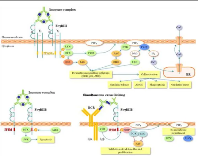
1.3.4. Fcg receptor signal transduction pathway
The clustering of these Fcg receptors with IgG1 or IgG3-coated particles or cells delivers an activation signal to phagocytes (Figure II.1-10). Activation signal requires cross-linking of the FcR a chains by several linked Ig molecules (e.g. Ig coated microbes, immuncomplexes). The signal transduction starts with Src kinase-mediated tyrosine phosphorylation of the ITAMs followed by SH2 domain-mediated recruitment of Syk family kinases to ITAMs , activation of PI-3 kinase, recruitment of adapter molecules like SLP-76 and BLNK, and the activation of enzymes like phospholipase Cg and Tec family kinases. Consequently, IP3 and DAG is generated and intracellular free Ca2+ is mobilized. These signal pathways in leukocytes induce gene transcription of cytokines, inflammatory mediators, microbicidal enzymes, activation of the cytoskeleton, alltogether leading to phagocytosis, cell migration and degranulation.
II Detailed (systematic) signal transduction
Figure II.1-10: Overview of Fcγ receptor signaling
1.4. II.1.4 Fce Receptor signaling
FcεRs have the ability to bind IgE. Although a lot is known about FcεRI function, the exact role of FcεRII still needs further studies.
1.4.1. The structure and expression of FcεRs
FcεRI (high-affinity IgE receptor) consists of α, β and γ chains (Figure II.1-11). It is expressed as an αβγ2 tetramer on mast cells and basophils, and as an αγ2 trimer on human antigen-presenting cells, monocytes, eosinophils, platelets and smooth-muscle cells. The α chain has 2 extracellular domains which are responsible for IgE binding. The intracellular parts of the β and γ chain contain ITAMs (immunoreceptor tyrosine-based activation motifs) being important in signal transduction.
Figure II.1-11: IgE bound FcεR I
FcεRII (CD23, the low affinity receptor) is structurally different from all immunoglobulin-binding receptors because it belongs to the C-type lectin superfamily (Figure II.1-12). It consists of 3 C-type lectin domain heads, C terminal „tails‟, an extracellular trimeric coiled coil „stalk‟ and a short N-terminal intracellular sequence that exists in two splice variants. The coiled coil „stalk‟ can undergo proteolysis resulting in soluble forms of CD23.
CD23 does not only bind IgE but also CD21 (expressed by B cells, follicular dendritic cells, activated T cells and basophils) that might be important in allergic processes and in the regulation of IgE through the complement system.
Figure II.1-12: IgE bound FceR II
1.4.2. FcεRI mediated signaling
Type I hypersensitivity reactions, for example anaphylaxis, hay fever, food allergies, other allergic diseases or asthma, are the most important pathological conditions mediated by FcεRI. Allergens like plant pollens, insect venoms are recognized by the immune system and IgE type antibody is produced against them. IgE then binds to the FcεRI receptors of mast cells, called sensibilisation. When the body meets the same allergen for the second time, crosslinking of the FcεRI-bound IgE molecules leads to activation of the cells (Figure II.1-13 and Figure II.1-14). Following FcεRI aggregation, ITAMs of the FcεRI become phosphorylated and protein tyrosine kinases Fyn and Lyn become activated, resulting in tyrosine phosphorylation of Syk non-receptor tyrosine kinase and Gab2 (Grb2-associated binding protein). These initial steps of FcεRI signaling share close homology with those of the TcR (Figure II.1-15). Gab2 binds to phosphatidylinositol 3-kinase (PI3K) and PI3K activation leads to Btk (Bruton's tyrosine kinase)-dependent phosphorylation of phospholipase C, that results in Ca2+
mobilization. PI3K might also enhance Ca2+ mobilization through phospholipase D mediated sphingosine- kinase activation. Parallel to PI3K, the MAP-kinase cascade is activated as well. Increased cytoplasmic Ca2+
leads to degranulation of the mast cells resulting in exocytosis of vasoactive amins (e.g. histamine) and proteases. The activation of the MAP-kinase cascade together with the increased Ca2+ signal enhances
II Detailed (systematic) signal transduction
Besides its pathogenic role, FcεRI has important physiological function in the immune response against parasites. Helminthes recognized by the immune system induce the production of IgE. IgE binds on the appropriate epitope of the parasite, thus, eosinophils can attack it through binding with their high-affinity FcεRI.
During the exocytosis of eosinophilic granules cationic granule proteins, like major basic proteins, eosinophil basic proteins, and enzymes, like eosinophil peroxidase, are released leading to the killing of the parasite.
Figure II.1-13: Biological effects of FcεR signaling
Figure II.1-14: FceRI mediated signaling
II Detailed (systematic) signal transduction
allergen from the gut lumen to the mucosa. The inhibitory function of CD23 suggests that it could serve as a basis for anti-allergic drug development in the future.
1.5. II.1.5 Cytokine signaling
1.5.1. Definition
Cytokines are small molecular weight glycoproteins that act at low concentrations on high affinity, specific cell surface receptors. In most cases they act on the cell(s) that are in the close vicinity of the producing cell (paracrine action), but some of them has autocrine (target cell = producing cell) or endocrine effects (via the circulation), too.
1.5.2. Division, groups
From a structural point of view, 3 groups were defined: (1) 4 α-helix bundle family (comprising from the IL-2-, IFN- and IL-10 subfamilies); (2) IL-1 family; and (3) IL-17 family.
From a functional point of view, we distinguish (1) haematopoetic cytokines (e.g. GM-CSF, G-CSF, M-CSF, erythropoetin, thrombopoetin); (2) cytokines that regulate lymphocyte activation and differentiation (immunoregulatory cytokines); and (3) inflammatory cytokines (IL-1, IL-6, TNFα). Immunoregulatory cytokines can be further classified based on the helper T cell subset that produces them:
a) Th1 cytokines: IL-2, TNFα, IFNγ;
b) Th2 cytokines: IL-4, 5, 13 c) Th17: IL-17A-F
d) Treg: TGFβ, IL-10
1.5.3. Receptors
The following cytokine receptor classes can be distinguished: class I (hematopoetin family), class II (IFN, IL- 10), and TNF-receptor family (Figure II.1-16). Class I receptors are heterodimer/trimer molecules that can be further divided into subgroups:
(1) receptors for erythropoetin, growth hormone and IL-13
(2) receptors with common β chain (IL-3, IL-5, GM-CSF);
(3) receptors sharing a common γ chain (IL-2, IL-4, IL-7, IL-9, IL-15);
(4) receptors sharing a common gp130 subunit (IL-6 receptor subfamily) (Figure II.1-17).
Figure II.1-16: Cytokine receptors
Figure II.1-17: Characteristics of multichain cytokine receptors
II Detailed (systematic) signal transduction
JH1: kinase, JH2: pseudokinase, JH3: SH2, JH4-7: FERM (=band 4.1, ezrin, radixin and moesin) domain (Figure II.1-18). The FERM domain binds to the proline-rich membrane proximal part of cytokine receptors.
Phosphorylation of two neighboring tyrosine residues in the kinase domain is critical in the activation of the molecule. Class I.1. and I.2. (see above) receptors associate with JAK2; Class I.3. receptors associate with JAK1 and JAK3; Class I.4. and Class II receptors associate with JAK1, JAK2 and TYK2.
Figure II.1-18: The structure of JAK and STAT proteins
1.5.5. Signal Transducer and Activator of Transcription (STATs)
STATs are the main target molecules of JAKs (Figure II.1-18). They contain an NH2 domain (Dimerization, DNA-binding and Nuclear transport), a coiled-coil (binding of regulator proteins), a DNA-binding domain (DBD), a linker (Lk), an SH2 (receptor recruitment and dimerization) and a transcriptional activation domain (TAD). Phosphorylation of the tyrosine residue between the SH2 and TAD domains is critical in the activation of the molecule. STAT1 and 2 are involved in IFN signaling; STAT3 mediates IL-6 & IL-10 family, IL-21 and IL-27 signaling controlling Th17 differentiation; STAT-4 mediates IL-12 and IL-23 signaling controlling Th1 differentiation; STAT5a & b mediate IL-3, IL-5 and GM-CSF signaling; and STAT-6 is involved in IL-4, IL-13 signaling driving Th2 differentiation and allergic immune responses.
1.5.6. Cytokine signaling
Upon ligand binding the receptor chains dimerize which leads to the activation of the associated JAKs (Figure II.1-19). Activated JAKs phosphorylate each other and the receptor chains. STATs bind to the phosphorylated receptors and, in turn, they are phosphorylated by JAKs. Activated STATs form dimers and translocate to the nucleus where they act as transcription factors. For example, type I IFNs (IFNα and IFNβ) activate STAT1/2 heterodimers which bind to ISRE (=IFN-sensitive response elements) sequences, whereas type II IFN (IFNγ) signaling activates STAT1 homodimers which bind to GAS (=IFNγ-activated site) sequences.
Figure II.1-19: Overview of cytokine signalling
1.5.7. Regulation of JAK/STAT signaling
The JAK/STAT pathway is controlled by four major mechanisms.
(1) Phosphatases SHP-1/2 and CD45 dephosphorylate JAK, whereas SHP-2, PTP1B, TC-PTP and PTP-BL dephosphorylate STAT proteins.
(2) Control of nuclear export/import by NES (nuclear export sequence) or NLS (nuclear localization sequence).
(3) SOCS (suppressors of cytokine signaling) e.g. PIAS=Protein Inhibitor of Activated STATs (4) Serine-phosphorylation, acetylation or O-glycosylation of TAD.
1.5.8. Clinical implications: JAK inhibitors
JAK inhibitors (e.g. Lestaurtinib; Tofacitinib; Ruxolitinib) are being tested in the treatment of hematological diseases e.g. polycythemia vera, thrombocytemia, myeloid metaplasia, myelofibrosis; and autoimmune diseases like psoriasis and RA.
1.5.9. TNF receptor signaling
Upon ligand binding, TNF receptor chains form trimers, which leads to conformational change and the subsequent dissociation of the inhibitor SODD (=silencer of death domains) from the intracellular “death domain”. The adaptor protein TRADD (=tumor necrosis factor receptor type 1-associated DEATH domain protein) binds to the death domains and serves as a platform for further protein association (Figure II.1-20 and Figure II.1-21).
II Detailed (systematic) signal transduction
Figure II.1-20: TNF receptor mediated apoptosis I
Figure II.1-21: TNF receptor mediated apoptosis II Three major signaling pathways are activated:
(1) NF-κB pathway: TRAF2 (=TNF receptor-associated factor 2) and RIP (=receptor interacting protein) are recruited to TRADD. RIP, a Ser/Thr kinase, activates IKK (IκB kinase), which, in turn, activates NF-κB. The activated NF-κB translocates to the nucleus and controls the transcription of cell survival, proliferation, inflammation and apoptosis genes (generally anti-apoptotic).
(2) Activation of MAPK pathways: From the three major MAPK pathways, strong activation of the stress response JNK pathway, moderate activation of p38 and minimal activation of the ERK pathway occurs after TNF receptor activation. TRAF2 recruits MEKK1 (=Mitogen-activated protein kinase kinase kinase 1) and ASK1 (=Apoptosis signal-regulating kinase 1), which phosphorylate MKK7 (=Mitogen-activated protein kinase kinase 7). MKK7 phosphorylates JNK (=c-Jun N-terminal kinase), which in turn, translocates to the nucleus and activates the transcription factors c-Jun and ATF2 (=Activating transcription factor 2). This pathway controls genes of cell differentiation, proliferation and apoptosis (generally pro-apoptotic).
(3) Death signaling (“Extrinsic apoptosis pathway ”, see Chapter II.4 for details): TNFR1 does not induce this pathway as strong as for example the Fas molecule. TRADD binds FADD, which recruits pro-Caspase-8.
Autocatalytic cleavage activates Caspase-8, which initiates the downstream events of the apoptotic cascade:
Caspase-3 and Bid (=BH3 interacting domain death agonist), a pro apoptotic member of the Bcl-2 family leading to Cytochrome C release from the mitochondria.