Full Terms & Conditions of access and use can be found at
https://www.tandfonline.com/action/journalInformation?journalCode=kvir20
Virulence
ISSN: (Print) (Online) Journal homepage: https://www.tandfonline.com/loi/kvir20
Deciphering of Candida parapsilosis induced immune response in Drosophila melanogaster
Katalin Csonka, Zsolt Tasi, Viktor Vedelek, Csaba Vágvölgyi, Rita Sinka &
Attila Gácser
To cite this article: Katalin Csonka, Zsolt Tasi, Viktor Vedelek, Csaba Vágvölgyi, Rita Sinka &
Attila Gácser (2021) Deciphering of Candida�parapsilosis induced immune response in Drosophila melanogaster, Virulence, 12:1, 2571-2582, DOI: 10.1080/21505594.2021.1980989
To link to this article: https://doi.org/10.1080/21505594.2021.1980989
© 2021 The Author(s). Published by Informa UK Limited, trading as Taylor & Francis Group.
View supplementary material
Published online: 27 Sep 2021.
Submit your article to this journal
View related articles
View Crossmark data
RESEARCH PAPER
Deciphering of Candida parapsilosis induced immune response in Drosophila melanogaster
Katalin Csonkaa, Zsolt Tasia, Viktor Vedelekb, Csaba Vágvölgyia, Rita Sinka b, and Attila Gácser c,d
aDepartment of Microbiology, Faculty of Science and Informatics, University of Szeged, Szeged, Hungary; bDepartment of Genetics, Faculty of Science and Informatics, University of Szeged, Szeged, Hungary; cHCEMM-USZ, Department of Microbiology, Faculty of Science and Informatics, University of Szeged, Szeged, Hungary; dMTA-SZTE Lendület Mycobiome Research Group, University of Szeged, Szeged, Hungary
ABSTRACT
Candida infections are the most prevalent cause of serious human mycoses and are the third most common pathogens isolated from bloodstream infections in hospitalized patients. C. parapsilosis is a member of the non-albicans spp., which have a predilection for causing life-threatening disease in neonates and hospitalized pediatric patients. In this study, we utilized a Drosophila melanogaster infection model to analyze the immunological responses to C. parapsilosis. Our results demonstrate that the Toll pathway in Drosophila controls C. parapsilosis proliferation as the Toll signaling mutant MyD88−/− flies are highly susceptible to C. parapsilosis. We also con- firmed that the MyD88−/− fly is a convenient invertebrate animal model to analyze virulence properties of different species and strains from the C. parapsilosis sensu lato complex as C. orthopsilosis, C. metapsilosis proved to be less virulent than C. parapsilosis sensu stricto and the N-mannan deficient C. parapsilosis och1Δ/Δ strain showed attenuated pathogenicity in this immunodeficient Drosophila background. We also found that Persephone protease is not required for detection and activation of Toll pathway during C. parapsilosis infection. Furthermore, we observed that Drosophila β-glucan receptor deficient flies where more sensitive to C. parapsilosis compared to wild-type flies; however, we could not find a clear dependence on the recognition of this receptor and the cell wall β-glucan exposure-induced host response. These studies establish this D. melanogaster infection model as an efficient tool in deciphering immune responses to C. parapsilosis as well as for assessing virulence factors produced by this emerging fungal predator.
ARTICLE HISTORY Received 1 June 2021 Revised 31 August 2021 Accepted 10 September 2021
KEYWORDS
Candida; innate immune response; virulence;
recognition; systemic infection; Drosophila melanogaste; cell wall; β- glucan
Introduction
Candida species are opportunistic fungal pathogens causing severe diseases in immunocompromised patients and are the third most common microorgan- isms responsible for healthcare-related bloodstream infections [1]. Although C. albicans is the most fre- quently isolated species globally as the causative agent for disseminated Candida diseases, the frequency of infections due to non-albicans Candida species con- tinues to increase [2,3]. C. parapsilosis is ranked as the second or third most common non-albicans spp [4]. This pathogen has a particular predilection for causing hospital-acquired infections in neonatal and pediatric patients as well as adult patients with intra- vascular catheters and other implantable devices [5,6].
Through investigations of C. parapsilosis biology, numerous factors have been identified that play roles in pathogenesis including extracellular lipases, tran- scription regulators, pseudohyphae and biofilm
production, antifungal resistance mechanisms, and iron metabolic processes [6–8].
Since virulence-related genes in various Candida spp. require validated models to define their function, experimental in vivo models are essential in pathogen- esis research. The fruit fly, Drosophila melanogaster is a remarkably flexible invertebrate model organism to study specific responses of innate immunity against microbial infections [9,10]. This mini-host has been applied to examine innate immune defense mechan- isms against certain Candida species, as flies deficient in the Toll signaling pathway are sensitive to fungal infections [11]. The Toll/Dif pathway responds to the presence of fungal and Gram-positive bacterial infec- tions and mediates the production of antimicrobial peptides (AMPs), such as Drosomycin, Metchnikowin, and Defensins [12–14]. After it is activated by a proteolytic cleavage cascade, Spätzle (spz) is a ligand for Toll and binds to the cell transmembrane receptor,
CONTACT Attila Gácser gacsera@bio.u-szeged.hu Supplemental data for this article can be accessed here.
2021, VOL. 12, NO. 1, 2571–2582
https://doi.org/10.1080/21505594.2021.1980989
© 2021 The Author(s). Published by Informa UK Limited, trading as Taylor & Francis Group.
This is an Open Access article distributed under the terms of the Creative Commons Attribution License (http://creativecommons.org/licenses/by/4.0/), which permits unrestricted use, distribution, and reproduction in any medium, provided the original work is properly cited.
triggering its dimerization, which leads to the recruit- ment of the adaptor, Drosophila myeloid differentia- tion factor 88 (dMyD88, homolog of mammalian MyD88). Upstream of Spätzle, immune detection of fungal determinants is regulated by the Gram-negative binding protein 3 (GNBP3) and the Persephone (psh) serine protease [15]. The GNBP3, a member of the GNBP/β-glucan recognition proteins (βGRP) family, has been reported to bind to fungal cell wall β-(1,3)- glucan and activates the antifungal Toll pathway in a Spätzle-dependent manner [15]. Indeed, GNBP3 con- tributes to controlling Candida infections, as C. albicans and C. glabrata challenged GNBP3 deficient flies display increased susceptibility/death events and impaired expression levels of the Toll-dependent Drosomycin gene [15,16]. Psh encodes a hemolymphatic serine protease belonging to a Drosophila danger pathway and becomes activated by proteolytic activities of microbes that induce Toll signaling [15]. The lack of psh causes a weak suscept- ibility of adult flies to C. albicans, but psh mutant flies are highly susceptible to C. glabrata challenge [16,17].
The contribution of these sensor molecules in Candida defense can vary depending on the Drosophila model selected. For example, gastrointestinal infection with C. albicans in Drosophila larvae generated a GNBP3 independent, but psh-Toll dependent systemic response, which required the presence of hemo- cytes [18].
A broad range of research has demonstrated that Drosophila models are reliable tools for screening new antifungal treatment options against C. albicans [11]
and C. auris [19] and investigating genes involved in Candida pathogenesis. Different virulence factors, such as C. albicans Cas5, a transcriptional regulator of genes involved in cell wall integrity [20] and secreted aspartyl proteases SAP4 and SAP6 [18], as well as C. glabrata Yapsins (secreted GPI-anchored aspartyl proteases) [16] and ADA2 for oxidative stress tolerance were identified in this mini-host [21].
In this study, we aimed to describe C. parapsilosis infection in an immunodeficient D. melanogaster fly model, which we adapted from the work previously performed to characterize C. albicans and C. glabrata induced specific immune responses [16]. We demon- strated that C. parapsilosis infection is highly regulated by the Drosophila Toll pathway, as MyD88 mutant flies succumbed to challenge with C. parapsilosis cells. We extended our studies to include additional members of the C. parapsilosis sensu lato complex, and demon- strated that this type of immunodeficient fly is suitable to analyze the differences in virulence of the C. parapsilosis sensu lato complex strains. We also
found, as reported in mouse models, that a C. parapsilosis without a proper N-mannan layer in the cell wall was significantly less virulent in our fly model. Our results show that D. melanogaster and mutants like MyD88−/- Drosophila are extremely useful model for identifying and analyzing C. parapsilosis virulence factors.
Materials and methods Drosophila stocks
Drosophila stocks were maintained on standard corn- meal agar medium at 25°C, in 12 h light/12 h dark cycle according to Drosophila Protocols, Chapter 35 (Sullivan, Ashburner, Hawley, Cold Spring Harbor Laboratory Press, 2000). The wild-type w1118 (BL3605) (from Bloomington Stock Center) and Drosomycin- GFP (GFP-Drs-fly) [22], GNBP3hades, psh, and MyD88 mutant flies (a kind gift from Jessica Quintin) were used in this study. Stocks have been described pre- viously [23].
Microbial strains
C. parapsilosis GA1 (SZMC 8110) [24], C. parapsilosis CLIB 214 (SZMC 1560) [25] C. parapsilosis SZMC 1592, C. parapsilosis SZMC 8050, C. albicans SC5314 (SZMC 1523), C. metapsilosis SZMC 1548, C. metapsilosis SZMC 8099, C. metapsilosis SZMC 8094, C. orthopsilosis SZMC 1545, C. orthopsilosis SZMC 8121 and C. orthopsilosis SZMC 8119 [26] wild- type strains, C. parapsilosis och1Δ/Δ, C. parapsilosis CPRI [27], a GFP-expressing derivative of C. parapsilosis CLIB 214 (genotype: CpNEUT5L/
CpNEUT5L::pECpOE-GFP-N-N5L) and C. albicans SC5314 (RPS1/RPS1::CIp10-PTDH3-GFP-CaNAT1) were used in this study and maintained on YPD agar plates (0.5% yeast extract, 1% peptone, 1% glucose, 2.5% agar) at 4°C. The C. parapsilosis CPRI strain was used as a reference for the analysis of infections with C. parapsilosis och1∆/∆ strain. Prior to use, Candida cells were grown in liquid YPD medium (0.5% yeast extract, 1% peptone, and 1% glucose supplemented with 1% Penicillin-Streptomycin) with shaking (200 rpm) at 30°C overnight. Micrococcus luteus SZMC 0264 (Szeged, Hungary), a Gram-positive bac- teria, was used as a reference for the Drosomycin induc- tion studies. M. luteus was grown in an overnight culture in LB broth (1% tryptone, 0.5% yeast extract and 0.5% NaCl) at 37°C with 200 rpm shaking. Prior to use, microbial cells were harvested by centrifugation, washed twice with PBS (phosphate-buffered saline;
137 mM NaCl, 2.7 mM KCl, 10 mM Na2HPO4, 2 mM KH2PO4; pH 7.4), counted using
a hemocytometer and adjusted to the proper concen- tration detailed for each experiment.
Survival study of flies
Batches of 15 (2- to 4-day-old females and males;
45 per experimental group) wild-type (wt) and mutant flies were infected by septic injury on the dorsal side of the thorax. The flies were injected using a 30 g needle previously dipped into PBS or a 2×10⁷/ml suspension of bacteria or yeasts. The vials containing the chal- lenged flies were housed in an incubator (29°C for fungal infections or 25°C for bacterial challenges).
Survival was assessed daily, and live flies were put into new vials containing standard cornmeal agar med- ium every second day. Results are expressed as a percentage of surviving flies at different days post- infection.
Even though it may not be ideal for examining the virulence of human pathogens at temperatures below 37°C, the incubation of flies at 29°C is a good compro- mise to avoid the physiological consequences of the heat-shock response. We choose this methodology according to our experiences and previous work of Davis et al., where they demonstrated that performance of Drosophila-fungus interaction at 29°C is suitable for examining C. albicans virulence factors and this tem- perature has no adverse effect on the yeast dissemina- tion and the development of pseudohyphae and hyphae [28].
Fungal burden assay
Groups of 10 infected flies were homogenized in PBS at specific times (right after the infection (input), 5 hours (0 day), 2 days, and 4 days) after the PBS and Candida (2×10⁷/ml) infection. The homogenates were serially diluted, plated on YPD agar plates, and incubated for 48 h at 30°C to enable colony growth for counting.
Yeast colonies recovered from flies were calculated and expressed as CFU/fly. Results are pooled data from five independent experiments.
In vivo phagocytosis assay
Flies were infected with 20 μl of a 1×10⁵/ml GFP- labeled Candida strain suspension using a sharpened glass capillary on the thorax and then the insects were incubated for 3 h at 25°C. Collection of hemocytes was performed according to a standard method [29].
Briefly, flies were anaesthetized and the last section of the abdomen was removed. The fly’s thorax was punc- tured with a sharpened glass capillary. Perfusion was performed through a capillary with a Schneider’s med- ium (Biowest, cat.: L0207) containing 1-phenyl- 2-thiourea (PTU, Sigma-Aldrich, cat.: P7629). The
samples collected from five flies per group were placed on glass slides and incubated for 30 min to allow hemocytes to adhere to the slides. After the incubation, the medium was removed, and non-phagocytosed yeasts were labeled with 5 μM of Calcofluor white (5 mM, Sigma-Aldrich, cat.: 18,909–100ML-F) at room temperature for 10 min and then washed two times with PBS to remove excess stain. Samples were fixed for 5 min in 4% formaldehyde in PBS, permeabi- lized for 5 min in 0.1% Triton X-100 and filamentous actin of Drosophila hemocytes was stained with Texas Red®-X Phalloidin (Thermofisher, cat.: T7471) (1:250) for 20 min. After washing steps with PBS, samples were covered with SlowFade mounting medium (Invitrogen, cat.: S36917) and the slides analyzed with a BX51 OLYMPUS microscope.
RNA isolation and qPCR
The measurement of Drosomycin mRNA level was designed and performed according to a standard method [30]. Samples of five flies/group were frozen in liquid nitrogen and total RNA was isolated using the Quick-RNA MiniPrep Kit (Zymo Research, cat.: R1054) according to the manufacturer’s instructions. The con- centration and integrity of isolated RNA were con- firmed by ND-1000 Spectrophotometer (Thermo Scientific). cDNA was synthesized from 2000 ng total RNA using the RevertAidTM First Strand cDNA Synthesis Kit (Thermo Scientific, cat.: K1622) accord- ing to the manufacturer’s protocol. qRT-PCR was per- formed using Maxima SYBR Green qPCR Master Mix (Thermo Scientific, cat.: K0242), in a C1000TM Thermal Cycler (BIO-RAD) equipped with a CFX96™ Real-Time Detector System (BIO-RAD). Ribosomal protein 49 (Rp49) was used as an endogenous control gene, and fold changes were calculated by the ΔΔCt method. PCR product specificity was confirmed by melting analysis.
Primer sequences were as follows: rp49: forward: 5’
GACGCTTCAAGGGACAGTATCTG 3’, reverse: 5’
AAACGCGGTTCTGCATGAG 3’; drosomycin: forward 5’ CGTGAGAACCTTTTCCAATATGATG 3’, reverse:
5’ TCCCAGGACCACCAGCAT 3’ [30].
Detection of Drosomycin production by microscopy GFP-Drs-flies were injected with 2×10⁸/ml Candida or bacterial suspension [22]. After 24 h of incubation, flies were anaesthetized for direct observation of Drosomycin induction. Microscopy was performed using an OLYMPUS SZX7 stereomicroscope.
Statistical analysis
Graphs represent at least three independent experi- ments (n ≥ 3 in each experiment) that yielded similar
results unless otherwise stated (see Results and Figure legends for details). Results from the fungal burden and real-time PCR analysis are expressed as mean ± SEM.
Diagrams were created and statistical analyses were performed with the GraphPad Prism 7.0 software.
Differences were considered statistically significant at p ≤ 0.05.
Results
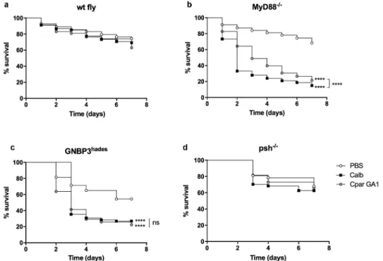
Toll pathway involvement in Candida infection To determine if D. melanogaster are susceptible to C. parapsilosis and whether the Toll signaling pathway regulates disease, we injected wild-type (wt fly) and MyD88−/- flies with C. parapsilosis GA1 or C. albicans SC5314 strain, as reference. As expected, there was no significant difference in survival rates of wt flies infected with the fungal species compared to the PBS injected flies (Figure 1(a)). Notably, MyD88−/- flies dis- played similar susceptibility to C. albicans and C. parapsilosis GA1. However, with a more in-depth examination of the mini-host’s survival, we observed that more MyD88−/- flies challenged with C. parapsilosis survived compared to C. albicans infected flies (Figure 1(b)). The lower virulence of C. parapsilosis in the Drosophila model was previously reported by Chamilos et al., where the Tl mutant flies died more from C. albicans or C. krusei than C. parapsilosis [11].
Therefore, our data further strengthened the observa- tion that the Toll pathway is required for defense against these opportunistic yeasts and the Drosophila model is suitable to distinguish between the different virulence potentials of distinct fungal species.
To detail the upstream events participating in the Toll pathway activation against C. parapsilosis, we also tested flies carrying a mutation in the GNBP3 receptor or the Persephone serine protease. During the monitor- ing of GNBP3 deficient flies’ survival, we noted that the absence of GNBP3 PRR markedly affected the fly’s fitness and viability, as the PBS injection alone caused death events in our experimental settings. As expected from the previous studies, GNBP3hades flies nevertheless showed increased susceptibility to C. albicans compared to the PBS injected fly groups. Our data also documents that this receptor takes part in detecting C. parapsilosis cells, as the GNBP3hades flies displayed increased mor- tality in response to this pathogen compared to the PBS injected fly group (Figure 1(c)). Furthermore, we observed that psh−/- flies were resistant to C. parapsilosis and C. albicans (Figure 1(d)).
Effect of Candida infection on antimicrobial peptide gene expression
Upon fungal challenge, the Drosophila pathogen recog- nition receptors trigger signaling pathways leading to the production of the antifungal peptide Drosomycin.
Figure 1. Comparison of the survival of wt (a), MyD88−/- (b), GNBP3hades (c) and psh−/- (d) flies after injection with PBS, C. albicans SC5314 or C. parapsilosis GA1. Infection dose 2 × 10⁷ yeast/ml. n = 45 fly/group/experiment. Results are representative of 4 independent experiments with statistical analysis by Mantel–COX test. P value style: GP: **** p < 0,0001; not significant (ns)<0.1234.
Drosomycin mRNA-level measurement has been used as a readout of Toll pathway activation, and its induc- tion was reported upon Candida infections [15].
Therefore, we challenged a transgenic Drosophila line expressing the GFP-Drosomycin fusion protein [22] to examine the response induced by C. parapsilosis. We used M. luteus as bacterial [31] and C. albicans as fungal reference. As survival of wt, MyD88−/- and GNBP3hades flies showed no significant difference between infection with C. parapsilosis GA1, C. parapsilosis CLIB 214 and C. parapsilosis CPRI strains (Figure S1(a-c)), we presented data performed with C. parapsilosis CPRI strain.
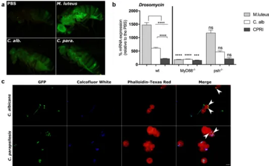
As expected, M. luteus injected flies exhibited a robust GFP-expression compared to the Candida infected fly groups (Figure 2(a)). In comparison to the PBS injected fly, C. albicans and C. parapsilosis chal- lenged flies presented a strong GFP-Drosomycin expression, suggesting that systemic infection with either of the two Candida species similarly induces the production of this AMP in the fruit fly (Figure 2(a)).
To depict the activation of the humoral response against C. parapsilosis, we measured the mRNA level of Drosomycin using quantitative real-time PCR method in the wt, MyD88−/- and psh−/- flies. In the GNBP3hades fly, PBS injection alone caused higher
Drosomycin mRNA expression in this background than the wt fly groups (Figure S1(d)); therefore, we did not include this fly strain in our analyses. After 24 h of the infection, wt flies showed the highest expression of AMP after challenge with M. luteus, whereas the yeast species induced lower mRNA levels.
Furthermore, C. parapsilosis provoked a significantly weaker humoral response compared to C. albicans (Figure 2(b)). In comparison to the wt fly, a mutation in the MyD88 adapter resulted in a significantly decreased level of Drosomycin after either the bacterial or fungal stimuli. Similarly, as reported for C. albicans [15,16], antifungal peptide gene induction by C. parapsilosis was not affected by lack of Persephone protease, as no differences were detected in its mRNA levels between the corresponding psh−/- and wt fly groups (Figure 2(b)).
These data confirmed the results presented by the survival experiments and indicate that C. parapsilosis infection cause Toll-mediated humoral defense in Drosophila.
Detection of phagocytosis upon Candida infection We next examined in vivo phagocytosis, which is one of the cellular responses of the Drosophila immune sys- tem. For this, we infected adult flies by septic injury
Figure 2. A. GFP-Drosomycin expression of flies after 24 h of injection with M. luteus, C. albicans or C. parapsilosis CPRI. Injection dose 1x10⁸/ml. B. Drosomycin mRNA induction in wt, MyD88−/- and psh−/- flies after 24 h of injection with M. luteus, C. albicans (C. alb) or C. parapsilosis CPRI. Injection dose 5x10⁷/ml. Data are represented as means with ± SEM from 3 independent experiments as determined by paired t-test. P value style: GP: **** p < 0,0001; *** p < 0,0002; not significant (ns)<0.1234. C. In vivo phagocytosis of GFP-C. albicans and GFP-C. parapsilosis. Flies (wt) were injected with 20 µl of 2×10⁵/ml of GFP-C. albicans or GFP-C. parapsilosis strains. Hemolymph was collected (5 fly/group) 3 h after the injection, non-phagocytosed yeast were labeled with Calcofluor White, and hemocytes were stained with Phalloidin-Texas Red. White arrows indicate engulfed Candida cells.
with a suspension of C. albicans or C. parapsilosis, and examined the phagocytosis capacity of hemocytes against the yeast cells. After 3 hours of incubation, we found that Drosophila blood cells effectively detected and engulfed both C. albicans and C. parapsilosis cells.
Representative pictures show phagocytosed yeast cells of C. albicans and partially enveloped cells of C. parapsilosis (Figure 2(c)). These results confirmed a systemic response after C. parapsilosis septic wound- ing and suggest a similar elimination mechanism against this yeast as to that described with C. albicans in Drosophila [32].
Assessment of virulence properties of different C. parapsilosis strains
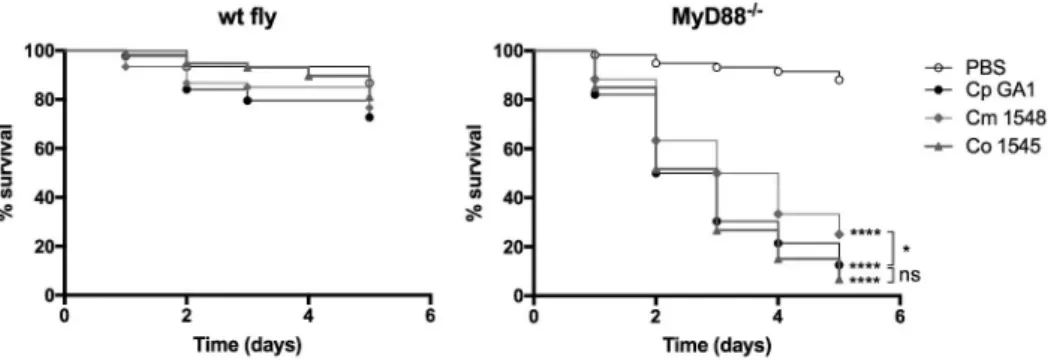
As our results showed that MyD88−/- flies are suscep- tible to C. parapsilosis challenge, we wanted to examine whether this fly group could be utilized to determine virulence differences in closely related C. parapsilosis sensu lato complex species. Therefore, we injected the wt and the MyD88−/- flies with three isolates each of C. parapsilosis, C. orthopsilosis or C. metapsilosis spe- cies, and found that the MyD88−/- flies display increased susceptibility to the members of the C. parapsilosis sensu lato group compared to the PBS injected fly group (data not shown). As next step, we selected one isolate of C. parapsilosis (Cp GA1), C. orthopsilosis (Co 1548) and C. metapsilosis (Cm 1546) and compared their virulence in the immune- deficient mini-host. In agreement with previous studies with another invertebrate model, Galleria mellonella larvae [26], our results presented that the C. metapsilosis infected MyD88−/- flies had significantly better survival rates than flies challenged with the other C. parapsilosis sensu lato species. Furthermore, no sig- nificant differences were detected in the death events caused by the C. parapsilosis sensu stricto and
C. orthopsilosis challenged MyD88 deficient flies (Figure 3).
Effect of C. parapsilosis cell wall integrity on pathogenesis in the D. melanogaster model
Next, to characterize the pathogenesis of C. parapsilosis in D. melanogaster and test whether this invertebrate model is suitable to assess differences in the virulence of mutant C. parapsilosis strains, we employed the C. parapsilosis CPRI reference and the mutant och1Δ/
Δ (Cpoch1∆/∆) for fly injection. The C. parapsilosis och1∆/∆ strain exhibits a severe defect in N-mannan content with elevated β-glucan and chitin levels in the cell wall. In previous studies, Cpoch1∆/∆ strain-induced alterations in the cytokine production in human mono- nuclear cells and displayed significantly decreased viru- lence in Balb/C mouse and neonate mouse model [27,33].
In agreement with the findings of systemic murine infection, our results revealed that the percentage of surviving MyD88−/- flies were significantly higher after challenged with the Cpoch1∆/∆ cells compared the C. parapsilosis CPRI infected fly groups (Figure 4). In comparison to PBS, the GNBP3hades flies died signifi- cantly faster after challenge with the cell wall mutant Candida strain, but mortality rate was similar to that caused by C. parapsilosis CPRI. As expected, the psh mutant flies were resistant to Cpoch1∆/∆ infection (Figure 4).
For a detailed assessment of the two C. parapsilosis strain’s virulence properties, we analyzed the prolifera- tion capacity of the fungi in flies using CFU determina- tions. As shown, the wt flies were resistant to C. parapsilosis infection, and the CFU results show that these insects can rapidly kill C. parapsilosis CPRI cells. However, C. parapsilosis CPRI that survived the initial immune response were able to proliferate in
Figure 3. Survival of wt, MyD88−/- flies after injection with PBS, C. parapsilosis GA1, C. metapsilosis, or C. orthopsilosis. Infection dose 2 × 10⁷ yeast/ml. n = 45 fly/group/experiment. Results are representative of 4 independent experiments with statistical analysis by Mantel–COX test. P value style: GP: **** p < 0,0001; * p < 0,0332; not significant (ns)<0.1234.
these flies, as increased numbers of yeast cells were observed during the infection period (Figure 5) The susceptibility of the MyD88−/- and GNBP3hades flies to C. parapsilosis infection was also strengthened by the significantly higher fungal loads at 2 and 4 days of infection compared to that observed in wt flies. The assessment of the colonization also supported the
resistance of psh mutant fly. The psh−/- flies showed that the yeast cells could survive within the mini-host, but the fungal loads were relatively low (Figure 5).
Notably, we detected a decrease in fungal colonies of Cpoch1∆/∆ infected wt and psh−/- flies from day 2 to day 4, suggesting an enhanced clearance of the mutant C. parapsilosis strain by the mini-host Figure 4. Survival of wt, MyD88−/-, GNBP3hades and psh−/- flies after injection with C. parapsilosis CPRI or Cpoch1∆/∆. Injection dose 2×10⁷/ml. Results are representative of 4 independent experiments with statistical analysis by Mantel-Cox-test. P value style: GP: ****
p < 0,0001; *** p < 0,0002; ** p < 0,0021; * p < 0,0332; not significant (ns)<0.1234.
Figure 5. CFU assessment of wt, MyD88−/-, GNBP3hades and psh−/- flies after injection with C. parapsilosis CPRI. Injection dose 2×10⁷/ ml. Data are presented as mean with ± SEM from 5 independent experiments as determined by paired t-test. P value style: GP: ****
p < 0,0001; *** p < 0,0002; ** p < 0,0021; * p < 0,0332; not significant (ns)<0.1234.
(Figure 6). In line with the survival data, as the Cpoch1∆/∆ challenged MyD88−/- and GNBP3hades flies showed an increment in the death events compared to the PBS injected fly groups, these genotypes of flies were unable to clear the Cpoch1∆/∆ cells. However, all groups of flies, either sensitive to the reference C. parapsilosis strain or not, were able to control the growth of the N-mannan mutant strain, as significantly lower CFUs of Cpoch1∆/∆ were obtained from each fly background at each time point of the experiments compared to C. parapsilosis CPRI (Figure 6).
Therefore, these CFU data also support the decreased virulence of Cpoch1∆/∆ in the immune-deficient Drosophila model.
Next, we tested whether the decreased virulence of the Cpoch1∆/∆ is commensurate with its induction of antifungal peptides. In comparison to the C. parapsilosis CPRI, Cpoch1∆/∆ induced a non- significant increase in Drosomycin mRNA level in the wt fly (Figure 7). When the MyD88−/- flies were infected with the Cpoch1∆/∆, we found significantly decreased mRNA level of the antifungal peptides fol- lowing challenge with CPRI or Cpoch1∆/∆ compared to levels in wt flies, but there were again no signifi- cant differences between the C. parapsilosis strains.
Drosomycin expression measured from psh−/- flies infected with either C. parapsilosis strain were similar to levels in wt insects (Figure 7). Therefore, these data Figure 6. CFU assessment of wt, MyD88−/-, GNBP3hades and psh−/- flies after injection with C. parapsilosis CPRI and Cpoch1∆/∆.
Injection dose 2×10⁷/ml. Data are represented as mean with ± SEM from 5 independent experiments, Paired t-test. P value style: GP:
**** p < 0,0001; *** p < 0,0002; ** p < 0,0021; * p < 0,0332.
Figure 7. Drosomycin mRNA induction in wt, MyD88−/- and psh−/- flies after injection with C. parapsilosis CPRI or Cpoch1∆/∆. Injection dose 5x10⁷/ml. Data are presented as means with ± SEM from 3 independent experiments as determined by paired t-test. P value style: *** p < 0,0002; ** p < 0,0021; not significant (ns)<0.1234.
further strengthened the results that the Toll pathway detects C. parapsilosis cells and the Persephone serine protease is not involved in its activation process.
Discussion
Here, we aimed to use D. melanogaster as a model to investigate the pathogenicity of C. parapsilosis. Our results indicate that the Drosophila Toll restrains C. parapsilosis proliferation as MyD88−/− flies display a significantly enhanced susceptibility to C. parapsilosis.
Our data also support an earlier study where C. parapsilosis showed lower virulence in D. melanogaster compared to C. albicans [11]. We have further explored the capacity of this mini-host to sense C. parapsilosis using flies lacking the GNBP3 β- glucan receptor or the Persephone protease required for Toll pathway activation during fungal invasion. We found that GNBP3hades flies displayed increased sus- ceptibility to C. parapsilosis, whereas psh mutants were resistant, which is similar to findings with C. albicans challenge in these fly strains [15].
Furthermore, we demonstrated that the MyD88−/- Drosophila strain could distinguish variations in viru- lence between the closely related Candida species, and that the characterizations were similar to that found in our prior work using a G. mellonella model where C. metapsilosis was the least virulent species of the psilosis group and no significant divergence was observed between the mortality rate of larvae infected with C. parapsilosis sensu stricto and C. orthopsilosis isolates [26].
Our study examined whether immune-deficient flies might be useful to identify and test the variation of C. parapsilosis strains’ pathogenesis. The α1,6-mannosyltransferase Och1 initiates N-glycan outer chain branch addition in the yeast cell wall and possibly regulates virulence in both C. albicans and C. parapsilosis [27,34]. In the MyD88 mutant D. melanogaster, the lack of N-mannan content in C. parapsilosis altered the survival rates of infected flies compared to insects infected with the reference yeasts. The decreased virulence of C. parapsilosis lack- ing N-mannan in this mini-host is similar to results found in studies using a systemic mouse infection model [27,33]. Albeit, the decreased virulence with this C. parapsilosis mutant was not paired with differ- ences in antimicrobial peptide induction in the fly.
A similar result was found in a gastrointestinal Drosophila larvae model where the C. albicans cell wall mutant PMR1, which has defects in both N- and O-linked mannosylation, activated Drosomycin to the
same extent as did the wild-type C. albicans counter- part strain [18].
C. parapsilosis induced Drosomycin at a significantly lower rate than C. albicans in wt and the psh−/- flies, and the flies were more resistant to C. parapsilosis.
Also, the Cpoch1Δ/Δ strain demonstrated attenuated virulence in the Drosophila model, but it induced an antimicrobial response that was similar to the reference C. parapsilosis strain. There are controversial results regarding the fungicidal activity of Drosomycin against yeasts. In vitro studies noted that Drosomycin has no fungicidal effect on C. albicans and C. glabrata [16,35,36]. A study using a knockout approach of dif- ferent AMPs deficient flies deduced that AMPs have may not individually be essential in defense against fungi and disclosed the additive cooperation of Drosomycin and Metchnikowin to restrain C. albicans infection [37].
Thus, our results indicate that it is not primarily the antimicrobial peptide production that performs the elimination function in Drosophila in controlling C. parapsilosis infection as it was correspondingly con- cluded for C. albicans and C. glabrata [38].
GNBP3 is essential for controlling C. albicans and C. glabrata infections, as deprivation of this receptor caused increased susceptibility of adult flies against these Candida species [16,32]. Unexpectedly, in our experiment settings, the GNBP3hades flies were extre- mely sensitive to injection as the survival proportion of the PBS treated flies was around 56%. However, the death events of the C. albicans or C. parapsilosis chal- lenged fly group were significantly higher compared to the PBS injection. We were surprised that och1Δ/Δ and the reference C. parapsilosis strain infection provoked similar survival curves in the GNBP3 deficient flies, albeit the wild-type produced significantly hihger CFUs compared to the mutant. Therefore, we could not find clear interdependence between the lack of the N-mannosyl residues and the higher β-glucan exposure in the cell wall of C. parapsilosis and ligand binding of this Drosophila receptor.
We also found elevated Drosomycin mRNA levels in GNBP3hades flies compared to the wild-type Drosophila after the PBS injection alone. This could suggest that the death events of the Candida-challenged GNBP3 receptor mutant flies were not necessarily the sole effect of the fungus, but the deficiency of this receptor could cause the lack of some specific response to the injury. Therefore, the combined effect of the fungus and the infection route may generate the phenomenon that no difference was detected between the survival of the C. albicans- and the C. parapsilosis strains- challenged GNBP3hades fly groups. Results from
gastrointestinal infection of Drosophila larvae also registered that absence of GNBP3 receptor did not influence systemic activation of Drosomycin and dou- ble mutant psh; GNBP3 larvae exhibited a similar decrease in the level of the antimicrobial peptide as psh mutants following infection with live C. albicans [18]. This study suggests the altered immune sensing processes, including the role of GNBP3 between the larvae and adult fly and the Drosophila gut and sys- temic infection model. The mammalian β-glucan receptor, Dectin-1, displays a similar feature. In mice, Dectin-1 is indispensable in regulating systemic infec- tion with C. albicans, but it performs a redundant role for the control of gastrointestinal colonization [39].
Furthermore, a comparative study established that Dectin-1 is essential for both innate and adaptive immune responses to C. albicans, C. glabrata, C. tropicalis and C. parapsilosis; however, its function in specific responses diverge between the different Candida species [40]. Overall, our data show decreased survival and reduced ability of GNBP3hades fly to clear C. parapsilosis cells, but, due to the confusing results described earlier, it is challenging to resolve the real effect of this receptor in Drosophila host response in the control of systemic C. parapsilosis dissemination.
As a measure of the adult Drosophila’s immune recogni- tion process, we examined whether blood cells circulating in hemolymph engulf C. parapsilosis cells after septic infection.
Representative microscopical pictures demonstrated that phagocytosis of C. parapsilosis and C. albicans cells occurred in vivo. It is interesting that GNBP3 is required for C. albicans cells agglutination, prophenoloxidase activation and forma- tion of attack complexes combating this pathogen. All the same time, phagocytosis of C. albicans cells was not affected by this sensor molecule’s presence or absence [32].
Meanwhile, these defense functions vary between different Candida strains, as C. glabrata cells are not agglutinated and they do not entirely trigger the PO cascade in a GNBP3- dependent manner, which occurs with C. albicans [16]. Our experiments have limitations as additional elements of the cellular arm of protection in Drosophila (e.g. agglutination or PO formation) were not examined. Our results could point to other recognition receptors that might be at play in regulating C. parapsilosis infection in Drosophila as the Persephone mutant flies were resistant to C. parapsilosis and according to a previous research engulfment of C. albicans cells was not dependent on the GNBP3 receptor [32]. More detailed studies are needed to obtain deeper insights and decipher the cellular arm of the Drosophila immune defense and elimination mechanisms against C. parapsilosis.
Taken together, our results demonstrate the impor- tance of a well-functioning Drosophila Toll pathway to
hinder C. parapsilosis infection, and we established the utility of the MyD88−/- Drosophila model to analyze differences in the virulence properties of C. parapsilosis and related strains.
Acknowlegments
We thank Csaba Gergő Papp for his practical assistance in generating pictures for microscopy analysis. We owe our gratitude to Renáta Tóth and to Joshua D. Nosanchuk for improving the manuscript.
Disclosure statement
The authors declare no competing interests.
Funding
A.G. was supported by grants 20391 3/2018/FEKUSTRAT, Hungarian National Research, Development and Innovation Office (NKFIH K 123952). A.G. and K.Cs were additionally funded by the Momentum Grant by the Hungarian Academy of Sciences (LP2018-15/2018). This work was also funded and supported by GINOP-2.3.2.-15-2016-00035;H2020 European Institute of Innovation and Technology [739593]; Hungarian Scientific Research Fund [K123952]; Nemzeti Fejlesztési Minisztérium [by GINOP-2.3.2.-15-2016-00035]; Nemzeti Fejlesztési Minisztérium [20391 3/2018/FEKUSTRAT];
Magyar Tudományos Akadémia [LP2018-15/2018]
Author contribution
K.CS., R.S. and A.G. designed the experiments. K.Cs., V.V., Zs.T. and R. S. performed all the experiments. K.Cs. per- formed statistical analyses and interpreted data together with R. S. and A.G. K.Cs., R. S. and A.G. coordinated the study and K.Cs. wrote the manuscript with contributions from V.Cs., V.V., R.S., and A.G.
Data availability statement
The authors confirm that the data supporting the findings of this study are available within the article and its supplemen- tary materials.
ORCID
Rita Sinka http://orcid.org/0000-0003-4040-4184 Attila Gácser http://orcid.org/0000-0003-2939-9580
References
[1] Wisplinghoff H, Bischoff T, Tallent SM, et al.
Nosocomial bloodstream infections in US hospitals:
analysis of 24,179 cases from a prospective nationwide surveillance study. Clin Infect Dis. 2004;39(3):309–317.
[2] Paramythiotou E, Frantzeskaki F, Flevari A, et al.
Invasive fungal infections in the ICU: how to approach, how to treat. Molecules. 2014;19(1):1085–1119.
[3] Goemaere B, Becker P, Van Wijngaerden E, et al.
Increasing candidaemia incidence from 2004 to 2015 with a shift in epidemiology in patients preexposed to antifungals. Mycoses. 2018;61(2):127–133.
[4] Pammi M, Holland L, Butler G, et al. Candida para- psilosis is a significant neonatal pathogen: a systematic review and meta-analysis. Pediatr Infect Dis J. 2013;32 (5):e206–16.
[5] Ozkaya-Parlakay A, Tezer H, Kazmacan T, et al.
Successful treatment of an infant infected with refrac- tory C. parapsilosis with caspofungin. J Trop Pediatr.
2014;60(4):329–330.
[6] Toth R, Nosek J, Mora-Montes HM, et al. Candida parapsilosis: from genes to the bedside. Clin Microbiol Rev. 2019;32(2):2.
[7] Chakraborty T, Toth Z, Toth R, et al. Iron metabolism, pseudohypha production, and biofilm formation through a multicopper oxidase in the human-pathogenic fungus Candida parapsilosis.
mSphere. 2020;5(3):3.
[8] Papp C, Bohner F, Kocsis K, et al. Triazole evolution of Candida parapsilosis results in cross-resistance to other antifungal drugs, influences stress responses, and alters virulence in an antifungal drug-dependent manner.
mSphere. 2020;5(5):5.
[9] Alarco AM, Marcil A, Chen J, et al. Immune-deficient Drosophila melanogaster: a model for the innate immune response to human fungal pathogens.
J Immunol. 2004;172(9):5622–5628.
[10] Lionakis MS, Lewis RE, May GS, et al. Toll-deficient Drosophila flies as a fast, high-throughput model for the study of antifungal drug efficacy against invasive aspergillosis and Aspergillus virulence. J Infect Dis.
2005;191(7):1188–1195.
[11] Chamilos G, Lionakis MS, Lewis RE, et al. Drosophila melanogaster as a facile model for large-scale studies of virulence mechanisms and antifungal drug efficacy in Candida species. J Infect Dis. 2006;193(7):1014–1022.
[12] Kaneko T, Silverman N. Bacterial recognition and sig- nalling by the Drosophila IMD pathway. Cell Microbiol. 2005;7(4):461–469.
[13] Hetru C, Hoffmann JA. NF-kappaB in the immune response of Drosophila. Cold Spring Harb Perspect Biol. 2009;1(6):a000232.
[14] Hoffmann JA. The immune response of Drosophila.
Nature. 2003;426(6962):33–38.
[15] Gottar M, Gobert V, Matskevich AA, et al. Dual detec- tion of fungal infections in Drosophila via recognition of glucans and sensing of virulence factors. Cell.
2006;127(7):1425–1437.
[16] Quintin J, Asmar J, Matskevich AA, et al. The Drosophila Toll pathway controls but does not clear Candida glabrata infections. J Immunol. 2013;190 (6):2818–2827.
[17] Issa N, Guillaumot N, Lauret E, et al. The circulating protease persephone is an immune sensor for microbial proteolytic activities upstream of the Drosophila toll pathway. Mol Cell. 2018;69(4):539–50 e6.
[18] Glittenberg MT, Kounatidis I, Christensen D, et al.
Pathogen and host factors are needed to provoke a systemic host response to gastrointestinal infection of Drosophila larvae by Candida albicans. Dis Model Mech. 2011;4(4):515–525.
[19] Wurster S, Bandi A, Beyda ND, et al. Drosophila mel- anogaster as a model to study virulence and azole treatment of the emerging pathogen Candida auris.
J Antimicrob Chemother. 2019;74(7):1904–1910.
[20] Chamilos G, Nobile CJ, Bruno VM, et al. Candida albicans Cas5, a regulator of cell wall integrity, is required for virulence in murine and toll mutant fly models. J Infect Dis. 2009;200(1):152–157.
[21] Kounatidis I, Ames L, Mistry R, et al. Screen identifies ada2 as a mediator of Candida glabrata defenses against reactive oxygen species. G3 (Bethesda). 2018;8 (5):1637–1647.
[22] Ferrandon D, Jung AC, Criqui M, et al. A drosomycin-GFP reporter transgene reveals a local immune response in Drosophila that is not depen- dent on the Toll pathway. EMBO J. 1998;17 (5):1217–1227.
[23] Tauszig-Delamasure S, Bilak H, Capovilla M, et al.
Drosophila MyD88 is required for the response to fungal and Gram-positive bacterial infections. Nat Immunol. 2002;3(1):91–97.
[24] Gacser A, Salomon S, Schafer W. Direct transforma- tion of a clinical isolate of Candida parapsilosis using a dominant selection marker. FEMS Microbiol Lett.
2005;245(1):117–121.
[25] Laffey SF, Butler G. Phenotype switching affects bio- film formation by Candida parapsilosis. Microbiology (Reading). 2005;151(Pt 4):1073–1081.
[26] Nemeth T, Toth A, Szenzenstein J, et al.
Characterization of virulence properties in the C. parapsilosis sensu lato species. PLoS One. 2013;8 (7):e68704.
[27] Perez-Garcia LA, Csonka K, Flores-Carreon A, et al.
Role of protein glycosylation in Candida parapsilosis cell wall integrity and host interaction. Front Microbiol. 2016;7:306.
[28] Davis MM, Alvarez FJ, Ryman K, et al. Wild-type Drosophila melanogaster as a model host to analyze nitrogen source dependent virulence of Candida albicans. PLoS One. 2011;6(11):e27434.
[29] Honti V, Csordas G, Kurucz E, et al. The cell-mediated immunity of Drosophila melanogaster: hemocyte lineages, immune compartments, microanatomy and regulation. Dev Comp Immunol. 2014;42(1):47–56.
[30] Troha K, Buchon N. Methods for the study of innate immunity in Drosophila melanogaster. Wiley Interdiscip Rev Dev Biol. 2019;8(5):e344.
[31] Troha K, Im JH, Revah J, et al. Comparative transcrip- tomics reveals CrebA as a novel regulator of infection tolerance in D. melanogaster. PLoS Pathog. 2018;14(2):
e1006847.
[32] Matskevich AA, Quintin J, Ferrandon D. The Drosophila PRR GNBP3 assembles effector complexes involved in antifungal defenses independently of its Toll-pathway activation function. Eur J Immunol.
2010;40(5):1244–1254.
[33] Csonka K, Vadovics M, Marton A, et al. Investigation of OCH1 in the virulence of Candida parapsilosis using a new neonatal mouse model. Front Microbiol.
2017;8:1197.
[34] Bates S, Hughes HB, Munro CA, et al. Outer chain N-glycans are required for cell wall integrity and virulence of Candida albicans. J Biol Chem. 2006;281(1):90–98.
[35] Lamberty M, Ades S, Uttenweiler-Joseph S, et al.
Insect immunity. Isolation from the lepidopteran Heliothis virescens of a novel insect defensin with potent antifungal activity. J Biol Chem. 1999;274 (14):9320–9326.
[36] Lamberty M, Zachary D, Lanot R, et al. Insect immu- nity. Constitutive expression of a cysteine-rich antifun- gal and a linear antibacterial peptide in a termite insect.
J Biol Chem. 2001;276(6):4085–4092.
[37] Hanson MA, Dostalova A, Ceroni C, et al. Correction:
synergy and remarkable specificity of antimicrobial peptides in vivo using a systematic knockout approach. Elife. 2019;26;8: PMID: 31135338; PMCID:
PMC6538370.
[38] Lin SJH, Cohen LB, Wasserman SA. Effector specificity and function in Drosophila innate immunity: getting AMPed and dropping boms. PLoS Pathog. 2020;16(5):
e1008480.
[39] Vautier S, Drummond RA, Redelinghuys P, et al.
Dectin-1 is not required for controlling Candida albi- cans colonization of the gastrointestinal tract. Infect Immun. 2012;80(12):4216–4222.
[40] Thompson A, Griffiths JS, Walker L, et al. Dependence on Dectin-1 varies with multiple Candida species.
Front Microbiol. 2019;10:1800.