parapsilosis virulence regulatory factors during host-pathogen interaction
Renáta Tóth
1, Vitor Cabral
2, Ernst Thuer
3,4, Flóra Bohner
1, Tibor Németh
1, Csaba Papp
1, Leonardo Nimrichter
6, Gergő Molnár1, Csaba Vágvölgyi
1, Toni Gabaldón
3,4,5,
Joshua D. Nosanchuk
2& Attila Gácser
1Invasive candidiasis is among the most life-threatening infections in patients in intensive care units.
Although Candida albicans is the leading cause of candidaemia, the incidence of Candida parapsilosis infections is also rising, particularly among the neonates. Due to differences in their biology, these species employ different antifungal resistance and virulence mechanisms and also induce dissimilar immune responses. Previously, it has been suggested that core virulence effecting transcription regulators could be attractive ligands for future antifungal drugs. Although the virulence regulatory mechanisms of C. albicans are well studied, less is known about similar mechanisms in C. parapsilosis.
In order to search for potential targets for future antifungal drugs against this species, we analyzed the fungal transcriptome during host-pathogen interaction using an in vitro infection model. Selected genes with high expression levels were further examined through their respective null mutant strains, under conditions that mimic the host environment or influence pathogenicity. As a result, we identified several mutants with relevant pathogenicity affecting phenotypes. During the study we highlight three potentially tractable signaling regulators that influence C. parapsilosis pathogenicity in distinct mechanisms. During infection, CPAR2_100540 is responsible for nutrient acquisition, CPAR2_200390 for cell wall assembly and morphology switching and CPAR2_303700 for fungal viability.
Based on our rapidly expanding knowledge about the function of signaling regulators and their applicability as novel drug targets, approaches of modifying the transcriptional factors (TFs) of parasites and pathogenic species have been investigated as an alternative method to treat infections1,2. In addition, due to the emerging resistance of pathogenic species against different antimicrobial drugs and to the fact that drug toxicity is still a concern, applying an alternative method for drug design is appealing. So far, hundreds of virulence related transcriptional regulators have been identified and characterized in both pathogenic bacterial and fungal species, and several of these are involved in multiple virulence regulatory mechanisms3–6. Although the incidence of invasive fungal infections is lower than those caused by certain bacterial species, the mortality rate is comparable, particularly as the populations most impacted by invasive mycoses are immunocompromised. For example, invasive candidiasis caused by Candida albicans and non-albicans Candida (NAC) species remains one of the most common fungal infection at intensive care units, with more than 400,000 new life-threatening cases occurring annually world- wide7. To support the applicability of transcriptional regulators as potential targets, the pathogenic yeast C. albi- cans has regulatory processes that connect distinct mechanisms of virulence (Supplementary Fig. 1)8–11. Although the major pathogenicity effecting mechanisms are well understood in C. albicans, only a few have been studied in NAC species, such as Candida parapsilosis, which is often the second most frequently isolated opportunistic
1Department of Microbiology, University of Szeged, Szeged, Hungary. 2Departments of Medicine and Microbiology and Immunology, Albert Einstein College of Medicine, New York, NY, USA. 3Centre for Genomic Regulation (CRG), Barcelona Institute of Science and Technology, Barcelona, Spain. 4Universitat Pompeu Fabra (UPF), Barcelona, Spain. 5Institució Catalana de Recerca i Estudis Avançats (ICREA), Barcelona, Spain. 6Laboratório de Glicobiologia de Eucariotos, Instituto de Microbiologia Professor Paulo de Góes, Universidade Federal do Rio de Janeiro, Rio de Janeiro, Brazil. Renáta Tóth and Vitor Cabral contributed equally to this work. Correspondence and requests for materials should be addressed to A.G. (email: gacsera@gmail.com)
Received: 19 September 2017 Accepted: 2 January 2018 Published: xx xx xxxx
www.nature.com/scientificreports/
Candida species. In light of previously revealed differences in epidemiology, antifungal resistance, virulence mechanisms, and triggered immune responses between C. parapsilosis and C. albicans, an in-depth examination of C. parapsilosis is urgently needed12. Horizontal transmission, lack of primary colonization, rapid growth in parenteral nutrition and its common occurrence among the neonates are additional major features of C. parapsi- losis13,14. Despite of the availability of applicable molecular tools for gene disruption15,16, only a limited number of studies are available about the virulence attributes of this species17–19. To date, extensive mutant libraries are now available for C. albicans, for the purpose of studying transcriptional regulators, although only one is accessible in C. parapsilosis. A C. parapsilosis deletion mutant library including more than a hundred regulatory ORFs, with approximately 37% of mutant strains shown to effect C. parapsilosis virulence either directly or indirectly was pre- viously generated; however the authors only further characterized regulators that impacted biofilm formation15.
In this study, we aimed to investigate virulence regulatory mechanisms in C. parapsilosis during host-pathogen interaction. We leveraged changes in whole transcriptomes of C. parapsilosis upon early engagement with host effector cells to identify potential fungal regulatory factors for subsequent gene disruption and characterization.
Following the generation of a targeted small mutant set based on the transcriptomic data, we aimed to charac- terize the mutant strains under various conditions that are thought to simulate pathogenesis with the purpose of searching for mutants with multiple defects. Among the identified regulators, we highlight novel transcriptional regulators that influence pathogenicity determining mechanisms in distinct ways in this species.
Results
Identification of C. parapsilosis virulence regulatory genes. In order to identify virulence regula- tory factors, THP-1 monocytes were infected with C. parapsilosis cells using a multiplicity of infection (MOI) of 5. Following co-incubation, host cells were removed after 1 and 6 hours post-infection and fungal RNA was isolated for whole transcriptome analysis using Illumina-based sequencing (see Materials and Methods). In addi- tion, we also considered yeast cells incubated in the same medium but in the absence of THP-1 cells as a con- trol. We used a state- of- the- art pipeline (see Materials and Methods) to analyze the RNA sequencing reads.
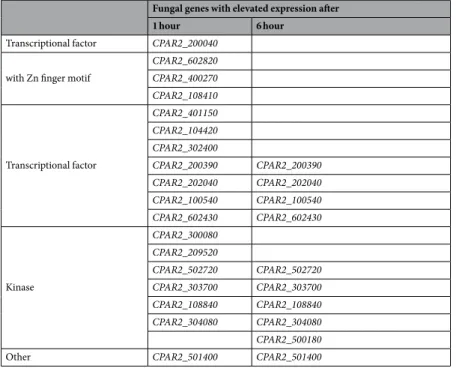
Our results show clear changes in expression upon incubation with THP-1 cells and during the monitored time course, with an increase in expression of fungal genes during host interaction, suggesting their involvement in virulence (see Materials and Methods, Supplementary Fig. S2 and Supplementary Table S1). A total fold change greater than 4 in gene expression (log2fold change greater than 2) was used to select genes for further analyses (Supplementary Table S1). The set of up-regulated genes includes several uncharacterized ORFs with hypothet- ical regulatory functions ranging from transcriptional factors to protein kinases, according to orthology-based functional assignment. Furthermore, the set of up-regulated genes included an additional ORF, CPAR2_501400, for which orthology-based functional assignment suggests a role in cell wall beta 1,6-glucan assembly. Based on their expression profiles and their putative biochemical activities, we aimed to generate a set of deletion mutant strains (Table 1).
Preparation of deletion mutant strains.
In order to study the role of the up-regulated hypothetical regulators in C. parapsilosis virulence, we established a deletion mutant collection of the identified ORFs. ORF deletion was performed using the fusion PCR method20, as adapted to C. parapsilosis by Holland et al.15. Due to its high specificity and speed, this method allowed us to generate a set of deletion mutant strains of the selectedFungal genes with elevated expression after
1 hour 6 hour
Transcriptional factor CPAR2_200040 with Zn finger motif
CPAR2_602820 CPAR2_400270 CPAR2_108410
Transcriptional factor
CPAR2_401150 CPAR2_104420 CPAR2_302400
CPAR2_200390 CPAR2_200390 CPAR2_202040 CPAR2_202040 CPAR2_100540 CPAR2_100540 CPAR2_602430 CPAR2_602430
Kinase
CPAR2_300080 CPAR2_209520
CPAR2_502720 CPAR2_502720 CPAR2_303700 CPAR2_303700 CPAR2_108840 CPAR2_108840 CPAR2_304080 CPAR2_304080 CPAR2_500180
Other CPAR2_501400 CPAR2_501400
Table 1. Targets of the C. parapsilosis deletion mutant library.
genes, each represented by two parallel homozygous deletion mutant strains. Following confirmation of the null mutants, strains were prepared for phenotypic assays.
Systematic screen of the generated mutant strains.
Viability testing and response to stressors.During invasion, fungal cells need to adapt to restrictive environmental conditions in order to survive and dis- seminate throughout a host. Such conditions include the presence of alternative nitrogen and carbon sources, elevated temperature, and a shift from neutral pH to slightly basic or acidic21–23. Pathogenic species also devel- oped strategies to degrade antimicrobial components present in the host serum19,24. Therefore, the deletion mutant strains were first analyzed in terms of fitness and viability under various conditions. Among the tested mutant strains, three showed a general growth defect on YPD complex media, indicating an adverse effect on fungal viability (Fig. 1/A). Furthermore, five showed reduced growth on minimal media (YNB + glucose), three on complex media set to pH 8, three in the presence of bovine serum albumin (BSA), and five on fetal bovine serum (FBS) supplemented plates, suggesting a potential defect in adaptation to alkaline conditions, in alterna- tive energy source utilization and serum protein degradation (Fig. 1/A). The response of each deletion mutant strain was also examined in the setting of various restrictive environmental conditions25. Survival of each mutant strain was evaluated in the presence of different stressors in liquid media. During fungal infection, phagocytic cells first distinguish cell wall surface components of the invaders via pattern recognition receptors, thus cell wall assembly significantly influences virulence26. To assess alterations in cell wall homeostasis calcofluor white, congo red and caffeine were used27,28. To identify mutants with potential glycosylation defects, Hygromycin B was also applied29. According to our results, eight strains showed altered response to the aforementioned stressors (Fig. 1/B). To evaluate the responses of the mutants to oxidative damage, we used H2O2 as stressor, to which two strains were susceptible (Fig. 1/B). In order to examine cell membrane integrity the membrane stressor SDS was used. During the analyses three mutant strains showed altered susceptibility to the membrane disturbing agent (Fig. 1/B). Altogether, these data suggest that 13 of the 19 examined genes may be involved in viability and stress tolerance regulation.
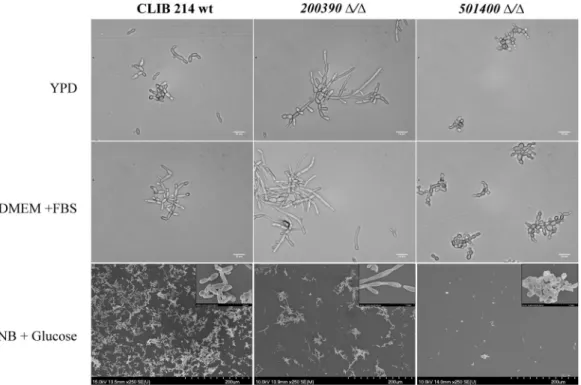
Morphology change. Although C. parapsilosis is unable to form true hyphae, the morphogenic shift to pseu- dohyphal forms has been associated with virulence13. When examining the ability of the mutants to form pseu- dohyphae, we found that CPAR2_200390Δ/Δ and CPAR2_501400Δ/Δ strains showed a remarkably different phenotype compared to the wild type. Yeast cells of CPAR2_200390Δ/Δ rapidly transitioned into extremely long and aggregating pseudohyphae, while CPAR2_501400Δ/Δ cells remained phenotypically locked in a yeast form (Fig. 2). Thus, these data suggest that CPAR2_200390 and CPAR2_501400 may be involved in the regulation and maintenance of morphology.
Biofilm formation and adhesive properties. C. parapsilosis effectively forms biofilms on intravenous catheters, prostheses, and other indwelling medical devices13. In order to examine the biofilm forming abilities of each deletion mutant strain we applied the FDA metabolic assay (Supplementary Fig. S3 and Supplementary Table S2).
Our results demonstrated that three strains showed a different biofilm profile when compared to the CLIB 214 wild-type strain (Fig. 3/A). Strains CPAR2_200390Δ/Δ, CPAR2_209520Δ/Δ and CPAR2_501400Δ/Δ displayed a lower capacity for biofilm formation, suggesting that the corresponding genes influence biofilm formation in C. parapsilosis.
Adhesion to various biotic and abiotic surfaces is a critical step for biofilm formation. While several key regulators of adhesion have been identified in C. albicans, only a few have been described in C. parapsilo- sis17. In order to search for such regulators, we tested adhesion to polystyrene plastic, the substrate used for the aforementioned biofilm formation assay (Supplementary Fig. S4 and Supplementary Table S2.). According to our results, six of the examined strains (CPAR2_100540Δ/Δ, CPAR2_200390Δ/Δ, CPAR2_300080Δ/Δ, Figure 1. Screening of the C. parapsilosis mutant strains. Summary table of the mutant phenotypes identified through viability testing on different solid media (A) and screening of stress responses in stressor supplemented liquid solutions (B). Mutant strains with altered phenotypes are shown only. Screening conditions are detailed in the Materials and Methods section and in Supplementary Materials and Methods. Defective growth, sensitive and resistant phenotypes were determined relative to the CLIB 214 wild-type strain.
www.nature.com/scientificreports/
CPAR2_302400Δ/Δ, CPAR2_303700Δ/Δ and CPAR2_400270Δ/Δ) showed decreased ability for adhesion (Fig. 3/B). As CPAR2_501400Δ/Δ cells were unable to significantly adhere to the polystyrene substrate, the results obtained with this strain were excluded from the final analyses.
Determining gene function - selected mutant strains.
Following the systematic screening of our mutant strains, three interesting ORFs with ≥6 altered phenotypes were selected for further in-depth analysis as potential regulators of distinct virulence-affecting mechanisms during host-pathogen interactions. These were nutrient acquisition (CPAR2_100540), morphology switch, cell wall reassembly (CPAR2_200390), and viability (CPAR2_303700).Nutrient acquisition by CPAR2_100540. During the general characterization of the deletion mutant strains, the CPAR2_100540Δ/Δ strain produced smooth colony morphology (Fig. 4/A), showed a mild growth defect on minimal media (Fig. 1/A) and appeared to be defective in terms of adhesion (Fig. 3/B) when compared to the wild type. CPAR2_100540Δ/Δ cells were also more susceptible to oxidative stress (Fig. 4/B) and alkaline envi- ronmental conditions (Fig. 4/C) than the wild type. While the inability to grow on pH 8 suggests a failure in trace element (e.g. ferric or ferrous iron) acquisition, oxidative stress susceptibility may indicate defects in respiration.
The orthologous gene of CPAR2_100540 in C. albicans (HAP5, 68.0% amino acid sequence identity) is known to play a role in both processes.
Iron acquisition: C. albicans Hap5, an indispensable subunit of the CBF (CCAAT-binding factor) complex, is required for the expression of the essential iron reductase FRP1 under iron-limited conditions30,31. Loss of CaHAP5 results in low FRP1 expression levels when cells are grown in inducing media with either pH 8 or an iron chelator30. Furthermore, iron chelators inhibit the growth of Cahap5Δ/hap5Δ cells30. Similarly, C. parapsi- losis CPAR2_100540Δ/Δ has decreased growth at pH 8 (Fig. 4/C) and on iron chelator (BPS)-supplemented media that also contained hemin (low-iron source) (Fig. 4/D). Gene expression analysis suggests that iron acqui- sition by C. parapsilosis is also dependent on a ferric reductase (CPAR2_402880) similar to C. albicans FRP1.
CPAR2_402880 expression in C. parapsilosis is also induced under both iron-limited conditions in the wild type strain (Fig. 4/E). In contrast, CPAR2_402880 expression levels remain relatively low in the CPAR2_100540Δ/Δ mutant strain, thus further supporting a role of CPAR2_100540 in C. parapsilosis iron acquisition (Fig. 4/E).
Alternative carbon source utilization: Carbon utilization via respiration is tightly related to available iron32, therefore the phenotypic traits observed in this section might be also linked to the altered iron homeostasis.
Previous reports suggested that HAP5 is involved in alternative carbon source utilization in S. cerevisiae and C. albicans33,34. Alternative energy source metabolism is usually associated with altered regulation of respiratory chain element encoding genes such as cytochrome c (CYC) subunits and/or cytochrome c oxidases (COX)33,34. Thus, growth deficiencies on alternative carbon sources are often related to defects in the respiratory chain35. Susceptibility of CPAR2_100540Δ/Δ cells to oxidative stressors further suggests a respiratory chain defect.
Figure 2. Pseudohyphal growth of the C. parapsilosis mutant strains. DIC and SEM images of pseudohyphae produced by the wild-type (CLIB 214) CPAR2_200390Δ/Δ and CPAR2_501400Δ/Δ strains 48 hours after cultivation in YPD, FBS supplemented DMEM and YNB + glucose liquid medium. Scale bars: 10 µm and 200 µm.
When studying the role of CPAR2_100540 in unfermentable carbon source metabolism, CPAR2_100540Δ/Δ cells showed decreased growth on both amino acids (Fig. 4/F) and lactate-supplemented media (Fig. 4/G).
Furthermore, gene expression analysis results suggest that the observed phenotype may be due to the misreg- ulation of ORFs equivalent to CYC1 (CPAR2_407500) and COX4 (CPAR2_207710) of C. albicans (Fig. 4/H).
Therefore, the obtained data led us to the conclusion that CPAR2_100540 is also involved in alternative carbon source utilization via regulating elements of the respiratory chain.
Contribution of CPAR2_100540 to virulence: Competition for the available iron sources and the ability to utilize alternative energy sources in the host are considered important virulence traits of pathogenic fungi36. To investigate the effects of the CPAR2_100540 ORF on virulence, we used both in vitro and in vivo infection models.
Killing assays performed with J774.1 macrophage-like cells indicated that more of CPAR2_100540Δ/Δ yeast cells were killed when compared to the wild-type strain both at 18 h and 24 h of the interaction (Fig. 4/I). Due to structural and functional similarities between G. mellonella and the mammalian innate immune system, this non-vertebrate model is frequently used as an alternative for virulence studies of Candida species37. Following inoculation with C. parapsilosis strains, survival of the individual larvae was monitored for 7 days. The survival results showed that the loss of CPAR2_100540 ORF resulted in significantly decreased virulence compared to the wild type strain (Fig. 4/J). We confirmed the attenuation of CPAR2_100540Δ/Δ in a murine infection model.
Infection with these cells resulted in significantly lower fungal burdens in the spleen and kidney 7 days after challenge, when compared to mice infected with the wild type strain (Fig. 4/K). These data confirm the role of CPAR2_100540 in C. parapsilosis virulence, possibly via the regulation of iron utilization and alternative carbon source acquisition.
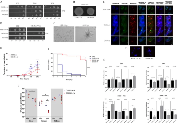
Morphology switch and cell wall assembly regulation by CPAR2_200390. Morphology transition: Our gen- eral characterization of the CPAR2_200390Δ/Δ strain revealed a retardation in growth (Fig. 5/A) along with Figure 3. Biofilm formation and substrate adherence of the C. parapsilosis mutant strains. (A) - Biofilm formation in microtiter plates was determined using FDA assay. For mutants N ≥ 16 and for CLIB wt N = 196, from at least 2 independent experiments per strain were used. Obtained data were analyzed by Kruskal-Wallis and Dunn’s multiple comparisons tests (***p ≤ 0.001; ****p ≤ 0.0001). (B) - Adherence to polystyrene plastic as a substrate in microtiter plates was also measured. For adherence assays N ≥ 12 for all mutant strains and N = 36 for the wild type strain were used from at least 2 independent experiments per strain and analyzed by Kruskal-Wallis and Dunn’s multiple comparisons test (****p ≤ 0.0001).
www.nature.com/scientificreports/
an irregular colony morphology (Fig. 5/B) and hyper-filamentation (Fig. 5/C), a phenotype set similar to that observed in C. albicans following the removal of its orthologous gene, SPT3 (81.8% amino acid sequence identity).
C. albicans SPT3 is a negative regulator of filamentous growth38. The obtained phenotypic traits suggested func- tional homology between SPT3 and the CPAR2_200390 ORF, supporting the inclusion of CPAR2_200390 as a reg- ulator of morphogenesis in C. parapsilosis. Interestingly, during the characterization of the CPAR2_200390Δ/Δ mutant strain we also observed low adhesive and biofilm forming capability (Fig. 3/A-B) along with resist- ance to the cell wall stressor calcofluor white (Fig. 5/D). The latter observation led us to the assumption that CPAR2_200390 in C. parapsilosis might also be involved in the maintenance of cell wall homeostasis.
Cell wall rearrangement: In order to further examine the cell wall stress resistant phenotype of CPAR2_200390Δ/Δ cells, different fluorescent dyes were used to investigate cell wall composition with calco- fluor white binding to chitin, Concanavalin A (ConA) to alpha mannan, and Wheat Germ Agglutinin (WGA) to chitin oligomer compounds. Fluorescent labeling revealed that the CPAR2_200390Δ/Δ strain displayed elevated chitin and chitin oligomer content in comparison with the wild type (Fig. 5/E). In addition to the accumulation of chitin oligomers around apical bud scars, the oligomers were also present along the longitudinal line of the mutant cells (Fig. 5/F) suggesting altered chitin homeostasis in CPAR2_200390Δ/Δ. To support this hypothesis, we examined the expression level of four chitinase and four chitin synthase encoding genes that were poten- tially involved in the dysregulation. Examined chitinase encoding ORFs included those equivalent to C. albicans Figure 4. Phenotypic traits of the CPAR2_100540Δ/Δ strain. (A) Colony morphology on YPD solid medium after 2 days of incubation at 30 °C. Growth of wild type (CLIB 214), CPAR2_100540Δ/Δ and CPAR2_100540 reintegrant (RI) strains on (B) 0.015 mM menadione containing solid medium after 3 days, (C) on pH 8 medium after 2 days, (D) on 500 mM BPS + 2 μM hemin supplemented media following 6 days, (F) on YNB + amino acid (AA) supplemented and (G) 2% lactate containing plates after 3 days of incubation, at the presented temperatures. Following growth assays, gene expression levels of the (E) C. albicans FRP1 ortholog, CPAR2_402880 was determined after cultivation of the three strains in both inductive (pH 8, BPS supplemented YPD) and non-inductive (YPD) conditions for 12 hours at 37 °C. (H) Relative normalized expression of CYC and COX gene orthologs (CPAR2_407500 and CPAR2_207710) was also examined following cultivation in YPD and amino acid supplemented (AA) YNB medium at 37 °C. Statistical significance was determined by unpaired two-tailed t-tests (**p ≤ 0.01; ***p ≤ 0.001, (****p ≤ 0.0001). (I) Interaction between J774.1 macrophage-like cells and yeast mutants was followed for 24 h, with determinations made at 2, 6, 18, and 24 h. For 2 h N ≥ 5, 6 h N = 8, and for 18 h and 24 h N = 4, from 2 independent experiments. Virulence properties of the mutant strains was also investigated in vivo, examining (J) G. mellonella larvae survival and (K) organ colonization of BALB/c mice after infection. Statistical significance was determined by two-way ANOVA and Dunnett’s multiple comparisons tests in case of J774.1, unpaired Mann-Whitney test in case of BALB/c mice infection and Mantel-Cox (Log-rank) test after G. mellonella infection (*p ≤ 0.05; ****p ≤ 0.0001).
CHS1 (CPAR2_805640), CHS2 (CPAR2_701490), CHS3 (CPAR2_801800) and CHS8 (CPAR2_502940), while chitinase encoding genes were CPAR2_800050 (CHT1), CPAR2_ 502140 (CHT2), CPAR2_ 200660 (CHT3) and CPAR2_211950 (CHT4)39–42. Real-time PCR analyses revealed altered chitinase and chitin synthase expression profiles for the mutant strain in both YPD complex and 10% FBS supplemented DMEM- medium when com- pared to the wild type (Fig. 5/G). These data suggest that the CPAR2_200390 ORF is also involved in chitin homeostasis regulation.
Role of CPAR2_200390 in virulence: During fungal infection, cell wall assembly has a major impact on rec- ognition by immune cells, while a morphology switch is associated with host cell and tissue disruption36. In kill- ing assays performed with J774.1 macrophage-like cells, the CPAR2_200390Δ/Δ strain had increased survival compared to wild-type cells both at 18 h and 24 h of interaction (Fig. 5/H). Interestingly however, G. mellonella infection studies suggested that CPAR2_200390Δ/Δ cells are avirulent in vivo, as survival rates were similar to that of PBS infected larvae (Fig. 5/I). Furthermore, following infection of BALB/c mice, the mutant cells had reduced CFUs present in the spleen 7 days after infection, compared to wild type-infected animals (Fig. 5/J).
Hence, CPAR2_200390 plays a role in morphology transition and cell wall homeostasis maintenance, and also contributes to virulence.
Figure 5. Characteristics of the CPAR2_200390Δ/Δ strain. (A) Growth of wild-type (CLIB 214), CPAR2_200390Δ/Δ and CPAR2_200390 reintegrant (RI) strains after 2 days on YPD solid media at selected temperatures. (B) Colony morphology on YPD plate following 2 days of incubation at 30 °C. (C) Pseudohyphae formation in 10% FBS supplemented DMEM medium after 24 hours of incubation at 37 °C.
(D) Growth on 60 μg/ml calcofluor white supplemented YPD medium at 30 °C. (E) Fluorescence microscopic images of calcofluor white, ConA (Concanavalin A) and WGA (wheat germ agglutinin) stained wild-type, CPAR2_200390Δ/Δ and CPAR2_200390 RI cells. (F) Localization of chitin oligomer accumulations in the wild-type and mutant cell wall. (G) Relative normalized expression of C. albicans chitinase and chitin synthase orthologs (CPAR2_805640 as CHS1; CPAR2_701490 as CHS2; CPAR2_801800 as CHS3; CPAR2_502940 as CHS8, CPAR2_800050 as CHT1; CPAR2_ 502140 as CHT2, CPAR2_ 200660 as CHT3 and CPAR2_211950 as CHT4) after cultivation in YPD and 10% FBS supplemented DMEM medium. Statistical significance was determined by unpaired two-tailed t-tests (*p ≤ 0.05; **p ≤ 0.01; ****p ≤ 0.0001). Virulence attributes of the CPAR2_200390Δ/Δ strain following (H) an in vitro killing assay with J774.1 macrophage-like cells and after (I) G. mellonella larvae and (J) BALB/c mice infection. Statistical significance was determined by two-way ANOVA and Dunnett’s multiple comparisons tests in case of J774.1, and unpaired Mann-Whitney test in case of BALB/c mice infection. Mantel-Cox (Log-rank) tests were applied in the case of G. mellonella infection (*p ≤ 0.05;
****p ≤ 0.0001).
www.nature.com/scientificreports/
Fitness regulation by CPAR2_303700. Viability regulation: Notable features of the CPAR2_303700Δ/Δ mutant strain included a growth defective phenotype (Fig. 6/A), low adhesive properties (Fig. 3/B), and susceptibility to the oxidative stressor H2O2 (Fig. 6/B) and to decreased temperature (Fig. 6/C). The closest characterized ortho- logue of CPAR2_303700 is S. cerevisiae CGI121, encoding a subunit of the KEOPS/EKC complex43–45. According to preliminary searches, this factor is conserved across the entire Candida clade and Cpar2_303700 also contains conserved domains of the CGI121 superfamily (NCBI conserved domain search). According to the performed in silico data analyses (Supplementary Materials and Methods), the two proteins share similar secondary and tertiary structures (Fig. 6/D, Supplementary Table S3) and Cpar2_303700 is likely to form a stable conformation with the closest interacting partner of Cgi121 in the S. cerevisiae KEOPS/EKC complex, Bud32 (Fig. 6/E, Supplementary Table S4.), suggesting that Cpar2_303700 and Cgi121 may also share similar properties. Structural similarity and the predicted interaction between Cpar2_303700 and Bud32 was supported by measurable values such as low average RMSD values used for distance comparison (0.61 ± 0.04 Å in case of structure comparison and 0.4 ± 0.2 Å in the predicted complex) and negative overall energy levels (Supplementary Table S4). In silico 7 H-bonds were observed between the residues of Cpar2_303700 and Bud32 that were stabilized by several hydrophobic inter- actions, further supporting a stable interaction (Fig. 6/E). These data also suggest that Cpar2_303700 might be a potential member of the C. parapsilosis KEOPS/EKC complex.
CPAR2_303700 and virulence: In A. nidulans, members of the KEOPS/EKC complex are associated with met- abolic processes that are known to influence the virulence properties of C. albicans. These mechanisms include the regulation of TUP1’s function as a key yeast to filamentous growth switch regulating transcriptional factor as well as the complex’s involvement in amino acid and carbon source acquisition46–49. Sensitivity to oxidative stress Figure 6. Phenotypic features of the CPAR2_303700Δ/Δ strain. (A) Growth kinetics of wild-type (CLIB 214), CPAR2_303700Δ/Δ and CPAR2_303700 reintegrant (RI) strains in YPD liquid medium, at 30 °C. (B) Growth on 10 mM H2O2 supplemented YPD medium, and (C) on YPD solid medium at different temperatures. (D) In silico 3D structure comparison of the hypothetical Cpar2_303700 and the previously described S. cerevisiae Cgi121 proteins. Scale bar: 10 ångström (Å). (E) 3D structure of the complex formed by Cpar2_303700 and Bud32 and its comparison to the Cgi121-Bud32 complex. Predicted H-bonds (bottom left) and the surrounding hydrophobic residues (bottom right) maintain the stability of the Cpar2_303700 – Bud32 complex. Moderately attenuated virulence of CPAR2_303700Δ/Δ (F) in vitro after J774.1 macrophage infection, following (G) G. mellonella larvae inoculation and (H) BALB/c mice infection. Statistical significance was determined by two-way ANOVA and Dunnett’s multiple comparisons tests in case of J774.1, unpaired Mann-Whitney tests in case of BALB/c mice infection and Mantel-Cox (Log-rank) test after G. mellonella infection: *p ≤ 0.05;
****p ≤ 0.0001.
multifunctional regulatory factors such as transcriptional factors and kinases are as important as virulence fac- tors themselves in determining the outcomes of host-pathogen interactions. Therefore, targeting the virulence regulatory machinery of pathogenic fungal species has been proposed as an innovative method for effectively combating fungi while concomitantly avoiding the toxicity and resistance mechanisms of the currently available antifungal compounds2.
In this study, we used a pre-selective approach for target selection in order to identify regulators potentially contributing to the virulence of an emerging human fungal pathogen, C. parapsilosis in various routes. To iden- tify such regulatory genes, whole RNA sequencing was performed shortly following host-pathogen interaction.
Among the ORFs upregulated after infection, transcriptional factor- and kinase-encoding genes were selected for further examination and a targeted deletion mutant collection was constructed. These deletion mutant strains were tested under various conditions designed to recapitulate environments the fungus would encounter under certain host conditions or that would likely influence C. parapsilosis pathogenicity in an indirect manner.
As a result we found that 84% of the characterized mutant strains had a phenotype different from wild-type (Supplementary Fig. S5, Supplementary Table S5). Furthermore, 32% of the tested mutant strains showed multi- ple phenotypic defects suggesting that the examined regulators have pleiotropic effects (Supplementary Fig. S5).
Out of the characterized ORFs, three with potentially distinct regulatory roles in virulence were further exam- ined: CPAR2_100540, CPAR2_200390 and CPAR2_303700.
During infection, pathogens continually compete with host cells for available iron sources. Although iron compounds are usually carried by high affinity proteins in the host, limiting access to these trace elements, path- ogenic fungi have developed various strategies in order to acquire these vital compounds52. In addition, carbon utilization via respiration is tightly linked to available iron - due to the presence of iron containing enzymes –, that further highlights the role of this trace element in viability32. Depending on the site of invasion, invading fungi face a limited access to glucose and often only alternative carbon sources are available for utilization. Under such circumstances, alternative carbon and nitrogen source metabolic pathways are activated, which usually require additional energy and, thus, alter the function of the mitochondrial respiratory chain49,53. In C. albicans both iron acquisition and alternative carbon source metabolism are influenced by the transcriptional factor, Hap5, a subunit of the conserved CBF complex. Hap5 controls iron homeostasis by regulating the ferric reductase FRP1, and also influences carbon source utilization via regulating respiratory chain elements cytochrome c and cytochrome c oxidases30,34. According to our results, the identified CPAR2_100540 ORF, an orthologous gene of HAP5, plays a similar role in both processes in C. parapsilosis. Moreover, results of the performed virulence studies indicate that the identified regulator further contributes to the virulence of this species in both in vitro and in vivo infection models. It is noteworthy to mention that S. cerevisiae HAP5, is also involved in alternative carbon source utiliza- tion, although, while CYC and COX gene expression was downregulated in the S. cerevisiae hap5Δ/hap5Δ strain, upregulation was observed in the respective mutant of C. albicans, which underscores the variability in gene regulation between different yeast species and supports further investigation of these processes in other fungi33,34. In the present work, our functional studies show that CPAR2_100540 is comparable to that of C. albicans HAP5;
thus, this identified regulator is likely required for C. parapsilosis virulence via the regulation of nutrient acquisi- tion and alternative carbon source utilization.
Yeast to pseudohyphal growth transition promotes C. parapsilosis invasion through several mechanisms, including host cell and tissue disruption, tissue penetration and biofilm formation13. In this study, loss of the CPAR2_200390 ORF resulted in a phenotypic feature set previously observed after the removal of SPT3 in C. albi- cans. Spt3, a subunit of the evolutionally conserved SAGA complex, is known to play a key role in the morpho- logical switching of both S. cerevisiae and C. albicans38. Although while SPT3 deletion resulted in a yeast-locked phenotype in S. cerevisiae, hyper-filamentous growth was observed in C. albicans, suggesting opposite regula- tory functions in the two species38. The obtained phenotype set of the CPAR2_200390Δ/Δ strain indicated that CPAR2_200390 function is equivalent to that of C. albicans SPT3. Interestingly, our further analyses revealed that CPAR2_200390 also influences the chitin homeostasis of the cell wall, adhesive properties and biofilm formation, suggesting a pleiotropic effect on C. parapsilosis virulence. These features have not been previously associated with SPT3. Furthermore, our virulence studies underscored the influence of this regulator on C. parapsilosis patho- genicity, although the outcomes were inconsistent, as the CPAR2_200390Δ/Δ strain was significantly attenuated in vivo, however less efficiently killed by murine macrophages in vitro, when compared to the wild type. These results suggest that at the cellular level, clearance of CPAR2_200390 Δ/Δ is less dependent on macrophages due to weak or no preference (decreased phagocytic activity, data not shown), although could be the result of an
www.nature.com/scientificreports/
alternative host response. In total, these data confirm that CPAR2_200390 regulates virulence in C. parapsilosis by mechanisms that affect mainly morphogenesis and cell wall assembly.
Several conditions, such as temperature and oxidative stress, are known to influence fungal viability, which in turn indirectly affects virulence. Removal of CPAR2_303700 resulted in various defects, mainly including a gen- eral growth defective phenotype coupled with sensitivity to low temperatures, suggesting the regulator’s involve- ment in fungal fitness. The closest ortholog to the identified C. parapsilosis ORF is S. cerevisiae’s CGI121, and there is no ortholog yet characterized among other Candida species. As part of the highly conserved KEOPS/EKC complex, Cgi121 is involved in functions such as transcription co-activation, tRNA modification, and telomere maintenance, although not required for survival in S. cerevisiae43–45. Although the performed in silico data analy- ses do not confirm functional similarity between Cgi121 and Cpar2_303700, they suggest that Cpar2_303700 is a protein structurally similar to Cgi121. Thus Cpar2_303700 may also be a subunit of the evolutionarily conserved KEOPS/EKC complex in C. parapsilosis.
Interestingly, disruption of 3 of the 5 KEOPS/EKC subunits in S. cerevisiae resulted in a serious growth defective phenotype and temperature sensitivity, features that are especially common among telomere defec- tive strains43. Mutants defective in telomere maintenance are also hypothesized to be more susceptible to oxida- tive stress, although this phenotype was not reported in with CGI121. In addition, removal of CGI121 rescued growth defective phenotypes of telomere defective mutant strains, which led to the conclusion that Cgi121 promotes telomere uncapping43. The evolutionally conserved hypothetical function and phenotypical features of CPAR2_303700Δ/Δ and Cgi121Δ/Δ strains suggest that there might be opposite regulatory mechanisms between C. parapsilosis and S. cerevisiae, similarly to what was observed in case of HAP5 and SPT334,38, although this hypothesis needs to be supported by further experiments and analyses. Although the exact function of CPAR2_303700 is not yet determined, according to our results, the identified ORF nevertheless contributes to virulence regulation, possibly via an indirect manner.
Taken together, in this study we identified several interesting C. parapsilosis mutant strains with relevant virulence-determining features and a relatively high yield of mutants with multiple defects that indicate the respective regulatory gene’s function in virulence. Out of the characterized genes, we describe three transcription regulators that influence C. parapsilosis pathogenicity in distinct ways and thus contribute to our better under- standing of virulence regulation in this species. These include Cpar2_100540, a transcriptional factor regulating nutrient acquisition, Cpar2_200390 involved in morphology switch and cell wall assembly, and Cpar2_303700, a protein kinase regulating fungal viability. Although more in-depth studies of the identified regulatory processes in C. parapsilosis are required, these data further support the idea that the origin of virulence in pathogenic species can be due to alterations in signaling and regulatory networks.
Materials and Methods
THP-1 cell line infection with C. parapsilosis cells. THP-1 monocytic cells (Sigma-Aldrich, 100 μl of 5 × 10⁶/ml) were seeded onto 96 well plates in 10% FBS (EuroClone) supplemented Dulbecco’s Modified Eagle Medium (DMEM, Lonza) and incubated overnight at 37 °C, with 5% CO2. Infection of monocytes with C. par- apsilosis CDC 317 cells was performed in 1:5 ratio. Cells were co-incubated at 37 °C with 5% CO2 and collected by centrifugation (1000 rpm, 5 min) at 1 or 6 h post-infection. Cells were washed once with ice-cold PBS and pellet was suspended in RNase-free distilled water. For fungal RNA isolation, monocytes were lysed through 27 G needles and RNA extraction was performed with the RNeasy Plant Mini Plus Kit (Qiagen) according to manu- facturer’s instructions.
RNA sequencing and data analysis.
RNA sequencing libraries were prepared according to protocols provided by Illumina. Sequencing was carried out in an Illumina HiSeq2000 sequencer with a depth of >20 mil- lion reads per sample at the CRG genomics facility. We sequenced paired-end 50bp-long reads RNAseq data for the above-mentioned time-points. The reference genomes for the reference strain CDC317 and the human hg19 were obtained from the Candida Genome Database (CGD)54 and UCSC (UCSC Genome Browser)55, respec- tively. Gene annotations were obtained from the gencode project, version 18 and from CGD. Read mapping and alignment were carried out using tophat2 v2.0.956 with default sensitivity and specificity conditions, based on the bowtie2 2.1.0 short read mapper57. Transcript abundance estimation was obtained via FluxCapacitor v1.5.2 with automatic annotation mapping. Gene normalization for thresholding was carried out via RPKM (Reads Per Kilobase of transcript per Million mapped reads) obtained from Flux Capacitor output. A minimum threshold of RPKM ≥10 was used to eliminate low expression transcripts and limit noise. Differential expression analysis was carried out via the DESeq2 v1.10.1 Differential Expression analysis package in R.Preparation of C. parapsilosis mutant strains.
Strains used in this study are listed in Supplementary Table S6. Two of the 19 deletion mutants were also part of the Holland deletion collection and analysis (CPAR2_202040 and CPAR2_100540). Homozygous mutant strains were generated according to methods described by Holland et al. using the C. parapsilosis CLIB 214 double auxotrophic strain (CPL2H1)15. Primers used for gene deletion and null mutant confirmation are presented in Supplementary Dataset 1. In each case, two homozygous deletion mutant strains were selected for phenotypic analyses.For the mutant strains CPAR2_100540Δ/Δ, CPAR2_200390Δ/Δ and CPAR2_303700Δ/Δ, one copy of the respective allele was introduced into the RP10 locus for complementation (see Supplementary Materials and Methods)58.
Strain cultivation.
Strains were grown in YPD liquid medium (1%D-glucose-1%peptone-0.5%yeast-extract) supplemented with 1% Penicillin-Streptomycin with shaking (200 rpm) at 30 °C overnight, unless stated otherwise.Detailed information about the characterization process is included in Supplementary Materials and Methods.
and 48 h of incubation using Leica light microscope (DMI4000B, Leica Microsystems) and HITACHI S-4700 cold field emission scanning electron microscope (see Supplementary Materials and Methods for sample preparation).
Three individual experiments were performed to confirm abnormal phenotypes.
Cell wall composition assay (microscopy).
To examine cell wall composition of C. parapsilosis mutant cells, ConA-FITC (Sigma-Aldrich), calcofluor white (Sigma-Aldrich) and WGA-TRITC (Sigma-Aldrich) were used. A detailed protocol of staining procedure is available in Supplementary Materials and Methods. Stained strains were imaged with a Zeiss Observer Z1 fluorescence microscope. At least three individual experiments were performed to confirm alterations in cell wall assembly.Adhesion and biofilm assay. Cultures were grown overnight (12–16 h) at 30 °C with shaking in YNB medium. Cells were washed with PBS and adjusted to 105cells/ml. In a microtiter plate, 100 µL of adjusted cul- tures were aliquoted per well and statically incubated at 37 °C for 90 min for adhesion to occur. Each well was washed once with 100 µL of PBS, and metabolic activity assay (FDA) was performed to assess adhesion capacity.
For adhesion assays, N = 36 for wild-type and N ≥ 12 for mutant strains was used from at least 2 independent experiments. In case of biofilm formation, following adhesion of seeded cells, each well was washed once with 100 µL of PBS, and 100 µL of fresh YNB media was added per well. Plates were incubated at 37 °C with a wet paper towel for 48 h for biofilms to form. After biofilm formation, each well was washed with 100 µL of PBS and biofilm levels were assessed using a metabolic activity assay (FDA). For biofilm assays, at least 2 independent experiments were performed for each mutant strain.
Fluorescein diacetate (FDA) Assay.
To each tested well, 100 µL of FDA solution (40 µg/mL, Sigma-Aldrich) was added. Plates were incubated at 37 °C for 1 h, protected from light, and supernatants were transferred to a new fluorescence-compatible plate and read at 485/535 nm (ex/em) in a Victor plate reader.Gene expression analyses.
For total RNA isolation, the Ambion, RibopureTM-Yeast RNA isolation kit (Invitrogen) was applied. Prior to examining expression levels in BPS, pH8, amino acid supplemented YNB and FBS-containing DMEM medium, 50 µl of 2 × 10⁷cells from overnight cultures were transferred into the respective media and incubated overnight at 37 °C. For gene expression analysis during biofilm formation, cells were grown in 1 ml of 0.5% glucose-supplemented YNB solution for 48 h.For cDNA synthesis, the RevertAid First Strand cDNA Synthesis Kit (Thermo Scientific) was applied accord- ing to the manufacturer’s guide. For expression analyses 100ng cDNA were used. PCR conditions were as follows:
95 °C for 3 minutes, followed by 50 cycles of 95 °C for 10 s, and 60 °C for 30 s. Primers used for gene expression studies are listed in Supplementary Dataset 1. TUB4 housekeeping gene was used as an internal control. Data were normalized to wild-type gene expression levels. Three individual experiments were performed to confirm alterations in gene expression.
Interaction between C. parapsilosis and J774.1 macrophage-like cells.
Killing assays were performed according to a standardized protocol with minor modifications59. Briefly, 5 × 10⁴ J774.1 mac- rophages per well (American Type Culture Collection) were infected with C. parapsilosis cells in a 5:1 ratio of yeast:macrophage-like cells. Control wells contained yeast cells only. Interactions were left for 0, 2, 6, 18, and 24 h at 37 °C, with 10% CO2. Contents were collected, washed, and passed through a 26 G needle. Suspensions were plated on Sabouraud dextrose agar plates and incubated at 30 °C for 24–48 h. Killing efficiency was calculated as described previously59. Graph values were normalized to the number of viable fungal cells in untreated wells at 24 h (100%). At 2 h N ≥ 5, 6 h N = 8, and at 18 h and 24 h N = 4 from 2 independent experiments were used.Galleria mellonella infection assay. Infection was achieved with 10 µl of 5 × 10⁸ C. parapsilosis cells/ml inoculated in the last pro-leg and 20 caterpillars (N = 20) were used per strain. Groups of PBS sham-infected and uninfected (witness group) larvae were also used. The Galleria were maintained at 30 °C and survival was monitored daily.
In vivo murine infection model. Female Balb/c mice (Jackson Laboratories, 6–8 weeks of age, NCI) were injected intraperitoneally with 107cells (N ≥ 6 per yeast strain). After 3 days of infection, 3 mice per condition were sacrificed and the remaining mice were sacrificed at day 7 post-infection. Liver, kidney, and spleen were
www.nature.com/scientificreports/
collected by necropsy, weighed, homogenized, and plated on Sabouraud dextrose agar plates. Colony forming units (CFUs) were counted after 24 h incubation at 30 °C, to assess infection levels.
Ethics Statement.
Animal experiments were performed according to the guide published by the Institute of Laboratory Animal Resources of the National Research Council. Animal care for this study was approved by the Animal Welfare and Research Ethics Committee at the Albert Einstein College of Medicine (Animal Use Protocol#20170503).
Statistical analyses.
Unpaired, two-tailed t-tests were applied for gene expression analysis, Kruskal-Wallis test with Dunn’s multiple comparisons test was performed for FDA assays, 2-way ANOVA and Dunnett’s test for multiple comparisons was applied for J774.1, and non-parametric Mann-Whitney tests were performed on BALB/c mice data assessment, and Mantel-Cox (Log-rank) tests were used for survival data evaluation. After unpaired, two-tailed t-tests, the mean ± SEM values, whereas in case of non-parametric tests, medians and the interquartile range are represented on the graphs. Statistical significance was determined by GraphPad Prism v5.0 or v6.0software. Significant differences were considered at P-values of ≤ 0.05(*p ≤ 0.05; **p ≤ 0.01; ***p ≤ 0.001;****p ≤ 0.0001).
References
1. Sparagano, O. A. Transcription Factors as a Target for Vaccination Against Ticks and Mites. Adv Protein Chem Struct Biol 107, 275–282, https://doi.org/10.1016/bs.apcsb.2016.11.004 (2017).
2. Bahn, Y. S. Exploiting Fungal Virulence-Regulating Transcription Factors As Novel Antifungal Drug Targets. PLoS Pathog 11, e1004936, https://doi.org/10.1371/journal.ppat.1004936 (2015).
3. Rodionov, D. A. Comparative genomic reconstruction of transcriptional regulatory networks in bacteria. Chem Rev 107, 3467–3497, https://doi.org/10.1021/cr068309+ (2007).
4. Schwarzmuller, T. et al. Systematic phenotyping of a large-scale Candida glabrata deletion collection reveals novel antifungal tolerance genes. PLoS Pathog 10, e1004211, https://doi.org/10.1371/journal.ppat.1004211 (2014).
5. Noble, S. M., French, S., Kohn, L. A., Chen, V. & Johnson, A. D. Systematic screens of a Candida albicans homozygous deletion library decouple morphogenetic switching and pathogenicity. Nat Genet 42, 590–598, https://doi.org/10.1038/ng.605 (2010).
6. Liu, O. W. et al. Systematic genetic analysis of virulence in the human fungal pathogen Cryptococcus neoformans. Cell 135, 174–188, https://doi.org/10.1016/j.cell.2008.07.046 (2008).
7. Brown, G. D. et al. Hidden killers: human fungal infections. Sci Transl Med 4, 165rv113, https://doi.org/10.1126/scitranslmed.3004404 (2012).
8. Liu, Y. & Filler, S. G. Candida albicans Als3, a multifunctional adhesin and invasin. Eukaryot Cell 10, 168–173, https://doi.
org/10.1128/EC.00279-10 (2011).
9. Naglik, J. R., Challacombe, S. J. & Hube, B. Candida albicans secreted aspartyl proteinases in virulence and pathogenesis. Microbiol Mol Biol Rev 67, 400–428, table of contents (2003).
10. Kadosh, D. & Johnson, A. D. Induction of the Candida albicans filamentous growth program by relief of transcriptional repression:
a genome-wide analysis. Mol Biol Cell 16, 2903–2912, https://doi.org/10.1091/mbc.E05-01-0073 (2005).
11. Knight, S. A., Lesuisse, E., Stearman, R., Klausner, R. D. & Dancis, A. Reductive iron uptake by Candida albicans: role of copper, iron and the TUP1 regulator. Microbiology 148, 29–40, https://doi.org/10.1099/00221287-148-1-29 (2002).
12. Bliss, J. M. Candida parapsilosis: an emerging pathogen developing its own identity. Virulence 6, 109–111, https://doi.org/10.1080/2 1505594.2015.1008897 (2015).
13. Trofa, D., Gacser, A. & Nosanchuk, J. D. Candida parapsilosis, an emerging fungal pathogen. Clin Microbiol Rev 21, 606–625, https://
doi.org/10.1128/CMR.00013-08 (2008).
14. Pammi, M., Holland, L., Butler, G., Gacser, A. & Bliss, J. M. Candida parapsilosis is a significant neonatal pathogen: a systematic review and meta-analysis. Pediatr Infect Dis J 32, e206–216, https://doi.org/10.1097/INF.0b013e3182863a1c (2013).
15. Holland, L. M. et al. Comparative phenotypic analysis of the major fungal pathogens Candida parapsilosis and Candida albicans.
PLoS Pathog 10, e1004365, https://doi.org/10.1371/journal.ppat.1004365 (2014).
16. Gacser, A., Trofa, D., Schafer, W. & Nosanchuk, J. D. Targeted gene deletion in Candida parapsilosis demonstrates the role of secreted lipase in virulence. J Clin Invest 117, 3049–3058, https://doi.org/10.1172/JCI32294 (2007).
17. Bertini, A. et al. Targeted gene disruption in Candida parapsilosis demonstrates a role for CPAR2_404800 in adhesion to a biotic surface and in a murine model of ascending urinary tract infection. Virulence 7, 85–97, https://doi.org/10.1080/21505594.2015.111 2491 (2016).
18. Grozer, Z. et al. Candida parapsilosis produces prostaglandins from exogenous arachidonic acid and OLE2 is not required for their synthesis. Virulence 6, 85–92, https://doi.org/10.4161/21505594.2014.988097 (2015).
19. Horvath, P., Nosanchuk, J. D., Hamari, Z., Vagvolgyi, C. & Gacser, A. The identification of gene duplication and the role of secreted aspartyl proteinase 1 in Candida parapsilosis virulence. J Infect Dis 205, 923–933, https://doi.org/10.1093/infdis/jir873 (2012).
20. Noble, S. M. & Johnson, A. D. Strains and strategies for large-scale gene deletion studies of the diploid human fungal pathogen Candida albicans. Eukaryot Cell 4, 298–309, https://doi.org/10.1128/EC.4.2.298-309.2005 (2005).
21. Barelle, C. J. et al. Niche-specific regulation of central metabolic pathways in a fungal pathogen. Cell Microbiol 8, 961–971, https://
doi.org/10.1111/j.1462-5822.2005.00676.x (2006).
22. Brown, A. J., Brown, G. D., Netea, M. G. & Gow, N. A. Metabolism impacts upon Candida immunogenicity and pathogenicity at multiple levels. Trends Microbiol 22, 614–622, https://doi.org/10.1016/j.tim.2014.07.001 (2014).
23. Vylkova, S. et al. The fungal pathogen Candida albicans autoinduces hyphal morphogenesis by raising extracellular pH. MBio 2, e00055–00011, https://doi.org/10.1128/mBio.00055-11 (2011).
24. Gropp, K. et al. The yeast Candida albicans evades human complement attack by secretion of aspartic proteases. Mol Immunol 47, 465–475, https://doi.org/10.1016/j.molimm.2009.08.019 (2009).
25. Perez-Garcia, L. A. et al. Role of Protein Glycosylation in Candida parapsilosis Cell Wall Integrity and Host Interaction. Front Microbiol 7, 306, https://doi.org/10.3389/fmicb.2016.00306 (2016).
26. Netea, M. G., Van der Meer, J. W. & Kullberg, B. J. Role of the dual interaction of fungal pathogens with pattern recognition receptors in the activation and modulation of host defence. Clin Microbiol Infect 12, 404–409, https://doi.org/10.1111/j.1469-0691.2006.01388.x (2006).
27. Ram, A. F. & Klis, F. M. Identification of fungal cell wall mutants using susceptibility assays based on Calcofluor white and Congo red. Nat Protoc 1, 2253–2256, https://doi.org/10.1038/nprot.2006.397 (2006).
28. Fuchs, B. B. & Mylonakis, E. Our paths might cross: the role of the fungal cell wall integrity pathway in stress response and cross talk with other stress response pathways. Eukaryot Cell 8, 1616–1625, https://doi.org/10.1128/EC.00193-09 (2009).
29. Dean, N. Yeast glycosylation mutants are sensitive to aminoglycosides. Proc Natl Acad Sci USA 92, 1287–1291 (1995).
cerevisiae and Candida albicans and is required for C. albicans virulence. Genetics 161, 509–519 (2002).
39. Dunkler, A., Jorde, S. & Wendland, J. An Ashbya gossypii cts2 mutant deficient in a sporulation-specific chitinase can be complemented by Candida albicans CHT4. Microbiol Res 163, 701–710, https://doi.org/10.1016/j.micres.2008.08.005 (2008).
40. McCreath, K. J., Specht, C. A. & Robbins, P. W. Molecular cloning and characterization of chitinase genes from Candida albicans.
Proc Natl Acad Sci USA 92, 2544–2548 (1995).
41. Mio, T. et al. Role of three chitin synthase genes in the growth of Candida albicans. J Bacteriol 178, 2416–2419 (1996).
42. Munro, C. A. et al. CHS8-a fourth chitin synthase gene of Candida albicans contributes to in vitro chitin synthase activity, but is dispensable for growth. Fungal Genet Biol 40, 146–158 (2003).
43. Downey, M. et al. A genome-wide screen identifies the evolutionarily conserved KEOPS complex as a telomere regulator. Cell 124, 1155–1168, https://doi.org/10.1016/j.cell.2005.12.044 (2006).
44. Kisseleva-Romanova, E. et al. Yeast homolog of a cancer-testis antigen defines a new transcription complex. EMBO J 25, 3576–3585, https://doi.org/10.1038/sj.emboj.7601235 (2006).
45. Srinivasan, M. et al. The highly conserved KEOPS/EKC complex is essential for a universal tRNA modification, t6A. EMBO J 30, 873–881, https://doi.org/10.1038/emboj.2010.343 (2011).
46. Braun, B. R., Head, W. S., Wang, M. X. & Johnson, A. D. Identification and characterization of TUP1-regulated genes in Candida albicans. Genetics 156, 31–44 (2000).
47. Dzikowska, A. et al. KAEA (SUDPRO), a member of the ubiquitous KEOPS/EKC protein complex, regulates the arginine catabolic pathway and the expression of several other genes in Aspergillus nidulans. Gene 573, 310–320, https://doi.org/10.1016/j.
gene.2015.07.066 (2015).
48. Heymann, P. et al. The siderophore iron transporter of Candida albicans (Sit1p/Arn1p) mediates uptake of ferrichrome-type siderophores and is required for epithelial invasion. Infect Immun 70, 5246–5255 (2002).
49. Ene, I. V. et al. Host carbon sources modulate cell wall architecture, drug resistance and virulence in a fungal pathogen. Cell Microbiol 14, 1319–1335, https://doi.org/10.1111/j.1462-5822.2012.01813.x (2012).
50. Wu, P., Nielsen, T. E. & Clausen, M. H. Small-molecule kinase inhibitors: an analysis of FDA-approved drugs. Drug Discov Today 21, 5–10, https://doi.org/10.1016/j.drudis.2015.07.008 (2016).
51. Berg, T. Inhibition of transcription factors with small organic molecules. Curr Opin Chem Biol 12, 464–471, https://doi.
org/10.1016/j.cbpa.2008.07.023 (2008).
52. Almeida, R. S., Wilson, D. & Hube, B. Candida albicans iron acquisition within the host. FEMS Yeast Res 9, 1000–1012, https://doi.
org/10.1111/j.1567-1364.2009.00570.x (2009).
53. Lorenz, M. C. & Fink, G. R. The glyoxylate cycle is required for fungal virulence. Nature 412, 83–86, https://doi.
org/10.1038/35083594 (2001).
54. Binkley, J. et al. The Candida Genome Database: the new homology information page highlights protein similarity and phylogeny.
Nucleic Acids Res 42, D711–716, https://doi.org/10.1093/nar/gkt1046 (2014).
55. Tyner, C. et al. The UCSC Genome Browser database: 2017 update. Nucleic Acids Res 45, D626–D634, https://doi.org/10.1093/nar/
gkw1134 (2017).
56. Kim, D. et al. TopHat2: accurate alignment of transcriptomes in the presence of insertions, deletions and gene fusions. Genome Biol 14, R36, https://doi.org/10.1186/gb-2013-14-4-r36 (2013).
57. Langmead, B. & Salzberg, S. L. Fast gapped-read alignment with Bowtie 2. Nat Methods 9, 357–359, https://doi.org/10.1038/
nmeth.1923 (2012).
58. Murad, A. M., Lee, P. R., Broadbent, I. D., Barelle, C. J. & Brown, A. J. CIp10, an efficient and convenient integrating vector for Candida albicans. Yeast 16, 325–327, https://doi.org/10.1002/(SICI)1097-0061(20000315)16:4<325::AID-YEA538>3.0.CO;2-# (2000).
59. Nemeth, T. et al. Characterization of virulence properties in the C. parapsilosis sensu lato species. PLoS One 8, e68704, https://doi.
org/10.1371/journal.pone.0068704 (2013).
Acknowledgements
RT and this research was supported by TÁMOP 4.2.4. A/2-11-1-2012-0001 „National Excellence Program – Elaborating and operating an inland student and researcher personal support system convergence program. The project was subsidized by the European Union and co-financed by the European Social Fund. AG was funded by NKFIH NN 113153, by GINOP-2.3.2-15-2016-00035, by GINOP-2.3.3-15-2016-00006. AG and LN were also founded by CNPq (Program Science without borders - 407380/2013-2). TN was supported by the Postdoctoral Fellowship by the Hungarian Academy of Sciences. Research at TG lab was partially funded by grants from Spanish Ministry of Economy and Competitiveness BFU2015-67107 cofounded by European Regional Development Fund (ERDF); from the European Union Seventh Framework Programme (FP7/2007-2013) under grant agreements FP7-PEOPLE-2013-ITN-606786 “ImResFun” and from the European Union’s Horizon 2020 research and innovation programme under the Marie Sklodowska-Curie grant agreement No H2020-MSCA- ITN-2014-642095. JDN is partially supported by US NIH grants R21 AI124797-02 and AI52733-06A2. We would like to thank Prof. Geraldine Butler for the KO strategy and parental strains necessary for mutant strain generation. We would like to thank Chetna Tyagi for the help with the in silico data analyses, Mónika Homolya for contributing to the chitinase and chitin synthase expression experiments, Dr. Sándor Kocsubé for the summary table and Tamás Petkovits for the help with the SEM experiments.
www.nature.com/scientificreports/
Author Contributions
A.G. and T.G. designed the study, J.D.N., C.V. and L.N. also contributed to the development of this project. R.T.
and V.C. carried out the majority of experiments with the help of E.T., F.B., G.M., T.N. and C.P. R.T., V.C. and E.T.
also analysed the data. R.T. prepared manuscript figures to which V.C. contributed, and R.T. with A.G. prepared the main manuscript text. All authors reviewed the manuscript, contributed to the discussion and approved the final version.
Additional Information
Supplementary information accompanies this paper at https://doi.org/10.1038/s41598-018-19453-4.
Competing Interests: The authors declare that they have no competing interests.
Publisher's note: Springer Nature remains neutral with regard to jurisdictional claims in published maps and institutional affiliations.
Open Access This article is licensed under a Creative Commons Attribution 4.0 International License, which permits use, sharing, adaptation, distribution and reproduction in any medium or format, as long as you give appropriate credit to the original author(s) and the source, provide a link to the Cre- ative Commons license, and indicate if changes were made. The images or other third party material in this article are included in the article’s Creative Commons license, unless indicated otherwise in a credit line to the material. If material is not included in the article’s Creative Commons license and your intended use is not per- mitted by statutory regulation or exceeds the permitted use, you will need to obtain permission directly from the copyright holder. To view a copy of this license, visit http://creativecommons.org/licenses/by/4.0/.
© The Author(s) 2018


