Cell Host & Microbe
Innovative anti-cytolytic screen identifies potent inhibitors of mycobacterial virulence protein secretion
--Manuscript Draft--
Manuscript Number: CELL-HOST-MICROBE-D-14-00341R1
Full Title: Innovative anti-cytolytic screen identifies potent inhibitors of mycobacterial virulence protein secretion
Article Type: Resource Article
Keywords: Tuberculosis, protein secretion, ESX-1 system
Corresponding Author: Stewart Cole, Ph.D.
Lausanne, FRANCE
First Author: Jan Rybniker
Order of Authors: Jan Rybniker
Jeffrey Chen Claudia Sala Ruben Hartkoorn Anthony Vocat Andrej Benjak Stefanie Boy-Röttger Ming Zhang
Rita Szekely Zoltán Greff László Örfi István Szabadkai János Pató György Kéri Stewart Cole, Ph.D.
Abstract: Mycobacterium tuberculosis (Mtb) depends on protein secretion systems like ESX-1 for intracellular survival and virulence. The major virulence determinant and ESX-1- substrate, EsxA, causes tissue damage and necrosis, thereby promoting pathogen spread and dissemination. We developed a fibroblast survival assay (FSA) that exploits this phenotype by selecting for molecules that protect host cells from Mtb-induced lysis without being bactericidal in vitro. Hit compounds identified in this high-throughput screen blocked secretion of EsxA thus promoting phagosome maturation and
substantially reducing bacterial burden in activated macrophages. Target identification studies led to the discovery of BTP15, a benzothiophene inhibitor of the histidine kinase MprB that indirectly regulates ESX-1, and BBH7, a benzyloxybenzylidine hydrazine compound. BBH7 affects metal ion homeostasis in Mtb and revealed zinc stress as a signal for EsxA secretion. This novel screening approach extends the target spectrum of small molecule libraries and will help to tackle the mounting problem of antibiotic-resistant mycobacteria.
Suggested Reviewers: Olivier Neyrolles olivier.neyrolles@ipbs.fr
Expert in intracellular pathogenesis of TB and heavy metal toxicity Charles Thompson
Powered by Editorial Manager® and ProduXion Manager® from Aries Systems Corporation
Expert in drug discovery and natural products with deep knowledge of actinobacteria Wilbert Bitter
w.bitter@vumc.nl
Expert in type 7 secretion systems and mycobacteriology Vojo Deretic
vderetic@salud.unm.edu
Expert in intracellular pathogenesis, autophagy and two component systems, particularly MprAB.
Patricia Champion champion.7@nd.edu
Expert in type 7 secretion systems and mycobacteria Miriam Braunstein
braunste@med.unc.edu Expert in protein secretion Sophie Lagrange
Sophie.Lagrange@sanofi.com
Expert in screening, drug discovery and drug development
Opposed Reviewers: Jeffery Cox
Jeffery.Cox@ucsf.edu Potential conflict of interest
Powered by Editorial Manager® and ProduXion Manager® from Aries Systems Corporation
FACULTE DES SCIENCES DE LA VIE GLOBAL HEALTH INSTITUTE UNITE DU PROFESSEUR COLE
EPFL SV / GHI / UPCOL SV 3531
Station nº19 CH-1015 Lausanne
Téléphone : Fax : E-mail : Site web :
+4121 693 1851 +4121 693 1790 stewart.cole@epfl.ch http://ghi.epfl.ch
Lausanne, 19 August 2014 Ella Hinson, Ph.D.
Scientific Editor Cell Host & Microbe Re. CELL-HOST-MICROBE-D-14-00341
Dear Dr. Hinson,
Thank you very much for sending the very positive reviews of our “Resource” paper that we have revised along the lines suggested by the reviewers.
Major changes to the manuscript are highlighted in the text whereas minor edits and shortening of the text are not. The manuscript currently contains 54,226 characters including spaces.
Detailed responses to the reviewers’ queries may be found in the accompanying rebuttal and we are confident that their comments have been more than adequately addressed by additional experimentation and careful editing of the text.
We would like to thank the reviewers for their helpful comments and constructive criticism, and you for handling the manuscript, which we trust will now be found acceptable for publication.
Since it was first submitted the authors have decided to file a patent to protect some of the compounds discussed in the paper. The filing has not yet been completed so I would like to request that the paper not be published online following acceptance if this is your policy. Thank you for considering this request and for providing a “conflict of interest form” for us to sign.
Sincerely yours,
Professor Stewart Cole FRS Director of the Global Health Institute
Cover Letter
1
Rebuttal CELL-HOST-MICROBE-D-14-00341
Reviewers' Comments:
Reviewer #1: The manuscript of Rybniker et al describes an elegant and interesting study for new compounds blocking M. tuberculosis virulence. They have developed and used a novel assay for this, by blocking the virulence mechanism of the crucial ESX-1 system. Using this system the authors have identified two new classes of compounds with interesting activities. Overall, the manuscript is interesting, novel and well written and as such important for the field and for a wider audience.
Suggestions for improvement:
The authors indicate that BTP15 could function as a kinase inhibitor through inhibition of the autophosphorylation of MprB, resulting in altered expression of espA. However, the difference in expression of espA is rather mild (factor 2) and also an espA mutant (fig. 1B) seems to have a more mild effect than compound BTP15. The authors should discuss these observations, which probably mean that the regulon of MprB could potentially contain more genes that have an influence on ESX-1 functioning.
We agree with reviewer 1 that dysregulation of the espACD operon alone would not fully explain the strong attenuation of BTP15-treated bacteria since the ΔespA strain is only moderately attenuated in the FSA. We have recently shown that stronger (BTP15-like) attenuation requires both dysregulation of the espACD regulon AND the ESX-1 core region (Chen et al., Mol. Micro. 2013). A typical sign for
dysregulation of the ESX-1 core region is inhibition of EspB secretion, a core protein that is still secreted in the espACD mutant (Chen et al., Mol. Micro. 2013). Interestingly, the ΔmprAB mutant is also defective in EspB secretion (Pang et al. JB, 195, 66-75, 2013). Furthermore, putative MprA binding sites have been identified in the esxA promoter (RD1 region). The whiB6 gene, which is involved in RD1 gene regulation, is also under control of MprA (Pang et al. JB, 195, 66-75, 2013, Pang et al. Microbiology 153, 3007). This indicates that MprAB not only regulates the espACD operon but also the ESX-1 core region. In the revised manuscript we included an anti-EspB Western blot of BTP1-treated samples showing that EspB-secretion is also affected by this compound (new Figure S3B), which explains the strong attenuation of BTP15- treated bacteria. This is discussed in the revised manuscript lines 328 – 332: “MprAB also seems to regulate the ESX-1 region itself since the mprAB mutant fails to secrete EspB, a protein that is not influenced by the espACD operon (Chen et al., 2013; Pang et al., 2013). We also found inhibition of EspB secretion upon BTP15 treatment (Figure S3B), accompanied by greater attenuation than seen with a ΔespA mutant in the FSA.”
The authors convincingly show that compound BBH7 affects cell wall permeability using the EtBr assay.
However, it would be interesting to determine whether also sensitivity to other antibiotics is altered due to this permeability defect.
Response to Reviewers
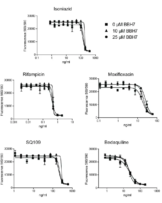
2 In the revised version of the manuscript we present the results of testing a panel of antimycobacterial drugs in the presence of 0, 10 and 25 µM of BBH7. Interestingly, the MIC of these compounds was not altered in the presence of these rather high concentrations of BBH7 in contrast to treatment of Mtb with broadly acting efflux pump inhibitors or protonophores such as CCCP, which have a major impact on MICs of anti-TB drugs (Rodrigues et al., BMC Microbiology 11:35, 2011; Gupta et al. Microbial Drug Resistance, Vol 16, 1, 2010). This indicates that BBH7-induced cell wall alterations are subtle leading to accumulation of selected molecules such as the cationic dye EtBr and well-defined alterations of the mycobacterial transcriptome (metal ion stress). However, this subtle alteration has a major impact on the intracellular survival of Mtb probably due to the effect on virulence protein secretion. The new data were added to the supplementary information (Figure S5) and to the main text, lines 246 – 247: “Interestingly, BBH7 concentrations as high as 25 µM had no impact on the activity of first- and second-line TB drugs (Figure S5).“
Furthermore, and perhaps more importantly, the authors mention that the alteration in cell wall permeability are leading to signs or zinc and copper stress (line 261). However, in my opinion the authors should be careful to what is the cause and the consequence of cell wall permeability, gene induction and zinc/copper stress. Zinc stress could also be achived for instance by blocking the ESX-3 secretion system (which should be mentioned).
Several publications link the essential ESX-3 system with iron and zinc stress and we agree with reviewer 1 that alterations of ESX-3 secretion through BBH7 may lead to transcriptional signs of zinc stress.
Although we were unable to identify differential secretion of the ESX-3 substrates EsxG/H (which carry a zinc binding site) in the mycobacterial secretome (see comment below), we did see down-regulation of the respective genes upon treatment (Figure 5, Table S3). This correlates well with published data showing that ESX-3 depletion leads to up-regulation of genes associated with zinc starvation (Serafini et al, PLOSone Vol8/10, 2013). Thus, the ESX-3 system responds to BBH7 treatment, but it is not known whether this happens in response to increased zinc levels in the mycobacterial cytosol or to direct alterations of ESX-3 through BBH7. Since ESX-3 seems to be involved in zinc uptake (depletion of ESX-3 leads to low intracellular zinc concentrations), blockage of ESX-3 by a small molecule will probably not lead to zinc accumulation as suggested by our RNA-seq data. Thus, it will rather be an increase of EsxG/H secretion that would cause the effects we saw for BBH7. We now discuss a possible role of ESX-3 and BBH7 function in lines 402 – 405: “Of note, there is evidence that the ESX-3 secretion system is involved in zinc homeostasis of Mtb (Serafini et al., 2013). Thus, interference with ESX-3 activity by BBH7 may cause for the zinc stress observed.“
- and have modified the sentence in lines 249 - 251 (previously 261) - this now reads: “Having established that BBH7 treatment leads to transcriptional signs of zinc and copper stress, we surmised that intracellular metal-ion stress might be the link to inhibition of mycobacterial protein secretion.”
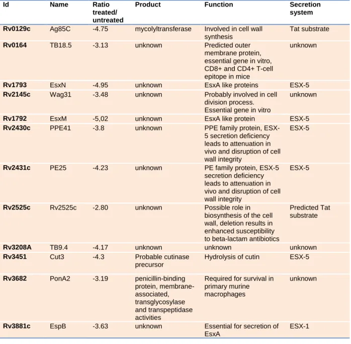
In Table S2 a number of proteins are listed that show reduced secretion upon addition of BBH7.
However, the fold reduction is not shown and also a list of proteins that are not affected by the compound would be helpful to understand the mechanism of action, i.e. are all ESX-1 and ESX-5
3 substrates blocked? Is also the secretion of all extracellular proteins with a signal sequence blocked?
Please also note that cut 3 has been suggested previously as a putative ESX-5 substrate (Abdallah et al., 2009).
In the revised version of Table S2 we now display the ratios of treated and untreated samples. For all of the chosen proteins, the amount of protein in the treated sample is heavily reduced.
In our MS experiments, we were able to identify 1140 proteins in the culture filtrate of Mtb.
Approximately 5% of these were quantified with lower amounts in the BBH7-treated samples. We were unable to show reduced secretion of all ESX-1 substrates. EspA and EspC for example were not
significantly reduced upon treatment. However, due to strong reduction of overall protein content in BBH7-treated culture filtrate (approx. 50% less total protein) it was difficult to normalize the MS data against the background. This led to a relatively stringent statistical data evaluation with better identification of differential secretion of the most abundant proteins. EspA and EspC are not very
abundant and though there was a clear trend towards reduction of these proteins in the treated samples, the p-value was not below the threshold of 0.05. This also applies to Tat secreted proteins.Ag85C carries a signal sequence for Tat secretion – this protein was clearly reduced in the treated samples. However, Ag85A, another Tat substrate, was significantly reduced in the forward experiment (p-value <0.05) but not in the reverse experiment (p-value = 0.06).
We performed the MS experiments primarily to confirm Western blot data, which indicated that the secretion of non-ESX-1 substrates is also affected by BBH7. We were able to identify at least 3 secretion systems that were significantly affected: ESX-1, ESX-5 and Tat. However, the strong effect of BBH7 on protein secretion heavily impairs the protein ratio density and thereby reduces the efficiency of statistical tools in detecting changes to all proteins.
The reference to cut3 has now been changed to ESX-5.
Please check the use of that/which.
This has been checked and some changes made.
Reviewer #2: This paper describes an anti-cytolytic screen conducted in MRC-5 lung fibroblasts infected with high doses of Mycobacterium tuberculosis (M.tb). The innovative aspect of this study is the identification of M.tb secreted proteins by screening for compounds that inhibit M.tb cytotoxicity in fibroblasts but are not bactericidal in vitro. The authors focus on inhibition of Esx-1 (ESAT-6), a secreted mycobacterial protein involved in membrane binding and host cell lysis. An input library of over 10,000 compounds yielded benzothiophene inhibitors and benzyloxybenzylidine hydrazine as two major classes of compounds that met hit criteria. The authors subsequently investigated the possible mechanism of action for two compounds with M.tb specific activity: BTP15 and BBH7. While they do not find a specific mode of action for BBH7, they do uncover a potential role for zinc as a signal for Esx1 secretion.
4 In general, the work is well done and the results are interesting. There is an unusual mix here - although labeled as a "resource" paper, much of the work involves investigating the mechanism of action of the compounds that they found in their screen, less of a resource and more of a science paper. However, whatever the designation, the results are interesting and publishable.
A couple of major items might make the work a bit more readable:
- The fibroblast assay will seem quite peculiar to those in the field and immediately raise red flags as there is little evidence that these are actually infected in vivo (and the MOI is certainly likely to be non physiologic). It would probably be worth framing the discussion around using these as a bioassay for ESX- 1 function rather than infection. This seemed to be the intention and the fibroblasts probably offer advantages over screening in macrophages (although, what those are should be enumerated).
We agree with reviewer 3 that the in vivo role of lung fibroblasts in mycobacterial pathogenesis is unknown although these cells are part of the Mtb-granuloma. In fact, in our screening assay the only purpose of these cells is quantification of Mtb induced cell-death. Though this works equally well with macrophages, one advantage of fibroblasts over these professional phagocytes is the failure to detect effects of host-modifying drugs like imatinib when using fibroblasts (see also comment 2 of the minor points). We tried to set up the assay in a sufficiently broad manner to target virtually all structural and regulatory ESX-1-components yet to be specific enough to exclude compounds with no or minor effects on this system (such as kinase inhibitors acting on the host), which makes target identification easier. This is now discussed in lines 310 – 314: “Another key factor for selectivity of the bioassay is our choice of lung fibroblasts for the quantification of Mtb-induced cell death. These non-professional phagocytes fail to detect host modifying agents that reduce the intracellular burden of Mtb in macrophages (Lechartier et al., 2014). This feature may be beneficial for target identification of these anti-virulence drugs.“
- The confirmatory macrophage assays represent something a bit more like a real infection. These should be featured more prominently in both the results and discussion.
This wise suggestion has been followed. We have modified the results section of the macrophage experiments by linking it to the intracellular Mtb-quantification data generated in fibroblasts (Figure 2D) and changed the presentation of Figure 6 to make it more readable. Highlighting the difference in
intracellular survival of BTP15-treated bacteria in fibroblasts vs. macrophages puts more emphasis on the importance of data generated in the activated THP-1 cells. All macrophage data are now presented in the main manuscript in lines 277 – 294 and not in the supplementary data section thereby ensuring greater prominence. Furthermore, the data are now referred to in the discussion lines 322 - 340.
- This might be a semantic point but I found it quite distracting. The compounds the authors describe are not clearly direct inhibitors of protein secretion. To the extent that their mechanism is clear, they produce broad transcriptional changes altering the production of some secreted proteins and of other proteins required for secretion. I find the title to be misleading.
5 We respectfully disagree with the reviewer and have retained the original title. At no point, do we state that the inhibitors directly target the protein secretion systems and on several occasions we indicate that BTP15 acts on an upstream kinase, MprB. Since we are not misleading the reader and the other
reviewers had no objection to the wording of the title we feel that our choice is justified.
Minor points:
1. The authors do an excellent job of describing the screen that pairs the fibroblast survival assay (FSA) with the resazurin reduction microtiter assay (M.tb REMA). They are missing a citation for the original fibroblast assay describing correlation between MOI and cytotoxicity by Takii et. al (AAC, 2002).
We have included this citation in line 104 of the revised version of the manuscript as well as the Hsu et al.
2003 citation as requested by reviewer 3.
2. The authors highlight one limitation of the FSA when they show that compounds blocking host components of programmed cell death fail to protect M.tb infected fibroblasts from lysis, even though they have been shown to do this in infected macrophages. While this is designed to be a rapid screen, with only 3 days incubation with infected fibroblasts, it would be useful to see a longer time course and potential interactions between host-modifying compounds and infected fibroblasts.
As suggested by the reviewer we repeated the FSA using lower MOIs (5 and 2 instead of 10) and a longer incubation time of 5 days instead of 3 days. At an MOI of 5, the panel of host modifying agents had no effect on fibroblast survival (not shown). At an MOI of 2 there was only a slight protective effect of imatinib, the Akt inhibitor H-89 and AX20017 (but not of nilotinib – another BCR-Abl inhibitor). This stands in contrast to macrophage data where similar compounds had potent impact on intracellular Mtb burden (between 0.5 and 1 log reduction for imatinib), which most likely correlates with good survival of infected macrophages. Thus we assume that the inactivity of these compounds in the FSA is primarily due to the choice of fibroblasts, which are not professional phagocytes, and to differences in cell
differentiation between the respective cell lines. This is discussed in lines 123 - 126 of the revised version:
“Reducing the MOI led to a minor protective effect of some kinase inhibitors (Figure S1A) indicating that their inactivity in the FSA is primarily due to the choice of cell: fibroblasts versus macrophages.“
and the new data may be found in Figure S1A.
3. Figure 2D shows that BTP15 is not toxic to GFP-M.tb in lung fibroblasts while BBH7 is. While this begins to show the different mechanisms of action of each drug, this experiment should be linked in the text to the comparable experiment done in macrophages in Figure 6C.
In the revised version of the manuscript we linked the experiment presented in Figure 2D with the section on macrophage experiments by recalling the fibroblast work at the beginning of the section presenting the macrophage results - lines 279 – 280: “Using GFP-expressing Mtb we had shown that BBH7 strongly affects viability of intracellular bacteria in MRC-5 fibroblasts whereas BTP15 does not (Figure 2D). “
6 4. The authors show clearly that treatment with BBH7 results in a dose-dependent inhibition of EsxA and Ag85 secretion, while BTP15 shows inhibition of EsxA and Ag85, but with a different pattern. Bacteria were grown in Sauton's medium for preparation of protein lists for the immunoblots, which nicely shows the efficacy of the compounds in different media.
We thank the reviewer for appreciating this point.
5. The authors show that mprA mRNA levels are downregulated with BTP15 treatment at late
timepoints, and then further pursue an argument for deregulation of the mprAB locus leading to reduced EsxA secretion. Were mRNA levels of mprB also downregulated?
Yes, this was the case. We found a 3-fold reduction in abundance of the mprB transcript after 48 hours of treatment with 10 µM of BTP15. This new set of data may be found in Figure S3A and lines 186 -187 of the main text: “BTP15 (10 µM) also decreased mprB transcript levels 3-fold after 48 h exposure (Figure S3A).”
6. EtBr assays are used to study membrane permeability as well as efflux. The authors conclude that the increased fluorescence curve acquired with BBH7 treatment is suggestive of a change in membrane permeability. If the authors add an efflux pump inhibitor to the system, does the fluorescence shift?
The EtBr assay we describe has been extensively used by several groups to determine efflux and altered cell wall permeability in mycobacteria (Rodrigues et al. AAC, vol 57/2, 2013; Rodrigues et al., BMC microbiology 11:35, 2011, Machado et al. PLOSone, vol 7/4, 2012). It was shown that efflux pump inhibitors as well as ionophores that alter cell wall permeability lead to EtBr accumulation. Since this has been extensively documented already we decided not to repeat the experiment using pump inhibitors such as verapamil. Furthermore, the EtBr assay cannot differentiate between altered influx or efflux across the mycobacterial cell wall. In the case of BBH7, the transcriptomic signature shows signs of metal ion accumulation and this could also be due to reduced efflux of these ions. Thus, in our manuscript, we use the expression “altered cell wall permeability” and prefer not to use the terms "efflux" or "influx". Of note, when we screened a panel of FDA-approved drugs such as verapamil in the FSA, there was no protective effect on fibroblast survival. In addition, BBH7 had no impact on the MIC of a panel of antimycobacterial drugs, which stands in contrast to treatment with efflux pump inhibitors (new Figure S5 in the revised manuscript).
7. The authors use microscopy to show that there is a reduced bacterial load in the macrophage upon treatment with BBH7 and BTP15. However, since this paper has focused on the Esx1 secretion system, it would be nice to se a comparison experiment with a GFP-labeled ESX1 knockout strain.
ESX-1 knockout strains of the RD1 region show a controversial pattern with regard to phago-lysosomal processing as published by McGurn et al. (Infection and Immunity, 75, 2007). The ESX-1 secretion
7 deficient ΔeccD1 (putative ESX-1 transmembrane channel) strain was transferred to the lysosome
whereas a ΔesxA strain was not. In another study an espL transposon mutant did not arrest phagosomal processing whereas wild-type bacteria did (Brodin et al., PLOSpathogens 2010), however, it is not known whether this mutant is deficient in EsxA secretion. In addition, there are several studies showing that the BCG vaccine strain (deficient in ESX-1 secretion) arrests phagosome maturation, possibly due to up- regulation of compensatory genes. Due to these discrepancies, we decided not to include a mutant strain as a control – furthermore, it is not clear which mutant would have been most appropriate for this experiment. To fully answer the question of ESX-1 dependent arrest in phagosome maturation, these experiments should be performed with a panel of different RD1 and non-RD1 mutants deficient in ESX-1 secretion followed by in-depth analysis of the mechanism behind the different phenotypes - obviously this is far beyond the scope of the present drug screening assay. Interestingly, the ΔphoP mutant (an EsxA secretion mutant) is clearly deficient in blockage of phagosome maturation indicating that sensor kinases impact phagosomal processing of Mtb (Ferrer et al., PLOSone, 2010).
8. While the finding that zinc may be a signal for Esx1 secretion is interesting, the model described in Figure 7 is perhaps beyond the scope of a Resource paper.
We have chosen to retain Figure 7 as it summarizes graphically several important findings.
Edits
1. The labels for Table S1 and Table S3 say RNA-sec, instead of RNA-seq.
This has been changed. Thanks for pointing it out.
2. The figure legend for Figure S3 states: "EsxA and GroEL were detected in the culture filtrate of Mtb Erdman treated with different cell wall biosynthesis inhibitors as well as BBH7 and BTP15". The immunoblot shown here has no visible band for EsxA under BBH7 treatment and a significantly reduce band with BTP15 treatment. This discrepancy should be addressed.
This corresponds to secretion inhibition by these compounds as is also shown in Figure 3 of the manuscript. We improved the legend for Figure S4C (previously Figure S3C) to make this clear.
3. Figure 5a. It would be helpful to label the chemical structure of BTP15.
This has been changed.
4. The manuscript describes the EtBr membrane permeability assays as being Figure 4b when they should be Figure 5b.
This has been changed. Thanks for the correction.
8 Reviewer #3: The Manuscript by Rybniker et al. is a well written manuscript describing an interesting new screen for novel compounds that alter the virulence of Mycobaterium tuberculosis. The authors take advantage of the observation that Mycobaterium tuberculosis will induce lysis of various cell lines and develop a high throughput screen to identify novel compounds from a 10,000 compound library that inhibit this lysis. They identify a number of interesting hits which they subsequently validate. In an excellent RNA-seq study preformed on Mtb treated with one of the compound they identify which demonstrates that one of these compounds regulates the transcription of the ESX-1 system. In addition they show other compounds attack diverse targets that affect phosphorylation, permeability or
membranes bound ATPses. Clearly this is one of the strengths of this paper that it reveals a number of different pathways important for the parthenogenesis and virulence of M. tuberculosis. including a kinase, while other compounds are shown to affect permeability. The authors conclude in the manuscript by saying that these compounds alter phagosome-lysosome fusion maturation for Mtb in macrophages. The work is clearly innovative and provides new tools to study the biology of Mtb. Further studies to asses their roles in animal models would be out of the scope of this original manuscript, but it clear the authors intend to explore these possibilities.
Specific Comments:
1) Line 77 should include a reference to Braunstein et al. 2003. Mol. Microbiol. 48:453-464.
This reference has been inserted.
2) Line 113: the end of the first sentence should include: "as first describes by Hsu et al." (From:
Proceedings of the National Academy of Sciences of the United States of America 100, 12420-12425, listed in references)
This suggestion has been adopted as mentioned above in response to Minor Point 1 of reviewer 2.
3) Line 118: Which ΔRD1 mutant are you using in the experiment? Was is made in the Erdman strain or the Mtb strain? Please provide the reference. This is important to know because the screen is done selective of the Erdman strain. I presume this is because the RV strain does not work. Is this true or not?
And having the matched isogenic strains could give you different results for this specific experiment.
We agree with reviewer 3 that strain specification is an important issue when investigating and discussing bacterial virulence and we failed to provide these data in the initial submission. The ΔRD1 strain we used in our study is based on the H37Rv genetic background and not on the Erdman strain. To ensure comparability of experiments we included FSA data of the well defined Tn::pe35 Mtb Erdman transposon mutant in Fig. 1B. This mutant carries a transposon insertion in a promoter upstream of the esxAB genes leading to full abrogation of ESX-1 dependent protein secretion, which is comparable to that seen with ΔRD1 strains (Chen et al. 2013, Mol. Micro. 89:1154-66). In the FSA, this mutant is attenuated
9 to the same extent as the H37Rv ΔRD1 strain. This indicates that once ESX-1-dependent protein secretion is fully blocked, the attenuation phenotype in the FSA is high and independent of the genetic background of the original strain used. The ΔespA strain we used in our study is an Erdman strain. The backgrounds of all strains used in this figure are now specified in the materials and methods section (lines 431 - 433).
To comment on the phenotype of the H37Rv strain in the FSA: during setup of the FSA we tested a panel of wild type Mtb strains for efficiency in fibroblast lysis. Though clearly inhibiting fibroblast survival, H37Rv displayed the lowest levels of cytolysis and this correlates well with recent findings of reduced EsxA secretion in H37Rv, which is most likely due to a mutation of the WhiB6 promoter upstream of the RD1 region (Solans et al., Infection and Immunity, 82/8, 2014). This confirms again, that the FSA is a highly sensitive tool to quantify EsxA secretion and justifies our choice of Mtb Erdman for drug screening.
4) Also the strains listed in lines 119 and 120 are neither references in the text nor the figure legends.
Please see the response to point 3 where clarification is now provided.
5) Line 123: Rather than say "all compounds," instead list Isoniazid, Rifampicin, Limezolid, Moxifloxacin, DMSO, Streptomycin, and Kanamycin.
This has been changed on line 117 as suggested.
6) In line 260-273: the zinc regulation of ESXA is a very interesting result. However, is it possible that this is a secondary response to regulation of ESX-3 which then leads to this altered response of ESX-1? Can you please comment?
Recent data suggest a role of ESX-3 in zinc and iron uptake. Key to this was the finding that the ESX-3 system is transcriptionally regulated by the zinc uptake repressor, Zur, and the iron dependent repressor, IdeR (Serafini et al, JB, Vol 191/20, 2009). To our knowledge, there are no data showing that these regulators also affect ESX-1 secretion. Furthermore, as might be expected, ESX-3 responds to low zinc conditions since it seems to be required for zinc uptake, probably due to up-regulation of its substrates, though this has not been shown yet. We observed the opposite for ESX-1 where high zinc levels led to hyper-secretion of EsxA. Thus, it is difficult to identify a link between the divergent responses of ESX-3 and ESX-1 secretion. ESX-1 regulation is complex and several regulatory and sensor proteins have been shown to target ESX-1 promoters. An environmental signal for most of these regulators has not been identified yet and zinc could be one of these signals. Indeed, zinc responsive two-component system such as CzcRS of Pseudomonas aeruginosa are well known regulators of bacterial virulence (Dieppois et al. Plosone 2012 http://www.ncbi.nlm.nih.gov/pubmed/22666466). In our view, the link between zinc, ESX-3 and ESX-1 could be evolutionary. It is possible that an ancestral ESX system was required for heavy metal uptake, and that after gene duplication, one copy diverged into a specialized host modifying ESX-1 system that still responds to zinc concentrations as well as to other stimuli. As a signal to differentiate between extracellular, cytosolic and intraphagosomal localization, responding to zinc levels is physiologically
10 relevant since these vary widely between these different compartments (Botella et al. 2011, Cell host and microbe 10, 248-259).
The role of ESX-3 and zinc homeostasis has been explained and cited in the revised version of the manuscript lines 403 – 405: “Of note, there is evidence that the ESX-3 secretion system is involved in zinc homeostasis of Mtb (Serafini et al., 2013). Thus, interference with ESX-3 activity by BBH7 may cause for the zinc stress observed.”
See also our response to comment 3 for reviewer 1.
Graphical abstract
Click here to download Graphical Abstract: graphicalabstract.tif
1
Innovative anti-cytolytic screen identifies potent inhibitors of
1
mycobacterial virulence protein secretion
2
Jan Rybniker1,2, Jeffrey M. Chen1, Claudia Sala1, Ruben Hartkoorn1, Anthony 3
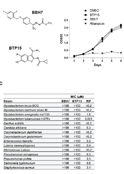
Vocat1, Andrej Benjak1, Stefanie Boy-Röttger1, Ming Zhang1, Rita E. Szekely1, 4
Zoltán Greff3, László Örfi3,4, István Szabadkai3, János Pató3, György Kéri3,5, 5
Stewart T. Cole1* 6
1 Global Health Institute, Ecole Polytechnique Fédérale de Lausanne (EPFL), CH-1015 7
Lausanne, Switzerland 8
2 1st Department of Internal Medicine, University of Cologne, D-50937 Cologne, Germany 9
3 Vichem Chemie Research Ltd, H-1022 Herman Otto u. 15, Budapest, Hungary 10
11
4 Semmelweis University, Department of Pharmaceutical Chemistry, H-1092, Hőgyes Endre u.
12
9, Budapest, Hungary 13
14
5 Semmelweis University, Department of Medical Chemistry, Pathobiochemistry and Molecular 15
Biology, H-1094 Tűzoltó u. 37-47, Budapest, Hungary 16
17
Running title: Targeting mycobacterial protein secretion 18
*Corresponding author:
19
Prof. Stewart T. Cole 20
Global Health Institute 21
Ecole Polytechnique Fédérale de Lausanne (EPFL) 22
Station 19 23
CH-1015 Lausanne 24
Switzerland 25
E-mail: stewart.cole@epfl.ch 26
Phone: +41-21-693 1851 27
Fax: +41-21-693 1790 28
29 30
Manuscript
2 SUMMARY
31
Mycobacterium tuberculosis (Mtb) depends on protein secretion systems like 32
ESX-1 for intracellular survival and virulence. The major virulence determinant 33
and ESX-1-substrate, EsxA, causes tissue damage and necrosis, thereby 34
promoting pathogen spread and dissemination. We developed a fibroblast 35
survival assay (FSA) that exploits this phenotype to select molecules that protect 36
host cells from Mtb-induced lysis without being bactericidal in vitro. Hit 37
compounds identified in this high-throughput screen blocked secretion of EsxA 38
thus promoting phagosome maturation and substantially reducing bacterial 39
burden in activated macrophages. Target identification studies led to the 40
discovery of BTP15, a benzothiophene inhibitor of the histidine kinase MprB that 41
indirectly regulates ESX-1, and BBH7, a benzyloxybenzylidine hydrazine 42
compound. BBH7 affects metal ion homeostasis in Mtb and revealed zinc stress 43
as a signal for EsxA secretion. This novel screening approach extends the target 44
spectrum of small molecule libraries and will help to tackle the mounting problem 45
of antibiotic-resistant mycobacteria.
46
HIGHLIGHTS 47
Mycobacterium tuberculosis causes EsxA-dependent host cell lysis 48
EsxA secretion can be targeted in high-throughput screens 49
Small molecules with diverse mechanisms of action inhibit EsxA secretion 50
Small molecule inhibitors abrogate ESX-1-dependent pathogenicity 51
52
3 INTRODUCTION
53
Tuberculosis, resulting from infection with Mycobacterium tuberculosis (Mtb), is a 54
serious global health problem accounting for 1.4 million deaths in 2011 (Lechartier et al., 55
2014). A major reason for the high morbidity and mortality caused by Mtb is the long 56
duration of therapy and increasing multidrug-resistance. Alternative therapeutic agents 57
are needed to combat drug resistance. By screening compounds in vitro for growth 58
inhibition of Mtb, some progress has been made towards clinically implementing 59
bioactive molecules with new mechanisms of action (Lechartier et al., 2014). However, 60
given the high attrition rate of lead compounds in preclinical and clinical development, 61
alternative screening approaches are needed. Traditional Mtb whole cell screens often 62
identify different inhibitors, with the same mechanism of action, of promiscuous targets, 63
a problem that may be solved by more sophisticated phenotypic screens (Lechartier et 64
al., 2014).
65
Targeting virulence protein secretion can extend the spectrum of existing 66
antibacterial libraries (Feltcher et al., 2010). This radically different approach has been 67
applied to the type III secretion systems (T3SS) of Gram-negative bacteria (Baron, 68
2010), which inject virulence determinants into eukaryotic cells. Several structurally 69
unrelated molecules block T3SS protein secretion leading to attenuation and bacterial 70
clearance by the immune system (Izore et al., 2011).
71
Mtb has two essential protein export systems that process most of the secretome:
72
the general secretory (Sec) and twin-arginine pathways (Tat), (Braunstein et al., 2003).
73
Five specialized ESX or type VII secretion systems export protein subsets such as 74
virulence determinants (Feltcher et al., 2010). Among these, ESX-1 is a major, well- 75
4
studied virulence protein secretion apparatus comprising several transmembrane 76
proteins, ATPases and essential accessory proteins. Additionally, there are several key 77
regulatory proteins that co-regulate ESX-1 secretion. Various ESX-1-dependent 78
substrates are essential for host-cell invasion, intracellular replication and inhibition of 79
phagosome maturation (MacGurn and Cox, 2007; Stoop et al., 2012). The best 80
understood ESX-1 substrate, EsxA, a 6 kDa protein, is capable of lysing cell membranes 81
leading to cytosolic escape and subsequent dissemination of Mtb (De Leon et al., 2012).
82
Loss of the ESX-1 genetic locus in Mycobacterium bovis accounts for attenuation of the 83
BCG vaccine (Pym et al., 2002).
84
The regulatory and core proteins of the ESX-1 and house-keeping secretion 85
systems comprise a multitude of interesting and thus far unexploited drug targets (Chen 86
et al., 2010; Feltcher et al., 2010). ESX-1 cannot be targeted by conventional whole cell 87
screens since it is not essential for bacterial viability in vitro. However, target-based 88
screens have largely failed to provide compounds with reasonable activity on Mtb 89
(Lechartier et al., 2014).
90
Here, we developed a robust, whole cell-based high-throughput screen (HTS) 91
broad enough to target essentially all structural and regulatory ESX-1-components yet 92
specific enough to exclude weak compounds. The screen exploited the EsxA-dependent 93
cytolytic activity of Mtb and uncovered small molecules that promote survival of Mtb- 94
infected human lung fibroblasts by inhibiting ESX-1-dependent protein secretion. The 95
transcriptomic signatures of the most potent hits indicate functions beyond ESX-1 96
inhibition including cell membrane transport, metal ion homeostasis and signal 97
transduction.
98
5 RESULTS
99
Development of a lung fibroblast based HTS to identify protein secretion 100
inhibitors 101
To screen small molecule libraries for inhibitors of mycobacterial protein secretion we 102
exploited the cytotoxicity of Mtb for eukaryotic cells at high multiplicities of infection 103
(MOI) as first described by Hsu et al. and Takii et al. (Hsu et al., 2003; Takii et al., 2002).
104
As proof of principle, MRC-5 lung fibroblasts were infected with the wild-type Erdman 105
strain and well-defined attenuated mutants deficient in ESX-1 secretion followed by 106
quantification of metabolic activity in fibroblasts (Figure 1A). Wild-type Mtb was highly 107
cytotoxic, markedly decreasing fluorescence compared to uninfected cells in this 108
fibroblast survival assay (FSA; Figure 1B). The ∆RD1 mutant, lacking core-genes in the 109
ESX-1 locus, as well as the Tn::pe35 mutant that does not produce EsxA due to an 110
upstream transposon insertion, failed to lyse MRC-5 fibroblasts. Furthermore, infection 111
with a deletion-mutant of the PhoPR two-component regulatory system or a ∆espA 112
mutant led to significantly less cytotoxicity due to impaired EsxA secretion (Figure 1B) 113
(Chen et al., 2013; Gonzalo-Asensio et al., 2008).
114
We also tested several compounds with known antimycobacterial activity for their 115
ability to protect MRC-5 cells from Mtb-induced cell death. As expected, drugs with 116
intracellular activity (rifampicin, isoniazid, linezolid and moxifloxacin) were highly 117
protective whereas aminoglycosides (streptomycin; kanamycin), which fail to penetrate 118
MRC-5 cells, were not (Figure 1C). Since the assay endpoint is survival of eukaryotic 119
cells, we tested host-modifying drugs that have been shown to reduce intracellular 120
bacterial load by inhibiting host kinases such as BCR-Abl, Akt or the secreted 121
6
mycobacterial kinase PknG (Lechartier et al., 2014). None of these compounds 122
protected fibroblasts from Mtb-induced host cell lysis (Figure S1A). Reducing the MOI 123
led to a minor protective effect of some kinase inhibitors (Figure S1A) indicating that 124
their inactivity in the FSA is primarily due to the choice of cell: fibroblasts versus 125
macrophages.
126
The FSA was adapted for 384-well plates giving a Z’-factor > 0.5 with rifampicin 127
and DMSO as controls (Figure S1B). To distinguish between anti-virulence compounds 128
and growth inhibitory drugs, all compounds were counter-screened against Mtb in the 129
resazurin reduction microtiter assay (REMA). A putative protein secretion inhibitor was 130
defined as a hit compound that protected fibroblasts from Mtb-induced cell death in the 131
FSA without affecting bacterial growth in the REMA (Figure 1D).
132
Outcome of the primary and confirmatory screens 133
A proprietary library of 10,880 synthetic compounds was screened at a concentration of 134
5 µM leading to the identification of 450 compounds that inhibited mycobacterial growth 135
in the REMA (Figure 2A). 137 compounds were protective in the FSA, 46 compounds 136
were active in both assays indicating that only 10% of the REMA hit compounds had 137
intracellular activity and were non-cytotoxic for fibroblasts. After a confirmatory screen, 138
55 of the 91 compounds, which impacted virulence without affecting mycobacterial 139
growth in the primary screen, were validated as true hits (Figure 2A). Cheminformatic 140
analysis identified 6 clusters and 9 singletons. Figure 2B correlates the potency of these 141
hits to the controls and displays the three most abundant core structures. Of note, 142
several analogs of the benzyloxybenzylidene-hydrazines and the benzothiophenes were 143
almost as efficient as rifampicin in protecting fibroblasts from Mtb-induced cell-death.
144
7
For further studies, we selected a benzyloxybenzylidene-hydrazine compound 145
(BBH7) and a benzothiophene compound (BTP15; Figure S2A) with particularly good 146
FSA activity and a favorable cytotoxicity profile. Both compounds protected fibroblasts in 147
a dose-dependent manner (Figure 2C) with an IC50 of 2.4 µM for BBH7 and 1.2 µM for 148
BTP15, while no growth inhibition of Mtb was observed in vitro at 25 µM concentration 149
(Figure S2B). The MIC99 for several other mycobacteria and bacterial pathogens was 150
>100 µM for both compounds (Figure S2C). Intracellular anti-mycobacterial activity was 151
determined by quantifying Mtb expressing GFP in infected fibroblasts and here the 152
compounds behaved divergently. BTP15-treated bacteria showed GFP fluorescence 153
comparable to the untreated control whereas no fluorescence was detected in the BBH7 154
and rifampicin-treated samples (Figure 2D). These data demonstrate that BTP15 did not 155
affect bacterial viability in the FSA, yet was highly protective for fibroblasts exposed to 156
Mtb, whereas BBH7 is a potent inhibitor of intracellular growth.
157
BBH7 and BTP15 inhibit mycobacterial protein secretion at nanomolar 158
concentrations 159
The main aim of the FSA is to identify potential inhibitors of ESX-1. We exposed Mtb 160
cultures to the compounds, harvested the culture filtrates and quantified EsxA by 161
immunoblotting. Intriguingly, both compounds showed dose-dependent secretion 162
inhibition of this major virulence protein (Figure 3). We also quantified Ag85 complex 163
proteins, since these are Tat-dependent substrates. At 5 µM, BBH7 fully blocked Ag85 164
secretion. For BTP15 we observed a different pattern as, at concentrations ≤10 µM, 165
Ag85 secretion was only slightly affected at best. However, 20 µM BTP15 reduced Ag85 166
secretion and blocked EsxA secretion fully (Figure 3).
167
8
BTP15 deregulates genes controlled by two-component regulatory systems 168
RNA-seq experiments with compound-treated Mtb provided mechanistic insight from 169
specific transcriptomic signatures . Only 35 genes were differentially regulated when Mtb 170
was exposed to 5 µM of BTP15 (Table S1). Surprisingly, all 18 significantly down- 171
regulated genes were in the DosR (DevR) regulon (Table S1, Figure 4A). This hypoxia- 172
induced regulon requires the two-component response regulator DosRS, which enables 173
the bacteria to enter a “dormant” non-replicative state ensuring long-term intracellular 174
survival and latency (Park et al., 2003).
175
In Mtb the response regulators PhoPR and MprAB are known to link the DosR- 176
regulon and transcriptional regulation of the ESX-1 secretion system via the distal 177
espACD locus (Gonzalo-Asensio et al., 2008; Pang et al., 2013; Pang et al., 2007).
178
Deletion of mprAB leads to upregulation of espA and reduced EsxA secretion (Pang et 179
al., 2013). In the primary RNA-seq experiment espA was up-regulated below the 180
threshold of 2 but on analysis by qRT-PCR, espA was among the genes with >2 fold 181
differential regulation (Figure. 4A). Thus, reduced EsxA secretion and the subsequent 182
loss of virulence observed could be caused by deregulation of the espACD locus. We 183
then quantified transcription levels of the regulatory genes dosR, phoP and mprA after 184
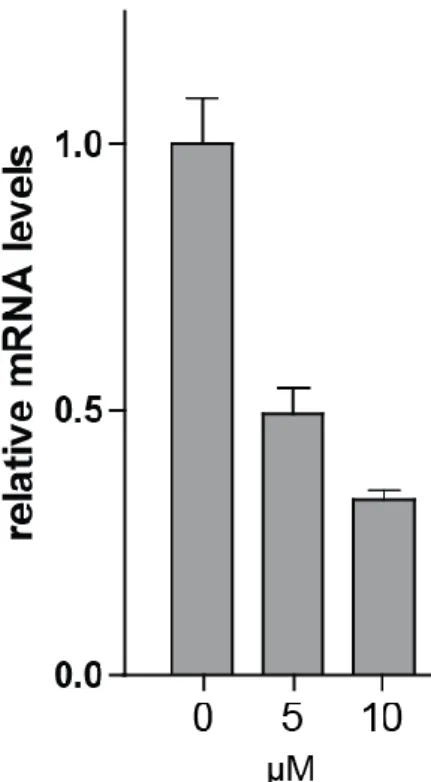
exposure to BTP15. Interestingly, mprA expression was significantly down-regulated 185
after 24 and 48 h of treatment (Figure 4B). BTP15 (10 µM) also decreased mprB 186
transcript levels 3-fold after 48 h exposure (Figure S3A). Since there is considerable 187
overlap among DosR- and MprA-regulated genes, we compared the BTP15 RNA-seq 188
transcript analysis with published gene expression data on mprAB deletion mutants and 189
found that the majority of the 35 deregulated genes (highlighted in Table S1) were also 190
9
differentially regulated in this mutant under different conditions (He et al., 2006; Pang et 191
al., 2007).
192
BTP15 is a kinase inhibitor that inhibits MprB autophosphorylation in vitro 193
Having found that treatment of Mtb with BTP15 leads to deregulation of genes controlled 194
by two-component regulatory systems, notably MprAB, we reasoned that the compound 195
might directly affect ATP-dependent signal transducing histidine kinases. Studying 196
histidine phosphorylation is extremely challenging due to the chemical instability of this 197
posttranscriptional modification (Kee and Muir, 2012). An MprB autophosphorylation 198
assay was established using purified truncated MprB as described (Zahrt et al., 2003).
199
Relatively large amounts of MprB (25 µM) were needed to detect the MprB 200
phosphohistidine (Figure 4C), as is common for histidine kinase phosphorylation assays 201
(Saini and Tyagi, 2005). Nonetheless, we demonstrated dose-dependent inhibition of 202
MprB auto-phosphorylation by BTP15 (Figure 4D) but could not accurately determine 203
the IC50 value due to the large amount of enzyme used. The non-hydrolyzable ATP 204
analog AMP-PNP can be employed to estimate the potency and specificity of histidine 205
kinase inhibitors having high in vitro IC50 values (Gilmour et al., 2005). When 10 mM 206
AMP-PNP (34x the in vitro IC50 of BTP15) was used only incomplete reduction of the 207
phosphohistidine signal was seen whereas 1 mM AMP-PNP had no effect on auto- 208
phosphorylation (Figure 4D) indicating that BTP15 is a much stronger inhibitor of MprB 209
auto-phosphorylation than the ATP-analog.
210
BBH7 has a pleiotropic inhibitory effect on mycobacterial protein secretion 211
By immunoblotting, BBH7 was found to impact two different protein secretion systems at 212
concentrations ≤ 5 µM (Figure 3); we also observed a 50% reduction of total culture 213
10
filtrate protein when bacteria were exposed to 5 µM BBH7 (not shown). To appreciate its 214
full impact on protein secretion we characterized and quantified the secretome of treated 215
and untreated bacteria by LC/MS-MS. These data confirmed the inhibitory effect of 216
BBH7 on the ESX-1 secretion system (Table S2). In addition, several substrates of the 217
ESX-5 secretion system such as EsxN, EsxM, PE25 and PPE41 were significantly 218
reduced in abundance upon treatment. Reduced secretion of virulence-associated 219
proteins of unknown export mechanism was uncovered showing that BBH7 affects 220
several independent lines of Mtb pathogenicity (Table S2).
221
BBH7 deregulates several transmembrane ATPases and alters mycobacterial cell 222
wall permeability 223
Since BBH7 substantially impacted mycobacterial protein secretion, we expected major 224
changes in the Mtb transcriptome after treatment. Indeed, RNA-seq experiments revealed 225
144 differentially regulated genes (≥ 2-fold) upon exposure to BBH7. Of these, 121 were 226
up-regulated and the gene expression signature mirrors changes primarily associated 227
with cell wall processes and transport (Figure 5A, Table S3, Figure S4). We found 228
positive regulation of the ESX transmembrane ATPase genes, eccCa1/eccCb1 and 229
eccA5/eccE5, in response to altered ESX-1 and ESX-5-dependent protein secretion. In 230
addition, strong up-regulation of the P-type ATPase genes, ctpC and ctpG, indicated 231
disturbed cell membrane/cell wall transport not only for secreted proteins but also for 232
ions such as zinc and copper. Several other signs for metal-ion overload were observed:
233
strong up-regulation of the metallothionein mymT, the multicopper oxidase mmcO, the 234
copper-dependent regulator ricR and the RicR-regulated gene lpqS, as well as 235
deregulation of the zinc stress responsive genes cadI, rv1993, cysK2, esxG and esxH 236
11
(Figure 4A, Table S3) (Botella et al., 2011; Serafini et al., 2013). Indirect targets for 237
metal-ion toxicity are Fe-S proteins, explaining the up-regulation of the Fe-S cluster 238
biogenesis operon SUF (rv1462–rv1466), and DNA damage leading to a LexA-driven 239
transcriptional response (Rowland and Niederweis, 2012).
240
To investigate whether BBH7 alters mycobacterial outer membrane permeability, 241
which might explain the transcriptomic pattern associated with metal-ion toxicity, we 242
performed ethidium bromide (EtBr) uptake assays after treatment with the compounds of 243
interest. Indeed, BBH7-treatment was found to increase EtBr accumulation and 244
fluorescence, a sign of perturbed membrane permeability (Figure 5B). This was not 245
observed with BTP15. Interestingly, BBH7 concentrations as high as 25 µM had no 246
impact on the activity of first- and second-line TB drugs (Figure S5).
247
Zinc stress augments EsxA secretion 248
Having established that BBH7 treatment leads to transcriptional signs of zinc and copper 249
stress, we surmised that intracellular metal-ion stress might be the link to inhibition of 250
mycobacterial protein secretion. Thus, we stressed Mtb with physiological 251
concentrations of zinc or copper, as encountered in the phagosome, and determined 252
EsxA secretion levels. Surprisingly, growing cells in media containing elevated levels of 253
ZnSO4 led to a significant and dose-dependent increase of EsxA secretion whereas 254
Ag85 secretion remained unchanged (Figure 5C). In the presence of 500 µM zinc, a 255
concentration measured in Mtb-infected macrophages (Botella et al., 2011), a six-fold 256
increase in EsxA secretion was observed (Figure 5C, lower panel). Elevated 257
concentrations of copper had no effect on EsxA secretion. These findings indicate that 258
BBH7 does not alter mycobacterial protein secretion by zinc or copper intoxication.
259
12
Furthermore, for the first time, we report an environmental signal (elevated zinc levels) 260
that augments EsxA secretion.
261
Since bacterial transport mechanisms depend on the proton motive force, which 262
is linked to intracellular ATP-levels, the intracellular ATP concentration was measured 263
after BBH7 treatment. Unlike treatment with the ATP-synthase inhibitor bedaquiline 264
(BDQ), ATP levels were not reduced by BBH7 (Figure 5D). To further distinguish BBH7 265
from well-known, mycobacterial cell wall inhibitors, we investigated whether such 266
compounds affect EsxA secretion. Isoniazid and ethambutol, as well as the thiourea 267
compounds ethionamide and thiacetazone, had no effect on EsxA secretion at 0.5 x MIC 268
(Figure S4C). At 5 x MIC, detection of the cytosolic heat-shock protein GroEL in the 269
culture filtrate indicated cell lysis, which was not observed after BBH7 and BTP15 270
treatment.
271
Taken together, these results indicate a novel mechanism of action for BBH7, 272
which alters cell-wall permeability for both export of proteins and import of small 273
molecules, leading to strong up-regulation of genes associated with metal ion overload.
274
However, blockage of EsxA secretion by BBH7 does not seem to be caused by 275
zinc/copper intoxication or ATP-depletion.
276
BBH7 and BTP15 reduce intracellular bacterial load and promote phagolysosomal 277
fusion in Mtb-infected THP-1 macrophages 278
Using GFP-expressing Mtb we had shown that BBH7 strongly affects viability of 279
intracellular bacteria in MRC-5 fibroblasts whereas BTP15 does not (Figure 2D). Since 280
the role of fibroblasts in in vivo infections is not clear, we also investigated the activity of 281
13
the compounds by infecting activated THP-1 macrophages and quantifying both 282
surviving macrophages and intracellular fluorescent mycobacteria. Treatment with BBH7 283
and BTP15 protected THP-1 cells from Mtb-induced cell death (Figure 6A) and greatly 284
reduced the intracellular bacterial load (Figure 6B, C). Since the ESX-1 secretion system 285
plays a decisive role in the arrest of phagosome maturation in Mtb-infected 286
macrophages (MacGurn and Cox, 2007) we investigated whether BBH7 and BTP15 can 287
reverse this phenotype. Activated THP-1 macrophages were infected at an MOI of 0.5 288
with Mtb expressing GFP and treated for 7 days. Subsequently, acidic compartments 289
were stained with Lysotracker Red and co-localization of the dye with fluorescent 290
mycobacteria quantified by confocal microscopy. Treated bacteria were found in acidic 291
compartments at significantly higher levels than untreated bacteria (Figure 6D, E) 292
indicating that reduction of intracellular bacterial load in macrophages is primarily 293
achieved through inhibition of Mtb-induced phagosome maturation arrest.
294 295
DISCUSSION 296
In this investigation, we developed and validated a novel phenotypic drug screen based 297
on ESX-1 secretion dependent cytotoxicity of Mtb. A HTS of >10,000 small molecules 298
identified two series of compounds that significantly reduced secretion of EsxA at 299
nanomolar concentrations without affecting mycobacterial growth in vitro, the 300
benzothiophenes and benzyloxybenzylidine hydrazines. In addition, less potent hit 301
compounds derived from the indoline-2-one core structure as well as several other 302
compounds also impacted ESX-1 function (not shown). This indicates that by selecting 303
14
for hits that abrogate cytotoxicity of Mtb, the chance of finding inhibitors of ESX-1- 304
dependent protein secretion is relatively high. The reason for this may lie in the nature of 305
the screen itself; Mtb-induced host cell lysis at high MOI is almost exclusively associated 306
with secretion of EsxA. Screening deep transposon libraries for mutants with impaired 307
cytolytic activity primarily detected insertions in genes required for EsxA secretion thus 308
highlighting the importance of this virulence determinant (Gao et al., 2004; Hsu et al., 309
2003). Another key factor for selectivity of the bioassay is our choice of lung fibroblasts 310
for the quantification of Mtb-induced cell death. These non-professional phagocytes fail 311
to detect host modifying agents that reduce the intracellular burden of Mtb in 312
macrophages (Lechartier et al., 2014). This feature may be beneficial for target 313
identification of these anti-virulence drugs.
314
The benzothiophene BTP15 is a kinase inhibitor that affects EsxA secretion most 315
likely by deregulating the espACD operon. Several transcriptional regulators control 316
ESX-1-dependent secretion by binding to this operon that is not part of the ESX-1 region 317
but nonetheless encodes EsxA co-secreted proteins (Blasco et al., 2012; Gonzalo- 318
Asensio et al., 2008; Pang et al., 2013). An mprAB mutant displayed up-regulation of 319
espA and greatly reduced EsxA secretion (Pang et al., 2013). Furthermore, MprA co- 320
regulates several DosR-regulated genes and SigE (Pang et al., 2007). BTP15 treatment 321
deregulates a similar set of genes and inhibits MprB auto-phosphorylation in vitro. Thus, 322
the two component regulatory system MprAB is the probable BTP-15 target. MprAB is 323
clearly associated with virulence since the corresponding mutants show impaired 324
survival in vivo, particularly during the chronic stage of infection (Zahrt and Deretic, 325
2001). Macrophages infected with a ΔmprAB strain elicit significantly lower levels of 326