Candida albicans Enhances the Progression of Oral Squamous Cell Carcinoma In Vitro and In Vivo
MátéVadovics,a,bJemimaHo,iNóraIgaz,b,cRóbertAlföldi,dDávidRakk,a,bEva Veres,a,bBalázsSzücs,aMártonHorváth,a RenátaTóth,aAttilaSzücs,aAndreaCsibi,aPéterHorváth,eLászlóTiszlavicz,fCsabaVágvölgyi,aJoshua D.Nosanchuk,g,h AndrásSzekeres,aMónikaKiricsi,cRhondaHenley-Smith,kDavid L.Moyes,iSelvamThavaraj,jRhysBrown,iLászló G.Puskás,d Julian R.Naglik,i AttilaGácserl,m
aDepartment of Microbiology, University of Szeged, Szeged, Hungary
bDoctoral School of Biology, University of Szeged, Szeged, Hungary
cDepartment of Biochemistry and Molecular Biology, University of Szeged, Szeged, Hungary
dAstridBio Technologies Ltd., Szeged, Hungary
eSynthetic and System Biology Unit, Biological Research Centre (BRC), Szeged, Hungary
fDepartment of Pathology, University of Szeged, Szeged, Hungary
gDivision of Infectious Diseases, Department of Medicine, Albert Einstein College of Medicine, Bronx, New York, USA
hDepartment of Microbiology and Immunology, Albert Einstein College of Medicine, Bronx, New York, USA
iCentre for Host-Microbiome Interactions, Faculty of Dentistry, Oral & Craniofacial Sciences, King’s College London, London, United Kingdom
jCentre for Oral, Clinical and Translational Science, Faculty of Dentistry, Oral & Craniofacial Sciences, King’s College London, London, United Kingdom
kKing’s Health Partners, Head and Neck Cancer Biobank, Guy's & St Thomas' NHS Foundation Trust, London, United Kingdom
lHCEMM-USZ Fungal Pathogens Research Group, Department of Microbiology, University of Szeged, Szeged, Hungary
mMTA-SZTE Lendület Mycobiome Research Group, University of Szeged, Szeged, Hungary Julian R. Naglik and Attila Gácser are co-principal authors.
ABSTRACT
Oral squamous cell carcinoma (OSCC) is associated with oral Candida albicans infection, although it is unclear whether the fungus promotes the genesis and progression of OSCC or whether cancer facilitates fungal growth. In this study, we investigated whether C. albicans can potentiate OSCC tumor development and progression. In vitro, the presence of live C. albicans, but not Candida parapsilosis, enhanced the progression of OSCC by stimulating the production of matrix metal- loproteinases, oncometabolites, protumor signaling pathways, and overexpression of prognostic marker genes associated with metastatic events. C. albicans also up- regulated oncogenes in nonmalignant cells. Using a newly established xenograft in vivo mouse model to investigate OSCC-C. albicans interactions, oral candidiasis enhanced the progression of OSCC through in
flammation and induced the overex- pression of metastatic genes and signi
ficant changes in markers of the epithelial- mesenchymal transition. Finally, using the 4-nitroquinoline 1-oxide (4NQO) murine model, we directly correlate these in vitro and short-term in vivo
findings with the progression of oncogenesis over the long term. Taken together, these data indi- cate that C. albicans upregulates oncogenes, potentiates a premalignant pheno- type, and is involved in early and late stages of malignant promotion and progres- sion of oral cancer.
IMPORTANCE
Oral squamous cell carcinoma (OSCC) is a serious health issue world- wide that accounts for 2% to 4% of all cancer cases. Previous studies have revealed a higher yeast carriage and diversity in oral cancer patients than in healthy individ- uals. Furthermore, fungal colonization in the oral cavity bearing OSCC is higher on the neoplastic epithelial surface than on adjacent healthy surfaces, indicating a positive association between oral yeast carriage and epithelial carcinoma. In addi- tion to this, there is strong evidence supporting the idea that Candida contributes
Invited EditorMichael J. McCullough, University of Melbourne
EditorMichael Lorenz, University of Texas Health Science Center
Copyright© 2022 Vadovics et al. This is an open-access article distributed under the terms of theCreative Commons Attribution 4.0 International license.
Address correspondence to Attila Gácser, gacsera@bio.u-szeged.hu.
The authors declare no conflict of interest.
Received27 October 2021 Accepted18 November 2021 Published
® 4 January 2022
to carcinogenesis events in the oral cavity. Here, we show that an increase in Candida albicans burden promotes an oncogenic phenotype in the oral cavity.
KEYWORDS
Candida albicans, cancer, oral squamous cell carcinoma, progression
T he head and neck account for 2 to 4% of all cancer cases, which includes neoplasms that affect several regions of the oral cavity, pharyngeal sites, and salivary glands.
Approximately 90% of oral neoplasms are squamous cell carcinomas (OSCC) (1). OSCC is the 16th most common cancer worldwide (2) and 6th in the United States (3). In Europe, the incidence of oral cancer is especially high in Central and Eastern Europe, and both morbidity and mortality rates are highest in Hungary (4). Risk factors for oral cancer include poor oral hygiene, tobacco use, alcohol use, and meat consumption (5). OSCC is treated by surgery, radiation, and chemotherapy. Chemotherapy and radiotherapy, when used simultaneously, provide a synergistic bene
fit against OSCC (6). Currently, the primary treatment mode for OSCC is surgery followed by radiotherapy or chemoradiotherapy depending on risk factors. Adverse effects include mucositis and myelosuppression (7), which also affect the composition, quantity, and complexity of the oral microbiota (8
–10).
Candida albicans is a highly prevalent yeast in the oral cavity (11
–13) which prolif- erates and invades host mucosal tissues upon epithelial barrier dysfunction or disrup- tion. C. albicans invades tissues via hypha formation and the production of associated hydrolytic enzymes and virulence factors. While these characteristics may endow Candida with a competitive advantage, it is the host's immune competence that ulti- mately determines whether clearance, colonization, or disease occurs (14).
There has long been a positive association between oral yeast carriage/dysbiosis and epithelial carcinoma (15
–19). Notably, higher yeast carriage and diversity are observed in oral cancer patients than in healthy individuals, and oral fungal coloni- zation in OSCC patients is higher on the neoplastic epithelial surface than on adja- cent healthy surfaces (20
–24). Furthermore, persistent oral candidiasis has been observed to lead to OSCC development in an elderly patient (25). Several other stud- ies have indicated that Candida invasion promotes a hyperplastic epithelial response and that untreated Candida epithelial lesions may become dysplastic and transform into carcinoma (reviewed in references 25 and 26). Thus, there is strong evidence supporting the idea that Candida promotes carcinogenic events in the oral cavity (16, 27
–30). However, Candida infection in cancer patients may also be considered the consequence of an altered immune status, because both myelosuppression and mucositis enable the development of oral candidiasis (8, 9, 31, 32).
Given the association of oral candidiasis and cancer, in this study we characterized the potential underlying mechanisms for Candida enhancing OSCC development and progression. Using in vitro and two in vivo models, we conclude that C. albicans can facilitate and enhance oncogenic mechanisms during oral cancer.
RESULTS
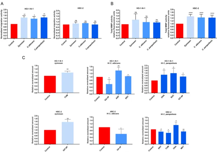
Heat-inactivatedCandidaand zymosan increasein vitro-related metastasis.
To examine whether increased fungal burden affects oral tumor progression, HSC-2 and HO-1-N-1 OSCC cells were treated with zymosan (cell wall component of Saccharomyces cerevisiae), heat-inactivated (HI) Candida albicans, or HI Candida parapsilosis yeast cells.
Initially, a wound healing assay at 24 h was used to analyze the invasive capacity of OSCC cells. Cellular movement of HO-1-N-1 cells was signi
ficantly enhanced by all three treat- ments compared to that of the untreated control (zymosan,1.445
60.076; HI C. albicans, 1.369
60.09; HI C. parapsilosis, 1.454
60.083). No signi
ficant differences in invasiveness were observed with HSC-2 cells (Fig. 1A). Next, epithelial proliferation during fungal expo- sure was performed using bromodeoxyuridine (BrdU) proliferation assays, which revealed that HI Candida cells do not affect OSCC cell proliferation (see Fig. S1A in the supplemen- tal material).
Remodeling of the extracellular matrix is a critical component of tumor cell adapta- bility and is attributed to the secretion of several proteases (serine, cysteine, threonine,
Vadovics et al. ®
aspartic acid, and metalloproteinases). In particular, matrix metalloproteinases (MMPs) are key enhancers of tumor dissemination (33). Notably, all fungal treatments signi
fi- cantly elevated secreted MMP activity in HSC-2 cells at 24 h compared to the untreated control (zymosan, 1.516
60.041; HI C. albicans, 1.437
60.06536; HI C. parapsilosis, 1.426
60.057). However, MMP activity was unaltered in HO-1-N-1 cells (Fig. 1B).
Metabolites generated by cancer cells in
fluence the metastatic cascade, affecting the epithelial-mesenchymal transition (EMT), the survival of cancer cells in circulation, and metastatic colonization at distant sites (34). Changes in metabolic activity were examined by analyzing the levels of glycolysis and tricarboxylic acid (TCA) cycle inter- mediates and certain amino acids (Fig. S1C) by high-performance liquid chromatogra- phy coupled with high-resolution mass spectrometry (HPLC-HRMS) after 24 h of treat- ment. For the HSC-2 cell line, zymosan treatment signi
ficantly increased the production of glucose/fructose 6-phosphate (glucose/fructose-6p) (1.564
60.132), while HI C. albicans treatment reduced the concentrations of glyceraldehyde 3-phos- phate (GA-3P) (0.605
60.142). HI C. parapsilosis treatment altered levels of erythrose- 4P (0.844
60.056), fumaric acid (0.763
60.065), oxaloacetic acid (1.289
60.117), and methionine (0.855
60.031) (Fig. 1C). Nonsigni
ficant changes are shown in Fig. S1C. In HO-1-N-1 cells, zymosan treatment produced a signi
ficant change in fumaric acid (1.189
60.044), while HI C. albicans treatment altered the levels of glyceraldehyde-3P (0.580
60.137), aspartic acid (1.374
60.096), and erythrose-4P (1.039
60.015). HI
FIG 1 Effects of HICandidaand zymosan on HO-1-N-1 and HSC-2 oral squamous cell carcinoma cellsin vitro.(A) Normalized migration activity of OSCC cells in the presence of HIC. albicans, HIC parapsilosis, and zymosan measured by a wound healing assay (n= 3). (B) Normalized total secreted matrix metalloproteinase (MMP) activity of OSCC cells in the presence of HIC. albicans, HI C. parapsilosis, and zymosan measured by a total MMP activity kit (n= 4). (C) Normalized amounts of metabolites of OSCC cells in the presence of HIC. albicans, HIC. parapsilosis, and zymosan as measured by HPLC-HRMS (n = 4). FUM, fumaric acid; GA-3P, glyceraldehyde-3P; ASP, aspartic acid; ERY, erythrose-4P; OXA, oxaloacetic acid; SUC, succinic acid; G/F-6P, glucose/
fructose-6p; MET, methionine; control, tumor cells without any treatment. Unpairedttest;*,P#0.05;**,P#0.01;****,P#0.0001. ns, nonsignificant.
C. parapsilosis treatment altered the production of oxaloacetic acid (1.463
60.214), succinic acid (1.586
60.199), and glucose/fructose-6p (1.317
60.118) (Fig. 1C).
Taken together, all three fungal treatments had a signi
ficant effect on migration, secreted MMP activity, and oncometabolite production of OSCC cells, which sug- gests interactions between the tumor cells and fungal components.
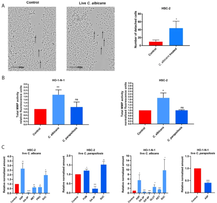
LiveCandidaenhances detachment, MMP activity, and metabolite production of OSCC cellsin vitro.
After assessing the effects of HI Candida infections, we further aimed to examine the potential outcome of live fungal stimuli. While during HI fungal treatment, the triggered host responses are mainly due to direct cell-cell contact, live Candida cells could also in
fluence host responses through other
“indirect
”stimuli, such as their secreted extracellular vesicles, released enzymes, etc. Therefore, we next assessed the effects of live C. albicans and C. parapsilosis on OSCC cultures and recorded the migration of cancer cells on time-lapse video. Live C. albicans, but not live C. parapsilosis, increased the numbers of detached, single HSC-2 cells compared to untreated controls (Fig. 2A; Video S1, S2, and S3). No changes were detected in the HO-1-N-1 cell line (data not shown). Similar to HI Candida, live C. albicans and C. para- psilosis did not alter OSCC proliferation as measured by a BrdU assay (Fig. S1B).
Secreted total MMP activity was increased in both cancer cell lines with live C. albicans (HSC-2, 1.918
60.209; HO-1-N-1, 1.918
60,183) but not live C. parapsilosis (Fig. 2B).
Next, metabolic changes in OSCC cells were measured following treatment with live C. albicans and C. parapsilosis at 24 h. Importantly, no fungal metabolites were detected using our extraction method. In HSC-2 cells, C. albicans signi
ficantly altered the secretion of aspartic acid (2.67
60.346), glyceraldehyde-3P (0.068
60.009), methi- onine (0.634
60.063), proline (0.666
60.087), and succinic acid (1.975
60.234), while C. parapsilosis altered fumaric acid (1.199
60.046), glyceraldehyde-3P (0.225
60.076), and succinic acid (1.523
60.101) secretion. In HO-1-N-1 cells, C. albicans signi
ficantly altered the secretion of aspartic acid (5.526
61.667), glyceraldehyde-3P (0.543
60.038), glucose/fructose-6p (0.288
60.047), glutamine (2.616
60.667),
a-ketoglutaric acid (1.532
60.051), and succinic acid (9.81
62.709), while C. parapsilosis treatment reduced the level of aspartic acid (0.408
60.024) (Fig. 2C). C. albicans was predomi- nantly in the hyphal form and C. parapsilosis in the yeast form when these assays were performed.
The data demonstrate that live C. albicans induced the most prominent changes in the movement, MMP activity, and metabolite production in OSCC cells compared to live C. parapsilosis or HI Candida and zymosan treatments.
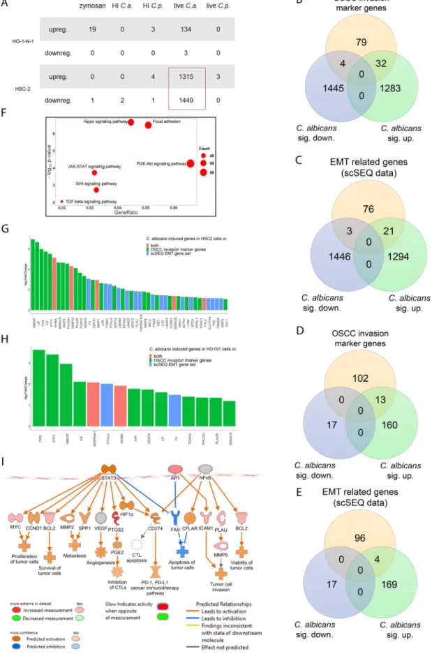
Live C. albicans activates genes and signaling pathways involved in OSCC invasion and metastasis.
To fully dissect the molecular mechanisms induced by fun- gal stimulation, whole-transcriptome analysis was performed with HSC-2 and HO-1-N-1 cells following exposure to zymosan, HI C. albicans, HI C. parapsilosis, live C. albicans, and live C. parapsilosis. Transcriptome analyses revealed that live C. albicans induced the most signi
ficant gene expression changes in OSCC cells (HSC-2, n = 2,764; HO-1-N- 1, n = 137), followed by zymosan (n = 19), while live C. parapsilosis and heat-inactivated fungal challenge did not trigger signi
ficant gene expression changes in either cell line.
C. albicans evoked a more signi
ficant response in HSC-2 (upregulated genes, n = 1,315; downregulated genes, n = 1,449) than HO-1-N-1 cells (upregulated, n = 134;
downregulated, n = 3) (Fig. 3A; Data Set S1A to J). Notably, in HSC-2 cells, C. albicans triggered signi
ficant changes in marker genes previously associated with OSCC inva- sion (32 upregulated, 4 downregulated) (Data Set S1L) (Fig. 3B). Moreover, another gene set (21 upregulated, 3 downregulated) overlapped with a characteristic pro
file of EMT derived from single-cell sequencing (scSeq) of 18 patients with head and neck squamous cell carcinoma (HNSCC) (35) (Fig. 3C). Five genes, INHBA, MMP10, MMP1, SEMA3C, and FHL2, were present in both the OSCC invasion marker gene and scSeq- derived gene data sets (Fig. 3G). Furthermore, even though the HO-1-N-1 response was modest compared to HSC-2, 13 OSCC invasion marker genes (Fig. 3D) and four EMT genes (Fig. 3E) were also upregulated after live C. albicans stimulus. Two of these
Vadovics et al. ®
genes, SERPINE1 and INHBA, overlapped with the OSCC invasion marker genes and EMT subsets (Fig. 3H).
KEGG pathway analysis of HSC-2 data sets revealed signi
ficant activation of several pathways associated with OSCC metastasis development, including Hippo signaling, focal adhesion, JAK-STAT, PI3K-Akt, Wnt, and TGF
bpathways (Fig. 3F) (36
–40).
Ingenuity pathway analysis (IPA) with built-in causal analyses was also used to investi- gate activation patterns of several intracellular signaling pathways based on the coher- ent regulation of their molecular elements. IPAs predicted the activation of tumor- related pathways, including the tumor microenvironment pathway, as well as the
FIG 2 Effects of liveCandidaon HO-1-N-1 and HSC-2 oral squamous cell carcinoma cellsin vitro.(A) Pictures from time-lapse videos of cellular migration of HSC-2 cells, with arrows pointing to detached cancer cells. The left picture shows the control cells, and the right shows the liveC. albicans-treated cells.
The graph shows the number of detached cells (n= 3). (B) Normalized total secreted matrix metalloproteinase activity of OSCC cells in the presence of live C. albicansandC. parapsilosisas obtained by a total MMP activity kit (n= 3). (C) Normalized amounts of metabolites of OSCC cells in the presence of live C. albicansand liveC. parapsilosisas measured by HPLC-HRMS (n= 3). ASP, aspartic acid; GA-3P, glyceraldehyde-3P; MET, methionine; PRO, proline; SUC, succinic acid; FUM, fumaric acid; G/F-6P, glucose/fructose-6p; GLUT, glutamic acid; KET,a-ketoglutaric acid. Control, tumor cells without any treatment.
Unpairedttest;*,P#0.05;**,P#0.01;***,P#0.001;****,P#0.0001.
FIG 3 In vitro transcriptomic analysis. Candida albicans activates genes and signaling pathways involved in the OSCC metastatic processes. (A) Number of up- or downregulated genes of HSC-2 and HO-1-N-1 cells after different fungal treatments (Continued on next page)
Vadovics et al. ®
signi
ficant activation of several prognostic features, such as metastasis, invasion, angio- genesis, and proliferation of tumors based on the C. albicans stimulus-derived differen- tially expressed genes (DEGs) (Fig. 3I). Interestingly, DLST and SUCLA gene expression was increased, and these genes are involved in succinic acid metabolism. Furthermore, ASNSD1 and GOT1 genes were also upregulated and are involved in aspartic acid syn- thetic processes (Fig. S4). Transcriptomic gene expression data was validated by quan- titative PCR (qPCR) (Fig. S2 and S4). Together, these data support the notion that live C.
albicans, but not live C. parapsilosis, HI Candida, or zymosan, enhances the metastatic features of OSCC cells.
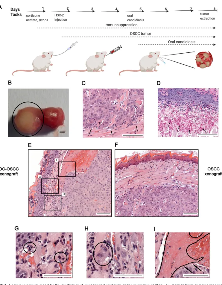
Establishment of a novel in vivomouse xenograft model of OSCC and oral candidiasis.
In order to validate our in vitro results, we developed a novel in vivo mouse model of OSCC and oral candidiasis. As C. albicans cells are not in direct contact with HSC-2 cells in this model, the indirect effect of oral candidiasis on OSCC progres- sion can be examined. To mimic the immunological condition of patients caused by chemoradiotherapy, cortisone acetate was administered to 6- to 8-week-old BALB/c mice to induce an immunosuppressed condition for subsequent tumor cell injection.
As HSC-2 cells were more responsive to fungal presence than HO-1-N-1 cells, 1 10
6HSC-2 cells were injected into mouse tongues to initiate OSCC development. A fully developed tumor formed by day 3 after HSC-2 injection. After con
firmation of tumor presence, calcium alginate swabs saturated in 1 10
9/mL C. albicans suspension for 5 min were placed under the tongue of each mouse for 75 min on day 5 (3 days after tumor cell injection). Mice were terminated on day 8 based upon clinical observation scores and weight loss (25%) (Fig. S5C). On day 8, the average tumor size was 5 mm in diameter (Fig. 4B). Hematoxylin-eosin (H&E) staining was performed for histopathologi- cal analyses (Fig. 4C). Fungal hyphae were detected in the mucosa following periodic acid-Schiff (PAS) staining (Fig. 4D). The oral C. albicans burden was 10
5to 10
6CFU per g tissue, which is comparable with that reported in the literature of oral candidiasis models (41).
Oral candidiasis enhances the progression of OSCCin vivo.
To investigate the effect of increased yeast burden on OSCC progression, two animal groups were com- pared: a control group received cortisone acetate and HSC-2 tumor cells (OSCC xeno- graft), while the other group received cortisone acetate, HSC-2 cells, and C. albicans for the development of oral candidiasis (OC-OSCC xenograft). Each group (OSCC xenograft and OC-OSCC xenograft) comprised 16 animals. Four mice were applied for transcrip- tome analysis and 4 for CFU analysis from each group (OSCC xenograft and OC-OSCC xenograft). For histopathological analysis, 8 mice were applied from each group. CFU analysis was applied for the validation of successful oral candidiasis establishment in the OC-OSCC xenograft group. Histopathological samples from both groups were ana- lyzed and scored manually in a blinded manner by a pathologist after H&E staining for the identi
fication of in
flammation, necrosis, in
filtrating or pushing tumor edge, EMT, invasion markers, and signs and symptoms of thrombosis and peritumoral in
flamma- tion (Fig. S5A). The EMT and budding score number of tumor cells were higher in 5/8 samples in OC-OSCC xenograft tumors than in OSCC xenograft samples, where no high
FIG 3Legend (Continued)
(n= 3). C.a.,C. albicans; C.p.,C. parapsilosis(B) Venn diagram of up- or downregulated genes in HSC-2 cells in the presence of live C. albicans and OSCC invasion marker genes found in the literature. sig., significantly. (C) Venn diagram of up- or downregulated genes in the HSC-2 cell line in the presence of liveC. albicansand EMT marker genes in HNSCC according to a single-cell sequencing study. (D) Venn diagram of up- or downregulated genes in HO-1-N-1 cells incubated with liveC. albicans and OSCC invasion marker genes found in the literature. (E) Venn diagram of up- or downregulated genes in the HO-1-N-1 cell line in the presence of liveC. albicans and EMT marker genes in HNSCC according to a single-cell sequencing study. (F) Signaling pathways that are key regulators of the OSCC invasion processes that were significantly activated in HSC-2 cells in the presence of liveC. albicans. (G) Graph showing log2fold change ofC. albicans-induced genes in HSC-2 cells involved in OSCC invasion according to the literature and of single-cell sequencing (scSeq) results from 18 patients with HNSCC. Red columns represent OSCC marker genes according to the literature and scSEQ data. (H) Graph showing log2fold change ofC.
albicans-induced genes in HO-1-N-1 cells involved in OSCC invasion according to the literature and scSEQ data. Red columns represent OSCC marker genes according to the literature and scSEQ data. (I) Causal analyses of the genes for which expression changed in HSC-2 cells after liveCandida albicanstreatment.
FIG 4 A newin vivomouse model for the investigation of oropharyngeal candidiasis on the progression of OSCC. (A) Schematicfigure of mouse xenograft for the investigation of the effect ofC. albicanson the progression of OSCC. Immunosuppression and injection of human HSC-2 OSCC cells into the tongue of mice (OSCC xenograft). OSCC xenograft and oral candidiasis (OC-OSCC xenograft). The cartoon was produced by BioRender. (B) Representative mouse tongue on the 8th day of the experiment (7 days after tumor cell injection). The circle highlights the tumor. Scale bar, 1 mm. (C) Histopathological image (Continued on next page)
Vadovics et al. ®
EMT/budding scores were detected. Thrombosis was also detected in 5/8 OC-OSCC xenograft tumors. Only 1 sample showed thrombosis in OSCC xenograft controls.
In
filtrating immune cells were detected within the OC-OSCC xenograft samples, indi- cating C. albicans-induced in
flammatory responses (Fig. 4G). Images indicative of EMT were identi
fied, as represented by the increased number of detached individual tumor cells (Fig. 4H). Ki-67 staining was performed to analyze the proliferation activity of the tu- mor cells in vivo. No difference could be detected between the C. albicans-infected (OC- OSCC xenograft) and OSCC xenograft group (data not shown). Taken together, histopath- ological scoring suggests that oral candidiasis may drive OSCC progression events.
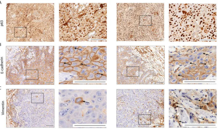
Oral candidiasis increases p63 and vimentin expression and decreases E- cadherin expression in OSCC histopathological specimens.
p63 expression is a reli- able indicator in histological grading and is an early marker of a poor OSCC prognosis.
Analysis of OC-OSCC xenograft samples showed that C. albicans increased p63 expres- sion and localization to the nucleus compared to uninfected tissues (Fig. 5A). Next, we analyzed for the expression of EMT markers E-cadherin and vimentin. Reduced E-cad- herin expression was observed in cell membranes of OC-OSCC xenograft samples com- pared with OSCC xenograft tumors (Fig. 5B). Correspondingly, vimentin-positive cells were notably increased in OC-OSCC xenograft sections compared with OSCC xenograft
FIG 4Legend (Continued)
of the tumor on the 8th day, with black arrows indicating the tumor edge. Scale bar, 100mm. (D) Histopathological examination of the tumor on the 8th day (7 days after tumor injection and 3 days postinfection), with black arrows indicating the fungal hyphae in the mucosa. Scale bar, 100mm. (E) Histopathological picture of the tumor on the 8th day after HSC-2 injection and oral candidiasis. Scale bar, 100mm. (F) Histopathological picture of the tongue on the 8th day after HSC-2 injection. Scale bar, 100mm. (G) Infiltrating immune cells in OC-OSCC xenograft samples indicating thatC. albicans caused inflammation. Scale bar, 100mm. (H) Detached budding tumor cells in OC-OSCC xenograft samples indicating epithelial-to-mesenchymal transition.
Scale bar, 100mm. (I) Thrombosis in OC-OSCC xenograft samples. Scale bar, 100mm. Created by BioRender.
FIG 5 Histopathological staining of OSCC and OC-OSCC xenograft tumor samples: p63 staining (A), E-cadherin staining (B), and vimentin staining (C). Scale bars, 100mm.n= 8/group. Squares indicate the magnified sections (right panels per mice model) of each tissue sample. Arrows indicate the E-cadherin positive (upper panels) and vimentin positive (lower panels) cells.
sections (Fig. 5C; Fig. S5D, E, and F). The data indicate that C. albicans can drive an EMT phenotype.
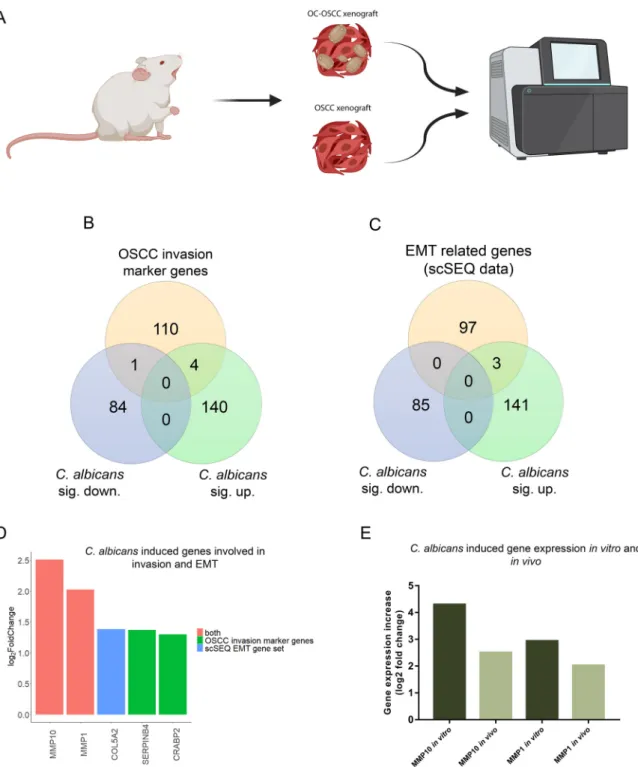
Oral candidiasis enhances the expression of genes involved in OSCC progression in vivo.
To analyze the molecular mechanisms behind the histopathological results, transcriptomic analysis of OC-OSCC and OSCC xenograft tumor samples was
FIG 6 Transcriptomic analysis ofin vivotumor samples followed by oral candidiasis. (A) Schematicfigure of mRNA sequencing of OSCC xenograft and OC-OSCC xenograft tumor samples. The cartoon was produced by BioRender. (B) Venn diagram of up- or downregulated genes in HSC-2 cells in the presence of liveC. albicansand OSCC invasion marker genes described in the literature.
(C) Venn diagram of up- or downregulated genes in HSC-2 cell line in the presence of liveC. albicansand EMT marker genes in HNSCC according to a single-cell sequencing study. (D) Graph showing log2fold change ofC. albicans-induced genes in HSC-2 cells involved in OSCC invasion according to the literature and single-cell sequencing data. Red columns represent OSCC marker genes according to both the literature and single-cell sequencing (scSeq) results from 18 patients with HNSCC. (E) Tumor invasion genes showing upregulated expression bothin vitroandin vivo. For transcriptomic analysis,n= 4. Created by BioRender.
Vadovics et al. ®
performed (Fig. 6A). OC-OSCC xenografts displayed expression changes in 229 genes (144 upregulated and 85 downregulated) (Data Set S1K). Among these, 5 genes (MMP10, MMP1, SERPINB4, and CRABP2 upregulated; MMP7 downregulated) are pre- dicted to be involved in OSCC invasion (Fig. 6B), while 3 genes (MMP10, MMP1, and COL5A2 upregulated) are associated with EMT regulation (Fig. 6C). Notably, MMP10 and MMP1 were present in both subsets (Fig. 6D) and were also upregulated in our in vitro and in vivo models (Fig. 6E). In vitro MMP1 and MMP10 expression increase was validated by Western blot analysis (Fig. S6C, D, E, and F). The data indicate that C. albi- cans induces a gene subset predicted to be involved in OSCC invasion.
C. albicansaffects genes related to carcinogenesis in OKF6/TERT2 nonmalignant epithelial cellsin vitro.
The above data show that C. albicans upregulates oncogenes in vitro and drives a more malignant phenotype in cancer cells in vitro and in vivo. We thus wanted to determine whether C. albicans also had carcinogenic potential in nor- mal epithelium. Therefore, we further investigated oral epithelial responses to C. albi- cans and C. parapsilosis using the oral epithelial cell line OKF6/TERT2, which is a telo- merase-de
ficient line derived from a healthy individual. We recently utilized OKF6/
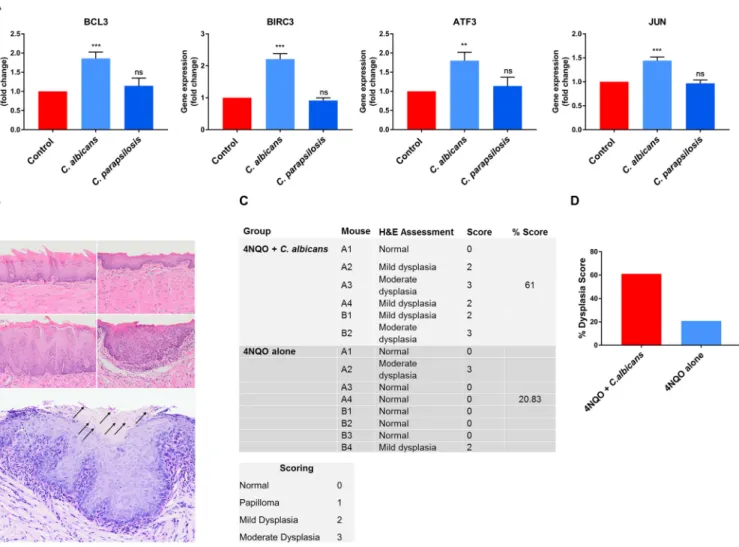
TERT2 cells to investigate non-tumor host cell responses and their microRNA (miRNA) regulatory processes (in epithelial cells) in the presence of C. albicans and C. parapsilo- sis. Based on these previously acquired sequencing data (42), we selected 4 oncogenes (BCL3, BIRC3, ATF3 and JUN) (37, 43, 44) potentially regulated by C. albicans and C. para- psilosis. The expression of these genes was analyzed in OKF6/TERT2 cells at 12 h by qPCR after C. albicans and C. parapsilosis stimulus. Only C. albicans signi
ficantly upregu- lated BLC3, BIRC3, ATF3, and JUN oncogenes, indicating that C. albicans also contributes to oncogenetic processes in nonmalignant oral epithelial cells (Fig. 7A).
C. albicansdrives oncogenic propertiesin vivobased on the 4NQO mouse model.
Our in vivo xenograft OSCC model data and in vitro transcriptomic and qPCR data strongly suggest that C. albicans may drive an oncogenic phenotype. However, the lim- itations of the xenograft OSCC model are its short time span (3 days) and that it addresses only tumor aggressiveness in already established cancer cells. While C. albi- cans may promote oncogenesis, in humans this is unlikely to occur during short-term colonization episodes, but chronic exposure to fungal virulence factors and metabo- lites during long-term infections may synergize with other risk factors to promote oncogenesis. To investigate this, we utilized the long-term 4-nitroquinoline 1-oxide (4NQO) mouse model to assess whether topical infection/colonization with C. albicans may directly potentiate an oncogenic phenotype (45). 4NQO is a carcinogen with a mode of activity similar to that of tobacco by-products but when used at low doses establishes conditions that allow us to investigate whether C. albicans can promote stepwise oncogenesis (from normal, through increasing severity of dysplasia, to inva- sive carcinoma).
BALB/c mice were given a low dose of 4NQO in the drinking water for 8 weeks, fol- lowed by 1 week of 0.1% tetracycline (TCN) to condition the epithelium. Mice were then infected with C. albicans (6 10
8yeast cells/mL) three times at week 10 to establish infec- tion. Tetracycline (0.01%) and C. albicans (6 10
4yeast cells/mL) were administered via drinking water for a further 10 weeks. At week 12 (2 weeks following C. albicans infection), 2 mice per group were euthanized and their tongues were assessed. Both C. albicans- infected mice showed mild dysplasia (Fig. 7B, middle panels) compared to the uninfected group (Fig. 7B, top panels). At week 20, the remaining mice were euthanized. Mouse tongues were assessed and graded for atypical architectural and cytological criteria by a quali
fied histopathologist (normal = 0, papilloma = 1, mild dysplasia = 2, moderate dys- plasia = 3), and a percentage score was calculated for each experimental group. Notably, 5/6 C. albicans-infected mouse tongues showed either mild or moderate dysplasia with a histological clinical score of 61%, with only 2/8 uninfected mice showing mild or moder- ate dysplasia with a histological clinical score of
;21% (Fig. 7C and D). When C. albicans- infected mouse tongues containing dysplasia were subjected to PAS-stained step section- ing, fungal hyphae were identi
fied in 1/5 mouse tongues. Interestingly, in this case, C.
albicans colocalized with a focus of moderate dysplasia (Fig. 7B). These
findings support
the notion that persistent infection with C. albicans can promote stepwise oncogenesis in healthy oral epithelial cells in the presence of predisposing environmental conditions.
DISCUSSION
OSCC is associated with the presence of oral candidiasis, but whether this is a cause rather than a causative relationship is unclear (15, 17
–19, 46
–48). We previously showed that the diversity of the oral fungal micro
flora of OSCC patients is remarkably different from that of healthy individuals, with signi
ficantly increased fungal burden and diversity in patients with oral tumors. Furthermore, oral fungal colonization in patients with OSCC is higher on neoplastic epithelial surfaces than on healthy surfaces, which indicates a positive association between oral yeast carriage and epithelial carci- noma (20). Candida might induce carcinogenesis by the production of carcinogenic compounds such as nitrosamines (49). These carcinogens bind to bases, phosphate residues, and/or hydrogen bonding sites of DNA that could interfere with DNA replica- tion. Induced point mutations might activate oncogenes and contribute to the devel- opment of oral cancer. Accumulation of acetaldehyde, a by-product of ethanol metab- olism, is also considered to be carcinogenic, and the induction of proin
flammatory
FIG 7 (A) Gene expression of carcinogens in OKF6/TERT2 immortalized cells; (B to D)C. albicans-infected mice exhibited enhanced dysplastic tongue features in the 4NQO model. (A) qPCR results of selected carcinogens in OKF6/TERT2 immortalized cells. (B) Representative photomicrographs of murine oral mucosa demonstrating normal dorsal tongue (top row, left), normal ventral tongue (top row, right), mild epithelial dysplasia on dorsal tongue (middle row, left), and moderate epithelial dysplasia on ventral tongue (middle row, right) (H&E; scale bar, 50mm).C. albicanshyphae (black arrows) were present in the keratin layer overlying a focus of moderate dysplasia (bottom panel). (C) Each mouse tongue was assessed and graded for atypical architectural and cytological criteria by a qualified histopathologist (normal = 0, papilloma = 1, mild dysplasia = 2, moderate dysplasia = 3), and the percentage score (sum of each group divided by the highest possible score) was calculated per group. (D) Graphical representation of panel C showing the dysplasia score.n= 6 for the 4NQO plusC. albicans group;n= 8 for the 4NQO-alone group. Scale bar, 50mm. Unpairedttest;**,P#0.01;***,P#0.001.
Vadovics et al. ®
cytokines could further contribute to oral cancer development (50), both of which are triggered by the presence of Candida cells.
C. albicans and C. parapsilosis both are common commensals of the oral cavity (51).
Both are also opportunistic human-pathogenic fungi, although C. albicans is more fre- quently associated with oral candidiasis than C. parapsilosis (52). C. albicans is polymor- phic, due to its ability to form hyphae and/or pseudohyphae (53). C. parapsilosis does not produce true hyphae but can generate pseudohyphae that are characteristically large and curved (54). Hypha formation is critical for host cell damage and immune activation, which are both driven by the secretion of candidalysin, a peptide toxin. Candidalysin damages epithelial membranes and activates several signaling cascades, including the epidermal growth factor receptor (EGFR) pathway (55) which is strongly associated with oral/epithelial cancers (56). Thus, in this study, we aimed to examine whether Candida pathogens and their components could potentiate OSCC progression.
In vitro experiments using two OSCC cell lines indicated that heat-killed fungi and zymosan are able to induce a moderate amount of cell migration, secreted MMP activ- ity, and oncometabolite production of OSCC cells. However, live C. albicans, but not live C. parapsilosis, was a major inducer of these oncogenic phenotypes. Notably, ma- trix metalloproteinases (MMPs) are essential for tumor invasion and metastasis, as their secretion degrades components of the extracellular matrix elements, facilitating the migration of individual malignantly transformed cells (57). The transcriptomic data also detected a signi
ficant increase in MMP1, MMP10, MMP3, and MMP9 expression. This suggests that C. albicans hypha formation, induction of cellular damage, and activation of epithelial in
flammatory and metabolic responses may be critical drivers of MMP ac- tivity, tumor invasion, and oncogenic progression. This is also supported by previous
findings indicating that the hypha-speci
fic toxin candidalysin induces MMP activity, leading to EGFR activation in the TR146 buccal carcinoma cell line (55).
Metastatic tumor cells have unique metabolic pro
files (58). For this reason, we examined the concentrations of glycolysis, TCA cycle intermediates, and some amino acids using HPLC-HRMS with and without exposure to fungal stimuli. Only live C. albi- cans had a signi
ficant effect on metabolic pro
files, leading to increased amounts of as- partic acid and succinic acid and decreased the amount of glyceraldehyde-3P (GA-3P) in both OSCC cell lines. Transcriptomic and qPCR data supported these
findings.
Notably, gene expression of GOT1, DLST, and SUCLA2, involved in aspartic and succinic acid synthesis, was signi
ficantly increased. Succinate may drive tumorigenesis through multiple mechanisms, and accumulated succinate can inhibit prolyl hydroxylase, which is responsible for hydroxylation of HIF1
a, causing its degradation. Therefore, succinate accumulation through inhibition of prolyl hydroxylase causes HIF1
astabilization and its translocation to the nucleus, which might enhance angiogenesis, resistance against apoptosis, and the activation of genes involved in tumor invasion (59). Furthermore, secreted tumor-derived succinate belongs to a novel class of cancer progression fac- tors, inhibiting tumor-associated macrophage polarization and promoting tumorigenic signaling through PI3K/AKT and HIF1
a(60). In addition, aspartate is a limiting metabo- lite for cancer cell proliferation and tumor growth under hypoxia (61), suggesting that C. albicans-induced aspartic acid increase may facilitate OSCC progression. We assume that the reduced GA-3P levels is the result of increased glyceraldehyde-3-phosphate dehydrogenase (GAPDH) activity. GAPDH catalyzes the redox reaction in the glycolytic pathway by converting GA-3P to 1,3-bisphosphoglycerate with a reduction of NAD
1to NADH. GAPDH promotes cancer growth and metastasis through upregulation of SNAIL expression (62). Therefore, the increase in succinic and aspartic acids, together with the decrease in GA-3P, suggests that C. albicans enhances OSCC progression by alter- ing complex metabolic processes in tumor cells.
Transcriptome analysis supported the in vitro
findings, with live C. albicans signi
fi-
cantly altering invasive features of OSCC cells. HSC-2 cells exhibited more prominent
responses to C. albicans than HO-1-N-1 cells. However, in both cell lines, we observed a
signi
ficant increase in expression of 14 genes involved in OSCC invasion and metastasis
regulation (ATF3, F3, FOS, FOXC2, HBEGF, IL-6, INHBA, JUN, LIF, PHLDA1, PLAUR, PTHLH, SEMA7A, and VEGFA). This conforms with and extends previous DNA microarray data describing the impact of C. albicans on reconstituted oral epithelium generated using the TR146 cell line (63), where similar signaling pathways (NF-
kB, MAPK, PI3K/Akt) and genes (ATF3, FOS, FOXC2, HBEGF, IL-6, JUN, LIF, PHLDA1) were signi
ficantly increased. All together, we found 1,020 genes and 19 cancer-promoting genes with an altered expression in the presence of C. albicans in both TR146 and HSC-2 cells. The data indi- cate that C. albicans induces a subset of genes and signaling pathways predicted to be involved in OSCC invasion and progression.
To con
firm whether C. albicans enhanced the invasive and potential metastatic ac- tivity of HSC-2 OSCC cells in vivo, we developed a novel mouse model of OSCC and oral candidiasis. While metastatic events could not be determined given the short time frame of this model (3 days), invasion and the initiation of metastatic events on the molecular level are detectable, namely, in gene expression change and detection of EMT markers. In the OC-OSCC tongues, in
filtrating immune cells with concomitant severe in
flammation caused by C. albicans was observed on the mucosa. Signs of in
flammation could also be observed in the tumor tissue. In
flammatory cells promote the development, advancement, and metastasis of cancer by producing tumor-pro- moting cytokines. Furthermore, in
flammation can alter the tumor microenvironment by inducing growth, survival, extracellular matrix, proangiogenic factors, and reactive oxygen species (64). Indeed, C. albicans and notably candidalysin can induce
fibroblast growth factor 2 release and angiogenesis (65).
EMT plays a vital role in invasion and metastasis of cancer cells (66). Thrombosis was also detected in
five OC-OSCC xenograft samples. The positive correlation between thrombosis and tumor invasion is well established, but the precise pathologi- cal processes are unclear (67). To evaluate whether C. albicans enhances metastatic events, we performed p63 staining on histopathological samples. p63 is the protein encoded by the TP63 gene, a TP53 gene homolog known for its role in cell cycle regula- tion and tumor differentiation. Its overexpression is associated with a poor prognosis of head and neck squamous cell carcinoma (68). p63 has two different promoter domains that produce TAp63, which includes an NH
2-terminal transactivation domain, and
DNp63, which lacks an NH
2-terminal domain.
DNp63 enhances EMT events during tumor progression by competing with TAp63 and p53 for binding sites (68, 69). In OC- OSCC xenografts, p63 expression and localization to the nucleus were higher than in the OSCC xenografts. We validated this by qPCR in HSC-2 cells in which the
DNp63 (lacks N-terminal domain) transcript variant was found (Fig. S6A and B). EMT progres- sion in OC-OSCC xenografts was con
firmed with vimentin and E-cadherin staining.
Vimentin is a cytoskeletal protein that is expressed in mesenchymal cells (
fibroblasts, endothelial cells, lymphocytes) but not in healthy epithelial cells, and its upregulation in tumors is linked with lymph node metastasis (70). Increased vimentin expression is associated with a poor prognosis in OSCC (71
–73). In OC-OSCC histopathological sec- tions, we detected more vimentin-positive cells than in OSCC samples. E-cadherin, a 120-kDa transmembrane receptor involved in cell-cell adhesion, plays an important role in cell polarity and is involved in several signal transduction pathways (e.g., induc- tion of apoptosis, growth factor receptor activation) (74, 75). Reduced expression of E- cadherin is a reliable indicator of increased invasiveness of OSCCs (76). OC-OSCC tumor samples showed reduced E-cadherin membrane positivity in comparison to OSCC tu- mor samples. The increased p63 and vimentin expression and decreased E-cadherin expression in the Candida-colonized tumors indicate that C. albicans can drive an EMT phenotype, which may lead to a poor prognosis for oral candidiasis-associated OSCC.
Importantly, transcriptome analysis of the in vivo OC-OSCC samples revealed that C.
albicans induced effects under these in vivo conditions similar to those in the in vitro setting. Also, the expression of MMP1, MMP10, COL5A2, SERPINB4, and CRABP2 was increased under both conditions, con
firming that oral candidiasis may drive OSCC pro- gression events in vivo.
Vadovics et al. ®
The mouse model of OSCC and oral candidiasis indicated that C. albicans could pro- mote invasion and the initiation of metastatic events on the molecular level. However, the disadvantage of this model is that oncogenic progression-related mechanisms induced by C. albicans could only be assessed over 3 days. Therefore, we additionally utilized the long-term 4NQO murine model (20 weeks) that enabled a more robust analysis of whether C. albicans can drive oncogenic mechanisms. Correspondingly, we demonstrated that C. albicans-infected mouse tongues showed either mild or moder- ate dysplasia with a histological clinical score of 60%, with uninfected mice showing a histological clinical score of
;21%. A previous study also showed that mice exposed to both 4NQO and C. albicans developed oral dysplastic lesions in this model, together with increased expression of Ki-67 and p16, two cell cycle-associated proteins fre- quently deregulated in oral dysplasia (45). Thus, the combined
findings of the two in vivo models support the notion that persistent infection with C. albicans promotes stepwise oncogenesis in healthy oral epithelial cells in the presence of predisposing environmental conditions.
The upregulation of oncogenes in OKF6/TERT2 cells and the increased dysplastic change of normal oral epithelium in 4NQO-treated mice by C. albicans demonstrated in our study have clinical implications. High fungal burdens and/or identi
fication of hyphal forms in dysplastic lesions may indicate a greater risk of malignant transforma- tion. Therefore, when present, effective elimination of C. albicans should form part of the preventative treatment regime in patients at risk of developing OSCC. We also show that C. albicans induces a more aggressive malignant phenotype in OSCC cells in vitro and in vivo, raising the possibility that the fungus may adversely affect the prog- nosis of patients with established tumors. Patients with OSCC are at particular risk of developing second primary head and neck tumors (77). Since these patients are also more susceptible to C. albicans infection following radiation or chemoradiation (7), the possible role of the fungus in second primary tumor formation warrants further investigation.
MATERIALS AND METHODS
Ethics statement.This study conformed with EU Directive 2010/63/EU and was approved by the re- gional Station for Animal Health and Food Control (Csongrád-Csanád, Hungary) under project license no. XXIX./4061/2020. The 4NQO model license was under United Kingdom Home Office license no.
P292BBCE6.
Cell lines and maintenance.Two human oral squamous cell carcinoma (OSCC) cell lines (HSC-2 and HO-1-N-1) and an artificially immortalized telomerase-deficient oral epithelial cell line (OKF6/TERT2) were used. HSC-2 (JCRB0622) cells were cultured in Eagle's minimum essential medium (EMEM; Lonza), while HO-1-N-1 (JCRB0831) cells were cultured in Dulbecco's modified Eagle medium–F-12 medium (DMEM/F-12; Lonza), both containing 10% heat-inactivated (56°C, 30 min) fetal bovine serum (FBS) (EuroClone) supplemented with 4 mM glutamine, 100 U/mL penicillin, and 100 mg/mL streptomycin.
OKF6/TERT2 cells were cultured in keratinocyte serum-free medium (KSFM) supplemented with 25mg/
mL bovine pituitary extract (BPE), 2 ng/mL recombinant epidermal growth factor (rEGF), 2 mML-gluta- mine, 100 U/mL penicillin, and 100 mg/mL streptomycin. Cells were maintained at 37°C in the presence of 5% CO2.
Wound healing assay to assess cellular migration.HSC-2 and HO-1-N-1 cells were seeded at 3105cells/well in 6-well plates per well. Cells were allowed to grow until 100% confluence. Scratches were made across the confluent cells using a P100 pipette tip. Zymosan (10-mg/mL working concentra- tion) or heat-killed (HI) (65°C, 2 h) or liveC. albicansSC5314 (SZMC 1523) orC. parapsilosisCLIB 214 (SZMC 1560) was used as the fungal treatments. In the case of HICandida, the multiplicity of infection (MOI, tumor cells to fungal cells) was 1:10. Images were taken at time point 0 h (immediately after treat- ment) and 24 h. Cell migration speed was analyzed using ImageJ software. The rapid hypha formation of liveC. albicanscells did not allow a comprehensive analysis of the extent of cancer cell movement af- ter 24 h. Thus, a 24-h time-lapse (CytoSMART) video was analyzed to examine the extent of tumor cell migration. For this experiment, 1105tumor cells were seeded into 24-well plates using 500-mm cul- ture inserts and grown until full confluence. On the following day, the insert was removed and live Candidacells (MOI, tumor cells to fungal cells; forC. albicans, MOI of 400:1; forC. parapsilosis, MOI of 1:4) were added and imaged by time-lapse video with CytoSMART Lux2.
BrdU incorporation assay.Cell proliferation activity was measured by a BrdU incorporation assay using a cell proliferation ELISA kit (Sigma-Aldrich). The wells of 96-well plates were seeded with 5,000 HSC-2 or HO-1-N-1 cells. The following day, cells were treated with zymosan (10mg/mL), HICandida (MOI, 1:10), liveC. albicans(MOI, 400:1), orC. parapsilosis(MOI, 1:4) for 24 h, and then the BrdU assay
was performed according to the manufacturer’s instructions. The experiment was performed in medium supplemented with 1% FBS, 4 mM glutamine, 100 U/mL penicillin, and 100 mg/mL streptomycin.
Sample preparation for metabolic analysis.For metabolomic analyses, 1.5105HSC-2 and HO-1- N-1 tumor cells were seeded per well in 6-well plates. On the following day, cells were treated with zymosan (10mg/mL) or HICandida(MOI, 1:10) for 24 h. After removal of medium, tumor cells were washed with phosphate-buffered saline (PBS) and extracted by the addition of 500mL ice-cold mixture of HPLC-grade methanol-water (4:6, vol/vol), and the remaining debris was removed by scraping (20- mm blade width). Cell lysates were transferred to microcentrifuge tubes and sonicated for 5 min at 23 kHz in an ice water bath. Sonicated samples were mixed for 15 s and centrifuged at 13,800gfor 10 min at 4°C. The supernatants were transferred to HPLC vials and stored at280°C.
Due to the rapid hyphal formation of the liveC. albicans, for the live yeast conditions, HSC-2 and HO-1-N-1 cells were seeded in 6-well plates (1.5105/well) in 5 technical replicates. After 4 h, the com- pletely attached cancer cells were treated with liveC. albicans(MOI, 400:1) orC. parapsilosis(MOI, 1:4) for 24 h. After removal of the culture medium, the cells were washed immediately one time in 37°C Ringer’s solution and then two times in 37°C HPLC-grade distilled water. Metabolite extraction was per- formed by incubation of the samples in 500mL HPLC-grade distilled water for 15 min, causing osmotic shock for the tumor cells, while the fungal cell wall remained intact according to the literature (78) and based on our measurement of the supernatants collected from theCandidacells incubated in distilled water for 15 min, in which no any metabolites were detected. Samples were then centrifuged at 13,800gfor 10 min at 4°C, and the supernatants were transferred to HPLC vials and stored at280°C.
HPLC-HRMS analysis of metabolites.The amounts of the intermediaries of glycolysis, TCA cycle, and amino acids were determined by high-performance liquid chromatography coupled with high-resolution mass spectrometry (HPLC-HRMS). The measurements were carried out on a Dionex UltiMate 3000 (Thermo Scientific) HPLC system coupled to a Q Exactive Plus (Thermo Scientific) HRMS, where eluent A was water and eluent B was methanol, both supplemented with 0.1% acetic acid. The applied gradient program was as follows on a Synergi Polar-RP (Phenomenex) 250- by 3-mm, 4-mm column for eluent B: 0 min, 20%;
2 min, 20%; 4 min, 30%; 6 min, 95%; 9 min, 95%; 9.5 min, 20%; and 15 min, 20%. Theflow rate and injec- tion volume were 0.2 mL/min and 5mL, respectively, while the column and the autosampler were thermo- stated at 30°C and 4°C, respectively. The HRMS was operated with a heated electrospray ionization (HESI) source in the parallel reaction monitoring (PRM) acquisition mode with polarity switching. During the measurements, the spray voltages were 4 kV and 3 kV in the positive and negative ionization modes, respectively. The sheath gas was 30 arbitrary units, the auxiliary gas was 15 arbitrary units, the auxiliary gas heater temperature was 250°C, and the ion transfer capillary temperature was 250°C in both ionization modes. The isolation window was 0.4m/z, and the resolution was 35,000 (atm/z200). The precursor mass, fragment ion mass, polarity, retention time, and fragmentation energy and the lower limit of determina- tion (LLOQ) of the examined metabolites are detailed in Data Set S1N in the supplemental material. For quantitative determinations, seven-level calibration curves were used in the case of each metabolite, in the range of 5 to 5,000 ng/mL. The calibration standard solutions were created in methanol (MeOH)-H2O (4:6) solution with the same amount of internal standard (250 ng/mL) as in the tumor cell extracts. For the liveCandidatreatment conditions, calibration standard solutions were created in HPLC-grade distilled water. Finally, the concentration values were compared with those of the control samples.
MMP enzymatic activity.Matrix metalloproteinase (MMP) activity was measured using the MMP ac- tivity assay kit (Abcam; ab112146), in accordance with the manufacturer’s instructions. For the experi- ments, 3105cells were seeded into T25flasks. On the following day, cells were treated with zymosan (10mg/mL), HICandida(MOI, 1:10), liveC. albicans(MOI, 400:1), orC. parapsilosis(MOI, 1:4) for 24 h in 4 mL serum-free medium. Next, the medium was collected and centrifuged for 5 min at 3,000g, and 4 mL of the supernatant was concentrated to approximately 200mL by centrifugation for 25 min at 7,500 gusing a centrifugalfilter (Amicon Ultra-4; UFC800324). The concentrated samples were adjusted to the same volume. The activities of the MMPs were measured with afluorescence plate reader at excitation/emission wavelengths of 490/525 nm.
RNA extraction for sequencingin vitrosamples.For RNA extraction, 1105HSC-2 and HO-1-N-1 cells were seeded into 24-well plates with three technical replicates. After 12 h, the tumor cells were treated with zymosan (10mg/mL), HIC. albicans(MOI, 1:10), HIC. parapsilosis(MOI, 1:10), liveC. albicans (MOI, 25:1), or liveC. parapsilosis(MOI, 1:4) for 12 h. After fungal treatment, mRNA was purified using the RNeasy Plus minikit (Qiagen) according to the manufacturer's protocol. RNA quality and quantity were analyzed using a Bioanalyzer instrument (Agilent). Library preparation and sequencing on a NovaSeq S4 platform was performed by Novogene.
RNA sequencing.The preparation of the mRNA sequencing library was done by external specialists at Novogene. Briefly, sequencing libraries were generated using the NEBNext Ultra RNA library prep kit from Illumina (NEB, USA) in accordance with the manufacturer’s recommendations. For the purification of mRNA samples, poly(T) oligonucleotide-attached magnetic beads were used, and then size-selected cDNAs were synthesized with the AMPure XP system (Beckman Coulter, Beverly, MA, USA).
Transcriptome analysis.RNA sequencefiles were also processed by Novogene; however, the analy- ses ofin vivosamples were repeated using our in-house protocol. According to the Novogene analysis pipeline, raw sequence reads processed for quality via fastp, reads with adaptor contamination, and a high percentage (N.10%) of misreads or reads with uncertain bases and low quality (phred,20) werefiltered out. Clean readfiles were aligned to the reference genome index (GRCh38) using HISAT2, with the parameters–dta–phred33. Read counts of known and novel genes were quantified as reads per kilobase of exon model per million mapped reads (RPKM) via Featurecounts. This step in the case of the in vivo samples was interchanged with the GenomicAlignments package, resulting in counts.
Vadovics et al. ®
Differential gene expression in logarithmic fold change (LFC) was then determined using the DeSeq2 tool. Objects with read counts lower than 1 ppm werefiltered out. In the experimentally derived gene list, differentially expressed genes (DEGs) were assigned above the absolute value of LFC of.1 and the adjustedPvalue of,0.05. The false discovery rate was minimized using the Benjamini and Hochberg approach.
Causal analyses.We employed causal analysis methods included in ingenuity pathway analysis (IPA), including (i) upstream regulatory analysis (URA) to identify probable upstream regulators and (ii) causal network analysis (CNA) to observe connections between these above-mentioned regulatory mol- ecules.A priori, an expression core analysis was run on the experimentally derived gene set to obtain a suitable input for these analyses, and predictions with aPvalue of overlap of,0.05 and a Z-score differ- ent than 0 were considered significant hits. We further specified that only experimentally proven or strongly predicted intermolecular relationships should be considered.
cDNA synthesis and reverse transcription-PCR for validation sequencing data.A total of 1mg RNA was used for cDNA synthesis using a RevertAidfirst-strand cDNA synthesis kit (Thermo Scientific) according to the manufacturer’s instructions. Real-time PCR was carried out in afinal volume of 20mL using Maxima SYBR green/fluorescein qPCR (2) master mix (Thermo Scientific). The reaction was per- formed in a C1000 thermal cycler (Bio-Rad) using the following reaction conditions: 95°C for 3 min, 95°C for 10 s, 60°C for 30 s, and 65°C for 5 s for 50 cycles. The fold change in mRNA expression was calculated by the threshold cycle (DDCT) method (real-time PCR applications guide; Bio-Rad) using theB2mhouse- keeping gene as an internal control.
Establishment of a novelin vivomouse xenograft model of OSCC and oral candidiasis.Sic- to eight-week old female BALB/c mice were immunocompromised with cortisone acetate for subsequent injection with human HSC-2 cells. Cortisone acetate was suspended in sterile Ringer’s solution containing 0.05% Tween 80 (vol/vol) and administered dailyper osat a concentration of 225 mg kg21 41in a total vol- ume of 0.2 mL using a sterile gavage needle (38 mm22 gauge curved). Because of the immunosuppres- sion, autoclaved rodent feed and bedding were used. Drinking water was supplemented with 1% 100 U/
mL penicillin and 100 mg/mL streptomycin.
Tumor cell injection was performed on the second day. HSC-2 cells were washed twice with 1PBS and trypsinized. Trypsin was then neutralized with complete growth medium, and the cell suspension was centrifuged at 400gfor 5 min. After removal of the supernatant, the cell pellet was suspended in serum-free medium. Anesthesia was performed by administration of 40 mg/kg pentobarbital intraperito- neally (i.p.), and 1106HSC-2 cells were injected into the apex of the tongue in a 50-mL volume con- taining 10% (vol/vol) Matrigel (Corning). Mice were continuously monitored until they recovered from anesthesia.
Oral candidiasis was induced on day 5 with minor modifications to the protocol described previously by Solis and Filler (41). Briefly,C. albicans(SC5314) cells were inoculated in 5 mL yeast extract-peptone- D-glucose (YPD) and cultured for 24 h with continuous shaking at 30°C overnight. Inoculation and cul- turing were performed 3 times.Candidacells were then washed 3 times in 1PBS and suspended in Hanks balanced salt solution (HBSS) to a concentration of 1109cells mL21. TheC. albicanssuspension was placed in a 30°C water bath for 5 min. Calcium alginate swabs were placed in the suspension approximately 5 min prior to use. Anesthesia was performed by administration of 40 mg/kg pentobarbi- tal i.p. Saturated calcium alginate swabs were placed under the tongue of each mouse for 75 min. The following day, cortisone acetate was administered i.p. to avoid damage in the mouth. No cortisone was given day 7 or 8 because cortisone acetate absorbs more slowly through i.p. injection, resulting in a pro- longed immunosuppressive effect. The weight of the animals was monitored every day. For validation of oral candidiasis, the tongue was excised and homogenized for approximately 8 to 10 s in PBS with a tis- sue homogenizer. Homogenates were used for determination of fungal burdens by colony counting af- ter plating serial dilutions on YPD agar plates per tissue. The CFU were counted after 48 h of incubation at 30°C and expressed as CFU/g tissue. For CFU analysis, 4 mice were applied from each group.
Sample preparation for sequencing fromin vivosamples.On the last day (8th) of thein vivo experiment, the mice were sacrificed and their tongues were removed for subsequent analyses. Mouse tongue tissues were removed from the tumor with the help of a scalpel. Next, 30 mg tumor tissue was homogenized (Bioneer) in RLT buffer, and RNA was extracted with an RNeasy Plus minikit (Qiagen) according to the manufacturer's instructions. RNA quality and quantity were analyzed using a Bioanalyzer instrument (Agilent). For transcriptome analysis, 4 mice were used from each group.
Histopathological analysis.The mice were sacrificed, and their tongues were removed from the base by use of dissecting scissors and forceps. Whole tongues werefixed in 4% formalin and kept at room temperature until specimen analysis. Fixed tongues containing tumor were sectioned and stained with periodic acid-Schiff (PAS) and hematoxylin-eosin (H&E) stains using conventional staining methods.
For E-cadherin staining, standard immunohistochemical procedures were applied using rabbit monoclo- nal primary antibody (dilution, 1:200; EP700Y; Cellmarque). For vimentin staining, rabbit monoclonal pri- mary antibody (dilution, 1:300; SP20; Cellmarque) was used. For p63 staining, mouse monoclonal pri- mary antibody (dilution, 1:100; DBR16.1; Hisztopatológia Kft., Pécs, Hungary) was used. Antigen retrieval was performed by using epitope retrieval (ER) 2 solution (Leica; pH 9). The slides were then incubated with anti-E-cadherin, anti-vimentin, or anti-p63 antibodies for 20 min. Staining was performed with a Leica Bond Max automatic staining system using bond polymer refine detection (Leica). The slides were mounted with coverslips and assessed by microscopic examination (BX51 Olympus or Zeiss Imager Z1).
For histopathological analysis, 8 mice were used from each group.
qPCR analysis of carcinogens in OKF6/TERT2 cell line.OKF6/TERT2, a telomerase-deficient oral epithelial cell line derived from a healthy individual, was used for the experiment and maintained as
described previously (79). A total of 3.5105cells were seeded into 12-well plates with three technical replicates. After 12 h, the tumor cells were treated with liveC. albicansandC. parapsilosisat an MOI of 200:1 (tumor cells to fungal cells) for 12 h. cDNA synthesis and qPCR were performed as mentioned above.
4NQO mouse model ofC. albicans-driven oncogenesis.BALB/c female mice were given 40mg/mL 4NQO in drinking water for 8 weeks (made up fresh every week), followed by 1 week of tetracycline (TCN; antibiotic, 0.1%, also in drinking water). Mice were infected withC. albicansBWP171CiP30 on week 10. To infect mice, mice were sedated (110 mg/kg ketamine and 8 mg/kg xylazine i.p.), and a cal- cium alginate urethral cotton swab soaked in 6108C. albicansyeast cells/mL was placed under the tongue for 75 min. This was done three times in week 10 at 2-day intervals to establish infection. Mice were continued on maintenance doses of both TCN (0.01%) and low-doseC. albicans(6104yeast cells/mL), delivered via drinking waterad libitum, for a further 10 weeks. At week 12 (2 weeks following C. albicansinfection), 2 mice per group were culled for initial assessment. The remaining mice were eu- thanized at week 20, and the tongues were harvested and halved longitudinally for histopathological assessment (n= 6 for the 4NQO plusC. albicansgroup,n= 8 for the 4NQO-alone group).
Histological assessment of dysplasia induced in 4NQO model.The entire intraoral mucosa (dorsal tongue, ventral tongue, buccal mucosa, palate, andfloor of mouth) was assessed for papillomas or dys- plasia. Epithelial dysplasia was graded as mild, moderate, or severe by assessing architectural and cyto- logical atypia criteria, including loss of basal nuclear polarity, expansion of the basal compartment, loss of intercellular cohesion, hyperchromasia, and increased nuclear-to-cytoplasm ratio. Aberrant stratifica- tion and maturation involving the basal third or two-thirds of the epithelium were graded as mild or moderate dysplasia, respectively. No foci of severe dysplasia were identified. Each mouse tongue was assessed and graded for atypical architectural and cytological criteria by a qualified histopathologist (normal = 0, papilloma = 1, mild dysplasia = 2, moderate dysplasia = 3), and the percentage score (sum of each group divided by the highest possible score) was calculated for each experimental group.
Specimens containing dysplasia were then subjected to six PAS-stained step sections, followed by PAS staining.
Western blot analysis of MMP1 and MMP10 protein.For the experiments, 8105HSC-2 cells were seeded into a 10-cm petri dish. On the following day, cells were treated with liveC. albicans(MOI, 400:1 and 1,600:1) andC. parapsilosis(MOI, 1:4) for 24 h. The amounts of MMP1 and MMP10 proteins were examined by Western blot analysis. Cells were washed with PBS, scraped, and collected by centrifu- gation (900g, 10 min). Cell pellet was resuspended in lysis buffer containing 50 mM Tris, 2 mM EDTA, 50 mM NaCl, 0.5 mM dithiothreitol (DTT), and protease inhibitor cocktail (cOmplete; Roche). Cells were mechanically lysed by 5 cycles of freezing and thawing in liquid nitrogen. The samples were then centri- fuged (16,000g, 10 min), and the supernatant, containing the proteins, was used for further investiga- tions. The protein concentration was measured with Bradford reagent (Bio-Rad) according to the manu- facturer’s instructions. Then, 30mg total protein of each sample was boiled for 10 min with protein loading buffer (60 mM Tris [pH 6.8], 2% SDS, 10% glycerol, 5%b-mercaptoethanol, 0.002% bromophenol blue) and loaded onto a 10% SDS-polyacrylamide gel. After separation (120 V, 90 min), the proteins were transferred to a nitrocellulose membrane (200 mA, 90 min), followed by blocking with 5% bovine serum albumin (BSA; Sigma) diluted in Tris-buffered saline containing 0.005% Tween (TBS-T). To detect the amount of MMP1 and MMP10, anti-MMP1 (ab137332; rabbit polyclonal antibody, 1:3,000; Abcam) and anti-MMP10 (ab261733; rabbit polyclonal antibody, 1:1,000; Abcam) antibodies diluted in 1% BSA– TBS-T were used overnight. To ensure equal loading of the samples, a GAPDH loading control was applied (G8795; mouse monoclonal antibody, 1:3,000; Merck).
On the following day, the membranes were washed 3 times in TBS-T and incubated with horseradish peroxidase (HRP)-conjugated goat anti-rabbit (Dako) secondary antibody in a 1:6,000 dilution in the case of MMP1 and in a 1:2,000 dilution for the detection of MMP10. For GAPDH, rabbit anti-mouse (Dako) sec- ondary antibody was applied in a 1:6,000 dilution. The chemiluminescent signals on the membranes were developed using ECL reagent (Millipore) according to the instructions of the manufacturer, and the signal was detected in a C-DiGit blot scanner (LI-COR).
Statistical analysis.Statistical analysis was performed using the GraphPad Prism 7 software. All experi- ments were performed at least three times. Each replicate was normalized to its own control value, when it was necessary, and then normalized data were statistically analyzed. Paired or unpairedttests were used to determine statistical significance (seefigure legends for details), and differences between groups were con- sidered significant atPvalues of,0.05 (*,P#0.05;**,P#0.01;***,P#0.001;****,P#0.0001).
Data availability.Processed and raw expression data are available through the Gene Expression Omnibus (https://www.ncbi.nlm.nih.gov/geo/) under accession numberGSE169278.
SUPPLEMENTAL MATERIAL
Supplemental material is available online only.
DATA SET S1, XLSXfi
le, 3.6 MB.
VIDEO S1, AVIfi
le, 13.3 MB.
VIDEO S2, AVIfi
le, 13.9 MB.
VIDEO S3, AVIfi
le, 9.2 MB.
FIG S1, PDFfi
le, 0.5 MB.
FIG S2, PDFfi
le, 0.4 MB.
FIG S3, PDFfi
le, 0.5 MB.
Vadovics et al. ®