Research Article
Tumor-produced, active Interleukin-1 β regulates gene expression in carcinoma-associated fibroblasts
József Dudás
a,⁎, Alexandra Fullár
a,b, Mario Bitsche
a, Volker Schartinger
a, Ilona Kovalszky
b, Georg Mathias Sprinzl
a, Herbert Riechelmann
aaDepartment of Otorhinolaryngology, Medical University Innsbruck, Anichstrasse 35, A-6020 Innsbruck, Austria
b1st Institute of Pathology and Experimental Cancer Research, Semmelweis University, Üllöi út 26, H-1085 Budapest, Hungary
A R T I C L E I N F O R M A T I O N A B S T R A C T
Article Chronology:
Received 3 April 2011
Revised version received 22 May 2011 Accepted 24 May 2011
Available online 2 June 2011
Recently we described a co-culture model of periodontal ligament (PDL) fibroblasts and SCC-25 lingual squamous carcinoma cells, which resulted in conversion of normal fibroblasts into carcinoma-associated fibroblasts (CAFs), and in epithelial–mesenchymal transition (EMT) of SCC- 25 cells. We have found a constitutive high interleukin-1β(IL1-β) expression in SCC-25 cells in normal and in co-cultured conditions. In our hypothesis a constitutive IL1-βexpression in SCC-25 regulates gene expression in fibroblasts during co-culture. Co-cultures were performed between PDL fibroblasts and SCC-25 cells with and without dexamethasone (DEX) treatment; IL1-β processing was investigated in SCC-25 cells, tumor cells and PDL fibroblasts were treated with IL1-β. IL1-β signaling was investigated by western blot and immunocytochemistry. IL1-β- regulated genes were analyzed by real-time qPCR.
SCC-25 cells produced 16 kD active IL1-β, its receptor was upregulated in PDL fibroblasts during co- culture, which induced phosphorylation of interleukin-1 receptor-associated kinase-1 (IRAK-1), and nuclear translocalization of NFκBα. Several genes, including interferon regulatory factor 1 (IRF1) interleukin-6 (IL-6) and prostaglandin-endoperoxide synthase 2 (COX-2) were induced in CAFs during co-culture. The most enhanced induction was found for IL-6 and COX-2. Treatment of PDL fibroblasts with IL1-βreproduced a time- and dose-dependent upregulation of IL1-receptor, IL-6 and COX-2. A further proof was achieved by DEX inhibition for IL1-β-stimulated IL-6 and COX-2 gene expression. Constitutive expression of IL1-βin the tumor cells leads to IL1-β-stimulated gene expression changes in tumor-associated fibroblasts, which are involved in tumor progression.
© 2011 Elsevier Inc.
Keywords:
Interleukin-1 receptor-associated kinase-1 (IRAK-1)
Nuclear factor kappa beta (NFκBα) Interferon regulatory factor 1 (IRF1) Interleukin-6 (IL-6)
Prostaglandin-endoperoxide synthase 2 (COX-2)
Carcinoma-associated fibroblasts (CAFs)
⁎Corresponding author.Fax: +43 512 504 23175.
E-mail addresses:Jozsef.Dudas@i-med.ac.at(J. Dudás),fullarsz@gmail.com(A. Fullár),Mario.Bitsche@i-med.ac.at(M. Bitsche), Volker.Schartinger@i-med.ac.at(V. Schartinger),koval@korb1.sote.hu(I. Kovalszky),Georg.Sprinzl@i-med.ac.at(G.M. Sprinzl), Herbert.Riechelmann@i-med.ac.at(H. Riechelmann).
Abbreviations: BDNF, brain-derived neurotrophic factor; CAFs, carcinoma-associated fibroblasts; DEX, dexamethasone; EMT, epithelial– mesenchymal transition; HNSCC, head and neck squamous cell carcinoma; IL1-β, interleukin-1 beta; IRAK-1, interleukin-1 receptor-associated kinase-1; IRF1, interferon regulatory factor 1; NFkBα, nuclear factor kappa beta; IL-6, interleukin-6; PAI1, Plasminogen-activator inhibitor-1; PDLs, periodontal ligament fibroblasts; COX-2, prostaglandin-endoperoxide synthase 2; SDF1, stromal-derived factor 1; TrkB, tropomyosin-like kinase B receptor; TNF-α, tumor necrosis factor alpha.
0014-4827 © 2011 Elsevier Inc.
doi:10.1016/j.yexcr.2011.05.023
a v a i l a b l e a t w w w . s c i e n c e d i r e c t . c o m
w w w . e l s e v i e r . c o m / l o c a t e / y e x c r
Open access under CC BY-NC-ND license.
Open access under CC BY-NC-ND license.
Introduction
Carcinoma-associated fibroblasts (CAFs), have been extracted from a number of invasive human carcinomas, which are competent to promote the growth of carcinoma cells [1]. A functional property of CAFs is the sustained expression of stromal derived factor 1 (SDF-1) [2,3], which plays a central role in the local invasion of cancer[4]. In tumor cells, stroma microenvironment induces an epithelial–mesenchymal transi- tion (EMT), which is considered as a major biological process in epithelial tumor invasion [5], progression and metastasis.
During this process invasive tumor cells tend to lose their epithelial antigens [6], their epithelial cell polarity and morphology, and acquire mesenchymal and stemness-related features [7–9]. EMT is implicated in the progression of primary tumors toward metastases [3]. In our recent report we described a co-culture model of periodontal ligament (PDL) fibroblasts and SCC-25 oral squamous carcinoma cells, which resulted in conversion of normal fibroblasts into CAFs.
In the same model EMT occurred in SCC-25 cells, representing its key-events: detection of snail-expression, increase of vimentin production and significant reduction of E-cadherin expression [3].We have identified CAFs as a major source of BDNF (brain-derived neurotrophic factor) [3], which specifi- cally binds to tropomyosin-like kinase B receptor (TrkB) and drives EMT in the tumor cells [10]. This finding described a novel mechanism for the involvement of the BDNF-TrkB-axis in tumor progression, as it was the first clear demonstration, which showed that conversion of oral fibroblasts into CAFs and EMT in oral carcinoma cells are simultaneous, coordinated events [3]. There are scarce reports about the regulation of BDNF-gene-expression. The role of the inflammatory cytokines in induction of BDNF-expression was described in astrocytes [11]. TNF-α represented a potential factor for regulating the induction of CAFs. TNF-α-expression was also significant in both oral fibroblasts and in carcinoma cells; moreover, in EMT-carcinoma cells its expression significantly increased [3].
A role of TNF-α in regulating EMT has been extensively reported recently [12,13]. In addition, in our previous report we described a constitutive high interleukin-1β (IL1-β) expression in SCC-25 cells in normal and in co-cultured conditions [3].
Interleukin 1 (IL1-β) is a protein with several biological activities regulating host defense and immune responses. Its' cDNA encodes a precursor polypeptide of 269 amino acids (30.747 kD), which is initially translated as a precursor molecule and subsequently processed into the 15–20 kD protein associated with IL-1 activity [14]. Several genes are regulated by IL1-β including interferon regulatory factor 1 (IRF1), interleukin-6 (IL-6), plasminogen- activator inhibitor-1 (PAI1) and prostaglandin-endoperoxide synthase 2 (COX-2)[15].
In our hypothesis human oral SCC cells produce processed, active IL1-β; CAFs react on this cytokine and produce IL1-β- regulated gene products. In this study we prove this hypothesis and provide evidences that oral squamous carcinoma cells, via IL1-β-gene expression, protein synthesis and procession induce the expression of IL1-β-regulated genes in CAFs, and initiate the release of major tumor prognostic factors of head and neck oncology into the stroma.
Materials and methods
Cell linesPDL fibroblasts were routinely cultured in DMEM-low glucose (PAA, Linz, Austria) supplemented with 10% fetal bovine serum (FBS) (PAA), 2 mM L-glutamine, 100 units/ml penicillin, and 100μg/ml streptomycin. SCC-25 cells were purchased from the German Collection of Microorganisms and cell cultures (Braun- schweig, Germany), and were routinely cultured in DMEM/F12 (PAA, Linz, Austria) supplemented with 10% FBS (PAA), 2 mM L- glutamine, 1 mM sodium pyruvate, 100 units/ml penicillin, and 100μg/ml streptomycin[3].
Co-culture
Co-culture between SCC-25 cells and PDL fibroblasts was detailed described before[3].
Briefly, PDL fibroblasts were plated in DMEM-low-glucose/10%
FBS at 104/ml on cell culture inserts containing a 0.45μm plastic membrane filter, and SCC-25 cells in DMEM/F12/10% FBS at 4 × 10⁴/ ml in wells of six-well plate (Greiner, Kremsmünster, Austria). Each experimental system contained controls, which were plated in the same way. After 48 h the cell culture inserts with the fibroblasts were put together with the wells containing SCC-25 cells. The direct contact of the cells was prevented by a 0.45μm plastic membrane, but there was a continuous communication between the upper and the lower cells. The cells in the filter received DMEM-low- glucose/10% FBS, the cells underneath in the wells received DMEM/
F12/10% FBS. In the controls the fibroblasts or SCC-25 cells were omitted, but the corresponding medium was always given to the inserts or to the wells. The medium in the co-cultures and in the controls was changed after 3 days. After 7 days co-culture the experiment was finished, the cells in the inserts and in the wells were used for RNA isolation, or for protein fractionation[3]. In some experiments the co-cultured cells were treated with 10−6mol/L dexamethasone (Sigma, Vienna, Austria) as previously reported[15].
In the co-culture system in some cases 1.5 cm in diameter iBidi Dishes (Gerasdorf, Austria) were placed into the 6-well plates under the inserts and PDL fibroblasts were plated into them in 10% fetal bovine serum- (FBS-) supplemented-DMEM-low glucose (PAA, Pasching, Austria) at 104/ml[3]. SCC-25 cells were plated into the cell culture inserts (Greiner) in DMEM/F12/—10% FBS (PAA) at 4 × 10⁴/ml[3].
The co-culture was timely performed as referenced[3]. The SCC-25 cells in the inserts were prepared for protein isolation, the iBidi Dishes were used for NFκBαimmunocytochemistry.
Treatment of PDLs with IL1-β
2 × 10⁵/ml PDLs were plated in 10% FBS-supplemented-DMEM-low- glucose (PAA, Pasching, Austria) in 10 cm culture plates (Unilab, Innsbruck, Austria). After 48 h, medium was replaced by 0.3% FBS- containing-DMEM. Cells were treated with IL1-βat 0.015–1.5 ng/ml concentration range for 4, 8 and 24 h as described before[16].
RNA extraction, reverse transcription and PCR
Total RNA was isolated and reverse transcribed from control and co-cultured cells as described before[3,17]. All used PCR primers
were published previously:β-actin[18], IL1-β[3], Interleukin-6 (IL-6)[19], Interferon regulatory factor 1 (IRF1)[20], Plasmino- gen-activator inhibitor-1 (PAI1)[21], and Prostaglandin-endoper- oxide synthase 2 (COX-2) [22]. β-actin functioned well as housekeeping gene[3], and did not show significant changes in co-culture conditions compared to controls. The relative gene expression was calculated as previously reported[17].
Protein fractionation, immunoprecipitation and western blot
Cells after co-culture and controls were scraped into 500μl extraction buffer/well or/insert, non-nuclear and nuclear protein fractionation were performed as described previously[23]. Non- nuclear protein fractions were subjected to protein concentration measurement[23], and 16μg proteins were used for western blots with mouse monoclonal antibodies for NFκBα(Cat. No: sc-8008) or for Interleukin-1 receptor-associated kinase-1 (IRAK-1) (Cat.
No: sc-5288) or with rabbit polyclonal antibody for phospho-IRAK (Cat No: sc-130197). All these primary antibodies were purchased from Santa Cruz Biotechnology (Santa Cruz, CA, USA) and used at 1μg/ml final concentration. Nuclear protein fractions were also subjected to protein concentration measurement, and 16μg proteins were used for western blot of NFκBα(Santa Cruz). Blots were processed in a western blot protocol previously detailed described[3].
Serum-reduced (0.3%) DMEM/F12 medium was used for 4 h incubation of SCC-25 cells plated at 105 cells/plate in 10 cm in diameter tissue culture dishes (Unilab) 48 h after plating. The conditioned medium was collected and the cells were lysed in an immunoprecipitation buffer (25 mM Tris, 150 mM NaCl, pH:7.5, proteinase inhibitors added as one tablet of Complete Mini/10 ml buffer (Roche, Mannheim, Germany)) following the instructions of the provider of Protein A/G agarose (Cat. No: 20421, Pierce, Rockford, IL, USA). In the conditioned cell supernatants and in the cell lysates immunoprecipitation was performed using a rabbit polyclonal antibody for IL1-β(Cat. No. PAI-28380, Pierce) accord- ing to the instruction of the provider of Protein A/G agarose.
Immunoprecipitated fractions of the conditioned cell supernatants and cell lysates were taken into 50μl 1 × Laemmli SDS-containing gel loading buffer, were subjected to vertical polyacrylamide gel electrophoresis (18% gels) [24], and were trans-blotted to nitrocellulose membranes. The membranes were reacted with polyclonal antibody for IL1-β(Pierce) (1:200 dilution), and were further processed in the western blot protocol as described before [3]. Non-binding fractions and 1μg pure rabbit IgG (Pierce) were used as controls for the immunoprecipitation.
If quantitative analysis was required, density of the detected bands was measured by the Image J software[3,17].
Immunocytochemistry and confocal microscopy
IBidi Dishes with control and co-cultured PDLs were used for NFκBαimmunocytochemistry after fixation with methanol for 20 mins at −20 °C. Blocking was performed with PBS-buffer containing 5% bovine serum albumin (BSA) (Roth, Karlsruhe, Germany) and 1% normal donkey serum (Jackson Immu- noscience, West Grove, PA, USA) for 2 h at room temperature, the primary antibody of mouse monoclonal anti-NFκBα(Santa Cruz) was used at 1:40 dilution (5μg/ml) at 4 °C, overnight.
Isotype matching control IgG (Santa Cruz) was used in control reactions; this IgG did not show detectable reactivity. Anti-mouse Alexa Fluor 647- (Invitrogen, Carlsbad, CA, USA) coupled secondary donkey immunoglobulin was used at 1:1200 final dilution in PBS-buffer containing 1% BSA for signal detection by 2 h at room temperature incubation. Finally, iBidi Dishes were covered with DAPI-containing Vectashield (Vector Laboratories, Burlingame, CA, USA) and observed in a Zeiss LSM 510 (Jena, Germany) confocal microscope.
Statistical analysis
Each experiment was performed in three independent sets containing at least three biological repeats/sets, altogether 9 repeats were performed. The relative gene-expression results were tested for normal distribution by D'Agostino & Pearson omnibus normality test using the Graphpad Prism 4.03 (Graphpad Software Inc). Significance of changes in co-culture vs. controls was tested by non-parametric tests (Mann-Whitney) and Stu- dents' t-tests depending on the distribution of the data, using Graphpad Prism. The independent experimental sets were then compared for reproducibility. Only reproducible significant changes were considered as “significant”. Significance was declared by the standard p < 0.05 level.
Results
Synthesis and procession of IL1-βin SCC-25 oral squamous carcinoma cells
Recently we reported a constitutive IL1-βmRNA expression not only in SCC-25 oral squamous carcinoma cells in co-culture with PDL fibroblasts, but also in control cells[3]. In the next step, we investigated the IL1-β protein synthesis and processing in the conditioned supernatant and cell lysate of SCC-25 cells by immunoprecipitation. The active IL1-βprotein was expected by 15–20 kD [14], which was found both in the conditioned supernatant and the cell lysate of SCC-25 (Fig. 1A arrow).
IL1-βsignaling in co-cultured fibroblasts
At first, the IL1 receptor gene expression was investigated in control and co-cultured SCC-25 cells and fibroblasts, in order to elucidate, which cells might react on the high expressed and processed IL1-βprotein of SCC-25 cells. SCC-25 possess low levels of IL1 receptor, which does not change during co-culture (p = 0.18; Fig. 1B). PDL fibroblasts express 4.56-times higher levels of IL1 receptor mRNA, which significantly (p < 10−4) further increases 3.37-times in co-culture with SCC-25 cells (Fig. 1B).
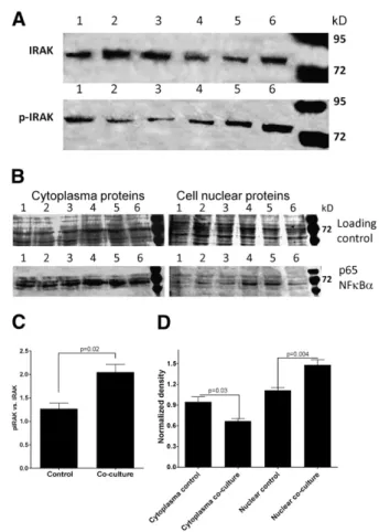
Following binding of IL1-βprotein to the receptor a key event is the phosphorylation of Interleukin-1 receptor-associated kinase 1 (IRAK1). Phosphorylated IRAK1 was also detected in control PDL fibroblasts, but IRAK phosphorylation was significantly induced (p = 0.02) in co-cultured fibroblasts (Figs. 2A and C).
After several steps and activation of multiple pathways a further, non-exclusive, but major event in IL1-βsignaling is the nuclear translocalization of the p65 c-Rel fragment of the transcription factor NFκBα. In co-cultured fibroblasts a significant increase of nuclear p65 was observed (p < 0.05) (Figs. 2B and D).
Using confocal microscopy a further proof was provided for the nuclear localization of p65 NFκBα(Figs. 3A-B).
Analysis of IL1-β-regulated genes in co-cultured fibroblasts
Following the investigation of the IL1-βsignaling, the expression of known IL1-β−regulated genes was analyzed in co-cultured fibroblasts vs. control cells. IRF1, IL-6 and COX-2 showed significant upregulation in co-cultured fibroblasts compared to control cells (p < 10−4), while PAI gene expression did not change significantly (p = 0.1). IRF1 increased 3-times, IL-6 60-times and COX-2: 222-times. The most prominent responsive genes for the presence of SCC-25 tumor cells in fibroblasts were: IL-6 and COX-2.
IL1-β-treatment of fibroblasts and SCC-25 cells
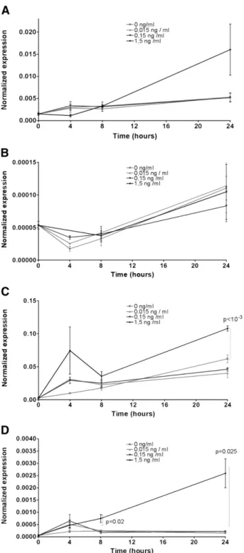
PDL fibroblasts were treated with IL1-βfollowing a previously described treatment schedule[16]. The gene expression of IL-1 receptor and of the previously found induced genes: IRF1, IL-6 and COX-2 was investigated. IL-1 receptor, IL-6 and COX-2 showed time- and dose-dependent upregulation, which was statistically significant (p < 0.05;Fig. 4) in the case of IL-6 and COX-2. In the same treatment schedule 24 h treatment of IL1-βat 1.5 ng/ml
induced a significant upregulation (p < 10−3) of COX-2 and of IL-1 receptor (p = 0.023) in SCC-25 cells. IL-6 was not significantly regulated by IL1-β(p > 0.05) in SCC-25 cells.
Dexamethasone treatment of the co-culture
IL-6 and COX-2 two extensively regulated genes in PDL fibroblasts upon IL1-β-treatment and in co-culture with SCC-25 were investigated in the same co-culture conditions as described before [3], and some of the co-cultures were treated with 10−6mol/L dexamethasone (DEX)[15]. The gene expression of IL-6 decreased to 30.85% (Fig. 5A), of COX-2 (Fig. 5B) to 0.3% in DEX-treated co- cultured fibroblasts of that of only co-cultured fibroblasts.
Discussion
Inflammation is commonly associated with cancer, and the upregulation of proinflammatory mediators has been observed Fig. 1–A) Detection of IL1-βprotein in cell lysate and supernatant
of SCC-25 cells by combined immunoprecipitation and western blot. 1–2: supernatant of SCC-25 cells, 3–4: cell lysate of SCC-25 cells, non-antibody-binding fraction of SCC-25 supernatant (5) and cell lysate (6), and1μg anti-IL1-βIgG (7). Arrow represents the active, processed 16 kD IL1-βdetectable both in the supernatant and cell lysate of SCC-25 cells. B) Normalized gene expression of IL-1 receptor in control and co-cultured fibroblasts and SCC-25 cells. In co-cultured fibroblasts IL-1R gene expression significantly increases.
Fig. 2–IL1-βsignaling in co-cultured fibroblasts. A) Detection of IRAK and phosphorylated-IRAK (p-IRAK) protein in cytoplasma extract of control (1–3) and co-cultured (4–6) fibroblasts. B) Detection of NFκBαprotein in cytoplasma and nuclear extracts of control (1–3) and co-cultured (4–6) fibroblasts. C) Densitometry of p-IRAK bands normalized to IRAK bands in cytoplasma extracts of control and co-cultured fibroblasts. D) Densitometry of NFκBαprotein in cytoplasma and nuclear extracts of control and co-cultured fibroblasts normalized to protein loading controls.
in HNSCCs[25]. A previous study reported that, IL-1βenhances activation of a cascade of proinflammatory cytokines and functions as an important autocrine and/or paracrine factor in coordinating expression of a repertoire of cytokines in HNSCC[26]. Authors found that IL-1β caused the downregulation of E-cadherin expression and upregulation of COX-2 expression, showing a direct relationship between EMT occurring in HNSCC tumor cells and inflammatory cytokines [26]. In our recent report we described a co-culture model of periodontal ligament (PDL) fibroblasts and SCC-25 oral squamous carcinoma cells, which resulted in conversion of CAFs from normal fibroblasts, and in EMT in SCC-25 cells[3]. In addition, we have identified CAFs as a major source of BDNF (brain-derived neurotrophic factor) [3], which specifically binds to tropomyosin-like kinase B receptor (TrkB) and drives EMT in the tumor cells[10]. The role of the inflammatory cytokines in induction of BDNF-expression was described before [11]. We also reported a constitutive interleukin-1β (IL1-β) expression in SCC-25 cells in normal and in co-cultured condi- tions[3]. In our hypothesis a constitutive IL1-βexpression in SCC- 25 regulates genes in fibroblasts during co-culture. We have proven this regulation step by step. SCC-25 cells produce active, processed IL1-β. PDL fibroblasts possess receptor for IL1-β, and its expression is increased 4.56-times in the presence of SCC-25 tumor cells. IL1-βreceptor expression in fibroblasts, especially in CAFs represents a major option in coordination of fibroblast and tumor behavior. A key event in IL1-βsignaling, the phosphoryla- tion of IRAK, occurred in co-cultured fibroblasts, which has lead to nuclear translocation of NFκBα, and finally to induction of several genes, including BDNF, IRF1, IL-6 and COX-2. The most enhanced induction was found for IL-6 and COX-2 (Fig. 6). Treatment of PDL fibroblasts with IL1-βreproduced a time- and dose-dependent upregulation of IL1-receptor, IL-6 and COX-2. A further proof was achieved by DEX inhibition of IL1-βstimulated gene expression, which was an adaptation of a previously published experimental system of human fibroblast-like synoviocytes[15]. Interestingly,
not only the IL1-β stimulated IL-6 expression, but also the stimulation of COX-2 was inhibited by DEX. All these proofs underline the main finding of this work that, HNSCC-tumor cells produce active IL1-βand stimulate extensive gene expression of IL-6 and of COX-2 in carcinoma-associated fibroblasts. All findings in this study were achieved by complementary methodology using RNA methods, western blots, immunoprecipitation and immuno- cytochemistry. IL1-β treatment and DEX-inhibition of IL1-β- stimulated gene expression were based on peer-reviewed and published references[15,16].
Both of the extensively regulated genes in CAFs, IL-6 and COX- 2, play major roles in the progression of HNSCC. Interestingly, SCC- 25 tumor cells might also respond to external IL1-βwith increased COX-2 expression. Nevertheless, IL1-βtreated tumor cells have no significant contribution to IL-6 levels.
Evidences were reported that IL-6 can directly influence cell proliferation and the invasion potential as the first step of tumor metastasis, in fact, IL-6 increased the invasion potential of several HNSCC cell lines[27]. Sensitive detection technologies have been developed[28]in order to measure serum IL-6, which is the only serum marker that significantly improved outcome prediction.
Higher levels of IL-6 were associated with a higher second primary cancer incidence [29]. Further studies revealed that serum IL-6 could be a valuable biomarker for predicting recurrence and overall survival among HNSCC patients. Using IL-6 as a biomarker for recurrence and survival may allow for earlier identification and treatment of disease relapse[30]. From a clinical point of view it is highly important that increased IL-6 levels were found in cachectic patients, whose quality of life was substantially reduced [31].
Based on experimental results in IL-6 gene knock-out mice it was suggested that, IL-6 secretion and cancer cachexia syndrome may be involved in the defense mechanism against tumor progression [32].
Similarly to IL-6 also COX-2 plays a major role in the progression of HNSCC. The overexpression of COX-2 is a frequent Fig. 3–NFκBα(red) immunocytochemistry in control (A) and co-cultured fibroblasts (B). Arrows indicate nuclear localization (blue DAPI-detection) of NFκBα(red) reaction. Panels are represented separately and merged. Bars: 100μm.
event in squamous cell carcinomas of the head and neck (HNSCC), and non-steroidal anti-inflammatory drugs, which are potent inhibitors of COX-1 and COX-2, exert chemopreventive effects on HNSCC cancer development. COX-2 promotes the release of the pro-inflammatory mediator prostaglandin E2 (PGE2), which acts on its cell surface G protein-coupled receptors EP1, EP2, EP3, and EP4. PGE2 produced in the tumor microenvironment by the
overexpression of COX-2 in tumor and stroma cells may promote the growth of HNSCC cells in an autocrine and paracrine fashion by acting on PGE2 receptors that are widely expressed in most HNSCC cancer cells[33].
COX-2 expression is closely correlated to VEGF expression and to tumor vascularisation. Patients with COX-2 tumor overexpres- sion and with higher PgE (2) tumor levels have significantly shorter overall survival estimates[34].
Conclusion
In our work we have provided further evidences that production of proinflammatory cytokines in HNSCC, which is associated with increased tumor growth or metastasis, is promoted by the interaction of neoplastic cells with factors and cells in the host environment. As it was previously reported, the expression of these cytokines by HNSCC cells occurs independently of T and B lymphocyte-mediated immunity [35]. The expression of some proinflammatory cytokines is detectable in the tumor cells and the one of other cytokines in the tumor-associated fibroblasts, and is promoted by tumor–host interactions[35]. Inflammatory regula- tory pathways are represented as a major therapeutic target in the HNSCC oncology.
Acknowledgments
Authors would like to acknowledge the contribution of Prof. Dr. N.
Miosge, for providing PDL fibroblasts for this study. This work was supported by the Austrian Science Foundation (FWF) Grant: P 22287-B13, and the Hungarian–Austrian Action Foundation, Grant number: 78öu1.
Fig. 5 – Expression of IL-6: (A) and COX-2 (B), in control, co- cultured and Dexamethason (DEX)-treated, co-cultured fibro- blasts. IL-6 decreased to 30.85% (A), COX-2 (B) to 0.3% in DEX- treated co-cultured fibroblasts of that of only co-cultured fibroblasts.
Fig. 4–Expression of IL1-β-regulated genes in PDL fibroblasts after IL1-β-tratment (IL1 receptor: A, IRF1: B, IL-6: C, COX-2:D).
IL1 receptor: (A), IL-6 (C) and COX-2 (D), showed time- and dose-dependent regulation.
R E F E R E N C E S
[1] A. Orimo, R.A. Weinberg, Stromal fibroblasts in cancer: a novel tumor-promoting cell type, Cell Cycle 5 (2006) 1597–1601.
[2] P.J. Mishra, P.J. Mishra, R. Humeniuk, D.J. Medina, G. Alexe, J.P.
Mesirov, S. Ganesan, J.W. Glod, D. Banerjee, Carcinoma-associated fibroblast-like differentiation of human mesenchymal stem cells, Cancer Res. 68 (2008) 4331–4339.
[3] J. Dudas, M. Bitsche, V. Schartinger, C. Falkeis, G.M. Sprinzl, H.
Riechelmann, Fibroblasts produce brain-derived neurotrophic factor and induce mesenchymal transition of oral tumor cells, Oral Oncol. 47 (2) (2011) 98–103.
[4] A.J. Daly, L. McIlreavey, C.R. Irwin, Regulation of HGF and SDF-1 expression by oral fibroblasts—implications for invasion of oral cancer, Oral Oncol. 44 (2008) 646–651.
[5] K. Higashikawa, S. Yoneda, M. Taki, H. Shigeishi, S. Ono, K.
Tobiume, N. Kamata, Gene expression profiling to identify genes associated with high-invasiveness in human squamous cell carcinoma with epithelial-to-mesenchymal transition, Cancer Lett. 264 (2008) 256–264.
[6] P. Paterlini-Brechot, N.L. Benali, Circulating tumor cells (CTC) detection: clinical impact and future directions, Cancer Lett. 253 (2007) 180–204.
[7] A.L. Kennedy, T. McBryan, G.H. Enders, F.B. Johnson, R. Zhang, P.D.
Adams, Senescent mouse cells fail to overtly regulate the HIRA histone chaperone and do not form robust Senescence Associated Heterochromatin Foci, Cell Div. 5 (2010) 16.
[8] K.E. Hoot, J. Lighthall, G. Han, S.L. Lu, A. Li, W. Ju, M. Kulesz-Martin, E.
Bottinger, X.J. Wang, Keratinocyte-specific Smad2 ablation results in increased epithelial–mesenchymal transition during skin cancer formation and progression, J. Clin. Invest. 118 (2008) 2722–2732.
[9] C. Casarsa, N. Bassani, F. Ambrogi, G. Zabucchi, P. Boracchi, E.
Biganzoli, D. Coradini, Epithelial-to-mesenchymal transition, cell polarity and stemness-associated features in malignant pleural mesothelioma, Cancer Lett. 302 (2011) 136–143.
[10] M.E. Kupferman, T. Jiffar, A. El Naggar, T. Yilmaz, G. Zhou, T. Xie, L.
Feng, J. Wang, F.C. Holsinger, D. Yu, J.N. Myers, TrkB induces EMT and has a key role in invasion of head and neck squamous cell carcinoma, Oncogene 29 (2010) 2047–2059.
[11] R.N. Saha, X. Liu, K. Pahan, Up-regulation of BDNF in astrocytes by TNF-alpha: a case for the neuroprotective role of cytokine, J.
Neuroimmune Pharmacol. 1 (2006) 212–222.
[12] C. Yan, W.A. Grimm, W.L. Garner, L. Qin, T. Travis, N. Tan, Y.P. Han, Epithelial to mesenchymal transition in human skin wound healing is induced by tumor necrosis factor-alpha through bone morphogenic protein-2, Am. J. Pathol. 176 (2010) 2247–2258.
[13] Y. Yamauchi, T. Kohyama, H. Takizawa, S. Kamitani, M. Desaki, K.
Takami, S. Kawasaki, J. Kato, T. Nagase, Tumor necrosis factor- alpha enhances both epithelial–mesenchymal transition and cell contraction induced in A549 human alveolar epithelial cells by transforming growth factor-beta1, Exp. Lung Res. 36 (2010) 12–24.
[14] P.E. Auron, A.C. Webb, L.J. Rosenwasser, S.F. Mucci, A. Rich, S.M.
Wolff, C.A. Dinarello, Nucleotide sequence of human monocyte interleukin 1 precursor cDNA, Proc. Natl. Acad. Sci. U.S.A. 81 (1984) 7907–7911.
[15] K. Miyazawa, A. Mori, H. Okudaira, Regulation of interleukin-1beta- induced interleukin-6 gene expression in human fibroblast-like synoviocytes by glucocorticoids, J. Biochem. 124 (1998) 1130–1137.
[16] C.M. Yang, S.F. Luo, H.L. Hsieh, P.L. Chi, C.C. Lin, C.C. Wu, L.D. Hsiao, Interleukin-1beta induces ICAM-1 expression enhancing leuko- cyte adhesion in human rheumatoid arthritis synovial fibroblasts:
involvement of ERK, JNK, AP-1, and NF-kappaB, J. Cell. Physiol.
224 (2010) 516–526.
[17] J. Dudas, T. Mansuroglu, F. Moriconi, F. Haller, J. Wilting, T. Lorf, L.
Fuzesi, G. Ramadori, Altered regulation of Prox1-gene-expression in liver tumors, BMC Cancer 8 (2008) 92.
[18] F. Haller, B. Kulle, S. Schwager, B. Gunawan, A. von Heydebreck, H.
Sultmann, L. Fuzesi, Equivalence test in quantitative reverse transcription polymerase chain reaction: confirmation of reference genes suitable for normalization, Anal. Biochem. 335 (2004) 1–9.
[19] F. Moriconi, D. Raddatz, N.A. Ho, S. Yeruva, J. Dudas, G. Ramadori, Quantitative gene expression of cytokines in peripheral blood leukocytes stimulated in vitro: modulation by the anti-tumor nerosis factor-alpha antibody infliximab and comparison with the mucosal cytokine expression in patients with ulcerative colitis, Transl. Res. 150 (2007) 223–232.
[20] L. Zheng, C. Yu, Z. Zhang, C. Yang, X. Cai, Expression of interferon regulatory factor 1, 3, and 7 in primary Sjogren syndrome, Oral Surg.
Oral Med. Oral Pathol. Oral Radiol. Endod. 107 (2009) 661–668.
[21] I.D. Witzel, K. Milde-Langosch, R.M. Wirtz, C. Roth, M. Ihnen, S.
Mahner, E.C. Zu, F. Janicke, V. Muller, Comparison of microarray- based RNA expression with ELISA-based protein determination of HER2, uPA and PAI-1 in tumour tissue of patients with breast cancer and relation to outcome, J. Cancer Res. Clin. Oncol. 136 (2010) 1709–1718.
[22] F.E. El Mansouri, N. Chabane, N. Zayed, M. Kapoor, M.
Benderdour, J. Martel-Pelletier, J.P. Pelletier, N. Duval, H. Fahmi, Contribution of H3K4 methylation by SET-1A to interleukin-1- induced cyclooxygenase 2 and inducible nitric oxide synthase expression in human osteoarthritis chondrocytes, Arthritis Rheum. 63 (2011) 168–179.
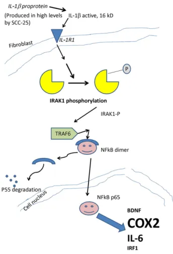
Fig. 6–Schematic summary of the main regulation mechanism of IL1βin CAFs. SCC-25 cells produce active, processed 16 kD IL1-β. PDL fibroblasts possess receptor for IL1-β, which leads to the phosphorylation of IRAK, and to subsequent nuclear translocation of NFκBα, and finally to induction of several genes, including BDNF, IRF1, IL-6 and COX-2.
[23] J. Dudas, G. Ramadori, T. Knittel, K. Neubauer, D. Raddatz, K.
Egedy, I. Kovalszky, Effect of heparin and liver heparan sulphate on interaction of HepG2-derived transcription factors and their cis-acting elements: altered potential of hepatocellular carcinoma heparan sulphate, Biochem. J. 350 (Pt 1) (2000) 245–251.
[24] U.K. Laemmli, Cleavage of structural proteins during the assembly of the head of bacteriophage T4, Nature 227 (1970) 680–685.
[25] A. Loercher, T.L. Lee, J.L. Ricker, A. Howard, J. Geoghegen, Z. Chen, J.B. Sunwoo, R. Sitcheran, E.Y. Chuang, J.B. Mitchell, A.S. Baldwin Jr., W.C. Van, Nuclear factor-kappaB is an important modulator of the altered gene expression profile and malignant phenotype in squamous cell carcinoma, Cancer Res. 64 (2004) 6511–6523.
[26] M.A. St John, M. Dohadwala, J. Luo, G. Wang, G. Lee, H. Shih, E.
Heinrich, K. Krysan, T. Walser, S. Hazra, L. Zhu, C. Lai, E. Abemayor, M.
Fishbein, D.A. Elashoff, S. Sharma, S.M. Dubinett, Proinflammatory mediators upregulate snail in head and neck squamous cell carcinoma, Clin. Cancer Res. 15 (2009) 6018–6027.
[27] T. Kanazawa, H. Nishino, M. Hasegawa, Y. Ohta, Y. Iino, K.
Ichimura, Y. Noda, Interleukin-6 directly influences proliferation and invasion potential of head and neck cancer cells, Eur. Arch.
Otorhinolaryngol. 264 (2007) 815–821.
[28] R. Malhotra, V. Patel, J.P. Vaque, J.S. Gutkind, J.F. Rusling, Ultrasensitive electrochemical immunosensor for oral cancer biomarker IL-6 using carbon nanotube forest electrodes and multilabel amplification, Anal. Chem. 82 (2010) 3118–3123.
[29] F. Meyer, E. Samson, P. Douville, T. Duchesne, G. Liu, I. Bairati, Serum prognostic markers in head and neck cancer, Clin. Cancer Res. 16 (2010) 1008–1015.
[30] S.A. Duffy, J.M. Taylor, J.E. Terrell, M. Islam, Y. Li, K.E. Fowler, G.T.
Wolf, T.N. Teknos, Interleukin-6 predicts recurrence and survival among head and neck cancer patients, Cancer 113 (2008) 750–757.
[31] L.M. Richey, J.R. George, M.E. Couch, B.K. Kanapkey, X. Yin, T.
Cannon, P.W. Stewart, M.C. Weissler, C.G. Shores, Defining cancer cachexia in head and neck squamous cell carcinoma, Clin. Cancer Res. 13 (2007) 6561–6567.
[32] A. Molotkov, M. Satoh, C. Tohyama, Tumor growth and food intake in interleukin-6 gene knock-out mice, Cancer Lett. 132 (1998) 187–192.
[33] A.C. Abrahao, R.M. Castilho, C.H. Squarize, A.A. Molinolo, D. dos Santos-Pinto Jr., J.S. Gutkind, A role for COX2-derived PGE2 and PGE2-receptor subtypes in head and neck squamous carcinoma cell proliferation, Oral Oncol. 46 (2010) 880–887.
[34] O. Gallo, E. Masini, B. Bianchi, L. Bruschini, M. Paglierani, A.
Franchi, Prognostic significance of cyclooxygenase-2 pathway and angiogenesis in head and neck squamous cell carcinoma, Hum. Pathol. 33 (2002) 708–714.
[35] C.W. Smith, Z. Chen, G. Dong, E. Loukinova, M.Y. Pegram, L.
Nicholas-Figueroa, W.C. Van, The host environment promotes the development of primary and metastatic squamous cell carcinomas that constitutively express proinflammatory cytokines IL-1alpha, IL-6, GM-CSF, and KC, Clin. Exp. Metastasis 16 (1998) 655–664.