Correlation and Immunolocalization of Substance P Nerve Fibers and Activated
Immune Cells in Human Chronic Gastritis
GA´ BOR SIPOS,1PE´ TER SIPOS,2KA´ ROLY ALTDORFER,3 E´ VA PONGOR,3ANDERZSE´ BET FEHE´R3*
1Department of Gastroenterology, Uzsoki Teaching Hospital, Budapest, Hungary
2Department of Surgery, Semmelweis University, Budapest, Hungary
3Department of Anatomy, Histology, and Embryology, Semmelweis University, Budapest, Hungary
ABSTRACT
Neuropeptides are able to modulate cytokine production by macro- phages in response to various stimulators and have a major role in inflammation of different organs. Mammalian poly (ADP-ribose) polymer- ase (PARP) and nuclear factor kappa B (NF-jB) both have been suggested to play a crucial role in inflammatory disorders. Unregulated increase of tumor necrosis factor-a (TNF-a) may also be pathogenic in inflammatory diseases. The aim of this study was to investigate the correlation between the number of Substance P (SP) containing nerve fibers and activated immune cells using immunohisto-, immunocytochemical (EM) and confo- cal laser microscopic methods. To investigate expression and activation of immune cells gastric biopsy samples from patients with chronic gastritis were used. The number of SP containing nerve fibers and activated immune cells increased significantly in gastritis. Using monoclonal p65 antibody, activated NF-jB was found in inflamed mucosa but was absent in uninflamed mucosa. Immunobinding for the activated form of p65 of NF-jB was found in 22% of macrophages and 45% of lymphocytes. The number of immune cells showing IR for NF-jB, PARP and TNF-a corre- lated with the increasing number of SP containing fibres. Confocal laser microscopy was used to confirm the colocalization of SP in TNF-a and NFjB positive lymphocytes and mast cells in inflamed mucosa. Immuno- electronmicroscopic investigation confirmed that these cells belong to lym- phocytes, mast cells and macrophages. Conclusions: The increase of SP in nerve fibers and in activated immune cells further activate the production of other proinflammatory mediators (e.g. TNF-a) and therefore generate the chronic inflammation. Anat Rec, 291:1140–1148, 2008. Ó2008 Wiley-Liss, Inc.
Key words: substance P; gastritis; neuroimmunomodulation;
TNF-alfa; NFkappaB
Neuropeptides are able to modulate cytokine produc- tion by macrophages in response to various stimulators.
Substance P (SP) and calcitonin gene-related peptide (CGRP) are important mediators of neuroimmunomodu- latory activity (Veronesi et al., 1999; Azzolina et al., 2003; Yarace et al., 2003). It was also demonstrated that SP has a major role in inflammation of the stomach (Mo´zsik et al., 2001). Previously we have shown that the
*Correspondence to: Prof. Erzse´bet Fehe´r, Semmelweis Uni- versity, Tuzolto u. 58, P.O. Box 95, H-1450, Budapest, 1450, Hungary. Fax: 36(1)215-51-58. E-mail: feher@anal.soe.hu
Received 7 February 2008; Accepted 24 April 2008 DOI 10.1002/ar.20737
Published online in Wiley InterScience (www.interscience.wiley.
com).
Ó2008 WILEY-LISS, INC.
mor necrosis factor-alpha (TNF-a) gene expression in dif- ferent mast cells (Ansel et al., 1993; Cocchiara et al., 1997). Unstimulated peritoneal mast cells spontaneously released a small quantity of TNF-a, whereas after SP stimulation the amount of released TNF-a was approxi- mately four times higher and this effect was inhibited only by pretreatment with SP antagonist (SPA, P101, CP-96345; Cocchiara et al., 1999). It is well known that TNF-a plays a key role in the immunopathogenesis of inflammatory bowel diseases, a fact which is overtly con- firmed by clinical effects observed in refractory Crohn’s disease patients treated with a chimeric monoclonal antibody against TNF (infliximab; Blam et al., 2001).
The binding of SP to tachykinin-1 receptor can result in the up-regulation of proinflammatory cytokines, such as TNF-a and interleukin-8 (IL-8), whose expression is controlled by the transcription factor NF-kB (Lieb et al., 1997; Marriott et al., 2000). Activation of the NF-kB/Rel transcription family, by nuclear translocation of cytoplas- mic complexes, plays a central role in inflammation through its ability to induce transcription of proinflam- matory genes (Baldwin, 1996). NF-kB exists in the cyto- plasm in an inactive form associated with regulatory pro- teins called inhibitors ofkB (IkB). Phosphorylation of IkB an important step in NF-kB activation, is mediated by IkB kinase. The dimer, typically composed of a p50 and p65 subunit, is translocated to the nucleus after degrada- tion of the inhibitory I-kB in response to a wide variety of stimuli (Ghosh et al., 1998). Varro et al. (2004) also dem- onstrated that in unstimulated cells, p65-dsRed was located in the cytosol, and stimulation by H. pylori caused translocation to the nucleus. The activation of transcrip- tion factor nuclear factor-kB (NF-kB) regulates various genes involved in the proliferation, invasion, angiogene- sis, and metastasis of cancer cells. Substantial in vitro data suggest that activation of NF-kB is a critical initial step in the inflammatory response. Several studies dem- onstrated a link between in vivo NF-kB activation accom- panied by cytokine production and the generation of inflammation in animal models of inflammatory diseases (Sakurai et al., 1996; Blackwell et al., 1997; Ellis et al., 1998). The identification of NF-kB as a key player in the pathogenesis of inflammation suggests that NF-kB tar- geted therapeutics might be effective in diseases like gas- tritis. Inflammation in any part of the gastrointestinal tract can profoundly influence the function of the mucosal layer that lies closest to the luminal contents. The inflam- matory response is coordinated to a large extent by an array of chemical mediators that are released from nerves, from the immune cells and epithelium.
Besides of NF-kB the mammalian poly (ADP-ribose) polymerase (PARP) has been also suggested to play a crucial role in inflammatory disorders (Hassa and Hot-
variety of inflammatory diseases (Szabo´ et al., 2004).
It is now clear that Helicobacter pylori(HP) activates the transcription factor NF-kB and this event plays a central role in the induction of the inflammatory reac- tion often associated with colonization with this bacte- rium. The increase of SP-IR in nerve fibers and immune cells that parallel with the grade of inflammation in the stomach has never been described previously. Although, in recently similar changes in other inflamed organs has been published, the correlated increase in the substan- ces described was not observed. Therefore, we have examined in this study the expression and activation of NF-kB, TNF-a, PARP and the correlation of SP immuno- reactive nerve fibers and immune cells in the control stomach and HP-associated gastritis using light and electron microscopy as well as confocal laser microscopy.
MATERIALS AND METHODS
Endoscopic biopsies of gastric antrum were obtained from 10 HP-positive patients (4 male; 6 female, ageing from 30 to 67 years old) who underwent esophagogastro- duodenoscopy for dyspeptic symptoms over a period of 1 year. None of the patients had neoplasmic disease or peptic ulcer. Antral samples of 5 dyspeptics (2 male; 3 female, ranging from 27 to 58 years) obtained with endos- copy and histologically normal HP negative stomach sam- ples were used as controls. HP positivity, assessed by rapid urease test, had been confirmed by histology and serology in all patients. Biopsies from HP positive patients showed histologically chronic gastritis with moderate or severe ac- tivity according to the Sydney’s system (Price, 1991). All patients and healthy volunteers gave their informed con- sent according to Semmelweis University guidelines for ethics in human tissue experiments (No: TUKEB 85/2006).
Immunohistochemical Analysis
Biopsy materials were fixed in Zamboni’s fixative con- taining 4% paraformaldehyde, 0.1% glutaraldehyde in 250 mL of 0.4 M phosphate-buffer, 150 mL picric acid (pH 7.3) for 6 hr and placed overnight in glutaralde- hyde-free fixative containing 20% sucrose at 48C. Sec- tions (40mm thick) were treated for 1 hr with 1% TRI- TON-X 100 to increase membrane permeability and for 15 min with 3% hydrogen peroxide to remove endoge- nous peroxidase activity. Incubation with primary anti- sera was performed for 48 hr at 48C. The Avidin-Biotin technique was employed using a commercially available kit (Vectastain Elite ABC, Vector Laboratories, Peterbor- ough, UK) for the immunostaining. All manipulations were performed at room temperature. Immunoreactivity was visualized with diamino-benzidine (DAB) chromogen reaction (Dako, Milan, Italy) (0.025% 3, 3-diamino-benzi-
dine, 0.0015% H2O2in 0.05 M Tris-HCl buffer, pH 7.5) for 8 to 10 min, at room temperature. For light micro- scopic examination the sections were mounted on gelati- nized slides, air-dried, cleared, and covered with Depex.
To block nonspecific binding of antibody, sections were preincubated with 1:10 diluted normal goat serum.
Antibodies
Antibody to Substance P (SP) was developed in rabbit (Peninsula Lab. Inc., San Carlos, CA), dilution:
1:10,000). Antisera to NF-kB subunits, anti-p65 mouse monoclonal IgG (Chemicon, International Inc., Teme- cula, CA), dilution: 1:1,500; which recognizes an epitope overlapping the nuclear location signal of the p65 subu- nit of NF-kB heterodimer. The anti-human TNF-a poly- clonal antibody developed in rabbit (Sigma-Aldrich), dilution: 1:8,000, anti-PARP mouse monoclonal antibody (Biomol Int. LP, Plymouth Meeting, PA), dilution:1:500.
For double staining the sections were also examined by confocal laser microscopy (Nikon Eclipse 800 microscope, Japan, Radiance 2100, Bio-Rad, LaserSharp2000 Soft-
ware, Bio-Rad House, Hertfordshire, UK). Frozen sections were washed in phosphate buffered saline (PBS) at room temperature and permeabilized for 20 min in PBS (23NaCl) containing 0.3% Triton X-100 and 2% normal serum; the same solution was used to dilute the antibod- ies. Afterward, they were sequentially incubated with anti-SP antiserum at dilution 1:5,000 overnight. Slides were washed in PBS and then incubated for 3 hr at room temperature with a secondary fluorescein (FITC, 1:100) conjugated donkey anti-rabbit IgG antibody (Jackson ImmunoResearch, West Grove, PA). The sections were washed with buffer and incubated with anti–TNF-a (1:8,000) for 24 hr, followed by secondary antiserum raised in donkey fluorescein-labeled anti-rabbit IgG, Alexa 594, diluted in 1:500, Molecular Probes, Eugene, OR) for 3 hr, mounted in anti-fade medium (Vectashield, Vector Laboratories, Peterborough, UK) and stored at 2208C until needed. The cell bodies and the nerve fibers that could be followed were then scanned with a confocal laser microscope equipped with a krypton–argon laser.
Fluorescent signals from FITC (green) and Alexa 594 (red) were sequentially detected on a Bio-Rad MicroRa- diance confocal laser system (Bio-Rad MRC1024).
For electron microscopic investigations the sections were post-fixed in osmium and embedded in Epon.
Fig. 1. Cross-section of the stomach glands from gastritis. Numer- ous immunoreactive SP nerve fibers (arrow) and SP-positive immuno- cytes (arrowheads) are located among the glands. Scale bar5100mm.
Fig. 2. Changes of the SP-IR nerve fibers in the human gastritis.
Fig. 3. Changes of the activated immune cells in the human gastritis.
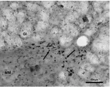
Fig. 4. A part of the tunica mucosa from gastritis. Arrows point TNF-a–IR immunocytes beneath the glands. Gl, glands; MM, lamina muscularis mucosae. Scale bar5100mm.
fields was taken as the activation score of the specimen.
To be able to compare the results between patients, it was ensured that all sections visualized the entire axis from the superficial epithelium to the muscularis mucosae.
Control Experiments
Specificity of the immunoreactivity was controlled by omission of the primary antiserum or, when the sections were incubated in antisera, preabsorbed with excess antigen, where no immunostaining appeared.
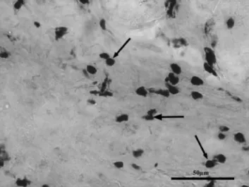
Fig. 5. Tunica mucosa from human gastritis. Arrows show the strong positive poly(ADP-ribose) staining in the cell nuclei of immune cells. Scale bar5100mm.
Fig. 6. A part of the mucosa of the inflamed human stomach. Arrows show the active NF-kB immu- noreactivity in the nucleus of the immune cells. Scale bar5100mm.
Statistical Analysis
Data are presented as mean6 scanning electron mi- croscopy. Comparisons between groups of data were made using a one-way analysis of variance followed by a Tukey post hoc test. P values < 0.05 were considered statistically significant.
RESULTS
All subjects were of adult age and no predominance of male or female sex was present. At endoscopy gastric hyperemia was the main feature of HP-related chronic gastritis.
The number of SP IR nerve fibers was increased sig- nificantly, also some immune cells (lymphocytes, plasma cells, macrophages, and mast cells) showed immunoreac- tivity for SP (Fig. 1). The gap between the nerve termi- nals and immune cells was approximately 1 mm or even less. The change of the number of SP-IR nerve fibers is shown on Figure 2.
In the control materials the number of PARP- and TNF-a–IR cells was very low, in some sections totally missing. However, in sections from chronic gastritis their number was significantly increased (P < 0.001) (Fig. 3). PARP and TNF-a staining in gastritis were localized primarily to the mononuclear cells and fibro- blasts in the lamina propria, with no detectable staining of the epithelium (Figs. 4, 5). These IR immune cells were in close contact with epithelial cells. The reaction
end-product of PARP was seen in the nuclei of the cells, while TNF-aimmunoreactivity was observed in the cyto- plasm of the positive cells. According to the size and form of these IR cells they belong to the lymphocytes, macrophages and polymorphonuclear cells (PMN).
In the normal stomach, low and moderate immunor- eactivity of NF-kB (p65 subunit) was found in the cyto- plasm of gastric glandular epithelial cells located in the deep region of the glands. In gastritis, the active NF-kB was observed in clusters of inflammatory cells, where NF-kB was detected not only in large amounts in the cytoplasm but also in the nucleus, suggesting activation of NF-kB in these cells (Fig. 6). The number of active NF-kB IR cells was markedly increased, as compared with uninflamed tissue, and they were mainly detected among the glands. 22% of mast cells and 45% of lympho- cytes were immunoreactive in gastritis.
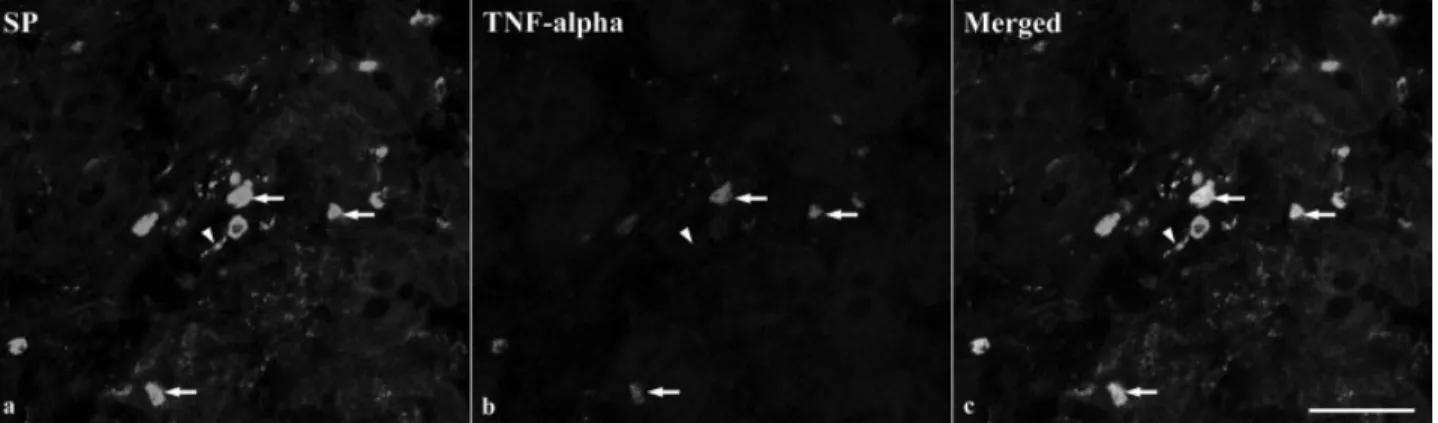
Confocal laser microscope investigations showed that dense green reaction end products (FITC) were distrib- uted in the SP containing nerve fibers and throughout the cytoplasm of immune cells (SP); some of these showed a red reaction (Alexa 594) for TNF-a (Fig. 7).
Fluorescent double-labeled immunostaining showed that SP (FITC, green) fluorescence positive immune cells also had immunostaining for p65 of NF-kB (Alexa 594, red), where their labeling was located in the nucleus of these cells (Fig. 8).
The electron-microscopic investigation proved that these cells belong to different immunocompetent cells (lymphocytes, macrophages and mast cells). The reaction Fig. 7. Confocal images illustrating of SP-IR nerve cells and acti-
vated immune cells in the human gastritis. a: SP-IR immune cells (arrows) in the lamina propria among the glands. Arrowhead shows the SP-IR nerve fibers close to the activated mast cells. b: The arrows
show the same mast cells showing immunoreactivity for TNF-a. c:
Arrows indicate double-labeling immune cells for SP and TNF-a. Scale bar550mm.
Fig. 8. Confocal images illustrating of SP and NF-kB immune cells in the human gastritis. Fluorescent double-labeled immunostaining showed that SP (FITC) fluorescence-positive immune cells were also labeled for NF-kB, where the immunostaining (Alexa 594, red) was located in the nucleus of these cells. In the insert, one of the double-
labeled mast cell.a:Arrows point the SP mast cells and arrowhead show a SP lymphocyte.b:The arrows and arrowhead show the same cells showing immunoreactivity for NF-kB. c: Arrows indicate the double-labeling cells for SP and NF-kB. Scale bar550mm.
end products were distributed in the cytoplasm and at the membranes of the TNF-a–IR cells (Figs. 9, 10), while in NF-kB–IR cells, the reaction end products were located in the nucleus of the cells (Fig. 11) reflecting activation of the NF-kB heterodimer.
DISCUSSION
SP is involved in the biological activities of the immune system, including the induction of cytokines in immune cells (Lee et al., 1994; Ho et al., 1996; Cocchiara et al., 1997; Maggi, 1997). It was demonstrated that the number of SP-IR nerve fibers and SP IR immune cells increased significantly during inflammation in the tongue and in the stomach (Batbayer et al., 2004; Sipos et al., 2006). SP degranulates mast cells and, therefore, further amplifies the inflammatory reactions. Capsaicin
pretreatment (which decreases or eliminates the func- tional contribution of C-fiber nociceptors) decreases phagocyte migration into inflammation sites and pre- vents degranulation of mast cells (Perretti et al., 1993).
Antagonists of pro-inflammatory peptides such as SP may control inflammatory diseases or processes in which these peptides have a primary pathogenic role (Frieri, 2003).
SP-stimulation of murine mast cells activates TNF-a gene expression and induces TNF-a secretion (Ansel et al., 1993). It was also demonstrated by Azzolina et al.
(2003) that the NFkB pathway is involved in the tran- scriptional regulation of the TNF-a and IL-6 over expression in SP-stimulated mast cells. SP also activates the transcription factor NFkB, a threefold increased nu- clear translocation being observed in alveolar macro- phages from healthy smokers (Bardelli et al., 2005). Our previous data and this study showed that Helicobacter pylori caused continuous infiltration of inflammatory cells and significant increase of SP IR nerve terminals as well as the SP immunoreactive immune cells (Sipos et al., 2006). Therefore, we have postulated that during the early events of an inflammatory process induced by neuropeptide SP could be the cause of the increase in TNF-a in the inflamed organs. The increased production of TNF-a might activate the NF-kB in the cells of the mucosa as well as it further activates the PARP which causes injury to the mucosal lining (Fig. 12).
PARP activation was identified as a key pathway in different pathophysiological conditions and disease states. Recent reports have shown that PARP can act as a coactivator of NFkB (Hassa and Hottinger, 2002). The cleaved form of PARP is catalytically inactive. Depend- ing on the severity of DNA damage, genotoxic stimuli can trigger three different pathways. In the case of mild DNA damage, PARP facilitates DNA repair and thus survival. More severe DNA damage induces enzymes of the apoptotic cell death during which caspases, the main executor enzymes of apoptotic process, inactivate PARP cleaving into two fragments (p89 and p24). The most severe DNA damage may cause excessive PARP activa- Fig. 11. Electron microscopic photograph of a NF-kB immunoreac- tive lymphocyte from the inflamed stomach. The reaction end-prod- ucts are located in the nucleus (arrow) showing the activation of this cell. Scale bar51mm.
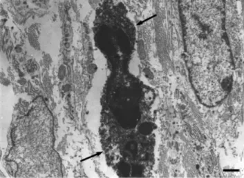
Fig. 10. Electron microscopic photograph of a TNF-a–IR mast cell from the gastritis. Scale bar51mm.
Fig. 9. Electron microscopic photograph of a TNF-a–IR granulo- cyte from the gastritis. Note that the reaction end-products are visible in the cytoplasm of the cell (arrows). Scale bar51mm.
tion inhibiting glycolysis and mitochondrial respiration and depleting NAD1and ATP stores (Szabo´ et al., 1997, 2002). Our histological examination clearly demon- strated that significant PARP activation occurs in the mucosa of the stomach during chronic inflammation. In- hibition of PARP reduces the infiltration of neutrophils, decreases inflammatory infiltrate, which would be asso- ciated with a reduction in both oxygen and nitrogen cen- tered free radical production (Szabo´ et al., 1997).
Our data indicate that immune cells (mast cells, lym- phocytes, macrophages) are the most prominent cell types exhibiting activated NF-kB in the inflamed mu- cosa, where their number was increased in chronic gas- tritis. This is in agreement with the findings of Rogler et al. (1998) in patients with inflammatory bowel dis-
eases, who showed that NF-kB was mainly activated in macrophages and epithelial cells. There is evidence of NF-kB activation in inflammatory bowel disease, where the lamina propria macrophages display activated p50, c-Rel, and p65 (Neurath et al., 1996). Reed et al. (2005) demonstrated that NF-kB activation increased signifi- cantly biphasically (temporal activation) in experimen- tal colitis. Helicobacter pylori-associated gastritis in previous studies exhibited increased NF-kB activity in gastric epithelial cells, where the number of NF-kB–
positive cells correlates with the degree of gastritis (Van Den Brink et al., 2000). It is now recognized that activation of NF-kB induces many different genes, some of which are associated with inhibition of apoptosis and others with innate immunity and inflammatory Fig. 12. Postulated mechanisms of gastric mucosal injury.
action of PARP-1 and NF-kB might inhibit the abnormal transcriptional activity of NF-kB, thereby reducing the inflammatory response at the level of transcription in instances where this process becomes chronic or dysregulated (Hassa and Hottinger, 2002).
NF-kB directed therapy was demonstrated to be effec- tive in a model of inflammatory bowel disease induced by 2,4,6-trinitrobenzene sulfonic acid. It was also shown that selective TNF receptor inhibition may be advantageous with anti-TNF treatments in combating chronic inflammatory disease (Kollias, 2005). The treat- ment of patients with rheumatoid arthritis with anti- bodies against TNF-a can control refractory disease (Elliott et al., 1994). Single intraperitoneal injection of anti–TNF-a monoclonal antibody treatment signifi- cantly reduced serum/tissue TNF-a and improved indo- methacin-induced enteropathy in rats by modulating iNOS expression (Saud et al., 2005). Our data indicate and suggest that TNF signaling system mediates muco- sal damage by the enhancement of NF-kB activity par- allel with increased number of SP nerve fibers and immune cells and that they may be the key pathoge- netic factor of gastritis. TNF-a may influence the sever- ity of disease, possibly by the persistent activation of NF-kB.
ACKNOWLEDGMENT
The authors thank Ms. E. Burka for assisting with the manuscript.
LITERATURE CITED
Ansel J, Brown RJ, Payan DG, Brown MA. 1993. Substance P selec- tively activates TNF-a gene expression in murine mast cell. J Immunol 150:4478–4485.
Azzolina A, Bongiovanni A, Lampiasi N. 2003. Substance P induces TNF-alpha and IL-6 production through NF-kappaB in peritoneal mast cells. Biochim Biophys Acta 1643:75–83.
Baldwin AS Jr. 1996. The NF-kappa B and I kappa B proteins: new discoveries and insights. Annu Rev Immunol 14:649–683.
Bardelli C, Gunella G, Varsaldi F, Balbo P, Del Boca E, Nernardone IS, Amoruso A, Brunelleschi S. 2005. Expression of functional NK1 receptors in human alveolar macrophages: superoxide anion production, cytokine release an involvement of NF-kappa B path- way. Br J Pharmacol 145:385–396.
Batbayar B, Nagy G, Kovesi G, Zelles T, Feher E. 2004. Morphologi- cal basis of sensory neuropathy and neuroimmunomodulation in minor salivary glands of patients with Sjogren’s syndrome. Arch Oral Sci 49:529–538.
Blackwell TS, Blackwell TR, Christman JW. 1997. Impaired activa- tion of NF-kB in endotoxin-tolerant rats in associated with down- regulation of chemokine gene expression and inhibition of neutro- philic lung inflammation. J Immunol 158:5934–5940.
1994. Randomized double-blind comparison of chimeric monoclo- nal antibody to tumor necrosis factor (alpha) CCA2) versus pla- cebo in rheumatoid arthritis. Lancet 334:1105–1110.
Ellis RD, Goodland JR, Limb GA, Powell JJ, Thompson RP, Pun- chard NA. 1998. Activation of nuclear factor kappa B in Crohn’s disease. Inflamm Res 47:440–445.
Frieri M. 2003. Neuroimmunology and inflammation: implications for therapy of allergic and autoimmune diseases. Ann Allergy Asthma Immunol 90(suppl 3):34–40.
Ghosh S, May JM, Kopp EB. 1998. NF-kB and Rel proteins: evolu- tionary conserved mediators of immune responses. Annu Rev Immunol 16:225.
Hassa PO, Hottinger MO. 2002. The functional role of poly (ADP- ribose)polymerase 1 as novel coactivator of NF-kB in inflamma- tory disorders. Rev Cell Mol Life Sci 59:1534–1553.
Ho WZ, Kaufmann D, Uvaydova M, Douglas SD. 1996. Substance P augmenti interleukin-10 and tumor necrosis factor-alpha release by human cord blood monocytes and macrophages. J Neuroimmu- nol 71:73–80.
Kahler C M, Sitte BA, Reinisch N, Wiedermann CJ. 1993. Stimula- tion of the chemotactic migration of human fibroblasts by sub- stance P. Eur J Pharmacol 249:281–286.
Kollias G. 2005. TNF pathophysiology in murine models of chronic inflammation and autoimmunity (review). Semin Arthritis Rheum 34(suppl 1):3–6.
Lee HR, Ho WZ, Douglas SD. 1994. Substance P augments tumor necrosis factor release in human monocyte-derived macrophages.
Clin Diagn Lab Immunol 1:419–423.
Lieb K, Fiebich BL, Berger M, Bauer J, Schulze-Osthoff K. 1997.
The neuropeptide substance P activates transcription factor NF- kappa B B-dependent gene expression in human astrocytoma cells. J Immunol 150:4952–4958.
Maggi CA. 1997. The effects of tachykinins on inflammatory and immune cells. Regul Pept 70:75–90.
Marriott I, Mason MJ, Elhofy A, Kenneth LB. 2000. Substance P activates NF-kB independent of elevations in intracellular cal- cium in murine macrophages and dendritic cells. J Neuroimmu- nol 102:163–171.
Mo´zsik GY, Vincze A, Szolcsanyi J. 2001. Four responses of capsai- cin-sensitive primary afferent neurons to capsaicin and its analog.
Gastric acid secretion, gastric mucosal damage and protection. J Gastroenterol Hepatol 16:1093–1097.
Neurath MF, Pettersson S, Meyer zum Buschenfelde KH, Strober W. 1996. Local administration of antisense phosphorothioate oli- gonucleotides to the p65 subunit of NF-kappaB abrogates estab- lished experimental colitis in mice. Nat Med 2:998–1004.
O’Connor TM, O’Connell J, O’Brien DI, Goode T, Bredin C, Shana- han F. 2004. The role of substance P in inflammatory disease. J Cell Physiol 201:167–180.
Perretti M, Ahluwalia A, Flower RJ, Manzini S. 1993. Endogenous tachykinins play a role in IL-1-induced neutrophil accumulation:
involvement of NK-1 receptors. Immunology 80:73–77.
Price AB. 1991. The Sydney System: histological division. J Gastro- enterol Hepatol 6:209–222.
Reed KL, Fruin AB, Gower AC, Gouzales KD, Stucchir AF, Andry CD, O’Brien M, Becker JM. 2005. NFkB activation precedes increases in mRNA encoding neurokinin-1 receptor, proinflamma- tory cytokines and adhesion molecules in dextran sulphate so- dium induced colitis in rats. Dig Dis Sci 50:2366–2378.
Rogler G, Brand K, Vogl D, Page S, Hofmeister R, Andus T, Uneu- chel R, Baeuerle PA, Scholmerich J, Gross V. 1998. Nuclear factor kB is activated in macrophages and epithelial cells of inflamed in- testinal mucosa. Gastroenterology 115:357–369.
Sakurai H, Hisada Y, Ueno M, Sugiura M, Kawashima K, Sugita T. 1996. Activation of transcription factor NF-kB in experimen- tal glomerulonephritis in rats. Biochim Biophys Acta 1316:132–
138.
Saud B, Nandi J, Ong G, Finocchiaro S, Levine RA. 2005. Inhibition of TNF-alpha improves indomethacin-induced enteropathy in rats by modulating iNOS expression. Dig Dis Sci 50:1677–1683.
Schratzberger P, Reinisch N, Prodinger WM, Kahler CM, Sitte BA, Bellmann R, Fischer-Colbri R, Winkler H, Wiedermann CJ. 1997.
Differential chemotactic activities of sensory neuropeptides for human peripheral blood mononuclear cells. J Immunol 158:3895–
3901.
Sipos G, Altdorfer K, Pongor E, Chen LP, Feher E. 2006. Neuroim- mune link in the mucosa of chronic gastritis with Helicobacter pylori infection. Dig Dis Sci 51:1810–1817.
Szabo´ C. 1998. Role of poly(ADP-ribose)synthase in inflammation.
Eur J Pharmacol 350:1–19.
Szabo´ C, Lim LH, Cuzzocrea S, Getting SJ, Zingarelli B, Flower RJ, Salzman AL, Perretti M. 1997. Inhibition of poli(ADP-ribose)syn- thetase exerts anti-inflammatory effects and inhibits neutrophil recruitment. J Exp Med 186:1041–1049.
Szabo´ G, Bahrle S, Stumpf N, Sonnenberg K, Szabo EE, Pacher P, Csont T, Schulz R, Dengler TJ, Liaudet L, Jagtap PG, Southan
GJ, Vahl CF, Hagl S, Szabo C. 2002. Poly(ADP-ribose) polymerase inhibition reduces reperfusion injury after heart transplantation.
Circ Res 90:100–106.
Szabo´ G, Soos P, Bahrle S, Zsengeller Z, Flechtenmacher C, Hagl S, Szabo C. 2004. Role of poly(ADP-ribose) polymerase activation in the pathogenesis of cardiopulmonary dysfunction in a canine model of cardiopulmonary bypass. Eur J Cardiothorac Surg 25:825–832.
Van Den Brink GR, ten Kate FJ, Ponsioen CY, Rive MM, Tytgat GN, van Deventer SJ, Peppelenbosch MP. 2000, Expression and activation of NF-kappa B in the antrum of the human stomach. J Immunol 164:3353–3359.
Varro A, Noble PJ, Pritchard DM, Kennedy S, Hart CA, Dimaline R, Dockray GJ. 2004. Helicobacter pylori induces plasminogen ac- tivator inhibitor 2 in gastric epithelial cells through nuclear fac- tor-kB and RhoA. Implications for invasion and apoptosis. Cancer Res 64:1695–1702.
Veronesi B, Carter JD, Devlin RB, Simon SA, Oortgresen M. 1999.
Neuropeptides and capsaicin stimulate the release of inflamma- tory cytokines in a human bronchial epithelial cell line. Neuro- peptides 33:447–456.
Vira´g L, Szabo C. 2002. The therapeutic potential of poly(ADP-ribo- se)polymerase inhibitors. Pharmacol Rev 54:375–429.
Yarace E, Ebtekar M, Ahmadiani A, Sabahi F. 2003. Neuropepti- des (SP, CGRP) augment pro-inflammatory cytokine production in HSV-infected macrophages. Int Immunopharmacol 3:1883–
1887.