FINE S T R U C T U R E
Elisabeth Reveling
I. Introduction 147 II. Cellular Structure of the Symbionts 148
A. Protoplast of the Lichenized Phycobionts 148 B. Protoplast of the Lichenized Mycobionts 157 C. The Cell Walls of Lichenized Algae and Fungi 161
D. The Cultured Symbionts 165 III. Symbiotic Relationship between Fungus and Alga 167
A. Intracellular Haustoria 167 B. Intramembranous Haustoria 170 C. Contact between Alga and Fungus without Haustoria 170
IV. Thallus Layers and Surface Structures as Viewed with SEM 170
A. Thallus Layers 171 B. Surface Structures 171
C. Rhizines 175 V. Some Aspects of Changes in Ultrastructure Due to Ecological
Conditions 177 VI. Conclusions 179 References 179
I. Introduction
Significant electron microscopic investigations of lichens did not begin until a few years ago. This is late compared with other organisms. The late start was due primarily to the technical difficulties in the preparation of lichen thalli. A lichen thallus is a heterogeneous structure that consists of two different symbionts, each one usually requiring a different preparative treatment. In addition, the fungal hyphae are arranged in layers of different densities and contain intercellular material which may be gelatinous, fibril- lar, or crystalline. Despite these obstacles, the progress of lichen ultrastruc- ture has been rapid and our knowledge in this area is approaching that in
147
148 ELISABETH PEVELING
lichen taxonomy and physiology. We know many details of the substructure of the vegetative organs of lichens. Of particular interest are cellular structures that are unique to the lichen symbiosis and those which constitute the new morphological entity we recognize as the lichen thallus.
In addition to cellular structures, the different number and composition of layers in the cell wall of the symbionts, the distribution of lichen acid crystals, and the arrangement of the algal cells and fungal hyphae in the thal- lus provide considerable variation among lichens. The scanning electron microscope (SEM) reveals more variety in the micromorphology of thalli.
The SEM gives a good impression of the three-dimensional inner organiza- tion of the thallus, its surface structure, and the structures by which thalli adhere to the substrate. The freeze-etch technique has been another method used to examine lichen ultrastructure (Ellis and Brown, 1972).
This chapter summarizes the submicroscopical investigations on lichens.
The substructure of phycobionts and mycobionts and their symbiotic rela- tionships are considered as well as changes in substructure due to different ecological conditions. Thallus surface structures are illustrated with the scanning electron microscope.
II. Cellular Structure of the Symbionts
A. Protoplast of the Lichenized Phycobionts
Of the 26 genera of lichenized algae (Ahmadjian, 1967), the common blue- green phycobionts Nostoc, Gloeocapsa, and Scytonema and the most impor- tant green phycobionts Trebouxia, Coccomyxa, Myrmecia, and Stichococcus
have been studied with the electron microscope. A general feature of these phycobionts is their smaller size relative to free-living or cultured forms.
An exception to this is Stichococcus which enlarges considerably when lichenized.
1. BLUE-GREEN PHYCOBIONTS
Nostoc phycobionts have been studied in Peltigera polydactyla (Peat, 1968), Peltigera canina, P. rufescens, and Leptogium hildenbrandii (Peveling, 1969c). Gloeocapsa has been examined in Gonohymenia mesopotamica and
G. sinaica (Paran et al., 1971). Scytonema was described in Heppia lutosa
(Ahmadjian, 1967) and Cora pavonia (Roskin, 1970).
In the phycobionts studied, a difference between centroplasm and chromatoplasm is visible. The two main components of the protoplast are not restricted completely to the central and peripheral areas of the cells. The chromatoplasm with its thylakoids can extend almost to the
middle of the cell. Thus, the thylakoids show two characteristic positions.
One part of the thylakoids has a tendency to run parallel to the cell wall (Fig. 1). Other thylakoids are directed in more or less distinct curves to the center of the cell. In this case four to seven single, long, parallel thy- lakoids often form layers of lamellae. Sometimes reticulations occur be- tween the lamellae parallel to the cell wall (Fig. 1). In mature cells of
Gloeocapsa less closely packed thylakoids were described (Paran et al.
1971). Also, the thylakoids in the Nostoc of Leptogiwn hildenbrandii may be swollen to small vesicles. Such vesicular thylakoids seem to be char- acteristic of degenerating cells since they appear regularly with vacuoles of different sizes in the protoplast.
In contrast to free-living blue-green algae, the lichenized forms have many osmiophilic globules scattered between the thylakoids. In the symbionts of
Peltigera canina, P. polydactyla, and P. rufescens the globules always are single and have a diameter of 60-100 nm. In the Nostoc phycobiont of Leptogium hildenbrandii, they are in groups of three to eight and their diameter is about 50 nm. Gloeocapsa cells have similar osmiophilic bodies with a less regular shape (Paran et al, 1971).
Besides the dense osmiophilic globules there are other positively or negatively stained bodies in the protoplast which appear mainly in the centroplasm. The function and origin of these different cellular inclusions are unknown.
Heterocysts were found occasionally in the Nostoc symbionts of Leptogium hildenbrandii (Peveling, 1969c). They differ from other cells by a poor differ- entiation of thylakoids. The lamellae are short and few in number but many osmiophilic globuli are present.
The main difference in protoplast differentiation between blue-green phycobionts and their free-living counterparts is the occurrence of osmiophi- lic globules.
2. GREEN PHYCOBIONTS
Among the Chlorophyceae, the chlorococcalean alga Trebouxia is the most frequent phycobiont. Table I lists those lichens with Trebouxia whose ultrastructure has been studied and all other lichens which have been investi- gated with the electron microscope. Trebouxia is a unicellular alga, and its cells are characterized by a large chloroplast surrounded by a small area of cytoplasm. Trebouxia has been divided into two groups according to the shape of the chloroplast and its position during division (Ahmadjian, 1960;
Jacobs and Ahmadjian, 1968). Trebouxia I, which is common in fruticose lichens, has an irregular chloroplast with deep inlets. During division the chloroplast segments take a parietal position against the cell wall. Trebouxia
II, which occurs mostly in foliose and crustose lichens, has a chloroplast
ELISABETH PEVELING 150
margin which is smoother than that of Trebouxia I and the chloroplast segments at division do not assume a parietal position.
A common feature of the Trebouxia chloroplast is the high number of thy- lakoids (Fig. 3). Examined in detail one can observe many differences in the arrangement of thylakoids. The lamellar system may consist of numerous long thylakoids, which cross the chloroplast as single structures or pile up to form stacks. Typical grana and stroma thylakoids are usually absent in these chloroplasts (Brown and Wilson, 1968; Jacobs and Ahmadjian, 1969;
Peveling, 1969a; Galun et al, 1970a). A thylakoid arrangement similar to that of higher plants was found in the Trebouxia of Caloplaca aurantiavar.
aurantia where a granalike organization of five to ten thylakoids was described (Ben-Shaul et al., 1969). In one Cladonia species the Trebouxia phycobiont has a chloroplast with a distinct differentiation of grana and stroma thy- lakoids (Peveling, 1969a). Most Trebouxia phycobionts have a chloroplast which is filled with many close-lying thylakoids. These lamellae are not long straight structures but always appear rather short and are often swollen to form irregular vesicles. At present, we are unable to say if the differences in the Trebouxia chloroplast reflect different species or if they are the result of specific physiological conditions.
All Trebouxia chloroplasts have in their center a large pyrenoid which is traversed by thylakoids. The thylakoids may cross the pyrenoid as parallel lines (Galun et al., 1970a), as long curves (Fig. 2) (Brown and Wilson, 1968;
Jacobs and Ahmadjian, 1969; Peveling, 1969a), or they may widen into broad channels (Fig. 3). Fisher and Lang (1971a) described pyrenoid vesicles in
Trebouxia of Ramalina menziesii and R. reticulata. These vesicles show a circular or a flattened profile and surround an electron transparent intra- vesicular space. Serial sections reveal that the vesicles are interconnected.
The pyrenoid matrix also contains dense osmiophilic globules which are aligned along the thylakoids. These globules—ranging in diameter from 40-100 nm—have been observed in all investigated Trebouxia cells except those of Usnea rockii and U. pruinosa (Chervin et al., 1968). The typical dense osmiophilic globular appearance becomes visible only after fixation with osmium tetroxide or glutaraldehyde followed by osmium tetroxide. With permanganate the globules are more or less electron transparent and become somewhat angular (Handley et al, 1969; Fisher and Lang, 1971a). After this fixation, the globules seem surrounded by a single membrane. Usually, the
FIGS. 1-3. Fig. 1, thylakoids parallel to the cell wall between two Nostoc cells in Peltigera canina. Arrows indicate reticulations of the thylakoids; Fig. 2, pyrenoid of Tre- bouxia cell from Cladonia fimbriata. Aligned on the curved thylakoids are pyrenoglobuli which are located directly opposite to each other. Figs. 1-2 are of material fixed with glutaraldehyde- osmium tetroxide; Fig. 3, Trebouxia cell from Cetraria pinastri. Note the central pyrenoid (P) which contains pyrenoglobuli along the widened thylakoids. Fig. 3 is of material fixed with osmium tetroxide.
152 ELISABETH PEVELING
T A B L E I
LIST OF PHYCOBIONTS A N D LICHENS T H A T H A V E BEEN EXAMINED WITH THE ELECTRON MICROSCOPE
Organism Reference
I. Blue-green phycobionts Calothrix sp. in
Lichina pygmaea Nostoc sp. in
Hydrothyria venosa Leptogium hildenbrandii Peltigera canina Peltigera polydactyla Peltigera rufescens Scytonema sp. in
Cora pavonia Heppia lutosa Gloeocapsa sp. in
Gonohymenia mesopotamica Gonohymenia sinaica II. Green phycobionts
Stichococcus diplosphaera in Endocarpon pusillum Trebouxia sp. in
Alectoria nidulifera Anaptychia palmatula Aspicilia sp.
Caloplaca aurantia var. aurantia Caloplaca erythrocarpa Caloplaca flavovirescens Caloplaca pyracea v. leucostigma Caloplaca velana
Cetraria islandica Cetraria pinastri Cladonia cristatella
Cladonia fimbriata Cladonia major Cladonia sp.
Cladonia rangiferina Cornicularia normoerica Lecanora muralis Lecanora olea Lecanora radiosa Lecanora rubina Lecanora subplanata Lecidea decipiens Lecidea limitata Lecidea olivacea
Peveling(1973
Jacobs and Ahmadjian (1973) Peveling (1969c)
Peveling (1969c) Peat(1968) Peveling (1969c) Roskin (1970) Ahmadjian (1967) Paran et at. (1971) Paran et al. (1971)
Ahmadjian and Jacobs (1970) Jacobs and Ahmadjian (1969) Jacobs and Ahmadjian (1969) Galun et al. (1970a,b) Ben-Shaul et al. (1969) Galun et al. (1971b) Galun et al. (1971b) Galun et al. (1971b) Galun et al. (1971b)
Jacobs and Ahmadjian (1969) Peveling (1969a)
Jacobs and Ahmadjian (1969, 1971a,b);
Moore and McAlear (1960) Peveling (1969a)
Peveling (1969a) Peveling (1969a)
Jacobs and Ahmadjian (1969) Walker (1968)
Peveling (1968a) Galun et al. (1970b) Galun et al. (1970c)
Jacobs and Ahmadjian (1969) Galun et al. (1970b)
Galun et al. (1971a)
Griffiths and Greenwood (1972) Galun et al. (1971a)
TABLE I (continued)
Organism Reference
Lecidea opaca Lecidea scalaris Lecidea sp.
Lepraria neglecta Parmelia caperata
Parmelia panniformis Parmelia pertusa Parmelia physodes Parmelia sulcata
Parmelia trichotera Physcia aipolia
Physcia pulverulenta Pseudevernia furfuracea Ramalina menziesii Ramalina maciformis Ramalina reticulata Ramalina siliquosa
Squamarina crassa var. crassa Usnea ceratina
Usnea comosa Usnea florida Usnea pruinosa Usnea rockii Usnea strigosa Xanthoria candelaria Xanthoria parietina
Coccomyxa sp. in Baeomyces roseus Baeomyces placophyllus Icmadophila aeruginosa Peltigera aphtosa Myremcia biatorella in
Dermatocarpon hepaticum Dermatocarpon miniatum Lobaria pulmonaria Maronella coricius Unidentified green algae in
Texosporium sancti-jacobi Verrucaria maura Verrucaria mucosa
Galun et al. (1971a) E. Peveling (unpublished) Moore and McAlear (1960) E. Peveling (unpublished) Jacobs and Ahmadjian (1969);
Peveling (1969a) Peveling (1968b) Fujita (1968) Peveling (1969a)
Jacobs and Ahmadjian (1969), Webber and Webber (1970) E. Peveling (unpublished) Brown and Wilson (1968);
Peveling (1969a) E. Peveling (unpublished) Peveling (1969a) Fisher and Lang (1971a) Peveling (1970b) Handley et al. (1969),
Griffiths and Greenwood (1972) Galun et al. (1970a,b)
Peveling (1969a) E. Peveling (unpublished) Griffiths and Greenwood (1972) Chervin et al. (1968)
Chervin et al. (1968) Jacobs and Ahmadjian (1969) Peveling (1969a)
Peveling (1968b);
Jacobs and Ahmadjian (1969) Jacobs and Ahmadjian (1969) E. Peveling (unpublished) E. Peveling (unpublished) E. Peveling (unpublished) Galun et al. (1971c) Peveling (1969b) Reznik et al. (1968) Ahmadjian (1967)
Tibell and van Hofsten (1968) Griffiths and Greenwood (1972) Griffiths and Greenwood (1972)
154 E L I S A B E T H P E V E L I N G
osmiophilic globules are described as very homogeneous structures. Re- plicas of freeze-etched cells of Trebouxia erici show the globules to be smooth and different from the surrounding pyrenoid matrix (K. A. Fisher, unpublished, according to Fisher and Lang, 1971a). Only at very high mag- nification and after chemical fixation did Fisher and Lang observe granules and fibrils in the globuli that were similar to those of the pyrenoid matrix.
The globules occur mainly in the pyrenoid although some may be scattered also in the stroma between the thylakoids adjacent to the pyrenoid. In
Squamarina crassa var. crassa the globules were located only around the pyrenoid (Galun et al, 1970a).
The chemical nature of these globules was tested with Sudan IV, which gave both negative (Brown and Wilson, 1968) and positive results (Jacobs and Ahmadjian, 1969). The smooth structure of the globules after freeze- etching was taken as additional evidence of their lipid composition (Fisher and Lang, 1971a). Because these globules are similar in size, contrast, and location with the lipoquinone-containing plastoglobuli of higher plants (Lichtenthaler, 1968), they were termed plastoglobuli (Peveling, 1968b, 1969a). Jacobs and Ahmadjian (1969) later named the globules pyreno- globuli on the basis of the position of the globules within the cell. The final decision on the name of these structures should not be made until we know more about the composition of the globules both inside and outside of the pyrenoid.
The lipid-containing globules are assumed to be storage products (Jacobs and Ahmadjian, 1969; Peveling, 1968b, 1969a). A study with high- resolution radioautography showed that the globules are later products of photosynthesis (Jacobs and Ahmadjian, 1971b). If so, then these globules probably have to undergo seasonal or, in culture, special physiological changes. Lichens kept dry for several years show a deterioration of the nor- mal lamellae structure and a corresponding increase in the number of plasto- globuli. Short-term desiccation which does not involve thylakoid changes causes a reduction of plastoglobuli (Peveling, 1968a). The degree of desicca- tion influences not only the number but also the location of plastoglobuli within a chloroplast. According to Jacobs and Ahmadjian (1971a), pyreno- globuli are concentrated at the periphery of the pyrenoid when a thallus is exposed to prolonged conditions of desiccation. Brown and Wilson (1968) found the opposite to be true. These authors reported pyrenoglobuli at the periphery of the pyrenoid under hydrated conditions.
Starch, the general storage product in plant cells, is found also in lichen algae. The occurrence of starch in lichens was discussed in detail by Geitler (1933) on the basis of light microscope observations. With the electron microscope large starch plates were found in the Trebouxia of
Physcia aipolia (Brown and Wilson, 1968) while Jacobs and Ahmadjian
(1969), in contrast, reported small disks of starch in the Trebouxia of Xan-
thoria parietina. In both cases, starch was located around the pyrenoid as well as being scattered in the stroma toward the periphery of the chloroplast.
Starch was not present in each investigated thallus. Ahmadjian (1966) re- ported the maximum activity for starch production to be in the spring and fall. The hydrated state of a thallus seems to be responsible for the occur- rence of starch (Brown and Wilson, 1968; Peveling, 1970b; Jacobs and Ahmadjian, 1971a). Jacobs and Ahmadjian (1971a) observed that thalli which were collected dry produced starch when wetted and incubated for 7 days under continuous illumination. When the lichens were dried again the starch disappeared within a short time.
Starch and pyrenoglobuli are considered to be storage products of the phy- cobionts. While starch is produced only in hydrated and illuminated lichens, the globules are found also in dried thalli. Recently, Jacobs and Ahmadjian (1971a) hypothesized that the lipids of the plastoglobuli may function as a water source. The authors supported their idea by the fact that respiration persists in dry lichens at water contents of about 1% of the thallus dry weight (Ahmadjian, 1967).
The nucleus and several mitochondria are located in the small cytoplasmic region between the chloroplast and plasmalemma. Dictyosomes have been described in the cultured Trebouxia of Cladonia cristatella (Jacobs and Ahmadjian, 1971a) and Ramalina menziesii (Fisher and Lang, 1971a). The dictyosomes were present most frequently during cell division. A typical endoplasmic reticulum could not be identified in these phycobionts. There were a large number of homogeneous globules with a diameter from 100-
1000 nm that were considered to be polyphosphate inclusions and cyto- plasmic lipid globules.
The ultrastructure of Stichococcus diplosphaera in squamules of Endo- carpon pusillum is very similar to that of Trebouxia. Stichococcus has a chloroplast which almost fills the cell and contains a central pyrenoid.
Ten parallel chloroplast lamellae pass through the pyrenoid. Pyreno- globuli were found at the periphery of the pyrenoid and in the stroma of the chloroplast. Starch was present also (Ahmadjian and Jacobs, 1970).
Other chlorophyceaen phycobionts whose ultrastructure has been studied include Myrmecia biatorella (Galun et al., 1971c) and representatives of the genus Coccomyxa in Baeomyces placophyllus, Icmadophila aeruginosa, and
Peltigera aphthosa (Peveling, in preparation). Myrmecia biatorella has a centrally located nucleus, few mitochondria, and, in young cells, a parietal chloroplast whose thylakoids are stacked in groups of three to four. The number of plastoglobuli is small in young cells but increases during enlarge- ment of the cells and decreases in aging cells (Galun et al., 1971c). The lichenized Coccomyxa has a cup-shaped chloroplast which lacks a pyrenoid
FIG. 4. Part of the algal layer from Baeomyces placophyllus showing close contacts between algae (A) and fungi ( F ) . Algal cells (Coccomyxa) contain a chloroplast (Ch) and osmiophilic storage bodies (SB). Material fixed with glutaraldehyde-osmium tetroxide.
and which is smaller than the chloroplast of Trebouxia (Fig. 4). The chloro- plast takes up only half of the protoplast and the cytoplasmic area is greater.
The chloroplast has long thylakoids of which three to six are usually piled up in stacks (Figs. 5 and 6). Scattered in the stroma are many plastoglobuli which can reach a diameter up to 300 nm. Starch grains also are present.
The cytoplasm contains a nucleus, mitochondria, and dictyosomes. The latter consist of up to six typical disks and show a number of golgi vesicles.
Many large globules of more or less dense contrast also are present in the cytoplasm. A single limiting membrane can be observed for many of these bodies, which are considered as storage products.
The Coccomyxa phycobiont of Icmadophila aeruginosa shows another structure which has not been described before. In ultrathin sections of cells one or several areas appear which are 1-2 ^m in diameter (Fig. 7). They are round to elliptical with several invaginations of their surrounding single membrane. Inside they posses a number of large vesicles 50-100 nm in dia- meter in a light ground substance. These vesicles are formed by one or two concentric membranes. Many vesicles show a dark center of the same con- trast as the surrounding membranes. Besides the vesicles two different stained inclusions also occur in these zones. One type shows the same reaction to fixation with osmium tetroxide as the plastoglobuli in the chloro- plast while the second type in its staining ability can be compared with the large storage bodies of low contrast in the cytoplasm. Often almost all the contents of these areas disappear and only small remnants of the three dif- ferent structures can be made out.
The unique features of the green phycobionts are as follows: (1) algae with a pyrenoid, i.e., Stichococcus and Trebouxia, have a large number of osmio- philic globules which are aligned along thylakoids; (2) Coccomyxa has in its cytoplasm areas which are separated by a single membrane and which in- clude large numbers of small vesicles as well as irregular inclusions of dif- ferent staining ability.
B. Protoplast of the Lichenized Mycobionts
Most of the mycobionts whose ultrastructure has been studied are asco- mycetes. The exceptions are Cora pavonia, whichisaBasidiomycete(Roskin, 1970), and Lepraria neglecta, which is an imperfect fungus (E. Peveling, un- published).
The ultrastructure of all the observed mycobionts is similar. The hyphae in the three layers of the lichen thalli, i.e., cortex, algal layer, and medulla, are more or less vacuolated. The cytoplasm has numerous ribosomes, often several nuclei, mitochondria with cristae, a rather extensive endoplasmic reticulum and, according to their staining ability, two types of granules which are considered to be storage products. The granules appear mainly
FIGS. 5-7. Micrographs of Coccomyxa phycobionts. Fig. 5, from Baeomyces placophyllus showing hyphal cell and in the algal wall channel-like (C) and myelinlike (M) structures;
Fig. 6, from Icmadophila aeruginosa showing long stacked thylakoids, plastoglobuli (GL), and
in the medullary hyphae (Fig. 8) but occur also in the cortex and algal layer.
Between the cell wall and cytoplasm appear lomasomes whose development in mycobionts was described by Jacobs and Ahmadjian (1969). These organelles are common to most nonlichenized fungi.
There are several structures which are characteristic of lichen fungi. The structure most often described is the ellipsoidal body (Brown and Wilson, 1968; Jacobs and Ahmadjian, 1969) or concentric body according to Peveling (1969b). These two terms express one difference between the otherwise similar structures. In the protoplast of Physcia aipolia (Brown and Wilson, 1968) and those lichens examined by Jacobs and Ahmadjian (1969; see Table I), these bodies show an elliptical shape whose long axes reach 100 nm.
The ellipsoidal bodies consist of an electron transparent core and two sur- rounding shells. "Fingerlike projections" are associated with the outer shell according to Brown and Wilson (1968). Because of the associations of these projections to surrounding membranes the authors discuss the role of these bodies as membrane synthesizers or organelles concerned with transport of materials. Ellis and Brown (1972)considerthesebodiestobe the functional equivalents of the Golgi apparatus. The importance of these structures for cell metabolism was discussed also by Peveling (1969b) in relation to a change in the development of the different shells. Peveling described the bodies as round and perfectly concentric structures with a diameter of 300-400 nm (Fig. 10). Fingerlike projections were seen as rays fixed at the inner shell if the outer one did not form. Griffiths and Greenwood (1972) in their study of concentric bodies agreed with Peveling's descriptions of these structures. They reported for the first time the presence of concentric bodies in two nonlichenized ascomycetous fungi and listed 43 lichens in which these bodies have been identified. The bodies are present in the hyphae of the different regions of the thallus, but they have not been found in the asci or ascospores. Jacobs and Ahmadjian (1973) did not find concentric bodies in the aquatic lichen, Hydrothyria venosa, and proposed that drying is a factor that influences the development of these bodies.
Another characteristic of mycobionts is the invaginated plasmalemma (Figs. 9 and 11) which has been reported for almost all lichenized fungi (Brown and Wilson, 1968; Jacobs and Ahmadjian, 1969, 1971a; Peveling, 1969b). The most extensive invaginations occur in fungal hyphae which are close to the algal cells. Because this differentiation enlarges the protoplast surface it is considered an adaptation that promotes the exchange between the symbionts.
starch (St) in the chloroplast and storage bodies (SB) in the protoplast; Fig. 7, from Icmadophila aeruginosa showing an area surrounded by a single membrane that contains many small vesicles and two types (I and II) of storage bodies. In addition, there are large storage bodies (SB) in the cytoplasm. Material fixed with glutaraldehyde-osmium tetroxide.
FIGS. 8-10. Hyphal protoplasts. Fig. 8, a medullary hypha of Dermatocarpon miniatum showing many storage globules (G) and nucleus (N). Freeze-etched; Fig. 9, invaginations (J).
A third unique structure in mycobionts are crystals. In Physciaaipolia they were observed in membranous sacs, but only after fixation in osmium te- troxide (Brown and Wilson, 1968). In Peltigera canina crystals were identified in glutaraldehyde fixed material but were not distinct and did not have a membranous sac (Peveling, 1969b). The possible relationship between such crystals and lichen acids has been raised by Brown and Wilson (1968).
Hyphal septa (Fig. 12) have been described in lichenized Ascomycetes (Peat, 1968; Jacobs and Ahmadjian, 1971a; Paran et al., 1971; Peveling, 1968a) and lichenized Basidiomycetes (Roskin, 1970). Septal pores are often plugged with Woronin-like bodies.
C The Cell Walls of Lichenized Algae and Fungi
The constitution of the cell walls of the algae and the fungi determines to a certain degree the growth form of a thallus.
1. CELL WALLS OF THE ALGAE
There is a fundamental difference in the cell walls of blue-green and green phycobionts. The blue-green cells have a sheath around their cell wall (Peat, 1968; Peveling, 1969c; P a r a n j a / . , 1971). For Nostoc cells in Peltigera polydactyla only an inner wall and a cell sheath that consisted of a fibrillar substance were described (Peat, 1968). The Nostoc cells in Leptogium hilden- brandii, Peltigera canina, and P. rufescens as well as Gloeocapsa in Gono- hymenia mesopotamica and G. sinaica showed a more detailed wall: one that consisted of four small zones of alternate contrast (Peveling, 1969c; Paran et al., 1971). The inner zone may consist of a mucopolymer as shown with cytochemical studies in Phormidium unicatum (Frank et al., 1962). In addition to the four layers, there is a fibrillar outer sheath. In Peltigera, the Nostoc
phycobiont has fibers which are packed tightly to form a 100-nm-thick sheath. The Nostoc cells in Leptogium hildenbrandii have their fibers in ths outermost layer much more extended so that their sheaths are 1 ^m wide (Peveling, 1969c). The whole sheath seems to be swollen which may account for the gelatinous character of this thallus.
The cell walls of Trebouxia vary in thickness from 100-500 nm and usually consist of two layers with different contrast (Brown and Wilson, 1968;
Jacobs and Ahmadjian, 1969). Occasionally, a small additional layer can be found which surrounds several algal cells (Chervin et al., 1968) or even
of the plasmalemma in Dermatocarpon miniatum. Material fixed with osmium tetroxide; Fig.
10, concentric bodies in Peltigera canina. Material fixed with glutaraldehyde-osmium tetroxide.
162 E L I S A B E T H P E V E L I N G
FIGS. 11-12. Fig. 11, ultrathin section of hyphae of Dermatocarpon miniatum showing invaginations (J) of the plasmalemma bordered by densely packed ribosomes (R); Fig. 12, two connecting hyphae in Peltigera rufescens. Note the Woronin-like bodies (W) close to the septal pore. Material fixed with osmium tetroxide.
algae and fungi (Peveling, 1969a). Probably this layer is a remnant of a spor- angial wall.
The Coccomyxa cell walls in Baeomyces placophyllus and Icmadophila aeruginosa show three layers (Fig. 4). The main layer is the middle one. It is 40-100 nm thick and has a high contrast and a fine granular appearance.
From this layer to the plasmalemma there is a space which varies in width, structure, and contrast. It has the same appearance as the middle zone and also a branched system of channel-like structures (Fig. 5). The width of such a structure, which is bordered by a membrane like the plasmalemma, varies from 20 to 100 nm. Occasionally, the plasmalemma is connected to the limiting membrane of the channel-like structures. Myelinlike figures and globules also occur in this zone. Finally, there is the outermost layer of the cell wall which is very irregular in width.
2 CELL WALLS OF THE FUNGI
The differentiation of the hyphal walls varies according to the position of the hyphae in the thallus. The simplest and thinnest walls are those of hyphae which are in contact with the algae. These walls have a fibrillar appearance and are only 150-400 nm thick (Chervin etal, 1968; Jacobs and Ahmadjian, 1969; Peveling, 1969b). Hyphae which make up the cortex are similar. In some lichens the hyphae in the cortex are arranged to form a paraplectenchyma. In such a case, only a small layer of extracellular sub
stance separates the hyphae. More often the hyphae of the cortex—those of the upper as well as of the lower cortex—are embedded in a thick extra
cellular substance that has a microfibrillar nature (Bednar and Juniper, 1964;
Brown and Wilson, 1968; Fujita, 1968; Handley et al, 1969). The density differs in a single thallus and also from species to species. Usually scattered between the submicroscopic fibers are crystals of lichen substances. The electron microscope generally shows only the outline of such crystals because they tend to be extracted during preparation (Fig. 13). Hyphae of the cortex can have multilaminated walls but this is more characteristic for the hyphae in the medulla. It is difficult to determine sometimes whether the different layers around a hypha are part of the proper cell wall or if they are extracellular additions to the wall (Fig. 14). All the layers of a single hypha form a structure as wide as 1.5 μπι. The basic elements of the multi- laminated layers are fibers which can run in concentric circles around the hypha protoplast or are perpendicular to it (Figs. 13 and 14). All the fibers are not packed tightly and crystals of lichenic acids may be deposited be
tween them or on the surface of the multilaminated layers. The thickness of the surrounding layers depends on the number of hyphae in the medulla. If
FIGS. 13-14. Fig. 13, part of the cortex of Usnea ceratina with hyphae (H) embedded in a thick fibrillar substance. Scattered between the hyphae are the outlines of crystals (Cr) of lichen acids left after fixation; Fig. 14, hyphae in the medulla of Xanthoria parietina.
Note the fibrillar material arranged concentrically around the hyphal (H) walls. Material fixed with glutaraldehyde-osmium tetroxide.
a medulla consists of a large number of hyphae, the walls of the individual hyphae are thinner than those in a medulla made up of fewer hyphae. By this distribution and differentiation the stability of a thallus is guaranteed.
D. The Cultured Symbionts
There are difficulties in isolating and culturing the symbionts of many lichens. For this reason, it is not possible to make exact comparisons between the fine structure of the lichenized algae and fungi and their isolated counter
parts. Nevertheless, we do have data about structures that seem to be com
mon to lichenized and isolated lichen symbionts and other structures that reflect differences between the two forms.
1. ISOLATED PYCOBIONTS
From Trebouxia, the most common phycobiont, the following six isolates have been studied: Trebouxia albulescens from Buelliapunctata, T. anticipata
from Parmelia rudecta, T. decolorans from Xanthoria parietina, T. erici from
Cladonia cristatella, T. gelatinosa from Parmelia caperata, and T. impressa
from Physcia stellaris (Laudi et al., 1969; Jacobs and Ahmadjian, 1971a;
Fisher and Lang, 1971b). These algae were cultured either in organic or inorganic media and either under illumination or in the dark. All the Tre
bouxia cells showed a pyrenoid in the center of a large chloroplast. The arrangement of the thylakoids showed the same variation as already described for lichenized cells. Trebouxia erici grown under 1075 and 3600 lux instead of 215 lux showed a decrease in stacked thylakoids (Fisher and Lang, 1971b). Trebouxia decolorans and T. albulescens were more sensitive to strong light (3000 lux) in their cultured state compared with their lichen
ized form.
While the lichenized Trebouxia usually have a single pyrenoid, several may occur in the cultured cells (Fisher and Lang, 1971b; Jacobs and Ahmadjian, 1971a). The pyrenoids are traversed by thylakoids whose appearance can be different even in one species, a fact that was noted also in lichenized
Trebouxia (Peveling, 1969a). The pyrenoglobuli, which are aligned along the intrapyrenoid thylakoids, measured 100 nm in diameter in cells of T. erici that were grown in inorganic culture. Cells grown in organic media had pyrenoglobuli that were only 30-50 nm in diameter and they were fewer in number (Fisher and Lang, 1971b; Jacobs and Ahmadjian, 1971a). Moreover,
T. erici grown in organic media at 215 lux showed more pyrenoglobuli than cells cultured at 1075 or 3600 lux. After transfer of Τ erici from organic to inorganic culture there was a marked increase of pyrenoglobuli (Jacobs and Ahmadjian, 1971a). Starch accumulation varies in lichenized and isolated phycobionts according to differences in the culture conditions.
166 ELISABETH PEVELING
Fisher and Lang (1971b) found that young cells grown at 215 lux contained larger amounts of starch than cells grown at 1075 or 3600 lux.
The cytoplasm forms a thin rim around the chloroplast and reveals endo- plasmic reticulum, mitochondria, and ribosomes. In the isolated T. erici more dictyosomes were observed than in the lichenized form (Fisher and Lang, 1971b). This is probably due to the higher division rate in culture. Two types of storage products are apparent in the cytoplasm of the cultured cells.
One product is an electron-dense spherical body, alveolate or mottled in appearance, which is commonly present in vacuoles. Such bodies are absent in cells cultured in low phosphate medium. According to Fisher accumula- tions of polyphosphate were demonstrated in these bodies. The second type of storage body are electron-transparent storage droplets similar to those in lichenized phycobionts.
In addition to allowing for observations of the vegetative cells, cultures present a chance to analyze the structure in dividing cells.
The building of aplanospores and zoospores by T. erici is described as very similar (Jacobs and Ahmadjian, 1971a). The division starts with an expan- sion of the pyrenoid and its fragmentation into smaller parts. Pyrenoglobuli and the osmiophilic globuli outside the pyrenoid fuse. At this stage, at least one dictyosome is evident in each cell. Two daughter chloroplasts move to a parietal position against the cell wall and several mitochondria become aligned along the edge of each chloroplast. These mitochondria then divide into many smaller ones. At this stage the nucleus has assumed a central position in the cell and becomes surrounded by electron-transparent storage droplets. Simultaneously, many dictyosomes are present and appear to be producing large numbers of electron-transparent vesicles, some with electron-dense cores. The daughter chloroplasts then divide successively many times. Nuclear division occurs before cytokinesis. The daughter cells as aplanospores or zoospores are released through an exit pore in the wall of the zoosporangium. A very typical structure of the zoospores is the eye spot. But this differentiation is found only in zoospores which originated from cells in inorganic medium. The eye spot when viewed in longitudinal section consists of one row of ten electron-dense droplets.
The cell walls of isolated Trebouxia cells grown in organic or inorganic media possess a fibrillar surface which becomes obvious by carbon replicas or after freeze-etching. This fibrillar component of the cell wall was not detected in lichenized Trebouxia (Jacobs and Ahmadjian, 1971a).
2. ISOLATED MYCOBIONTS
Only very few isolated mycobionts (Cladonia cristatella, Cora pavonia, Herpothallon sanguinium, and Lecanora dispersa) have been studied to date.
In the case of these few samples, it was observed that in cultured myco- bionts the cell wall with its configuration of three layers is similar to that in lichenized mycobionts. The plasmalemma of the free-living mycobiont cells is relatively smooth in contrast to the highly invaginated plasma- lemma of the lichenized forms. The ellipsoidal or concentric bodies could not be detected in the cultured mycobionts. The only exception was in pycnidia-producing cultures of the mycobiont Cladonia cristatella (Jacobs and Ahmadjian, 1971a). This colony had been soaked in citrate-phosphate buffer solution at pH 2.8 for 5 days and then placed onto a surface of agar made up with the same buffer solution. Whether the concentric bodies found in the hyphae of this colony formed in response to the developmental process, that is to the formation of pycnidia, or to the special conditions of the culture is not known.
One more characteristic feature of isolated mycobionts is their self- infection. Young hyphae penetrate older hyphae by haustoria. During this process the older cells were devoid of cytoplasmic contents or filled with a granular electron-dense material. According to J. B. Jacobs (personal communication), this fact may explain the relatively slow growth rates of lichen fungi. Intrahyphal hyphae also represent a form of autophagy where- by under poor nutrient conditions these fungi can cannibilize themselves as a means of persisting through difficult conditions.
III. Symbiotic Relationship between Fungus and Alga
Contacts between the lichen symbionts are either by closely appressed fungal hyphae or by haustoria. There are two types of haustoria according to light microscope investigations. An intramembranous haustorium pene- trates only the algal cell wall and does not extend much beyond the wall.
An intracellular haustorium penetrates deeply into the cell. Lichens with a poorly defined thallus usually have intracellular haustoria while those with a well-structured thallus generally have only intramembranous haustoria (Tschermak, 1941; Geitler, 1963; Plessl, 1963).
A. Intracellular Haustoria
Haustoria were the first lichen structures demonstrated with the electron microscope (Moore and McAlear, 1960; Durrell, 1967). These early investi- gations on lichen ultrastructure confirmed only the existence of haustoria and did not reveal many details of haustorial structure and the infected algal cells. Prominent intracellular haustoria were observed in Maronella laricina
(Ahmadjian, 1966), Lecanora muralis (Peveling, 1968b), Lecanora rubina
168 ELISABETH PEVELING
(Jacobs and Ahmadjian, 1969), Corapavonia (Roskin, 1970), Parmelia sulcata
(Webber and Webber, 1970), Lecanora olea (Galun et al, 1970b), Lecanora radiosa (Galun et al, 1970c), and occasionally in Squamarina crassa (Galun et al, 1970a).
The haustorium is always represented by a fungal tip whose diameter is smaller than that of the main hyphal branch. In one algal cell up to three haustoria were found in a single ultrathin section. The haustoria form a single tip or they become bifurcated. The cell wall of a haustorium is usually thinner than that of the hyphal walls outside the algal cell. While penetrating into the alga the haustorial cell wall remains intact and does not show any visible changes in structure (Figs. 15 and 16). However, the cell wall of the alga is ruptured. In some lichens only the outer layer of the algal cell wall breaks and the inner one is invaginated by the haustorium (Jacobs and Ahmadjian, 1969). In other lichens, the algal wall material forms a "short collar" around the penetrating haustorium (Roskin, 1970). There are only fine fibrillar structures between the cell wall of the haustorium and the plasmalemma of the algal cell. Possibly, these are remnants of the algal cell wall (Peveling, 1968b).
The haustorium contains mesosomes and "lysosomelike organelles"
(Webber and Webber, 1970) that may produce lytic enzymes necessary for penetrating the algal cell wall. The other alternative of a mechanical rupture of the algal cell also has been proposed.
Following the penetration of a haustorium, changes in the ultrastructure of the algal protoplast can be observed (Peveling, 1968b). In Lecanora
muralis the following alterations of the phycobiont were observed. First, the protoplast contracted. The plasmalemma was not penetrated by the haustorium but remained intact until a late stage in degeneration. Myelinlike structures appeared between the algal cell wall and the plasmalemma. The main degeneration of the protoplast began with the dissolution of the pyre- noid in the chloroplast. Then the thylakoids puffed up or adhered together.
Plastoglobuli of various sizes and number were scattered between the thyla- koid remnants. In a later stage the chloroplast matrix and the cytoplasm became lighter and all the various protoplast structures including the plasmalemma were no longer visible as distinct bodies (Fig. 17). Finally, the cell wall of the alga ruptured and the remaining contents of the alga, i.e., parts of its wall, lamellae, vesicles, and globular particles, were distributed between the intact algae and fungi.
Similar effects of degeneration were observed in uninfected Trebouxia
of an Aspicilia (Galun et al, 1970a). From their observations the authors concluded that the degenerative changes were part of the normal life cycle of the alga and not due to any influence of the haustorium. Therefore, according to our present knowledge, it can be assumed that there are two types of morphological degeneration of the algae: the normal life cycle of an
cross section of haustorium (H) within an algal cell; Fig. 17, part of a degenerated phycobiont.
Material fixed with glutaraldehyde-osmium tetroxide.
170 E L I S A B E T H P E V E L I N G
alga with its decaying process and the degeneration caused by fungal haustoria.
A different type of algal cell penetration was described in Dermatocarpon hepaticum (Galun et al, 1971 c). In the zone of contact between the symbionts an electron lucent area arises after disintegration of the fungal and algal cell walls. The naked fungal protoplast grows into this area while the algal proto- plast retreats. The space between the two protoplasts becomes filled with poorly defined material that integrates with the algal wall.
B. Intramembranous Haustoria
Intramembranous haustoria have been observed in Cladonia cristatella, Lecidea sp. (Moore and McAlear, 1960), Gonohymenia mesopotamica, G.
sinaica (Paran et al., 1971), Usnea pruinosa, and U. rockii (Chervin et al.,
1968). These haustoria show the same thinner walls in the contact region as the intracellular haustoria. The only striking difference observed in these haustoria was an increased number of mitochondria (Chervin et al., 1968).
The augmented appearance of mitochondria in a haustorium seems reason- able although they have not been described in haustoria of other lichens.
We can expect that intramembranous haustoria, which are rather difficult to detect with the light microscope, will be found more frequently with the electron microscope. Such observations already have been made with Endo- carpon pusillum (Ahmadjian and Jacobs, 1970).
C Contact between Alga and Fungus without Haustoria
Fungi which do not penetrate their symbiotic algal cells are closely attached to them. According to light microscope observations, the hyphal parts lying on the algal cells can develop special shapes (Bornet, 1873).
Ultrastructural observations show that the protoplast of such appressed hyphae have an enlarged surface. Such a differentiation can favor the flow of material from alga to fungus which has been demonstrated in labeling experiments to be very rapid (Smith and Drew, 1965; Drew and Smith,
1967a,b; Jacobs and Ahmadjian, 1971b).
Recent observations of Lichina pygmaea indicate that special structures of the algae also may favor a transport of metabolic products (Peveling, 1973).
The plasmalemma and outer cell wall of the Calothrix phycobiont can evaginate. At the same time numerous small vesicles appear in the fibrillar sheath of this blue-green symbiont.
IV. Thallus Layers and Surface Structures as Viewed with SEM
Classification is the main emphasis in lichenology along with studies on the symbiotic way of life. Determination of the large number of lichen
species requires an exact description of thallus morphology and reproduc- tive organs. To a great extent this has been done by macroscopic and light microscope observations. Today, the scanning electron microscope (SEM) allows for a more detailed analysis of thallus structures. This instrument with its high resolution and a resulting image of a marked three-dimensional character shows even the smallest differentiation of lichen morphology. The orientation of phycobiont and mycobiont within a thallus can be studied as well as the different surface bodies and the structures by which a thallus is fixed to the substrate.
A. Thallus Layers
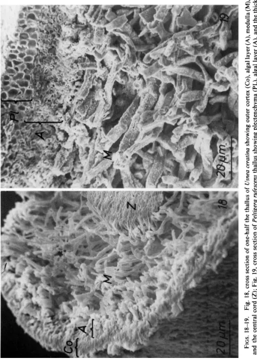
Fungi and algae are either scattered regularly in a thallus (homoiomerous lichen) or they are restricted to special layers (heteromerous lichen). Study- ing this arrangement with the light microscope may be difficult because of the thin sections needed to obtain good resolution. In such sections the symbionts tend to loosen so that their natural arrangement is no longer evident. With the SEM such difficulties are overcome and the real distribu- tion of the symbionts can be observed (Figs. 18 and 19). A disadvantage of this method is that the protoplasts are not preserved so that details of the symbiont cells are not revealed.
Particularly, the SEM gives an excellent view of hyphal distribution. The cortex can be discerned as a plectenchyma (Fig. 19) or as hyphae embedded in a thick gelatinous substance (compare Figs. 13 and 18). Hyphal orienta- tion in the medulla is clearly evident. Numerous small and tightly inter- woven hyphae can be seen as well as hyphae which are thicker in diameter but fewer in number. Within the thalli the distribution of crystals of lichen acids also can be studied with the SEM. The crystals are attached directly to the walls (Figs. 26 and 28) or they are scattered between the symbionts (Fig. 27).
B. Surface Structures
The surface of a lichen thallus consists of the cortical layer and structures which appear above the cortex as asexual and reproductive structures. They are best visible on air-dried thalli. Water-saturated lichens have uniformly swollen surfaces and structural details after freeze-drying are observable only in some soaked objects (Peveling and Vahl, 1968).
1. THE CORTICAL SURFACE
The lichen surface varies from smooth to roughly sculptured (Peveling, 1970a). In the extremly smooth surface not even the orientation of the corti- cal hyphae can be made out (Fig. 20). Only occasionally do nodulelike
172 ELISABETH PEVELING
FIGS. 18-19. Fig. 18, cross section of one-half the thallus of Usnea ceratina showing outer cortex (Co), algal layer (A), medulla (M), and the central cord (Z); Fig. 19, cross section of Peltigera rufescens thallus showing plectenchyma (PL), algal layer (A), and the thick interwoven medullary hyphae (M). Material air-dried.
cortex of Roccella sp.; Fig. 2 3 , furrows and ridges in the thallus of Collema tenax. Material air-dried.
FIGS. 24-29. Crystals of lichenic acids and other hyphal adherents. Fig. 24, rhombic and cubic-shaped crystals on the surface of Physcia dimidiata; Fig. 25, sticklike crystals embedded in the uppermost cortical layer of Xanthoria parietina; Fig. 26, small flake-shaped crystals on the medullary hyphae in Peltigera rufescens; Fig. 27, needle-shaped crystals between
elevations appear. Moreover, very small apertures are scattered in varying distance all over such a thallus. In some lichens which have a thick external homogeneous layer the direction of the cortical hyphae is still evident. A parallel orientation of the hyphae to the thallus surface is seen typically in the beard lichens where the hyphae also run parallel to the long axis of the thallus (Fig. 21). Apertures of round or elliptical shape occur between the visible hyphae in the thallus surface. Hyphae parallel to the thallus surface were clearly demonstrated in representatives of Cornicularia and Alectoria
(Hawksworth, 1969).
There are hyphae which are arranged perpendicular to the surface (Fig.
22). This type of orientation can be seen with the light microscope, but to bring out all the particles and their distribution between the ends of the hyphae higher magnification is needed.
Finally, there are lichens which do not have a typical cortex. If such a thallus is dry its surface shows alternating furrows and ridges (Fig. 23).
2. ADHERENTS TO THE CORTICAL SURFACE
Crystals of lichen acids which may be rhombic, cubic, flake, or needle- shaped (Figs. 24-28) cover parts of the thallus or the whole surface. These crystals may be scattered on the surface (Fig. 24) or embedded in the upper- most homogeneous cortical layer (Fig. 25). In addition to the crystals of lichen acids, threadlike forms occur occasionally (Fig. 29). They seem twisted and differ distinctly from crystals.
Tiny hairs occur at the brink of a thallus in some lichens. They are very often overlooked and only the SEM shows that they consist of several hyphae which form this brittle structure. At the end of such a hair several hyphae form a sharp tip while along its length short, single hyphal ends come out from the main structure (Fig. 30).
The other surface structures such as cyphellae, cephalodia, isidia, soredia, pycnidia, and apothecia have not been explored in detail by the SEM. One illustration of isidia is seen in Fig. 31.
C Rhizines
Lichens are attached to their substrate by different means. The most common structures of attachment are the rhizines, which are conglomerates of fungal hyphae. On the basis of observations with the SEM, three main forms of rhizines can be described.
hyphae of a soralium in Cetraria pinastri; Fig. 28, flake-shaped crystals between medullary hyphae of Parmelia carporrhizans; Fig. 29, threadlike adherents to hyphae in the uppermost cortex of Collema tenax. Material air-dried.
Figs. 3 0 - 3 1 . Fig. 30, small brittle hairs at the end of the thallus of Anaptychia ulotrichoides;
Fig. 31, isidia on the thallus of Pseudevernia furfuracea. Material air-dried.
The first type are those which grow out of the lower cortex and form a tip (Fig. 32). These rhizines become columnlike but still show a tip at their end. After touching the ground the tip enlarges to a footlike structure which consists of very homogeneous material and is adapted exactly to the ground (Fig. 33). The footlike structure of several rhizines can fuse. Such rhizines are especially common in Parmelia.
A second type of rhizine is formed by bundles of hyphae which are not closely connected. Single hyphae in these bundles can be recognized and at the end of such a bundle, where it touches the ground, the hyphae radiate outward (Figs. 34 and 35). Cross sections of such rhizines reveal that those which are loosely organized have hyphae with thick hyphal walls while the tightly formed rhizines have hyphae with thin walls and between them are thick layers of a homogenous substance (Reznik et al, 1968).
A third type of rhizine is that which appears as a single structure but consists of several hyphae (Fig. 36). The surface of these rhizines is smooth so that they appear as a single structure. At their ends these rhizines can be bifurcated.
V. Some Aspects of Changes in Ultrastructure Due to Ecological Conditions Lichens as well as higher plants show different ecotypes within one species. The main factors which produce ecotypes are climate and substrate.
Ecotypes can differ in their morphological appearance and show specific physiological characteristics.
Concerning lichens, it is of special interest if ecotypes are characterized by different forms in their symbiotic relationship. Ben-Shaul et al (1969) observed ecotypes of Caloplaca aurantia var. aurantia from the Mediter- ranean seashore, a mountainous region in the Mediterranean, and from desert locations in the Negev. In the specimens from the Mediterranean area fungi and algae were in close contact, but the authors could not observe any penetration of algal cells by haustoria. Lichens from the desert, however, did have algal cells penetrated by haustoria. Similar results were obtained with Lecanora radiosa (Galun et al, 1970c). In thalli collected in the moder- ate climate of the Mediterranean region haustoria were found only in senes- cing and already disorganized algal cells and they were rare. Thalli from the desert showed many haustoria in senescing as well as in young algal cells.
In two Gonohymenia lichens, one from a xeric habitat and the other from a moderate climate, no differences in the fungus-alga relationship were found (Paran et al, 1971).
The reported studies were done with material which was soaked for 2-10 days. Therefore, the question may be raised if under natural environ-
FIGS. 3 2 - 3 6 . Different forms of rhizines. Fig. 3 2 , young rhizines on the lower cortex of Parmelia caperata; Fig. 3 3 , an older enlarged tip of a rhizine of Parmelia caperata; Fig. 3 4 , view of the end of a hyphal bundle from a rhizine of Lobaria pulmonaria; Fig. 3 5 , rhizines
mental conditions the same differences are present. It is known that increas
ing humidity quickly stimulates photosynthetic and respiratory activity (Lange, 1969a,b) as well as starch production (Brown and Wilson, 1968;
Peveling, 1970b) and that the development of haustoria depends on the condition of a thallus (Tschermak, 1941).
VI. Conclusions
The recent studies on lichens with the scanning and transmission elec
tron microscopes have revealed new and interesting structures in the symbiotic algae and fungi. These protoplasmic structures, besides providing new aspects of micromorphology, are of special interest because they are not present in the free-living counterparts of the lichen symbionts. These struc
tures are considered to be important for the metabolism of the thalli. In the future, specific labeling of substances followed by electron microscopic autoradiography should reveal the exact mechanism of the cellular and intercellular flow of metabolic products. These studies should lead us to a better understanding of the physiology of lichens and will help to explain the unique characteristics of these mutualistic associations.
Acknowledgment
Acknowledgment is made to the Deutsche Forschungsgemeinschaft for support of this work.
References
Ahmadjian, V. (1960). Some new and interesting species of Trebouxia, a genus of lichenized algae. Amer. J. Bot. 47, 677-683.
Ahmadjian, V. (1966). Lichens. In "Symbiosis" (M. Henry, ed.), Vol. 1, pp. 35-97. Academic Press, N e w York.
Ahmadjian, V. (1967). "The Lichen Symbiosis." Ginn (Blaisdell), Boston, Massachusetts.
Ahmadjian, V., and Jacobs, J. B. (1970). The ultrastructure of lichens. III. Endocarpon pusil- lum. Lichenologist 4, 268-270.
Bednar, T. W., and Juniper, Β. E. (1964). Microfibrillar structure in the fungal portions of the lichen Xanthoria parietina (L.) Th. Fr. Exp. Cell Res. 36, 680-683.
Ben-Shaul, Υ., Paran, N., and Galun, M. (1969). The ultrastructure of the association between phycobiont and mycobiont in three ecotypes of the lichen Caloplaca aurantia var.
aurantia. J. Microsc. (Paris) 8, 415-422.
made up of hyphal bundles on the lower cortex of Lobaria pulmonaria; Fig. 36, rhizines of Umbilicaria crustulosa. Material air-dried.