New cellular level effects of vitamin D in healthy and pathological tissues
Ph.D. Thesis
Péter Horváth
Semmelweis University
Clinical Medicine Doctoral School
Supervisor: Dr. Lakatos Péter András, DSc.
Opponents: Dr. Bittner Nóra, PhD.
Dr. Reusz György, DSc.
Chairman of Final Exam Comittee: Dr. Gerő László, DSc.
Members of Final Exam Comittee: Dr. Szalai Csaba, DSc.
Dr. Kiss Csaba, PhD.
Budapest
2017
2 Introduction
Structure and effects of vitamin D, vitamin D deficiency
Cholecalciferol is a ring structured, lipophilic molecule. As synthesis of the compound could happen entirely in the human body the classification „vitamin” is not accurate, some scholars identify it as a hormone. Synthesis starts in the skin from 7-dehydrocholesterol (7-DHC). UVB radiation transforms 7-DHC into pre-vitamin D3. This reaction takes place in the epidermal layers of the skin, especially in the stratum basale and stratum spinosum. After that pre-vitamin D3 turns into 25-hydroxi-cholecalciferol. This reaction is catalyzed by CYP27A1 enzyme in the liver. Serum 25-OH-D3 becomes active vitamin D3 after hydroxylation by CY27B1.1,25-OH-D3 exerts its effect by binding to VDR (Vitamin D receptor) in the cell nucleus. Inactivation happens by CYP24A1, which turns the active vitamin D3 into calcitrol acid, which is excreted with urine through the kidneys.
Vitamin D deficiency is a widespread phenomenon. In Hungary during winter season the proportion of people with vitamin D deficiency is as high as 70 percent, and this figure is not substantially decreased during summer. Sunscreens, air pollutions and windows sift the UVB component of solar radiation. A well-known consequence of vitamin D deficiency is the decreased mineralization of bones. However, vitamin D deficiency has an established role in extra skeletal diseases such as several tumors, cardiovascular diseases and infections. Indian surveys show that vitamin D deficiency has an important role in the comorbidities of the local population.
The enzyme CYP27B1 is responsible for the synthesis of the active metabolite of vitamin D3, 1α, 25-dihydroxi-cholecalciferol. The enzyme hydroxylates 25-Oh-D3 in the 1α position. PTH and calcitonin increase the enzyme’s expression while active vitamin D3 decreases it through a negative feedback loop.
CYP24A1 is a member of the CYP450 family of enzymes. These enzymes have a hem- domain, and they play a role in the oxidation of organic compounds. They are very important in cell metabolism from steroid synthesis to drug metabolism. They are monooxidases, they bind one O atom to their target molecule, while the other O atom turns into water.
3 Vitamin D and WNT signaling in bone metabolism
The most important link between vitamin D and the WNT pathway is the Dickkopf protein (DKK). Vitamin D causes a substantial increase in the level of DKK2 in normoxic and hypoxic environment, however in differentiating osteoblasts, the vitamin decreases DKK2 levels. These results suggest that there are complex and not entirely understood interactions between the two pathways. The WNT pathway might be important in the early differentiation of osteoblasts, while vitamin D is important in the formation of the inorganic extracellular matrix.
Vitamin D and tumorigenesis
Several studies proved the anti-tumor effect of vitamin D. The serum level of the vitamin shows an inverse correlation with the prevalence of several tumors. The proportion of patients with malignant diseases increases from the equator to the poles, therefore increases by the decrease of sunny hours. This observation suggests some correlation between sun exposure and cancers which is mediated by vitamin D. The tissue level of vitamin D is also influenced by the local level of the enzymes in the vitamin D metabolic pathways. The main place of vitamin D activation are the kidneys; however, several tissues can produce these enzymes locally. In the colon we can detect both CP27B1 and CYP24A1. CYP27B1 increases while CYP24A1 decreases the local level of active vitamin D3 therefore altering the level of VDR’ substrate and changing local gene expression patterns. In early CRC we can detect an increase in CYP27B1. This is an early anti proliferative response from the tumor tissue. However later this effect fades.
The role of vitamin D metabolism in the evolution of thyroid cancer is not entirely clear.
Sharma et al. showed that in thyroid cancer cell lines the basal level of CYP24A1 is high resulting in a low local D3 level and the lack of growth inhibition. The most sensitive cell line was the papillary thyroid cancer line TPC1. Adding vitamin D3 to TPC1 cells result in a surge of CYP24A1 expression.
Aims
4
In our studies we looked into tumors – as they are increasing in incidence and mortality- and bone metabolic diseases – as they affect most women in post menopause.
We wanted to answer the following questions:
• Is vitamin D capable of cell growth inhibition?
• Are cancer cells able to deflect anti-proliferative effects of vitamin D?
• Can we selectively inhibit the defense of cancer cells without damaging healthy tissue?
• Can we observe the effects in vivo in histologic samples?
• How the expression pattern of CYP27B1 and CYP24A1 change in papillary thyroid cell lines?
• Are there any correlation between CYP24 A1 levels and demographic, histologic and clinical properties of patients?
• How the CYP24A1 expression changes in the functional subgroups of papillary thyroid carcinomas?
• Are there any correlations between vitamin D expression and WNT pathway in bone metabolic disorders?
• Are there any correlations between WNT polymorphisms and serum vitamin D levels?
Methods
Gene expression studies of CYP24A1 and CYP27B1 in papillary thyroid tumors
Study population
The gene expression studies were carried out on specimens from 100 white, Caucasian non-related participants. We collected biopsies from the tumor and the surrounding healthy thyroid tissue. We studied the somatic mutation state of BAF codon 600, HRAS codon 61, KRAS codon 12 and 13, NRAS codon 61, ELE/RET and CCD6/RET rearrangements.
RNA isolation and quantitative real time PCR
5
We had 31 fresh, frozen and 69 formalin fixated paraffin embedded samples. From all the specimen pair we isolated total RNA using Roche High Pure RNA kit (Roche, Indianapolis, IN, USA) following the manufacturer’s protocol. For the FFPE samples we used the Roche High Pure Total RNA isolation kit (Roche, Indianapolis, IN, USA). The quantity and the quality of the RNA was determined using a NanoDrop spectrophotometer (Nanodrop Technologies, Montchanin, DE, USA). 500 ng of RNA were reverse transcribed into cDNA. The expression difference was determined using Taqman based real-time PCR technique. Multiplication of DNA and detection of fluorescence were carried out using an ABI Prism 7500 RealTime PCR system meg (Applied Biosystems, Foster City, CA, USA). We used a 7500 System SDS Software to perform relative quantification. We used a two-fold change as a cut-off between the healthy and tumor samples.
Genomial DNA isolation and analysis of somatic mutations and gene rearrangements
Genomial DNA was isolated using Roche High Pure PCR template preparation. We measured DNA quantity with Qubit dsDNA HS Assay Kit.
To identify the genetic alterations, we used a version of the method described by Nikiforov et al. We identified single nucleotide variations BRAF (rs113488022), NRAS (rs79057879), HRAS (rs28933406), KRAS (rs121913535) using real-time PCR.
The ELE1/RE and the CDC6/RET rearrangements were identified using RT-PCR technique. The probes were designed to recognize the fusion points on the mRNA.
Immunohistochemical detection of CYP24A1 protein
Immunohistochemical detection of the protein was carried out on FFPE samples with purified anti human CYP24A1 rabbit antibodies (Prestige Antibodies, Sigma-Aldrich).
Detection was performed with Novolink polymer kit. We used hematoxylin for nucleus staining. A four-channel, automatic Freedom EVO pipetting system was used for the immunohistochemistry staining (TECAN, Mannerdorf, Switzerland).
Statistical analysis
6
We tested the significance level of relative changes in CYP24A1 and CYP27B1 genes expressions in 100 human papillary tumors vs. their own control thyroid tissues with a nonparametric method, Mann–Whitney U test. Results with a p value of 0.05 or lower were considered statistically significant. Pearson’s correlation coefficient was used to measure correlation between continuous variables (in our work between CYP24A1 expression and age at the time of PTC diagnosis). Four functional subsets of PTC samples have been created for evaluating the rate of CYP24A1 overexpressed, down-regulated and unchanged tumors. The subgroups were defined by the following criteria: (1) the presence of somatic oncogene mutation (BRAF, HRAS, KRAS, NRAS) and/or rearrangement (ELE1/RET, CCDC6/RET); (2) conventional PTC or other PTC histological variants (follicular, Hürthle-cell, tall-cell, encapsulated and microcarcinomas); (3) other thyroid disease is present in addition to PTC (Hashimoto, hypothyroidism, hyperthyroidism); (4) lymph node metastasis and/or vascular invasion are confirmed. The distribution of tumor samples exhibiting increased, decreased or unchanged CYP24A1 expression levels in the four different subsets was analyzed using Chi square test. The univariate Mann–Whitney U test cannot fully recover the information hidden in the data, and so more exhaustive multivariate procedures are called for. We used centered principal components analysis (PCA) to summarize multivariate data structure in terms of a few important and uncorrelated dimensions, called the components.
Each component is extracted such that its share from the total variance is maximized.
Coordinates of observations are obtained from the eigenvectors to show the positions in the component space, whereas coordinates of variables are their correlations with components, multiplied by an arbitrary scale factor to enhance visibility in the diagram.
In the graphical display the variables are emphasized by lines pointing to their positions.
The directions and lengths of lines pointing to positions representing the variables in the diagram are informative on the correlations and relative importance of variables, respectively. The overall picture provided by PCA is also useful in selecting variables that best reflect trends and differences among patient groups. Computations were performed by the SYNTAX 2000 program package.
Vitamin D treatment and CYP24A1 inhibition in vitro on human colorectal cancer cells
7 Compounds
There are two distinct groups of CYP450 inhibitors. One of them are the azol-type inhibitors. These compounds bind to the hem iron of the CYP450 molecules with a N- atom containing heterocyclic ring. The so called non-azole inhibitors exert their effects by forming hydrogen bonds at the active site of the enzyme. Because this is a more formidable mode of action we can achieve better selectivity though the inhibitory effect is inferior to the azol-type inhibitors.
We used nonazol inhibitors as selectivity is a key issue in our study. Most of the compounds were tetralone based chemicals, while a smaller proportion belonged to the sterane family.
The tetralone based compounds were synthesized by condensating 6-metoxi-1-tetralone with different benzaldehydes.
The inhibitors were produced by the Department of Organic Chemistry at the University of Szeged. The compounds were dissolved in dimethyl-sulfoxide (c=10-3 mol/l), and were stored at 4 °C. Right before the experiments the solutions were dissolved in sterile cell culture solution to reach the desired concentration (GIBCO’S OPTI-MEM).
Cell line
We used the human derived CACO-2 cell line in our experiments. The cell line was provided by the ATCC (American Type Culture Collection, Manassas Virginia, USA).
Cells were passaged after reaching a 90% confluence. Cells were cultured on 96 well plates. 24 hours before the experiments the cell supernatant was changed from DMEM to GIBCO’S OPTI-MEM.
Cell proliferation assays
Our initial aim was to select the compounds from our pool that can decrease the number of viable cells in the presence of vitamin D but not without it. For this purpose, we utilized Sulforhodamine-B dying, which is a cheap and reliable method. The principle of the test is to dye the protein content of the cells with SRB. After resolving the protein dye, we
8
can semiquantitatively measure the amount of proteins using opticodensitometry, which is in a linear relationship with the number of viable cells. The bound protein dye was resolved in Trisma-Sol and measured on 520 nm on an ELISA plate reader (ThermoFisher Luminoskan Ascent, Waltham, MA, USA).
This way we could semiquantitatively measure whether the compound decreases the number of viable cells in the presence of Vitamin D (100 nmol/l).
Cytotoxicity was measured by quantifying LDH activity in the cell supernatant. The materials needed for the measurement were available through Cytotoxicity Detection KitPLUS (LDH) (Roche, Indianapolis, IN, USA). We followed the manufacturer’s protocol.
To determine whether the effect was cytostatic, we measured 5-BrDU incorporation. We used the Cell Proliferation ELISA, BrdU (colorimetric) kit (Roche, Indianapolis, IN, USA). We followed the manufacturer’s protocol.
RNA isolation and quantification
We isolated total RNA using Roche High Pure RNA kit (Roche, Indianapolis, IN, USA) following the manufacturer’s protocol. The expression difference was determined using Taqman based real-time PCR technique. Multiplication of DNA and detection of fluorescence were carried out using an ABI Prism 7500 RealTime PCR system (Applied Biosystems, Foster City, CA, USA). We used a 7500 System SDS Software to perform relative quantification. All measurements were carried out in duplicates.
Statistics
We compared the different groups using ANOVA testing, we chose Tukey HSD test for post hoc testing. We used SPSS 18 for Windows.
WNT signaling in Vitamin D and bone metabolism
Study population
9
We recruited 932 non-related postmenopausal women. Inclusion criteria were as follows:
Age more than 40 years, no menstruation for at least one year. We measured height, weight and age of subjects. We measured total hip and spine (L2-L4) BMD values using Lunar Prodigy DXA scanner (GE Healthcare, Little Chalfont, UK) then calculated T- score values. Patients were assigned to three groups based on the recommendations of WHO. We classified non- vertebral osteoporotic fractures as ones caused by low trauma after the age of 40 and do not affect the skull, hands, toes or spine. In this study we did not include compression vertebral fractures. We measured vitamin D levels in a subgroup of patients (146 subjects). All participants gave written consent.
SNP selection
We used publicly available online databases (http://genome.ucsc.edu/, http://www.genome.gov/gwastudies/, http://www.ncbi.nlm.nih.gov/omim) to select SNPs of the following genes: LRP5, GPR177, SP7.
Nine SNPs were chosen based on their function or previous appearance in GWAS studies.
Based on the first criteria, missense variations were chosen which cause a change in the amino acid sequence of the encoded protein. Regarding the second criteria, we chose SNPs that have shown a p value less than or equal to 5 x 10⁻⁸.
Genotyping
We collected genomic DNA from the patients’ buccal mucosa, brushing off the superficial layer of cells lining the oral cavity. DNA was extracted using High Pure PCR Template Purification kit (Roche Diagnostics, GmbH, Mannheim, Germany), and genotyping was performed on a Sequenom MassARRAY Analyzer 4 (Sequenom, San Diego, CA, USA).
Statistical analysis
The subjects were assigned to different groups based on the genotyping results, meaning that 3 groups (homozygote recessive, homozygote dominant and heterozygote) were created for each SNP. We utilized Analysis of Covariance (ANCOVA) to test the
10
different genotypes against each other, and Bonferroni method was used to adjust for multiple testing. All tests were performed using SPSS 21 (SPSS Inc., Chicago, Illinois, USA). Linkage disequilibrium plots based on our own data were created using HaploView 4.0 (Broad Institute, Cambridge, Massachusetts, USA). Interactions of different genes on phenotype were calculated with SNPassoc [24], haplotype analyses were carried out using haplo.stats. Both are R packages (R Foundation for Statistical Computing, Vienna, Austria). We chose an alpha value of 0.05.
Results
Changes of CYP241 and CYP27B1 expression in tumor tissues compared with healthy control
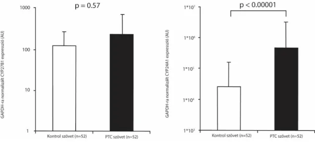
CYP24A1 expression significantly elevated in 52 patients with papillary carcinoma compared to tumor free thyroid gland tissue. There were tumor samples with a thousand-fold increase in CYP24A1 expression. The relative increase of CYP24A1 expression was significantly elevated, the p-value was less than 0.00001. There was no significant difference in the expression pattern of CYP27B1 in our samples (Fig. 1.).
CYP24A1 expression using immunohistochemistry
Fig. 1: Difference in CYP27B1 (left) and CYP24A1 (right) expression in control tissues and tumors. There is a significant increase in CYP24A1 expression, while CYP27B1 expression does not change.
11
In samples with detectable CYP24A1 mRNA we performed immunohistochemistry.
Tumor tissue showed strong staining compared to the surrounding healthy tissue in cases with elevated CYP24A1 mRNA levels.
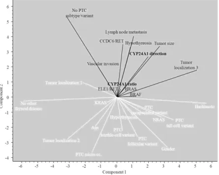
The PCA diagram shows the connection between the 26 studied demographic, clinical, histologic and genetic parameters included in our study of the 86 PTC samples (Fig. 2.).
CYP24A1 inhibition a CRC cell growth
Changes in CYP24A1 expression after vitamin D treatment on CACO2 cell
There was a 6-fold increase in CYP24A1 mRNA expression after short incubation. We could detect the increase 30 minutes after incubation which reached its peak after 12-16 hours after the addition of vitamin D. 4 hours after the incubation started the mRNA
Fig. 2: Ordination diagram of principal component analysis (PCA). Variables shown in black are associated with CYP24A1 exoression. We found a correlation between CYP24 A1 expression and clinical parameters of malignity (vascularization, metastases, tumor size), ELE1/RET, HRAS, BRAF mutations and classical PTC histology
Fig. 3: CYP24A1 mRNA levels in CACO2 cells after addition of vitamin D. The expression of CYP24A1 mRNA proportionately increaes with vitamin D
concentration.
12
levels of CYP24A1 were 311,405 and 612,801 times higher than in controls when 1 nmol/l and 10 nmol/l vitamin D were added respectively (Fig. 3).
Effect of tetralone derivatives and the CYP24A1 inhibiting KD35 on CACO2 cell line
Our preliminary test showed that some of the tetralone derivative compounds can inhibit the decrease the number of CACO2 cells in the presence of vitamin D. These compounds were applied to the cell cultures in different concentrations (1 nmol/l to 10 umol/l) for different incubation periods (1 to 4 days). We chose the compound KD35 for the subsequent studies.
Measuring cell viability using SRB dying
0 100000 200000 300000 400000 500000 600000 700000 800000
0 nM 1,25-D3 1 nM 1,25-D3 10 nM 1,25-D3
CYP24A1 mRNA level (arb. unit)
KD35 0 umol/l KD35 2umol/l
13
CACO2 cells were incubated for 4 days with 100 nmol/l vitamin D in the presence of 0.1, 0.3, 1,0 ill. 3,0 umol/l KD-35. After the treatment we observed a decrease in SRB incorporation proportionate to the concentration of KD-35 compared to cells treated with vitamin D only (2.17, 5.07, 6.18 and 10.93% decrease in viable cells). (Fig. 4.)
Results of cytotoxicity test
To determine the nature of cell decrease we performed cell proliferation testing. We measured the concentration of LDH in the cell supernatant No changes were detected in the cell supernatant of cells treated with KD-35 compared to controls treated with vitamin D in any setup. These results suggest that the nature of cell decrease is antiproliferative rather than cytotoxic.
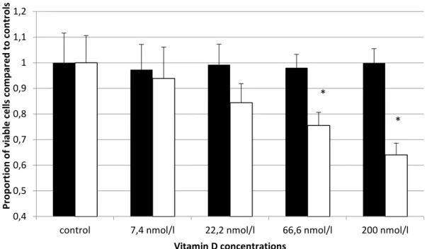
Results of cell proliferation assay
We used the following vitamin D concentrations on the CACO2 cell line with 2 umol/l KD35 concentration: 7,4; 22,2; 66,6 and 200 nmol/l. Control cells were treated with KD-
0,8 0,85 0,9 0,95 1 1,05
control 0,1 umol/l 0,3 umol/l 1 umol/l 3umol/l Proportion of viable cells compared to controls
KD-35 concentration KD-35 KD-35 + 100 nmol 1,25-D3
*
*
Fig. 4: Results of SRB toxicity test. There was a concentration dependent decrease in the number of viable cells in the presence of KD-35 and vitamin D. KD-35 alone did not change cell number.
14
35 only. We incubated cells for 4 days. After that we labeled the cells with BrDU for 2 hours. The relative decrease in cell proliferation were 3,43, 14,81, 22,49, and 35,81%.
The decrease was proportionate with the increasing concentration of vitamin D. (Fig. 5.)
There was no significant change in mRNA level of CYP24A1 in the cells treated with KD35 compared to the cells treated with only vitamin D. KD35 did not change the expression of CYP24A1. In the presence of vitamin D, the expression of CYP24A1 sharply increased. The level of CYP24A1 did not depend on the concentration of KD35 or incubation period.
Role of WNT pathway in the bone metabolism of postmenopausal women
We found that SNPs rs4988300 and rs64008 showed a correlation with total hip BMD of postmenopausal women. Effect of rs4988300 was still significant after Bonferroni correction. Women who carry the heterozygous phenotype of this SNP had significantly
0,4 0,5 0,6 0,7 0,8 0,9 1 1,1 1,2
control 7,4 nmol/l 22,2 nmol/l 66,6 nmol/l 200 nmol/l
Proportion of viable cells compared to controls
Vitamin D concentrations 1,25-D3 only 2 uM KD-35 + 1,25-D3
*
*
Fig. 5.: Proportion of viable cells after BrDU incorporationKD-35 decreases the number of viable cells in a concentration dependent manner. Above 66.6 nmol/l the decrease is significant.. (p<0,05)
15
higher total hip BMD. We could not find any significant relationships between osteoporotic fractures and LRP5 SNP genotypes.
Conclusions
Our studies conclude that the vitamin D metabolizing CYP24A1 enzyme plays a pivotal role in tumorigenesis. There are several articles in the literature proving a correspondence between low level of vitamin D and the occurrence of lung, prostate, thyroid, gastric and colorectal cancers. These tumors are capable of inactivating vitamin D by overexpressing the CYP24A1 enzyme even with vitamin D levels in the normal range.
We showed that in thyroid cancer patients the level of CYP24A1 shows a correlation with the invasiveness, vascularization and metastizing capabilities of the tumor. There was no correlation with CYP27B1 levels. The kidneys produce an adequate level of active vitamin D, therefore the level of active vitamin D in the tumor tissue does not differ from healthy tissues. Thus, the decreased expression of CYP27B1 would not change the vitamin D milieu of the cells. However increased expression of CYP24A1 lowers the active vitamin D level in the cell, therefore inhibiting the VDR-vitamin D binding As we saw in the introduction, there is a relationship between vitamin D pathway and the WNT pathway through the DKK protein. We investigated whether vitamin D levels show an association with polymorphisms of certain key genes in the WNT pathway, namely SP7, GPR177 and LRP5. We also studied if these polymorphisms affect the BMD and fracture risk of postmenopausal women. We concluded that there is no significant correlation between these polymorphisms and vitamin D levels. However, the rs4988300 polymorphism shows a correlation with total hip BD of postmenopausal women. It is known that this gene has an important role in the remodeling of bone tissue.
Heterozygosity of this polymorphism was associated with significantly higher total hip BMD. A possible explanation could be that this SNP is not responsible for the biologic effect. It could be a tag SNP for another polymorphism in the same haplotype block. This SNP could exert its effect by causing alternative splicing on the intron-exon border modulating the splicing RNAse. This should be a subject of further investigations on this issue.
16
In the light of our results it is obvious that the widespread vitamin D deficiency not only increases the prevalence of osteoporosis, it is an important factor in the evolution of tumors as well. The scientific literature has numerous studies suggesting a role of vitamin D in the regulation of circadian rhythm, in the pathogenesis of cardiovascular diseases and an important immunomodulating role in granulomatous disease. For these reasons it is very important for doctors in hospital care and general practitioners to have a broad understanding of vitamin D metabolism, vitamin D deficiency and the importance of vitamin D supplementation
Index of author’s publications
Publications that form the basis of the thesis
Kósa, J. P., P. Horváth, J. Wölfing, D. Kovács, B. Balla, P. Mátyus, E. Horváth, G. Speer, I. Takács, Z. Nagy, H. Horváth and P. Lakatos (2013). "CYP24A1 inhibition facilitates the anti-tumor effect of vitamin D3 on colorectal cancer cells." World Journal of Gastroenterology 19(17): 2621-2628.
Horvath, P., B. Balla, J. P. Kosa, B. Tobias, B. Szili, G. Kirschner, G. Gyori, K. Kato, P. Lakatos and I. Takacs (2016). "Strong effect of SNP rs4988300 of the LRP5 gene on bone phenotype of Caucasian postmenopausal women." J Bone Miner Metab 34(1): 79- 85.
Balla, B., B. Tobias, J. P. Kosa, J. Podani, P. Horvath, Z. Nagy, J. Horanyi, B. Jaray, E.
Szekely, L. Krenacs, K. Arvai, M. Dank, Z. Putz, B. Szabo, B. Szili, Z. Valkusz, B. Vasas, G. Gyori, P. Lakatos and I. Takacs (2014). "Vitamin D-neutralizing CYP24A1 expression, oncogenic mutation states and histological findings of human papillary thyroid cancer." J Endocrinol Invest.
Publications independent from the thesis
17
Laszlo Kunos, Zsofia Lazar, Fruzsina Martinovszky, Adam D. Tarnoki, David L.
Tarnoki, Daniel Kovacs, Bianka Forgo, Peter Horvath, Gyorgy Losonczy, Andras Bikov: Overnight Changes in Lung Function of Obese Patients with Obstructive Sleep Apnoea. Beiträge zur Klinik der Tuberkulose 10/2016; DOI:10.1007/s00408-016-9957-1
Péter Horváth, Zsófia Lázár, Rita Puskás, Gabriella Gálffy, György Losonczy, László Kunos, András Bikov: The role of the complement system in the pathomechanism of obstructive sleep apnea syndrome. European Respiratory Journal 09/2016; 48(suppl 60).
DOI:10.1183/13993003.congress-2016.PA2297
Zsófia Lázár, Péter Horváth, Rita Puskás, László Kunos, György Losonczy, Gabriella Gálffy, András Bikov: Bronchial and alveolar nitric oxide levels in healthy volunteers and patients with severe asthma. European Respiratory Journal 09/2016; 48(suppl 60).
DOI:10.1183/13993003.congress-2016.PA1070
Gyöngyi Kirschner, Bernadett Balla, János Kósa, Péter Horváth, Andrea Kövesdi, Gergely Lakatos, István Takács, Zsolt Nagy, Bálint Tóbiás, Kristóf Árvai, Péter Lakatos:
Az onkohematológiai betegségek kezelésében használt tirozinkináz-gátló imatinib és nilotinib csonthatásainak irodalmi áttekintése és a saját kutatási eredmények bemutatása.
Orvosi Hetilap 09/2016; 157(36). DOI:10.1556/650.2016.30525
Kristóf Árvai, Péter Horváth, Bernadett Balla, Bálint Tobiás, Karina Kató, Gyöngyi Kirschner, Valéria Klujber, Péter Lakatos, János P. Kósa: Next-generation sequencing of common osteogenesis imperfecta-related genes in clinical practice. Scientific Reports 06/2016; 6. DOI:10.1038/srep28417
Dániel Németh, Kristóf Árvai, Péter Horváth, János Pál Kósa, Bálint Tobiás, Bernadett Balla, Anikó Folhoffer, Anna Krolopp, Péter András Lakatos, Ferenc Szalay: Clinical Use of Next-Generation Sequencing in the Diagnosis of Wilson’s Disease.
Gastroenterology Research and Practice 01/2016; 2016(4). DOI:10.1155/2016/4548039 Bálint Tobiás, Csaba Halászlaki, Bernadett Balla, János P Kósa, Kristóf Árvai, Péter Horváth, István Takács, Zsolt Nagy, Evelin Horváth, János Horányi, Balázs Járay, Eszter Székely, Tamás Székely, Gabriella Győri, Zsuzsanna Putz, Magdolna Dank, Zsuzsanna Valkusz, Béla Vasas, Béla Iványi, Péter Lakatos: Genetic Alterations in Hungarian Patients with Papillary Thyroid Cancer. Pathology & Oncology Research 08/2015; 22(1).
DOI:10.1007/s12253-015-9969-9
18
Balint Tobias, Bernadett Balla, P Janos Kosa, Istvan Takacs, Zsolt Nagy, Peter Horvath, Balazs Jaray, Eszter Szekely, Roland Istok, Tamas Szekely, Peter Lakatos: Detection of somatic oncogene alterations in FNA samples of cold nodules and 3 years follow-up of patients in Hungary. 05/2015; DOI:10.1530/endoabs.37.EP853
Bernadett Balla, Peter Horvath, Balint Tobias, Gabriella Gyori, Balazs Jaray, Janos Kosa, Peter Lakatos: Vitamin-D neutralising CYP24A1 gene expression in thyroid fine- needle aspiration biopsy samples. 05/2015; DOI:10.1530/endoabs.37.EP855
H.N.A. Jozilan, P. Horvath, J.P. Kosa, P. Lakatos, D. Nemeth, J. Wölfling, D. Kovacs, B. Bodnar, P. Matyus, E. Horvath, I. Kovalszky, F. Szalay: P0321: Increased anti-tumor effect of vitamin D after CYP24A1 inhibition on HCC cell lines. Journal of Hepatology 04/2015; 62. DOI:10.1016/S0168-8278(15)30536-5
D. Németh, J. Pál Kósa, K. Árvai, P. Horváth, B. Tobiás, B. Balla, A. Folhoffer, A.
Krolopp, P. András Lakatos, F. Szalay: P1224: Next-generation sequencing for the diagnosis of wilson's disease. Journal of Hepatology 04/2015; 62. DOI:10.1016/S0168- 8278(15)31420-3
Kristóf Arvai, Péter Horváth, Bernadett Balla, Anna M Tőkés, Bálint Tobiás, István Takács, Zsolt Nagy, Péter Lakatos, János P Kósa: Rapid and cost effective screening of breast and ovarian cancer genes using novel sequence capture method in clinical samples. Familial Cancer 05/2014; 13(4). DOI:10.1007/s10689-014-9730-7
D Németh, A Folhoffer, A Krolopp, J Kósa, K Árvai, P Horváth, P Lakatos, Z Gerlei, L Kóbori, D Görög, M Szathmári, F Szalay: New mutation of ATP7B gene detected by Ion Torrent in a Wilson patient with acute on chronic liver failure, transplanted via Eurotransplant. Zeitschrift für Gastroenterologie 05/2014; 52(05). DOI:10.1055/s-0034- 1376107
Bernadett Balla, Kristóf Arvai, Péter Horváth, Bálint Tobiás, István Takács, Zsolt Nagy, Magdolna Dank, György Fekete, János P Kósa, Péter Lakatos: Fast and Robust Next- Generation Sequencing Technique Using Ion Torrent Personal Genome Machine for the Screening of Neurofibromatosis Type 1 (NF1) Gene. Journal of Molecular Neuroscience 03/2014; 53(2). DOI:10.1007/s12031-014-0286-7
19
Balint Tobias, Bernadett Balla, Janos P Kosa, Janos Horanyi, Istvan Takacs, Zsolt Nagy, Peter Horvath, Balazs Jaray, Eszter Szekely, Roland Istok, Tamas Szekely, Peter Lakatos: Detecting somatic oncogene mutations in FNA samples of cold nodules in Hungary. 03/2013; DOI:10.1530/endoabs.32.P1108
Bernadett Balla, Janos Kosa, Balint Tobias, Istvan Takacs, Zsolt Nagy, Peter Horvath, Janos Horanyi, Eszter Szekely, Balazs Jaray, Peter Lakatos: Examination of CYP24A1 and ’three-genes' (SFN, MRC2, HMGA2) expressions in different pathological subgroups of human papillary thyroid cancer. 03/2013; DOI:10.1530/endoabs.32.P1098
P Horváth, JP Kósa, J Wölfling, B Balla, D Kovács, P Mátyus, E Horváth, G Speer, I Takács, Z Nagy, P Lakatos: D-hormon és CYP24A1-gátlás: új megközelítés a colorectalis daganatok kezelésében. Magyar Belorvosi Archivum 01/2011; 64(5).