79
OVERVIEW: THE MACROPHAGE IN CELL BIOLOGY
David Vaux Siamon Gordon
I. INTRODUCTION
Because of its varied function and remarkable adaptabili- ty, the macrophage is not only of interest to the immunologist and pathologist, concerned with its special role in host de- fense and injury, but also to the cell biologist, who can use macrophages to study many processes common to all cells. Since macrophages can be obtained in good yield and purity and since they maintain many of their differentiated properties in cul- ture, a great deal has been learned in the past two decades about their physiology, especially with regard to endocytosis, lysosomal function, and the synthesis and secretion of various biologically active molecules.
In this chapter, we discuss selected aspects of macrophage cell biology to illustrate the versatility of the cell and the ingenuity of some of its investigators. We have chosen to present the spectrum of research with the aid of several sum- mary tables, to conserve space and for ease of reference.
METHODS FOR STUDYING Copyright © 1981 by Academic Press, Inc.
MONONUCLEAR PHAGOCYTES 8 2 1 All rights of reproduction in any form reserved.
ISBN 0-12-044220-5
Earlier reviews provide useful information and several topics are dealt with in greater detail elsewhere in this volume
( 1 - 4 ) .
II. SOURCES AND HETEROGENEITY
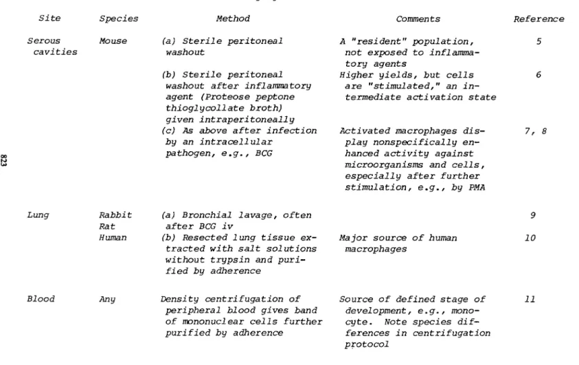
Mononuclear phagocytes can be obtained from various sources for experimental purposes (Table I) . It is important to recog- nize that cells from different species and from different sites in the body differ in development and state of activation.
Heterogeneity of this kind makes it possible to compare the structure and function of closely related types of cell. Al- though cell lines provide a more homogeneous cell population, their origin and stage of differentiation are usually ill- defined and the cells could change their properties after long periods in culture. Primary cells may also show properties reflecting their adaptation to conditions in vitro rather than those within the animal.
III. GROWTH AND DIFFERENTIATION
Like other hemopoietic cells, precursors for macrophages originate in bone marrow in the adult animal and undergo several divisions during differentiation. Cells enter the blood stream as monocytes and are then widely distributed in peripheral tissues, especially the liver (Kupffer cells), lung
(alveolar macrophages), spleen, and serosal cavities. Some of the methods used to study the origin and growth of macrophages are listed in Table II. Development of differentiated mouse macrophages in culture depends on a specific growth factor, colony-stimulating factor(s) (CSF), which is produced by mono- nuclear phagocytes under certain conditions or by other cells such as mouse fibroblasts (26). Methods have been developed for clonal analysis of cell growth in soft agar and for mass culture in liquid systems (see Chapter 2)·
Mouse bone marrow contains only a small proportion of colony-forming precursors (CFU-C), approximately 1 per 1000.
Cell culture with CSF initially favors production of polymor- phonuclear leukocytes that die rapidly, and, subsequently, after 2 - 7 days, of a relatively homogeneous population of adherent macrophages. Such bone marrow cultured macrophages
(BMCM) continue to proliferate for some weeks if the CSF is
renewed.
TABLE I. Sources of Mononuclear Phagocytes Site Species Method Serous Mouse (a) Sterile peritoneal
cavities washout (b) Sterile peritoneal
washout after inflammatory agent (Proteose peptone thioglycoll ate broth)
given intraperitoneally (c) As above after infection
by an intracellular pathogen, e.g., BCG
Lung Rabbit (a) Bronchial lavage, often Rat after BCG iv
Human (b) Resected lung tissue ex- tracted with salt solutions without trypsin and puri-
fied by adherence
Blood Any Density centrifugation of peripheral blood gives band of mononuclear cells further purified by adherence
Comments Reference A "resident" population,
not exposed to inflamma- tory agents
Higher yields, but cells are "stimulated," an in-
termediate activation state
Activated macrophages dis- 7,8 play nonspecifically en-
hanced activity against microorganisms and cells, especially after further stimulation, e.g., by PMA
9
Major source of human 10 macrophages
Source of defined stage of 11 development, e.g., mono-
cyte. Note species dif-
ferences in centrifugation protocol
matous organs Bone
marrow
Mouse Rat Human
fied by adherence
Sterile washout of bone marrow cultured with source of CSF
00
Cell Mouse In vitro or in vivo passage lines of transformed cell lines
carrying macrophage markers
Good yields of proliferating 13 cells. Differentiation
series present in culture.
Population initially hetero- geneous
High yields of homogeneous 14 population, but macrophage
status may be uncertain.
Viral antigens present
X. MONONUCLEAR PHAGOCYTES AS TOOLS IN CELL BIOLOGY
825 TABLE II. Macrophage Precursors and Growth
Method of study Purpose In Vivo
Parabiosis
Adoptive transfer into syngeneic, irradiated recipient using chromo- somal marker; colony formation in spleen by limiting dilution
Reference
Hematogenous origin of tissue macro- phages. Bone marrow origin of stem cell precursor for tissue macrophages and other hemopoietic cells
(CFU-S).
15 16 17
In Vitro
Cloning in suspension in a semisolid medium, in liquid culture or after adherence, all in presence of CSF
Mass liquid culture of bone marrow, in pres- ence of CSF
Mass liquid cultures of macrophages
Culture of bone marrow with a bone marrow derived feeder system, under special condi- tions, without exo- genous CSF
Assay CFU-C and pro- liférât ive capacity of precursors in bone marrow and of tissue and exudate macrophages.
DNA synthesis and proliferation in macrophages gener- ated in culture.
DNA synthesis and proliferation.
Cycling of CFU-C in vitro, without dif- ferentiation.
18 19 20
13 21
22, 23
24, 25
Although tissue macrophages are typically in the resting (GQ) phase of the cell cycle, up to 20% of macrophages ob- tained from inflammatory exudates, e.g., after stimulation with thioglycollate broth, give rise to colonies in agar in the presence of CSF, after a lag period of up to 4 weeks (20).
In general, the speed and extent of macrophage proliferation diminish as the cells mature from precursor to end cell.
CSF has been purified considerably, partially character-
ized as a glycoprotein approximately 70,000 MW and is composed
of two identical subunits (27). The effect of CSF is mostly
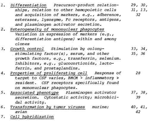
TABLE III. Studies with Primary Cultures of Proliferating Macrophages
Purpose of study Reference 1. Differentiation Precursor-product relation- 29, 30,
ships, relation to other hemopoietic cells 31, 13, and acquisition of markers, e.g., adherence, 32 esterases, lysozyme, Fc receptors, antigens,
and plasminogen activator secretion.
2. Heterogeneity of mononuclear phagocytes Variation in expression of markers (e.g., differentiation antigens) within and among clones
3. Growth control Stimulation by colony- 33, 34,
„stimulating factor(s), serum, and other 35, 36 growth factors, e.g., transferrin, seleniurn.
Inhibitors, e.g., glucocorticoids, lacto- ferrin, and prostaglandins.
4. Properties of proliferating cell Response of 28 target to CSF varies, BMCM > inflammatory >
resident. CSF receptors specifically found on mononuclear phagocytes.
5. Associated phenotype Plasminogen activator 37, 38, secretion. Cytostatic activity ; microbici- 39 dal activity.
6. Transformation by tumor viruses murine; 40, 41,
avian 42 7. Cell hybridization
restricted to target cells of the same species, and a specific receptor for CSF has been identified on cells of the mononuclear phagocyte system (28).
Dexter and his colleages have developed a specialized cul- ture system, not completely defined, to maintain some precursor cells in a cycling state in culture, without differentiation.
CSF is not added and adherent cells, often laden with lipid, perform a poorly understood accessory function (25) . Such ac- cessory cells can be obtained from mouse bone marrow by culti- vation at a lower temperature, 33°C, or by using horse serum supplemented with glucocorticoids.
As shown in Table III, these culture systems have been used
to define the stages of macrophage differentiation and the ac-
quisition of cell-specific markers in relation to other hemo-
poietic cells. A question that has not been resolved concerns
the nature of the heterogeneity observed among different mono-
nuclear phagocytes. Variation in expression of various markers
X. MONONUCLEAR PHAGOCYTES AS TOOLS IN CELL BIOLOGY
827 can be ascribed to differences in the stage of development and growth or in the degree of cell activation by various inflam- matory, immunologie, and endocytic stimuli. The role of un-
usual factors in the local environment, e.g., in the lung, has not been defined. Although no evidence for independent subsets of mononuclear phagocytes has yet been obtained, analysis of markers such as monoclonal antigens within and among indepen- dent clones of macrophages derived from bone marrow or tissue sources will provide a powerful test for their existence.
An unusual feature of proliferation and its control in the MPS is the ability of immature precursors to form colonies in semisolid medium, a trait often associated with malignancy in the case of other cells. Maturation of mononuclear phagocytes results in loss of this property. Unlike the growth of malig- nant cells, growth of macrophage precursors is tightly con- trolled and dependent on the continuous presence of CSF. Other molecules have been reported, which regulate growth when CSF is not limiting.
Very little is known about the properties that determine the response of a macrophage to CSF or about the complex phenotype associated with proliferation. BMCM are exquisitely sensitive to CSF compared with resident macrophages. Produc- tion and secretion of plasminogen activator are coordinated with cell proliferation and CSF is able to enhance both activi- ties in close sequence (37). Microbicidal and cytocidal activi- ties, other characteristic effector functions of activated macrophages, may be induced at the same time.
Two limitations to the present use of bone marrow cultures should be noted. The preadherent precursors of macrophages are poorly characterized and present in small numbers in the heterogeneous cell populations found early in culture. More- over, differentiation in vitro may differ from that in vivo, with the role of CSF or surface adherence unlike that of héma- tologie and local influences in the body.
IV. STRUCTURE AND FUNCTION OF MACROPHAGE MEMBRANES
The membranes of the macrophage, both surface and intra- cellular, are of central interest because so many of the specialized functions of macrophages are membrane mediated.
These include all types of endocytosis, associated delivery of
vesicles to the lysosomal compartment by a specific intracellu-
lar membrane fusion, and possible retrieval and redeployment of
any receptors involved. Exocytosis is another important mem-
brane-dependent event and intracellular membranes are important
in the complex oxidative killing systems delivering high-energy
TABLE IV. Plasma Membrane Components of the Macrophage
Class Example Characteristics Reference
"Receptor" Fc receptor
C3b recep- tor
"Denatured surface receptor"
Mannosyl/
fucosyl terminal receptor
ot,2~Macro- globulin receptor Lymphokine
receptor CSF receptor
Receptor molecules specific 1, 2, for the Fc portion of some 44, 45 subclasses of immunoglobu-
lin G. Trypsin-resistant and trypsin-sensitive re-
ceptors have been des- cribed. Mediate phagocy- tosis of antibody-coated particles and antigen-
antibody complexes
Receptor for cleaved third 2 component of complement
cascade, mediates phagocy- tosis of IgM-coated par- ticles in the presence of complement. Trypsin sensi- tive.
Immunologically nonspecific 46, 47
"receptors" recognizing chemically modified bio- logical particulates and nonphysiological stimuli, e.g., oil red 0 droplets or latex microspheres.
Pinocytosis-mediating re- 48, 49, ceptor-binding mannose or 50, 51 fucose terminated glyco-
proteins (including various lysosomal glycosidases).
Requires Ca
2+for binding.
Trypsin sensitive. May function as retrieval path- way for lysosomal glycopro- teins and may also be in- volved in clearance of antigen IgM antibody com- plexes.
Mediates clearance of 52 protease complexes.
Reported to be blocked by 53 fucose in guinea pig
macrophages.
Restricted to phagocytic 28
cells and their precursors
X. MONONUCLEAR PHAGOCYTES AS TOOLS IN CELL BIOLOGY 829
Table IV (Cont'd.)
Class Example Enzymes ATPase
5'Nucleo- tidase
Alkaline phospho- diester- ase I
Protein kinase
Characteristics Reference Macrophages are rich in a 54
divalent cation-dependent ATPase activity of unknown function.
Plasma membrane marker 55 [although also present on
some intracellular mem- branes in polymorphs (87)]
which is internalized during phagocytosis.
Function unknown, but present in much higher ac- tivity on resident peri- toneal macrophages than thioglycollate elicited macrophages.
Macrophages carry a mem- 56 brane-associated activity,
which liberates 5'nucleo- side monophosphates from polyribonucleotides with free 3'-OH. Most activity on plasma membrane, but
significant intracellular activity representing either a second site of localization or internali- zation of plasma membrane.
Reduced half-life for this activity in activated cells suggests the latter, show- ing endogenous pinocytic rate as an important de- terminant of the enzyme turnover.
Plasma membrane associated 57 activity capable of trans-
ferring y-phosphate of ATP
to specific membrane com-
ponents or exogenous accep-
tor has been described in
guinea pig peritoneal macro-
phages. Such selective
phosphorylation may be
fundamental in control of
Table IV (Cont'd.)
Class Example Characteristics Reference
Specific adherence compo- nents
Antigens
MHCla
Mac I
F4.80
Lipids
membrane functions.
Macrophages are highly 58, 58a adhesive to glass and
culture plastic, spread- ing widely over the sur- face and exhibiting mem- brane ruffling. Mouse peritoneal macrophages express a trypsin-resis- tant surface iodinatable 195,000 Mr component that appears with development of adhesive phenotype in culture. Human mono- cytes express surface re- ceptors for fibronectin.
Antigens coded by the 59 major histocompatibility
complex are present on the macrophage.
The presence and possible 60, 61 functional significance
of la and DRw antigens on the macrophage mem- brane remain controver- sial
Monoclonal antibodies are 62, 63 now being used to probe
the macrophage membrane, no function has been assigned to the antigens recognized by these an- tibodies, but their al-
most complete restriction to cells of the mono-
nuclear phagocyte series suggests the possibility of interesting specialized functions.
The phospholipid composi- 64 tion of the macrophage
plasma membrane may be modified by culturing
cells in serum-free medium
containing fatty acid-
X. MONONUCLEAR PHAGOCYTES AS TOOLS IN CELL BIOLOGY 831
Table IV (Cont'd.)
Class Example Characteristics Reference bovine serum albumin.
Analysis reveals in- crease in ratio of saturated/unsaturated
fatty acid and reduction in both phagocytic and pinocytic activity.
oxygen metabolites to the lumen of the phagosome without caus- ing damage to the cell.
This breadth of membrane activity is both an important reason for using macrophages to study problems in membrane biology and the biggest drawback that hinders such work. In addition to the common elements such as plasma membrane, nu- clear membrane, rough and smooth endoplasmic reticulum, Golgi and mitochondrial membrane, macrophages contain large popula- tions of lysosomes, secretory vesicles, phagosomes, and pino- somes. This has meant that it is very difficult to isolate in- dividual types of membrane for biochemical characterization.
It is partly for this reason that the modification of the technique of Wetzel and Korn (43) for flotation of latex-filled phagolysosomes on discontinuous sucrose density gradients dominate several areas of membrane research.
The composition of the various membrane compartments of the macrophage remains largely unknown, although a wide variety of plasma membrane components has been described (Table IV) and some differences have been noted between plasma membrane and phagolysosomal membrane. It is to be expected that the compo- sition of the plasma membrane will be further complicated by changes due to differentiation and activation of the cell.
The composition of intracellular membranes may be substantially different again, and the plasma membrane interiorized in endo- cytosis could represent an intermediate state. Studies in the fibroblast have revealed plasma membrane specializations in- volved in endocytosis, which take the form of areas of high receptor density in the membrane with a web of filaments be- neath (65). It may be that lateral differentiations in the plasma membrane are widespread, and experiments aimed at demonstrating selective internalization of plasma membrane components during endocytosis could be the first to show this
(see Selective internalization, Table VI).
It is a characteristic of the mature macrophage to be
highly adherent to tissue culture plastic and glassware.
Fibroblast adherence appears to be dependent on fibronectin (cell adherence substance, LETS) on the cell surface. Recently, it has been established that macrophages synthesize fibronectin
(66) and express receptors for fibronectin on the cell surface (58a). Other components of the plasma membrane have been impli- cated in the adhesion process in macrophages (see Table IV).
Among the components described on the plasma membrane of the macrophages are a variety of receptors for specific ligands mediating their endocytic uptake into the cell. The best
studied of these is the Fc receptor, but there are also recep- tors for the cleaved third component of the complement cascade and "nonspecific receptors" for such artificial polymers as latex or cross-linked cell surfaces (e.g., after glutaraldehyde fixation), which the macrophage shares with other cell types, such as fibroblasts. There is also a receptor for synthetic chemotactic peptides (see Chapter 85).
Mature macrophages express receptors capable of binding the Fc portion of certain classes of immunoglobulin G. Although present on a variety of cell types (including B lymphocytes, stimulated T lymphocytes, PMN, and mononuclear phagocytes), Fc receptors appear to mediate immune phagocytosis only in "pro- fessional" phagocytes, the macrophage, and PMN. The Fc recep- tor is an important marker because it is possessed by all mac- rophages and its expression is stable in long-term culture.
The Fc receptor has been intensively studied, and the results are prototypical of those that may eventually be achieved for other components. Table V sets out these studies, and reveals the wide range of cell biological fields that can be examined using the macrophage Fc receptor.
Macrophages also express a receptor for mannose/fucose ter- minal moieties, which mediates binding and internalization of glycoproteins, glycoconjugates, and lysosomal glycosidases.
This receptor, probably involved in retrieval of secreted lyso- somal enzymes, is of considerable interest since it appears to recycle from the cell surface to the interior and back again.
The rapid influx of macrophage plasma membrane and the constant
volume of intracellular membrane compartments in resting and
endocytosing cells have been interpreted for some time as a
bulk recycling of membrane, despite labeling studies assigning
long half-lives to many plasma membrane components. The funda-
mental question remains whether the bulk of the plasma membrane
recycles, tracing the path revealed by the mannosyl/fucosyl re-
ceptor, or is degraded after internalization, leaving only
certain components to escape and recycle. If the latter is
true, then it implies the existence of hitherto unknown mecha-
nisms for singling out receptors that are to recycle from the
rest of the membrane. Either way, further study in this area
should provide new insights into the dynamics of plasma mem-
brane movement within the cell.
X. MONONUCLEAR PHAGOCYTES AS TOOLS IN CELL BIOLOGY 833
TABLE V. The Fc Receptor as a Tool in Cell Biology Use
Cell Marker
Differentia-
tion Marker
Membrane marker
FcR structure
Characteristic Reference (a) Depletion of Fc receptor (FcR) 67
positive cells by density centri- fugation after rosetting with an- tibody-coated sheep red blood cells. Important technique for evaluating role of FcR bearing cells in in vitro immunologieal systems.
(b) Drug targeting to FcR bearing 68 cells, for example, depletion of
FcR positive cells with antibody- coated sheep red blood cells pre- loaded with tubercidin or by up- take of immune complexes of the toxic chain of ricin.
(c) Selection of macrophage vari- 69 ants in cell lines, mutagenesis
followed by selective toxicity as in (b) .
(a) FcR is an easily demonstrated 70 macrophage marker in heterokaryons
and hybrids.
(b) Appearance of FcR bearing cells 29 can be used to follow the differen-
tiation of in vitro cultures of bone marrow cells.
(a) Plating macrophages onto immo- 71, 72 bilized immune complexes clears
the free cell surface of the ap-
propriate FcR, allowing examination of receptor mobility in the plane of the plasma membrane.
(b) Monoclonal antibodies against 73 the FcR give plasma membrane
marker and enable measurements of receptor at different stages of differentiation and activation.
(a) FcR can be precipitated from 74, 75, 76 macrophages and analyzed by poly-
acrylamide gel electrophoresis or density gradient centrifugation.
(b) Development of monoclonal 73
Table V (Cont'd.)
Use
Characteristic Reference
antibodies against FcR have en- abled more rapid isolation of FcR for structural characterization.
(c) The FcR is thought to be hetero- geneous , with trypsin-sensitive and trypsin-resistant types exhibiting different subclass binding specifi- city.
77
Role of FcR (a) FcR and C3b receptors mediate in phagocy- phagocytosis of antibody-coated tosis particles via a distinctive
mechanism. Receptor mobility and continued sequential ligand-recep- tor interaction are important dur- ing internalization.
(b) 2 Deoxy-glucose blocks ingestion of particles via the FcR and C3b
receptor without affecting the non- specific ingestion of latex par- ticles. This observation may lead to a further dissociation of the sequence of events during internali- zation.
(c) FcR facilitates the uptake of certain viral-antibody complexes with resultant enhancement rather than neutralization of virus growth in macrophage cell lines or mono- cytes.
78
79
Immunological
function (a) FcR mediates immune phagocyto- sis, endocytic clearance of circu- lating immune complexes as well as extracellular antibody dependent lysis of appropriate target cells.
(b) Binding of immune complexes to the FcR can elicit increased super- oxide and H 2O 2 activity ; this sys- tem may be used to examine the genesis of the metabolic burst as- sociated with phagocytosis as well as the mechanisms of oxidative killing.
80
81
TABLE VI. Analysis of Endocytosis Phenomenon Experimental techniques Receptor (a) Capping of membrane glyco-
mobility proteins by lectin, e.g., concanavalin A.
(b) Loss of appropriate FcR on free plasma membrane surface when macrophages are plated on immobilized immune complexes.
(c) Macrophage x nonmacrophage heterokaryons exhibit FcR and ATPase on all plasma membrane soon after formation
Membrane (a) In pinocytosis, small pino- flow cytic vesicles may be visualized
by loading with horse radish peroxidase, detected with dia- minobenzidine, and quantified by stereology.
(b) In phagocytosis, uptake may be estimated by counting in the cells, by extraction of par- ticle material from cells or by using labeled particle for rate of uptake.
Comments Reference
8271
70
Kinetic, cytochemical, and biochemi- cal criteria allow differentiation between phagosome and secondary ly- sosome. Technique reveals destina- tion for contents of most pinocytic vesicles is the lysosomal compart- ment, and rate of membrane flux high.
Distinction between binding and up- take can be difficult.
83
46, 47, 84
Selective (a) Involvement of whole plasma internal!- membrane in internalization at zation endocytosis may be examined by
looking for changes in specific activity of membrane functions after extensive endocytosis.
(b) Labeling of plasma membrane, followed by extraction of phago- some membrane and comparing labeling pattern with plasma membrane
(c) Labeling of phagosome mem- brane from within, compared with plasma membrane label
Fate of in- (a) Marking plasma membrane, and ternalized tracing label after internaliza- membrane tion
(b) Stereological measurement of surface area of intracellular membrane compartments during endocytosis
Vmax
and K
mof adenosine and lysine 85, 86 plasma membrane transport sites
unaffected by large ingestion of latex beads, exclusion from phago-
some probably involves microtubules.
Macrophage 5' nucleotidase is in- ternalized during phagocytosis of latex beads.
In L cells all surface iodinatable 87 components of plasma membrane can
be isolated from phagosomes. In PMN, selective exclusion and inclu- sion of plasma membrane components into phagosomes have been claimed.
All surface iodinatable components 88 appear to be accessible to labeling
by iodination catalyzed by lacto- peroxidase immobilized on latex beads, after phagosome formation.
Enzyme marker (5' nucleotidase) or 89, surface iodination reveals that most
internalized components enter a ly- sosomal compartment and are degraded with a half life of a few hours.
Measurements of area of lysosomal 83 compartment during HRP-marked pino-
cytosis reveals constant value des-
pite plasma membrane internalization
Table VI (Cont'd.)
Phenomenon Experimental techniques Comments Reference
(c) Iodination of phagosomes from within intact cells followed by autoradiograph time course shows subsequent path for these mem- brane components
(d) Use of probes for specific membrane species, for example, labeled ligand to known receptor or monoclonal antibody against a membrane constituent, to trace dynamics of expression of these components within the various membrane compartments of the cell.
within 30 min. Unlikely de novo synthesis could account for re- placement at this rate.
Despite low levels of labeling, auto- radiography at the light or electron microscope level supports redeploy- ment of phagosomal membrane compo- nents back to the plasma membrane.
Continuous substantial reexpression of proteolytically removed surface component in conditions of no pro- tein synthesis reveal either a large intracellular pool or a recycling system. Comparison of uptake rate with surface density of receptor
(i.e., binding capacity) may confirm existence of recycling. Monoclonal antibodies have yet to be exploited in this area.
88
50
Involvement (a) In vivo, clearance role of of the ly- macrophages suggest lysosomal sosomal degradation of phagosome con-
compart- tents as the usual conclusion to ment phagocytosis.
Lysosomal hydrolases may be as- sayed for in fractions of discon- tinuous sucrose gradients used to isolate latex phagosomes.
Sensitive assays for lysosomal hydro- lases, based on appropriate umbel- liferyl derivatives and fluorescent detection, reveal very rapid asso- ciation of lysosomes with the pha- gosome fraction.
Single and double label studies of turnover of membrane proteins re- veals short half lives for each com-
91
Pulse chase studies using surface radioiodination can be used to estimate half lives for many membrane components.
(b) Essential for any degradation of phagosomal contents is phago- lysosomal fusion.
Phagolysosomal fusion may be ex- amined by preloading the lyso- somes with a marker such as acridine orange or colloidal thorium (Thorotrast) and moni- toring its subsequent transfer to the phagosomes.
Presence of lysosomal hydrolases in phagosomes is diagnostic of phagolysosomal fusion.
In vitro, fusion of two vesicle populations may be monitored by preloading each separately with one of a pair of Synergistic fluorochromes.
ponent in some cells, and a hetero- 88, 89, 90 geneous distribution of half-lives
in other cell types. Only the for- mer result is consistent with bulk delivery of membrane proteins to lysosomes, since iodinated components internalized on latex show short ho- mogeneous half lives.
It is relatively easy to distinguish 92, 93 transfer of lysosomal marker such as
the fluorescent acridine orange into phagosomes if the test particles are larger than lysosomes, for example, yeasts, latex beads or antibody-
coated erythrocytes.
Pinolysosomal fusion is harder to demonstrate since both vesicle popu- lations are similar in size. How- ever, appearance of .HRP after DAB staining reveals pinosomes with a rim of staining, whereas after fusion with lysosomes staining was diffuse and throughout the vesicle.
Synthetic fluorochromes have been de- 94 veloped, which only emit at a parti-
cular wavelength when both compo- nents are adjacent in the same mem- brane, so fusion may be monitored by recording emission at this wavelength.
Techniques like this may enable a
Table VI (Cont'd.)
Phenomenon Experimental techniques
(c) Inhibition of phagolysosomal fusion may be fundamental to the survival of many intracellular parasites.
Experimental inhibitors of phago- lysosomal fusion are of two
types, lysosomotropic polyaniomic compounds such as poly-D-glutamic acid and the semisynthetic chlor- ite oxidized amylose, and plasma membrane active compounds like concanavalin A.
Role of (a) In pinocytosis, receptor-
receptors ligand interaction required for internalization (N.B. Fluid phase pinocytosis does not in-
volve receptors.)
(b) In phagocytosis, segmental response of membrane to phago- cytic stimulus can be demon-
Comments Reference separation of fusion event from
its cellular control.
It is possible that both types of 95, 96, 97 inhibitor function in basically
the same way, by multivalent at- tachment to membrane components
(in the pinosome for concanavalin
A, and in the lysosome for thepolyanions) compromising normal membrane fluidity by this extensive cross-linking. Recent demonstra-
tions of the inhibition of phago- lysosomal fusion by ammonia suggest that the mechanism may be more subtle than this.
Receptor - ligand interaction con- 2 fers specificity and ability to
concentrate ligand from the extra- cellular medium.
In nonactivated macrophages binding via C3b receptor does not lead to
2
strated using two separate test particles ingested via different receptors.
(c) Internalized particles/mole- cules are normally delivered to the lysosomes.
For peptide hormone receptors, de- gradation of both the ligand and the receptor probably results and may account for down regulation.
For particles internalized by
"immune" phagocytosis, total ly- sosomal degradation occurs, with additional possibility of activi- ty of oxidative killing pathway.
Recently, comparison of surface binding with uptake rate in the presence of cycloheximide have suggested for a variety of re- ceptors (LDL receptors in fibro- blasts, mannose-fucose receptors in macrophages, mannose-6-phos-
phate receptors in fibroblasts) that lysosomal degradation is not inevitable.
internalization, surrounding mem- brane can ingest particles via Fc receptors without affecting C3b- bound markers.
Possible difference between receptors for uptake, which appear to recycle, and receptors for triggering meta- bolic changes, which may be "one- shot . "
Macrophages achieve intracellular (and extracellular) killing by means of high-energy oxygen metabolites
(e.g., Superoxide, H2O2) and this pathway is activated by phagocytosis.
Evidence is now compelling that a variety of receptors in several cell types engage in continuous recycling from plasma membrane to intracellu-
lar membrane compartment and back again. Since ligand, if present, must be removed and probably degraded, it seems likely that a traverse of the lysosomal compartment is part of the recycling route; the survival of the receptor is then an intriguing problem.
98, 99
100, 81
101, 50
Table VI (Cont'd.)
Phenomenon Experimental technique (d) Measurement of surface
binding of mannose receptor using -Mannose-BSA, a
"neoglycoprotein." Time course of recovery at 37°C after clear- ance of surface receptor at 4°C suggests ligand-independent re- cycling of receptors to the plasma membrane of alveolar macrophages.
(e) In fibroblasts, similar be- havior is found for the LDL re- ceptor, movement of which may be visualized using ferritin-LDL, revealing association of LDL receptor with specialized re- gions of membrane, having a novel cytoskeletal underlay of cl athrin.
(f) Redeployment of mannose re- ceptors to the plasma membrane can be inhibited with chloro- quine and ammonium ions that are known to dissipate lysosomal pH gradients.
Comments Reference
Essential to these studies is the 50 temperature-dependence of receptor-
mediated endocytosis, so that measurements at 4°C record only surface binding.
It is not yet known whether macro- 65 phage receptors are concentrated
into specialized coated pits prior to internalizat ion or role of
clathrin in macrophages.
These inhibitors have no effect on 102 surface binding, or internalizat ion
at 37°C of ligand prebound at 4°C, but reduce 37°C recovery of surface receptors after trypsin clearance at 4°C.
Conditions within the lysosomes may
influence survival and redeployment
of receptors.
Membrane (a) Electron microscopy reveals signal assembly of complex web of cy- trans- toskeleton adjacent to site of duction surface binding of particle
destined for phagocytosis.
(b) Use of voltage-sensitive chemical probes or direct re- cording of potential using intracellular micro elect rodes reveals hyperpolarization.
(c) Measurement of changes in phospholipid turnover with en- docytic activity in macrophages.
Invol vement of the contrac- tile system
(a) Biochemical characterization of lysate of macrophages by
SDS-PAGE and in vitro re- assembly of components.
There must be some signal for the 103 activation of the intracellular
contractile system as a result of binding to receptors present on the outer face of the plasma mem- brane.
Lipophilic cation triphenylmethyl 104, phosphonium ion distributes 105, 106
across membranes according to the potential across them and has been used to show hyperpolari- zation responses to endocytic stimuli in human PMN. This has been confirmed by intracellular mi- croelectrode recording in rat macrophages phagocytosing latex beads.
Phagocytosis of immune stimulants 107 (e.g., C. parvum) leads to increased
phosphatidylinositol turnover in mouse resident peritoneal cells.
Mononuclear phagocytes contain actin, 108, 109 myosin, an act in-binding protein
and a cofactor required for activa- tion of actomyosin ATPase, which may be a myosin light chain kinase.
Actin-, myosin-, and act in-binding protein together gel at room tem-
perature and exhibit Mg^
+-sensitive
contraction.
Table VI (Cont'd.)
Phenomenon Experimental techniques (b) Examination of control of
contraction in gels formed in tubes, looking for messenger- linking surface-binding of phagocytic target with adjacent actomyosin contraction.
(c) Electron microscopic mor-
phology of phagocytosing cells.
oo (d) Immunocy to chemical local iza-
<*» tion of contractile system com- ponents within phagocytosing
cells.
Role of (a) Biochemical search for
ionic macrophage cytosolic equivalents calcium of muscle Ca^*-dependent contrac-
tion triggers.
(b) Addition of troponin and tro- pomyosin to macrophage actomyo-
Comments Reference Recently, a new component has been 110
isolated from macrophages. Named gelsolin,,it has the ability to
confer Ca^+-dependent directionality of contraction on the gel matrix.
The pseudopodia that engulf phago- 103 cytic targets contain networks of
interdigit ating filaments, some of which bind exogenous heavy mero- myosin.
Immunofluorescence localization of 111 myosin and actin-binding protein
shows highest concentrations within pseudopodia engaged in phagocytic engulf ment.
Macrophages do not contain tropinin 110 or tropomyosin, which connect
changes in (Ca2+) to changes in con- traction in muscle systems.
Macrophage actomyosin ATPase activity 110 remains Ca2+-independent suggesting
that control of the contractile sys- tem in macrophages is fundamentally different from that in muscle.
However, see (b) of previous section.
(c) Ultrastructural cytochem- ical localization of divalent cations (e.g., by pyroanti- monate complexes) during phagocytosis.
(d) X-Ray microprobe localiza- tion of Ca
2+in conjunction with electron microscope.
(e) Microinjection of Ca
2+- sensitive photoprotein into phagocytosing cells.
Energy re- (a) Phagocytosis: Examination quire- of temperature dependence of ments of internalization of test par- phagocy- tides
tosis
(b) Temperature dependence of HRP-marked solute-phase pinocytosis.
(c) Effects of metabolic in- hibitors.
None of these techniques 112, 113,
have yet been applied to 114 macrophages, but it seems
possible that they will prove important in elucidating role of Ca
in macrophage endocytosis.
Binding to macrophage surface occurs down to 4°C, but in- ternal izat ion can only result above a critical temperature of 18 - 21°C.
No critical temperature found:
Pinocytic rate is linearly related to temperature from 2 - 38°C.
Phagocytosis is reduced by inhibitors of glycolysis
(NaF, iodoacetic acid) but
not inhibitors of oxidative
metabolism (CN"). Conclusion
from these and other results
suggested phagocytosis to be
an ΆΤΡ-dependent energy re-
quiring process.
Table VI (Cont'd.)
Phenomenon Experimental techniques (d) Dissection of metabolic in-
hibitor effects with a competi- tive glucose analog, 2-deoxy- glucose
(e) Estimates of ΆΤΡ turnover during phagocytosis by meas- uring incorporation of \^^P]P^
into ΆΤΡ
Comments Reference
Like glycolytic inhibitors, 2- 78 deoxyglucose causes large falls
in cellular (ΆΤΡ), but does not affect uptake öf inert particles
(however it does reduce phago-
cytosis via Fc and C3b receptors) . Glycoprotein synthesis may also be affected.
Reduction of specific activity of 115 ΆΤΡ labeled in macrophages under-
taking phagocytosis compared to controls suggests existence of another high-energy phosphate reservoir apart from ATP.
Creatine phosphate is present in
three- to fivefold molar excess
over ATP and its turnover is sub-
stantially increased during phago-
cytosis. It is possible that
creatine phosphate replenishes the
ATP pool such that it does not ap-
pear to change in size during
phagocytosis.
The mononuclear phagocytes are highly active endocytic cells and have been extensively used in the elucidation of the mechanisms involved in this process. The wide area covered by this research is outlined in Table VI. Although many of the techniques mentioned have not been exploited to the limit, there is one in particular which seems likely to contribute very much more in the future. The complexity of macrophage membranes has made isolation or marking of individual compo- nents difficult. This problem can be overcome by utilizing the high specificity of an antigen-antibody reaction by rais- ing antisera. However, antisera have the disadvantage of polyspecificity, small yields, and lack of reproducibility be- tween batches. The recent advent of the spleen cell-myeloma fusion technique (116) for production of clonal hybrid lines producing large quantities of pure monospecific antibody has profound implications in this field.
Early studies of macrophages using monoclonal antibodies have centered mainly on recognition and immunoprecipitation of specific plasma membrane components such as the Fc receptor or putative differentiation markers. Clearly, this will revo- lutionize the characterization of specific components of the plasma membrane, but it is far from the limit of exploitation of the technique. The production of monoclonal antibodies in other fields has involved use of indirect radioimmunoassay to detect active supernatants containing antibodies binding to cell surface determinants. However, it is possible to employ a functional assay so that selection is based on an antibody interfering with some cellular process, rather than simply the presence of a determinant on a complex cell surface. A selec- tion protocol of this sort, based on inhibition of rosetting of antibody-coated erythrocytes on macrophages, has been used to select a monoclonal antibody against the mouse macrophage Fc receptor (73). A second substantial area in which mono-
clonal antibodies have yet to be used is in the examination of intracellular components. This is somewhat more difficult as the indirect radioimmunoassay on whole cells is no longer ade- quate for screening of fusion supernatants for activity against intracellular components. In order to use a radioimmunoassay, then the intracellular components of interest must be isolated in considerable quantity, the latex phagolysosome is a good candidate here. Another approach is to use a functional in- hibition assay in a cell-free system, for example, to select
for an antibody recognizing a determinant on a molecule in- volved in intracellular membrane fusion, inhibition of an in vitro assay measuring fusion of two vesicle populations (by fluorescence enhancement, double labeling, single label trans- fer, see role of lysosomal compartment, Table VI) could be em- ployed as a supernatant screen.
One drawback of making antibodies against intracellular
X. MONONUCLEAR PHAGOCYTES AS TOOLS IN CELL BIOLOGY 847 components is the difficulty of getting them into intact cells to test their effects. Microinjection has been extensively used to introduce fluorescent antibodies into intact live cells (117), this gives a good way of examining antigen distri- bution in a small number of cells, but to observe biochemical effects it is necessary to introduce antibody into a large num- ber of cells. An as yet little-utilized technique here is the virally induced fusion of antibody-preloaded red cells with the cells of interest (118); there is now evidence to suggest de- livery of functionally intact antibody into the cytoplasm of the target cells (119).
A number of obligate intracellular parasites that penetrate macrophages appear to do so via a specific surface receptor.
The fact that the cells continue to express such an apparently disadvantageous receptor suggests that it has some normal physi- ological role and binding of the microorganism is the result of an adaptive modification by the microorganism or parasite. For example, Toxoplasma and Chlamydia enter the cell by an apparent- ly normal endocytic process, prevent phagolysosomal fusion and replicate within the phagosome until the cell is lysed (20).
Recent evidence suggests that survival of Toxoplasma gondii in- side cells may be due to a failure to trigger the burst of oxi- dative metabolism normally associated with immune phagocytosis
(121). Since it is clear that intracellular parasites are using isolated elements of the normal endocytic pathway to gain entry and to survive within macrophages, thes^e microorganisms may prove very important in the dissection of the endocytic process and the analysis of its component parts.
The consequences of endocytosis are far-ranging and the exact sequence of events is uncertain. Electrical changes are briefly discussed in Table VI. The reader is referred to Chapter 51 for a brief discussion of the oxidative metabolic burst.
V. SOMATIC CELL GENETICS
Since the macrophage phenotype remains stable in culture, this cell is suitable for analysis by cell fusion and related techniques. Some examples of studies of this kind are listed in Table VII. The study of human inborn errors has lagged, in part because of the difficulty of obtaining adequate yields of proliferating human macrophages, e.g., from bone marrow, in part because of the paucity of defined genetic disorders with exclusive involvement of cells of the mononuclear phagocyte system.
TABLE VII. Somatic Cell Genetics of the Macrophage Category
1. Heterokaryons
2. Enucleation
3. Cell hybridi- zation
4. Cell lines
5. Inborn errors of metabo- lism
Resistance to virus infec- tions
Use Reference Expression of plasma membrane
receptors (fluidity, mask- ing) and control of DNA syn- thesis in Go cell.
Role of nucleus in expression 122 of plasma membrane recep-
tors .
Isolation of intraspecific 37 hybrids (macrophage x non-
macrophage), which ex- tinguish or express a mac- rophage specific phenotype
(lysozyme, Fc receptors with variant function).
Rescue of rat alveolar macro- phage receptors for glyco- conjugates by fusion with receptor negative mouse mac- rophage cell line,
Mapping of mouse chromosome 123 loci by segregation analysis
Variant expression of macro- 124 phage phenotype; Membrane
antigens, adenyleyeläse.
Complement deficiencies (C4); 125, 126 chronic granulomatous dis-
ease, Chediak - Higashi
syndrome and beige mutation, and myeloperoxidase defi-
ciency.
Genetic control of host sus- 127, 128 ceptibility is expressed by
the macrophage.
Heterokaryon studies with mouse macrophages have been used
to study the fate of plasma membrane markers such as ATPase
and Fc receptors after Sendai-virus induced fusion with mouse
melanoma cells. Membrane reorganization is complex and in-
volves a rapid intermixing of markers, probably due to difffu-
sion within the plane of the membrane, and concurrent masking
of macrophage Fc receptors by surface proteins under control
of the melanoma nucleus. The ability of such heterokaryons to
continue to synthesize macrophage specific receptors has not
X. MONONUCLEAR PHAGOCYTES AS TOOLS IN CELL BIOLOGY 849
been demonstrated, because of the difficulty in distinguishing new and old receptor molecules at the level of the single cell.
Macrophage heterokaryons have also proved suitable to study cytoplasmic reorganization, especially the role of microtu- bules, and the reactivation of DNA synthesis in the Go macro- phage nucleus, a process controlled by the melanoma cell nucleus.
VI. MACROPHAGE HYBRIDIZATION
Permanent cell lines of macrophage hybrids have been used to map the mouse genome and to study expression of a macro- phage- specific phenotype. Intra- and interspecies hybrids have been produced by using primary macrophages or macrophage- like lines with suitable drug markers. Primary macrophages offer the advantage that the parental cells cannot proliferate indefinitely so that only half-selection systems are required to eliminate the other unfused parent line. An important problem is caused by the presence of other cells like fibro- blasts when primary populations are used for fusion. Thus, hybrids obtained by fusing peritoneal cells with human SV40- transformed cell lines express a mouse fibroblast and not a macrophage phenotype and are likely to originate from con- taminant cells (129).
Thioguanine-resistant variants of mouse macrophage lines, J774 and P388D1, have been prepared for use with selection in HAT medium. Ouabain resistance provides another suitable method to isolate mouse macrophage X human fibroblas hybrids.
These markers involve metabolic products found in all cell types. Macrophage specific markers, e.g., lysozyme secretion and Fc receptors, provide simple nondestructive screening sys- tems to identify hybrids that express a macrophage phenotype.
The use of surface markers with FACS and cell panning methods permit some enrichment of macrophagelike hybrids, but lack efficiency. Improved methods of positive and negative selec- tion are needed to isolate rare hybrids.
Little is known about stability of the karyotype of macro- phage hybrids. Intraspecies hybrids with primary diploid cells may be less stable than those with macrophage cell lines and a pattern of reverse segregation of mouse chromosomes in interspecies hybrids with human cells could occur (130).
Early experiments in which mouse peritoneal macrophages
were fused with mouse L cell fibroblasts or melanoma cells
yielded hybrids in which the macrophage-specific traits were
often extinguished. More recently we have isolated hybrids of
this type which continue to secrete lysozyme and to express Fc
receptors and cell-specific antigens. Such hybrids, however, show a variant phenotype in that Fc receptors result in bind- ing of antibody-coated red cells, but fail to internalize these particles. The defect in ingestion is selective since the hybrids take up latex and immune complexes. These vari- ants should provide useful insights into normal cellular func- tion.
The lack of subset heterogeneity in mononuclear phagocytes makes it difficult to distinguish the parental origin in mac-
rophage X macrophagelike hybrids. Species differences can be useful in this regard. The absence of a receptor for glyco- conjugates on the mouse macrophage line J774 has recently made it possible to rescue this marker by hybridization with rat alveolar macrophages.
Although several examples are known in which a genetically determined deficiency in the host, such as complement bio- synthesis or susceptibility to virus infection, is expressed by macrophages in culture, the cellular mechanisms remain to be characterized. The macrophage cell lines now available al- so lend themselves to mutagenesis and selection of variants with an interesting phenotype. It is, however, important to recognize that such variants are often complex and may express a variety of defects in addition to the one under investiga- tion.
REFERENCES
R. van Furth, ed. "Mononuclear Phagocytes in Immunity, Infection and Pathology." Blackwell Scientific Publica- tions, England, 19 75.
S. C. Silverstein, R. M. Steinman, and Z. A. Cohn. Endo- cytosis. Ann. Rev. Biochem. 46: 669-722, 1977.
S. Gordon and Z. A. Cohn. The macrophage. Int. Rev.
Cytol. 36: 171-214, 1973.
S. Gordon. Regulation of enzyme secretion by mononuclear phagocytes: Studies with macrophage plasminogen activator and lysozyme. Fed. Proc. 37: 2754-2758, 1978.
Z. A. Cohn and B. Benson. The differentiation of mononu- clear phagocytes: Morphology, cytochemistry, and biochem- istry. J. Exp. Med. 121: 153-170, 1965.
Z. A. Cohn. The activation of mononuclear phagocytes:
Fact, fancy, and future. J. Immunol. 121: 813-816, 1978.
S. Gordon and Z. A. Cohn. Bacille Calmette-Guérin infec- tion in the mouse. Regulation of macrophage plasminogen activator by T lymphocytes and specific antigen. J. Exp.
Med. 147: 1175-1188, 1978.
X. MONONUCLEAR PHAGOCYTES AS TOOLS IN CELL BIOLOGY 851 8. C. F. Nathan and R. K. Root. Hydrogen peroxide release
from mouse peritoneal macrophages: Dependence on se- quential activation and triggering. J. Exp. Med. 146:
1648-1662, 1977.
9. Q. N. Myrvik, E. S. Leake, and S. Oshima. A study of macrophages and epithelioid-like cells from granulomatous
(BCG-induced) lungs of rabbits. J. Immunol. 89: 745, 1962.
10. R. Mason, F. Brodsky, J. Austyn, and S. Gordon. Sub- mitted.
11. A. Boyum. Separation of leukocytes from blood and bone marrow. Scand. J. Clin. Lab. Invest. 21 (Suppl. 97):
1, 1968.
12. R. Seljelid. Properties of Kupffer cells. In "Mononu- clear Phagocytes; Functional Aspects" (R. van Furth, e d . ) , pp. 157-199. Martinus Nijhoff, Boston, The Hague,
London, 1980.
13. H-S. Lin and S. Gordon. Secretion of plasminogen activa- tor by bone marrow-derived mononuclear phagocytes and its enhancement by colony-stimulating factor. J. Exp. Med.
150: 231-245, 1979.
14. P. Ralph. Functions of macrophage cell lines. In "Mo- nonuclear Phagocytes, Functional Aspects" (R. van Furth, ed.), pp. 439-456. Martinus Nijhoff, Boston, The Hague, London, 1980.
15. A. Volkman and J. L. Gowans. The origin of macrophages from bone marrow in the rat. Br. J. Exp. Pathol. 46:
62, 1965.
16. J. E. Till and E. A. McCulloch. A direct measurement of the radiation sensitivity of normal mouse bone marrow cells. Radiât. Res. 14: 213, 1961.
17. M. Virolainen. Hematopoietic origin of macrophages as studied by chromosome markers in mice. J. Exp. Med. 127:
943, 1968.
18. T. R. Bradley and D. Metcalf. The growth of mouse bone marrow cells in vitro. J. Exp. Biol. 44: 287, 1966.
19. D. H. Pluznik and L. Sachs. The cloning of normal 'mast1 cells in tissue culture. J. Cell. Physiol. 66: 319, 1965.
20. C. C. Stewart, H. Lin, and C. Adles. Proliferation and colony-forming ability of peritoneal exudate cells in liquid culture. J. Exp. Med. 141: 1114-1132, 1975.
21. M. A. Sumner, T. R. Bradley, G. S. Hodgson, M. J. Cline, P. A. Fry, and L. Sutherland. The growth of bone marrow cells in liquid culture. Br. J. Haematol. 23: 221, 1972.
22. M. Virolainen and V. Defendi. Dependence of macrophage growth in vitro upon interaction with other cell types.
Wistar Inst. Symp. Monogr. 7: 67, 1967.
23. J. Mauel and V. Defendi. Regulation of DNA synthesis in
mouse macrophages- Studies on mechanisms of action of the macrophage growth factor. Exp. Cell Res. 65: 377-385, 1971.
24. T. M. Dexter, T. D. Allen, L. G. Lajtha, R. Schofield, and B. I. Lord. Stimulation of differentiation and prolifera- tion of hemopoietic cells in vitro. J. Cell Physiol. 82:
461-474, 1973.
25. J. S. Greenberger, P. B. Davisson, and P. J. Gans. Murine sarcoma viruses block corticosteroid-induced differentia- tion of bone marrow preadipocytes associated with long- term in vitro hemopoiesis. Virology 95: 317-333, 1979.
26. B. Clarkson, P. A. Marks, and J. E. Till, eds.). "Dif- ferentiation of Normal and Neoplastic Hematopoietic Cells."
Cold Spring Harbor Conferences on Cell Proliferation No. 5, 1978.
27. E. R. Stanley and P. M. Heard. Factors regulating macro- phage production and growth. Purification and some proper- ties of the colony stimulating factor from medium condi- tioned by mouse L cells. J. Biol. Chem. 252: 4305, 1977.
28. L. J. Guilbert and E. R. Stanley. Specific interaction of murine colony-stimulating factor with mononuclear phago- cytic cells. J. Cell Biol. 85: 153-159, 1980.
29. M. J. Cline and M. A. Sumner. Bone marrow macrophage precursors. I. Some functional characteristics of the early cells of the mouse macrophage series. Blood 40:
62, 1972.
30. T. J. L. M. Goud, C. Schotte, and R. van Furth. Identi- fication and characterization of the monoblast in mono- nuclear phagocyte colonies grown in vitro. J. Exp. Med.
142: 1180-1199, 1975.
31. L. Sachs. Control of normal cell differentiation and the phenotypic reversion of malignancy in myeloid leukaemia.
Nature 274: 535-539, 1978.
32. E. M. Rabellino, G. D. Ross, H. T. K. Trang, N. Williams, and D. Metcalf. Membrane receptors of mouse leukocytes.
II. Sequential expression of membrane receptors and pha- gocytic capacity during leukocyte differentiation. J.
Exp. Med. 147: 434-445, 1978.
33. L. J. Guilbert and N. N. Iscove. Partial replacement of serum by selenite, transferrin, albumin, and lecithin in haemopoietic cell cultures. Nature 263: 594, 1976.
34. Z. Werb. Biochemical actions of glucocorticoids on mac- rophages in culture. Specific inhibition of elastase, collagenase, and plasminogen activator secretion and ef- fects on other metabolic functions. J. Exp. Med. 147:
1695-1712, 1978.
35. J. I. Kurland, L. M. Pelus, P. Ralph, R. S. Bockman, and M. A. S. Moore. Induction of prostaglandin E synthesis in normal and neoplastic macrophages. Role for colony-