pneumoniae in Anatolia, screened via phenotypic and genotypic testing
SERRA ORSTEN €
1* , SELAY DEMIRCI-DUARTE
2, TU GÇE UNALAN-ALTıNTOP €
2, ASLı ÇAKAR
2, BANU SANCAK
2, KORAY ERG UNAY €
2and CUMHUR OZKUYUMCU €
21Vocational School of Health Services, Hacettepe University, Ankara, Turkey
2Department of Medical Microbiology, Faculty of Medicine, Hacettepe University, Ankara, Turkey
Received: January 19, 2020 • Accepted: February 03, 2020 Published online: July 03, 2020
ABSTRACT
HypervirulentKlebsiella pneumoniae(hvKP) strains are associated with vigorous clinical presentation and relapses. Initially reported from Asia, these variants have spread globally and become an emerging agent of significant health threat. This study was carried out to identify hvKP strains in a previously uninvestigated region and to evaluate the impact of commonly-employed phenotypic and genotypic markers as diagnostic assays. A total of 111 blood culture isolates, collected at a tertiary care center was investigated. The hvKP strains were sought by a string test and the amplification of partialmagA,rmpA, iucAandpeg344. All products were characterized via sequencing. Evidence for hvKP was observed in 10.8% viaiucAamplification (7.2%), string test (2.7%) andmagAamplification (0.9%). Specific products were not produced by assays targeting rmpA and peg344 genes. Antibiotic susceptibility patterns compatible with possible extensive or pan-antimicrobial resistance was noted in 66.7% of the hvKP candidate strains. Capsule type in themagApositive strain was characterized as K5. We have detected hvKP in low prevalence at a region with no prior documentation. Targetting the aerobactin gene via iucAamplification provided the most accurate detection in this setting. The epidemiology of hvKP in Anatolia requires elucidation for effective control and management.
KEYWORDS
Klebsiella pneumoniae, hypervirulent, hypermucoviscous, capsule
INTRODUCTION
Klebsiella pneumoniae is a global agent of clinically relevant bacterial infections with sig- nificant health impact [1]. The majority ofK. pneumoniae infections are hospital or insti- tution-acquired, with a propensity for antimicrobial resistance obtained via plasmid-based determinants, resulting in serious challenges for control via antibiotics [2]. In addition to the strains referred as the “classical” K. pneumoniae (cKP), infections caused by particular bacterial variants with different biological and epidemiological properties have been docu- mented [2]. Originally reported from Taiwan, they were generally community-acquired and frequently presented with clinical features involving tissue invasion in otherwise healthy, immunocompetent individuals. Intraabdominal abscesses in multiple sites with metastatic spread are common, as well as endophthalmitis, necrotizing fasciitis, pneumonia and men- ingitis occuring in individual cases [3–6]. These variants are frequently designated as
“hypervirulent” K. pneumoniae (hvKP) and despite vigorous clinical presentation and re- lapses, they often lack the antimicrobial resistance patterns observed in cKP [6]. These strains also frequently demonstrate increased production of capsule polysaccharide, leading to a
Acta Microbiologica et Immunologica Hungarica
67 (2020) 2, 120-126 DOI:
10.1556/030.2020.01143
© 2020 Akademiai Kiado, Budapest
ORIGINAL ARTICLE
*Corresponding author.
Tel.:þ90 312 305 14 33;
fax:þ90 312 310 27 30.
E-mail:serraorsten@gmail.com
hypermucoviscous phenotype and biofilm formation [7].
Following initial description, hvKP strains have been sub- sequently reported from many Asian countries, as well as in South Africa, Australia, America, and Europe [8]. However, as the worldwide spread of hvKP becomes evident, so does the prevalence of antimicrobial resistance [9, 10], making hvKP an emerging infectious agent of significant concern.
The capacity to identify and differentiate hvKP is, there- fore, likely to impact outcome and overall patient care enormously. It would not only enable rapid diagnosis of possible unrecognized sites of infection but facilitate optimal surgical or antimicrobial therapy as well. Unfortunately, a diagnostic assay for accurate identification of hvKP is currently lacking [11]. Clinical and epidemiological features, as well as formation of hypermucoviscous colonies in agar plate, due to prominent polysaccharide capsule formation, have been used to identify hvKP stains [5, 12]. Since the biological behaviour of the hvKp strains is partly mediated by genes on a large virulence plasmid or within chromosomal islands [13–17], some of these regions including mucovis- cosity-associated gene A (magA), regulator of mucoid phenotype (rmp) A and A2 (A), iron-acquisition systems aerobactin biosynthetic gene (iucABCD), and putative trans- porterpeg344have been suggested as markers [2, 11, 18, 19].
However, no specific assay that can be employed for the definitive identification of hvKP could be established. In this study, we aimed to investigate the occurence and prevalance of hvKP strains and evaluate the impact of commonly- employed phenotypic and genotypic markers as potential diagnostic assays in a microbiology laboratory setting.
METHODS
Bacterial strains
Blood culture isolates of K. pneumoniae, identified from January, 2015 to March, 2018 at Hacettepe University Hospital, Ankara, Turkey were included in the study. The strains were characterized using VITEK2 ID/AST system and MALDI-TOF MS (Biomerieux, France). Baseline anti- biotic susceptibility patterns were evaluated by VITEK2 ID/
AST and/or BD Phoenix NMIC-400/ID panel (Becton Dickinson Diagnostic Instrument Systems, Sparks, Md, USA) when required. The tested antibiotics were Amoxi- cillin/Clavulanate, Amikacin, Aztreonam, Cefepime, Cefta- zidime, Ceftriaxone, Cefuroxime, Ciprofloxacin, Ertapenem, Gentamicin, Imipenem, Meropenem, Piperacillin, Piper- acillin/Tazobactam, Tigecycline, Trimethoprim/Sulfameth- oxazole. Bacterial isolates were stored at 80 8C until use.
Different isolates from the same individual obtained during one round of admission were omitted. Further information and patient medical histories were retrieved from hospital records. As the study involved evaluation of bacterial strains exclusively and no identifying information of the infected individuals were disclosed, an institutional/regional ethics approval was not sought.
Phenotypic testing
String test was employed for the phenotypic evaluation of hypermucoviscosity associated with hvKP. For this purpose, eachK. pneumoniaeisolate was grown overnight at 378C on blood agar plates. A positive assay result was defined as the formation of a viscous string > 5 mm in length while taking a colony with a loop wire, as described previously [12].
Three colonies from individual plates were at least tested.
Genotypic testing
Previously-described markers associated with hvKP were screened in the isolates by polymerase chain reaction (PCR).
Bacteria were cultured overnight, 300
m
l nuclease-free water were added on single colonies and boiled for 15 min to release DNA template. The supernatant was, used for downstream amplification steps, following centrifugation at 4,000 rpm for 5 min. Partial magA (mucoviscosity-associ- ated gene A), rmpA (regulator of mucoid phenotype A), iucA (aerobactin siderophore biosynthesis) and peg344 (putative transporter) genes were amplified, as previously reported [1, 11, 12]. Standard precautions to prevent carry- over contamination were strictly followed. Amplified PCR products were visualized under ultraviolet light via ethidium bromide staining after electrophoresis in 1.0–1.7% agarose gels, depending on the amplicon size. The expected products of the assays were 1,282 (magA), 535 (rmpA), 583 (iucA) and 411 (peg344) basepairs (bp). The hvKP strain DNAs, kindly provided by Thomas Russo and Stephen Libby, were used for optimization and as positive controls.All amplicons produced by individual PCR assays were characterized by sequencing. The products were cleaned up using PureLink PCR Purification Kit (Thermo Fisher Sci- entific, Hennigsdorf, Germany). The forward and reverse primers of the particular assay and the BigDye Terminator v3.1 Cycle Sequencing Kit (Thermo Fisher Scientific) were employed for sequencing via an ABI PRISM 3500xL Dx genetic analyzer (Thermo Fisher Scientific). Obtained se- quences were handled using Geneious software v11.1.5 (Biomatters Ltd, Auckland, New Zealand). BLASTn and BLASTp algorithms were employed for similarity searches in the NCBI website (www.ncbi.nlm.nih.gov/blast/) [20].
Nucleotide and putative amino acid alignments and pairwise sequence comparisons were generated via the CLUSTAL W program, implemented in the Geneious software [21].
Evolutionary history was inferred via the maximum-likeli- hood method based on the estimated optimal substitution model according to the Bayesian information criterion and conducted using MEGA-X [22].
RESULTS
Clinical isolates
A total of 111K. pneumoniaeisolates were screened, which originated from pediatric (33/111, 29.7%) and adult patients (78/111, 70.3%). No repeat isolate from the same individual
was identified. Thirty-eight (38/78, 48.7%) isolates were from adults admitted at internal medicine departments (including oncology, gastroenterology, nephrology, neurology, infectious diseases, rheumatology, hematology and emergency medicine clinics) whereas 23 (23/78, 29.5%) individuals were located at intensive case units (ICU). Ten (12.8%) and 7 (8.9%) adults with K. pneumoniaeinfection were hospitalized at surgery wards (including general, gy- necologic, cardiovascular and neurosurgery) and associated ICUs, respectively. The patients of the pediatric age group originated from internal medicine (22/33, 66.7%), surgery (3/33, 9.1%) and ICUs (8/33, 24.2%).
Amplification of the virulence-associated genes
TheiucA PCR produced detectable products in 8 isolates (8/111, 7.2%), which yielded identical 510–553 bp segments of the aerobactin synthase gene via sequencing. The se- quences were aligned to homologous sequences from various strains of Klebsiella, Citrobacter, Escherichia and Salmonellaspecies, that formed well-demarcated clusters in the maximum likelihood analysis (Fig. 1). The characterized sequences grouped with severalK. pneumoniaestrains and pairwise nucleotide comparisons demonstrated a maximum of 0.5% divergence. The 553 bp sequence was submitted to GenBank (GenBank accession: MK714037).
Amplification of the partial magA region produced products of the expected range in 8 (8/111, 7.2%) isolates.
In one strain originating from a pediatric case with intes- tinal obstruction and subsequent bacteremia, sequencing provided a section of the wzc gene encoding the inner membrane tyrosine autokinase, within the capsule poly- saccharide synthesis gene cluster (GenBank accession:
MK695698). The capsule type was identified as K5 and pairwise alignments showed 19.3%–10.3% nucleotide and deduced amino acid divergence, respectively, when compared to the available complete sequence data (AB371292 and LT174568). However, sequences obtained from remaining strains were unrelated to the expected target and comprised highly-similar sequences in the K.
pneumoniae chromosome (located at 4119593–4120709 and 4281574–4282595 positions on the strain AR0158, CP021696). The virulence-associated genes peg344 and rmpA, were negative in all isolates.
Patient features and assay evalution
Pheotypic or genotypic evidence for hvKP could be iden- tified in 12 isolates (12/111, 10.8%) (Table 1). The isolates originated from individuals from a diverse age range (1–77 years, female/male ratio: 7/5) and from outpatient clinics as well as internal medicine or surgery wards. Various un- derlying conditions were noted in each patient (Table 1). All hvKP isolates were tested against the following antibiotics;
Amoxicillin/Clavulanate, Amikacin, Aztreonam, Cefepime, Ceftazidime, Ceftriaxone, Cefuroxime, Ciprofloxacin, Erta- penem, Gentamicin, Imipenem, Meropenem, Piperacillin, Piperacillin/Tazobactam, Tigecycline, Trimethoprim/Sulfa- methoxazole. In 8 isolates (8/12, 66.7%), antibiotic
susceptibility patterns compatible with possible extensive drug resistance (XDR) or possible pan-antimicrobial drug resistance (PDR) was observed [23], leaving only 4 isolates (33.3%) to be susceptible to all tested antibiotics. Intra- abdominal or metastatic abscesses frequently noted in initial hvKP reports were observed in 3 individuals (3/12, 25%), with all isolates were found to be positiveiucAPCR with being possible PDR. String test was reactive in 3 iso- lates (25%). Among four virulence-associated genes screened, iucA and magA PCRs yielded positive results, confirmed via product sequencing. No agreement among phenotypic or genotypic tests were observed and all isolates were reactive via a single assay only, however clinical findings and iucA results of 3 isolates were consistent with the hvKP phenotype (Table 1). TheiucAPCR provided the majority of the positive results, producing detectable Fig. 1.Evolutionary history of the partial aerobactin operon (iucA) sequences, inferred by maximum likelihood method and Kimura 2- parameter model. The tree with the highest log likelihood (2,460.71) is shown. Bootstrap values lower than 60 are omitted.
A discrete Gamma distribution was used to model evolutionary rate differences among sites (5 categories (þG, parameter53.8621)).
The tree is drawn to scale, with branch lengths measured in the number of substitutions per site. The sequences are identified wth GenBank accession number, strain name and identifier. The
sequence characterized in the study is given in bold
products in 8 isolates, comprising 88.9% (8/9) of the genotypic assays and 66.7% (8/12) of the total hvKP de- tections.
DISCUSSION
Serious health threats are posed by the increasing incidence and potential antimicrobial resistance of hvKP strains [24].
A reliable biomarker or laboratory assay, that can be used for detection, differentiation and screening for hvKP, is there- fore urgently needed. However, despite evaluation of several approaches based on phenotypic properties or amplification of particular virulence associated genes, no specific target could so far fulfill main requirements of a surrogate biomarker for hvKP detection [24]. In this study, we have evaluated frequently employed assays based on phenotype or genotype, for the identification of hvKP in a clinical microbiology laboratory setting.
The string test, an easy and practical method that can be robustly performed during routine diagnostic bacteriology applications, was initially suggested for hvKP identification [5]. This approach was used to define hvKp in some of the
initial studies. However, bacteria with the hypermucoviscous phenotype, as observed via the string test, have not been universally associated with hvKP and the test itself has been shown to be suboptimal, especially for screening in regions with lower prevalence [11]. In this study, we have observed a detection rate of 2.7% (3/111) for hypermucoviscous K. pneumoniaein our cohort. This prevalence is consider- ably lower compared to thefindings in reports from single centers China, where detection rates of 33%–45.7% were documented [8, 25]. The performance characteristics of the string test was previously evaluated in detail and observed to be overall inferior to the genotypic biomarkers [11]. More- over, a discrepancy between hypermucoviscosity phenotype and related virulence genes was observed [26]. Recently, an infection model in Galleria mellonella larvae was used to assess hvKp virulence where the strains could be clustered in high and moderate virulence groups, unrelated to the hypermucoviscosity phenotype [26]. Therefore, the string test cannot be considered a proper method for screening or virulence assessment in hvKP, despite its simplicity and ease of performance [27]. This is also indirectly observed in our study, where no association of string test reactivity and genotypic target detection was noted (Table 1).
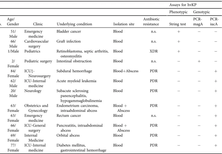
Table 1.Features of the individuals with hypervirulentKlebsiella pneumoniae(hvKP) detection Assays for hvKP Phenotypic Genotypic
No.
Age/
Gender Clinic Underlying condition Isolation site
Antibiotic
resistance String test
PCR- magA
PCR- iucA
1 51/
Male
Emergency medicine
Bladder cancer Blood n.s. þ
2 66/
Male
Cardiovascular surgery
Graft infection Blood n.s. þ
3 1/Male Pediatrics Retinoblastoma, septic arthritis, osteomyelitis
Blood XDR þ
4 2/
Female
Pediatric surgery Intestinal obstruction Blood n.s. þ
5 84/
Female
ICUz-
Neurosurgery
Subdural hemorrhage BloodþAbscess PDR þ
6 62/
Male
ICU-Internal medicine
Acute myeloid leukemia Blood PDR þ
7 20/
Male
Neurology Subacute sclerosing panencephalitis,
hypogammaglobulinemia
Blood PDR þ
8 63/
Female
Obstetrics and Gynecology
Endometrium carcinoma, intraabdominal abcess
Bloodþ Abscess
PDR þ
9 63/
Female
Emergency medicine
Rectum cancer Blood n.s. þ
10 66/
Female
ICU-General surgery
Pancreatitis, intraabdominal abcess
Bloodþ Abscess
PDR þ
11 69/
Female
Internal Medicine
Orbital abcess Blood PDR þ
12 77/
Female
ICU-Internal medicine
Diabetes mellitus,
gastrointestinal hemorrhage
Blood PDR þ
n.s.: not significant; ICU: intersive care unit; XDR: extensive drug resistance, PDR: pan-antimicrobial drug resistance.
Several loci on the Klebsiella genome, directly or indi- rectly associated with virulence or hypermucoviscous col- onies on agar plates have been suggested as biomarkers to characterize hvKP strains [11]. In addition to the commonly-used regions such as iroBCD, iucABCD and rmpA/A2, several other genes have been identified as candidate virulence factors ofK. pneumoniae, some of which were also associated with virulence in bacteria species other than Klebsiella[28]. We have selected some of these genes for PCR amplification, namely rmpA, iucA and peg344 in this study, as they detect different targets and have been previously used in endemic regions. Despite lack of specific detection viarmpAandpeg344PCRs, we could amplify and successfully characterize partialiucAsequences in 7.2% of all isolates (8/111), providing direct evidence for hvKP in the study cohort. The gene iucA is located in the operon encoding for the aerobactin, the abundant siderophore produced by hvKP strains, also a critical mediator for enhancing ex vivo and in vivo virulence [29, 30]. The amplification of iucA has been previously shown to have high accuracy for hvKp identification and could effectively differentiate hvKP from cKP in a murine sepsis model [11].
Therefore, it is widely considered as a stable genetic biomarker for hvKP strains. It has also been reported that a combination of iucA and peg344 amplification increases diagnostic accuracy [11], an observation that could not be reproduced in this study.
We have further performedmagAamplification in ourK.
pneumoniae cohort, using previously-published primers and identified one positive strain, comprising 0.9% of all isolates tested. The sequencing of the amplicon provided a section of the wzc gene and revealed the capsule type as K5, via pairwise sequence comparisons and BLAST analysis. It is known that 79 distinct capsular types exist inKlebsiellaspp. and recent efforts provided a complete sequence repertoire of coding regions for each [31]. Based on this data, it is documented that wzy-based capsular genotyping could reliably differentiate major capsule types except for those lacking or identical wzy sequences [31].
The initial hvKP clones were mostly hypermucoviscous and K1 and K2 type capsules, prevalent in Asia and Europe/America, respectively, were linked to hypervirulence [27]. Moreover, capsule types K5, K20, K54 and K57 have also been frequently- detected in human infections caused byK. pneumoniae. It is known that hvKP strains does not always confer to K1/K2 and other capsule types can be associated with the hypervirulent phenotype [9, 27]. Serological screening have previously revealed the circulation of variousK. pneumoniaecapsular types in Turkey [32, 33] but PCR-based investigations for virulence factors associated with hvKP and capsule typing have failed to identify positive strains [34, 35].
The rmpA and magA amplifications we carried out in this study also produced non-specific products, sometimes within the expected amplicon range. Sequencing of all detectable products revealed that the corresponding primers could bind and amplify various regions of the bacteria genome. This indicates a need for revisiting previously- published primers with the current genome data and updating as necessary. Given the advent of whole genome
sequencing for pathogenic bacteria and the subsequent availability of sequences [36], specificity and accuracy of a nucleic acid based assay should be improved, especially for the task of developing a universal assay for hvKP detection with FDA clearance.
The epidemiological impact of our findings require further elucidation. Originating from Turkey, an Eurasian country with no prior documentation of hvKP circulation, our data indicate an overall low prevalence. However, the observed antibiotic resistance patterns in individuals with evidence for hvKP infection is alarming, with possible PDR in 88.9% of the bacteria with detectable iucA sequences. So far, hvKP strains have mostly retained antimicrobial susceptibility to multiple drug classes [24]. However, recent reports describe hvKP clones obtaining resistance plasmids or acquisition of hvKP virulence plasmid by resistant cKP strains with serious outcomes [17, 37]. Therefore, active measures are required for hvKP screening, control and management. The main limitation of this effort is the single center origin of the evaluated strains. Given thefindings of this pilot study, hvKP circulation in community and other health- care settings should be closely monitored, with the imple- mantation of appropriate infection control precautions. For this purpose, an internationally acclaimed sensitive, accurate and robust diagnostic assay is urgently needed.
In conclusion, we have identified hvKP strains, at a tertiary care institution from a previously unexplored region, in low prevalence via genotypic and phenotypic testing.
Significant antibiotic resistance patterns were recognized in probable hvKP strains, and circulation of K. pneumoniae capsular type K5.
Conflict of interest:The authors have no conflicts of interest to declare.
ACKNOWLEDGMENTS
The authors are grateful to Dr. Thomas Russo and Dr.
Stephen Libby for providing control DNAs for assay opti- mization. This study was supported by the Hacettepe Uni- versity Research Project Funds (Project ID: 15401). The funders had no role in study design, data collection, analysis or interpretation and decision to publish. Preliminary find- ings of the study were accepted as a poster at the 29th European Congress of Clinical Microbiology and Infectious Diseases (ECCMID) 13th–16th April, 2019 in Amsterdam, the Netherlands.
REFERENCES
1. Yeh KM, Kurup A, Siu LK, Koh YL, Fung CP, Lin JC, et al.
Capsular serotype K1 or K2, rather than magA and rmpA, is a major virulence determinant forKlebsiella pneumoniaeliver abscess in Singapore and Taiwan. J Clin Microbiol 2007; 45: 466–71, https://doi.org/10.1128/JCM.01150-06.
2. Yan Q, Zhou M, Zou M, Liu WE. HypervirulentKlebsiella pneu- moniaeinduced ventilator-associated pneumonia in mechanically ventilated patients in China. Eur J Clin Microbiol Infect Dis 2016;
35: 387–96,https://doi.org/10.1007/s10096-015-2551-2.
3. Liu YC, Cheng DL, Lin CL. Klebsiella pneumoniaeliver abscess associated with septic endophthalmitis. Arch Intern Med 1986; 146:
1913–6.
4. Lee HC, Chuang YC, Yu WL, Lee NY, Chang CM, Ko NY, et al.
Clinical implications of hypermucoviscosity phenotype inKlebsiella pneumoniaeisolates: association with invasive syndrome in patients with community-acquired bacteraemia. J Intern Med 2006; 259:
606–14,https://doi.org/10.1111/j.1365-2796.2006.01641.x.
5. Fang CT, Lai SY, Yi WC, Hsueh PR, Liu KL, Chang SC.Klebsiella pneumoniae genotype K1: An emerging pathogen that causes septic ocular or central nervous system complications from pyogenic liver abscess. Clin Infect Dis 2007; 45: 284–93,https://doi.org/10.1086/519262.
6. Shon AS, Bajwa RP, Russo TA. Hypervirulent (hypermucoviscous) Klebsiella pneumoniae: a new and dangerous breed. Virulence 2013;
4: 107–18,https://doi.org/10.4161/viru.22718.
7. Wu MC, Lin TL, Hsieh PF, Yang HC, Wang JT. Isolation of genes involved in biofilm formation of a Klebsiella pneumoniae strain causing pyogenic liver abscess. PLoS One 2011; 6,https://doi.org/
10.1371/journal.pone.0023500.
8. Li W, Sun G, Yu Y, Li N, Chen M, Jin R, et al. Increasing occurrence of antimicrobial-resistant hypervirulent (hypermucoviscous) Klebsiella pneumoniaeisolates in China. Clin Infect Dis 2014; 58: 225–32,https://
doi.org/10.1093/cid/cit675.
9. Liu YM, Li BB, Zhang YY, Zhang W, Shen H, Li H, et al. Clinical and molecular characteristics of emerging hypervirulentKlebsiella pneumoniae bloodstream infections in mainland China. Anti- microb Agents Chemother 2014; 58: 5379–85,https://doi.org/10.
1128/AAC.02523-14.
10. Siu LK, Huang DB, Chiang T. Plasmid transferability of KPC into a virulent K2 serotypeKlebsiella pneumoniae. BMC Infect Dis 2014;
14: 176,https://doi.org/10.1186/1471-2334-14-176.
11. Russo TA, Olson R, Fang CT, Stoesser N, Miller M, MacDonald U, et al. Identification of biomarkers for differentiation of hyperviru- lent Klebsiella pneumoniae from classical K. pneumoniae. J Clin Microbiol 2018; 56:pii: e00776.
12. Fang CT, Chuang YP, Shun CT, Chang SC, Wang JT. A Novel virulence gene inKlebsiella pneumoniaestrains causing primary liver abscess and septic metastatic complications. J Exp Med 2004; 199:
697–705,https://doi.org/10.1084/jem.20030857.
13. Nassif X, Sansonetti PJ. Correlation of the virulence ofKlebsiella pneumoniaeK1 and K2 with the presence of a plasmid encoding aerobactin. Infect Immun 1986; 54: 603–8.
14. Nassif X, Fournier JM, Arondel J, Sansonetti PJ. Mucoid phenotype of Klebsiella pneumoniae is a plasmid-encoded virulence factor.
Infect Immun 1989; 57: 546–52.
15. Lin TL, Lee CZ, Hsieh PF, Tsai SF, Wang JT. Characterization of integrative and conjugative element ICEKp1-associated genomic heterogeneity in a Klebsiella pneumoniae strain isolated from a primary liver abscess. J Bacteriol 2008; 190: 515–26,https://doi.org/
10.1128/JB.01219-07.
16. Tang HL, Chiang MK, Liou WJ, Chen YT, Peng HL, Chiou CS, et al. Correlation betweenKlebsiella pneumoniaecarrying pLVPK- derived loci and abscess formation. Eur J Clin Microbiol Infect Dis 2010; 29:689–98,https://doi.org/10.1007/s10096-010-0915-1.
17. Gu D, Dong N, Zheng Z, Lin D, Huang M, Wang L, et al. A fatal outbreak of ST11 carbapenem-resistant hypervirulent Klebsiella pneumoniae in a Chinese hospital: a molecular epidemiological study. Lancet Infect Dis 2018; 18: 37–46,https://doi.org/10.1016/
S1473-3099(17)30489-9.
18. Siu LK, Yeh KM, Lin JC, Fung CP, Chang FY.Klebsiella pneumo- niaeliver abscess: a new invasive syndrome. Lancet Infect Dis 2012;
11: 881–7,https://doi.org/10.1016/S1473-3099(12)70205-0.
19. Chou A, Nuila RE, Franco LM, Stager CE, Atmar RL, Zechiedrich L.
Prevalence of hypervirulentKlebsiella pneumoniae-associated genes rmpAandmagAin two tertiary hospitals in Houston, TX, USA. J Med Microbiol 2016; 65: 1047–8, https://doi.org/10.1099/jmm.0.
000309.
20. Altschul SF, Gish W, Miller W, Myers EW, Lipman DJ. Basic local alignment search tool. J Mol Biol 1990; 215: 403–10,https://doi.org/
10.1016/S0022-2836(05)80360-2.
21. Thompson JD, Higgins DG, Gibson TJ. CLUSTAL W: Improving the sensitivity of progressive multiple sequence alignment through sequence weighting, position-specific gap penalties and weight matrix choice. Nucleic Acids Res 1994; 22: 4673–80,https://doi.org/
10.1093/nar/22.22.4673.
22. Kumar S, Stecher G, Li M, Knyaz C, Tamura K. MEGA X:
Molecular evolutionary genetics analysis across computing platforms.
Mol Biol Evol 2018; 35: 1547–9, https://doi.org/10.1093/molbev/
msy096.
23. Magiorakos AP, Srinivasan A, Carey RB, Carmeli Y, Falagas ME, Giske CG, et al. Multidrug-resistant, extensively drug-resistant and pandrug-resistant bacteria: an international expert proposal for interim standard definitions for acquired resistance. Clin Microbiol Infect 2012; 18: 268–81,https://doi.org/10.1111/j.1469-0691.2011.
03570.x.
24. Marr CM, Russo TA. HypervirulentKlebsiella pneumoniae: a new public health threat. Expert Rev Anti Infect Ther 2019; 17: 71–3, https://doi.org/10.1080/14787210.2019.1555470.
25. Liu C, Guo J. HypervirulentKlebsiella pneumoniae(hypermucoviscous and aerobactin positive) infection over 6 years in the elderly in China:
antimicrobial resistance patterns, molecular epidemiology and risk factor. Ann Clin Microbiol Antimicrob 2019; 18: 4,https://doi.org/10.
1186/s12941-018-0302-9.
26. Shi Q, Lan P, Huang D, Hua X, Jiang Y, Zhou J, et al. Di- versity of virulence level phenotype of hypervirulent Klebsiella pneumoniae from different sequence type lineage. BMC Microbiol 2018; 18: 94, https://doi.org/10.1186/s12866-018- 1236-2.
27. Catalan-Najera JC, Garza-Ramos U, Barrios-Camacho H. Hyper- virulence and hypermucoviscosity: Two different but complemen- taryKlebsiellaspp. phenotypes?. Virulence 2017; 8: 1111–23,https://
doi.org/10.1080/21505594.2017.1317412.
28. Ye M, Tu J, Jiang J, Bi Y, You W, Zhang Y, et al. Clinical and genomic analysis of liver abscess-causing Klebsiella pneumoniae identifies new liver abscess-associated virulence genes. Front Cell Infect Microbiol 2016; 6: 165,https://doi.org/10.3389/fcimb.2016.
00165.
29. Russo TA, Olson R, Macdonald U, Metzger D, Maltese LM, Drake EJ, et al. Aerobactin mediates virulence and accounts for increased siderophore production under iron-limiting conditions by hyper- virulent (hypermucoviscous)Klebsiella pneumoniae. Infect Immun 2014; 82: 2356–67,https://doi.org/10.1128/IAI.01667-13.
30. Russo TA, Olson R, MacDonald U, Beanan J, Davidson BA. Aero- bactin, but not yersiniabactin, salmochelin and enterobactin, enables the growth/survival of hypervirulent (hypermucoviscous)Klebsiella pneumoniaeex vivo and in vivo. Infect Immun 2015; 83: 3325–33, https://doi.org/10.1128/IAI.00430-15.
31. Pan YJ, Lin TL, Chen CT, Chen YY, Hsieh PF, Hsu CR, et al.
Genetic analysis of capsular polysaccharide synthesis gene clusters in 79 capsular types of Klebsiella spp. Sci Rep 2015; 5: 15573, https://doi.org/10.1038/srep15573.
32. Ozenci H, Karaaslan A, Ozsan M.Klebsiella pneumoniaecapsular type 48 isolated for thefirst time in Turkey. Mikrobiyol Bul 1990;
24: 91–2.
33. Ozenci H, Karaaslan A, Tuncer I, Erboyaci A.Klebsiella pneumo- niae capsular type 25 and type 16 isolated for the first time in Turkey. Mikrobiyol Bul 1990; 24: 93–4.
34. Candan ED, Aks€oz N. Klebsiella pneumoniae: characteristics of carbapenem resistance and virulence factors. Acta Biochim Pol 2015; 62: 867–74,https://doi.org/10.18388/abp.2015_1148.
35. Kus¸ H, Arslan U, T€urk Dagı H, Fındık D. Investigation of various virulence factors of Klebsiella pneumoniae strains iso- lated from nosocomial infections. Mikrobiyol Bul 2017; 51:
329–39, https://doi.org/10.5578/mb.59716.
36. Besser J, Carleton HA, Gerner-Smidt P, Lindsey RL, Trees E. Next- generation sequencing technologies and their application to the study and control of bacterial infections. Clin Microbiol Infect 2018; 24: 335–41,https://doi.org/10.1016/j.cmi.2017.10.013.
37. Zhang R, Lin D, Chan EW, Gu D, Chen GX, Chen S. Emergence of carbapenem-resistant serotype K1 hypervirulentKlebsiella pneumo- niaestrains in China. Antimicrob Agents Chemother 2015; 60: 709–11, https://doi.org/10.1128/AAC.02173-15.