ANTIBIOTIC RESISTANCE, VIRULENCE AND GENETIC DIVERSITY OF KLEBSIELLA
PNEUMONIAE IN COMMUNITY- AND HOSPITAL-ACQUIRED URINARY TRACT
INFECTIONS IN IRAN
FATEMEH EGHBALPOOR1, MEHRI HABIBI1, OMIDAZIZI2, MOHAMMADREZAASADIKARAM1* and SAEIDBOUZARI1
1Department of Molecular Biology, Pasteur Institute of Iran, Tehran, Iran
2Health Sciences Research Center, Torbat Heydariyeh University of Medical Sciences, Torbat Heydariyeh, Iran
(Received: 20 November 2018; accepted: 14 January 2019)
Klebsiella pneumoniae is among the most important causes of urinary tract infection (UTI). The aim of this study was to investigate the prevalence and correlation of antibiotic resistance with virulence characteristics and genetic diversity in K. pneumoniaeisolated from UTIs in Iran. Phenotypic tests and antibiotic suscepti- bility were carried out on the isolates. Detection of the virulence and extended- spectrumβ-lactamase (ESBL) genes was performed by polymerase chain reaction.
Pulsed-field gel electrophoresis (PFGE) was used for exploring the genomic related- ness. Hemolysin, biofilm, and hypermucoviscosity formation were observed in 87.1%, 86.4%, and 12.1% of isolates, respectively. The antibiotic resistance rate of K. pneumoniaeisolates ranged from 12.1% for meropenem to 100% for amoxicillin.
The prevalence of virulence genes ranged from 1.4% forcnf-1to 100% formrkD, fimH,kpn, andentBgenes. In this study, 91.7%, 33.3%, and 4.2% of phenotypically ESBL-producers were positive forblaCTX-M,blaTEM, andblaSHVgenes, respectively.
An association was observed between the presence oftraT,fyuA,orcnf-1 genes with antibiotic resistance. Two clone types were obtained by PFGE that indicate different K. pneumoniaeclones in community- and hospital-acquired UTIs. Thefindings of this study are valuable in development of treatment strategies against UTIs in Iran.
Keywords: urinary tract infections, Klebsiella pneumoniae, antibiotic resistance, ESBL, virulence factors, PFGE
*Corresponding author; E-mail:m_asadi12@yahoo.com
First published online February 26, 2019
Introduction
Urinary tract infections (UTIs) are among prevalent infections acquired both in community and in hospitals. It is estimated that 40% of women will have a UTI with a rate of 25% for recurrent UTIs [1].Klebsiella pneumoniaeis recognized as a common pathogen associated with both community- and hospital-acquired UTIs [2]. Its pathogenicity is related to expression of a variety of virulence factors includingfimbriae, toxins, iron acquisition systems, lipopolysaccharide, and capsule [2]. Fimbriae type 1 and type 3 as mannose-sensitive (MSHA) and mannose- resistant hemagglutination pili (MRHA) play a major role in adherence and colonization ofK. pneumoniaein the urinary tract [3]. FimH and MrkD composed the tip adhesin of types 1 and 3 pili inK. pneumoniae, respectively, and play relevant roles in biofilm formation [3]. Biofilms have known roles in pathogenesis of K. pneumoniaestrains, including the resistance to immune responses and acquisition of antibiotic resistance [4]. Capsule is among the main virulence factors of K. pneumoniae isolates for protection against host immune system. K1 and K2 capsular serotypes containing the mucoviscosity-associated gene A (magA) were associated with severe UTI infections [5]. K. pneumoniae strains also have two potential toxins, a cytolytic pore forming toxin (α-hemolysin) and cytotoxic- necrotizing factor 1 (CNF1) that cause damage to tissues in the host [6].
Antibiotics are used routinely for therapy of UTIs caused byK. pneumoniae strains. To date, K. pneumoniae is known worldwide for rapid acquisition of resistance to different antibiotics, including resistance toβ-lactams,fluoroquino- lones, and carbapenems [7, 8]. Antibiotic resistance among the K. pneumoniae strains poses a great threat to public health and imposes a high cost to the UTI patients [9]. Resistance of these isolates to antibiotics is acquired by different mechanisms such as production of β-lactamase enzymes named as extended- spectrum β-lactamases (ESBLs) [10]. TEM, SHV, and CTX-M as the most common ESBL types confer resistance to all β-lactam antibiotics, except for carbapenems, andβ-lactamase inhibitors. Furthermore, ESBL-producing isolates usually carry other resistance genes that confer resistance to other classes of antibiotics, such as aminoglycosides and quinolones [10, 11]. Unfortunately, many physicians prescribe that antibiotics before the results of antibiotic sensitivi- ty can be evaluated by the laboratories. Thus, the choice of effective antibiotics against UTIs caused by K. pneumoniae should be based on determination the causative agent and knowledge of the resistance patterns of the isolate [12].
K. pneumoniae is recognized as one of the most important opportunistic pathogens in UTIs; however, there are only a few studies about pathogenicity and correlation between the virulence factors with antibiotic resistance in these isolates in Iran. Thus, the objectives of this study were to investigate the prevalence and
correlation of the virulence factors with antibiotic resistance in K. pneumoniae isolated from outpatients and inpatients in Tehran, Iran. Then, the clonal related- ness among the K. pneumoniae isolates was evaluated by pulsed-field gel electrophoresis (PFGE) technique.
Methods
Collection of K. pneumoniae isolates from UTI patients
From July to December 2017, 140K. pneumoniaeisolates were collected from the urine samples of outpatients (n=80) and inpatients (n=60) in several hospitals in Tehran, Iran. Patients were informed about the project and provided the informed consent. The exclusion criteria in this study were the presence of fungi, mixed cultures, and age less than 18 years. The urine samples were collected from the clean catch mid-stream urine of the patients. After collection of the urine with colony count≥105CFU/ml, identification ofK. pneumoniaeisolates in these samples was performed on the basis of standard microbiological and biochemical tests.
Phenotypic determination of virulence factors
Hemagglutination assay.K. pneumoniaeisolates were screened for type 1 (MSHA) and type 3fimbriae (MRHA). Briefly, bacterial suspensions and human erythro- cytes of blood group O (3%) were added to the 96-well microtiter plates in the presence and absence of D-mannose 2%. The plates were incubated at room temperature and reported as MRHA when agglutination was observed in the presence of mannose and MSHA when it was not occurred with mannose.
The wells containing erythrocytes in the presence and absence of mannose were applied as negative controls andEscherichia coliATCC 25922 as positive control for MRHA phenotype.
Hemolysin production.The phenotypic ability of theK. pneumoniaeisolates in production of α-hemolysin was evaluated in culture media. The isolates were cultured in 5% sheep blood agar (Merck, Germany) and incubated overnight at 37 °C. Production of a clear zone of lysed erythrocytes around the colonies was considered as hemolysin producer isolate.
Biofilm formation testing.The ability of theK. pneumoniaeisolates to produce biofilm was monitored by a microtiter plate test. Briefly,K. pneumoniaeisolates were subcultured in lysogeny broth, diluted at 1:100 ratio, and incubated for 48 h.
After several washes, 0.5% of crystal violet was added to plates and incubated for 15 min at room temperature. The plates were washed and a mixture of 33% acetic
acid and 80% ethanol was added to plates to dissolve the crystal violet and plates read at absorbance 590 nm with ELISA reader (BIO-TEK, USA). The K. pneumoniaeisolates were classified as strong biofilm producers [(2.5≤optical density (OD)], moderate producers (1.5≤OD<2.5), weak producers (0.7≤OD≤ 1.5), and non-biofilm producers isolates (OD≤0.7). E. coli ATCC 25922 and Pseudomonas aeruginosastrains were used as positive controls andE. coliDH5αas a negative control.
Hypermucoviscosity (HMV) test. The string test was applied to evaluate the HMV phenotype of the isolates [13]. TheK. pneumoniaeisolates were cultured on 5% sheep blood agar plates and incubated for 18–24 h at 37 °C. When an inoculation loop was touched onto the surface of the colonies and slowly raised, the formation of a viscous string of at least 5 mm in length was considered as a HMV-positive phenotype.
Antibiotic susceptibility test.Antimicrobial susceptibility patterns of the isolates to different antibiotics were evaluated using the disk diffusion method using Mueller–Hinton agar (Merck). The tested antibiotics (Mast, Co., Merseyside, UK) were amoxicillin (10μg), ticarcillin (75μg), ampicillin–sulbactam (10+10μg), cephalotin (30μg), cephalexin (30μg), ceftazidime (30μg), cefotaxime (30μg), meropenem (10 μg), nalidixic acid (30 μg), norfloxacin (10 μg), gentamicin (10μg), tobramycin (10μg), and trimethoprim–sulfamethoxazole (SXT; 30μg).
The results were interpreted according to the Clinical and Laboratory Standards Institute (CLSI) recommendations [14]. The E. coli ATCC 25922 was used as control strain. Isolates that were resistant to at least three different classes of antimicrobial agents were determined to be multidrug-resistant (MDR) phenotypes.
Screening of ESBL isolates.TheK. pneumoniaeisolates were screened for ESBL production by combined disk method according to CLSI guidelines [14]. The phenotypic confirmatory test was performed by ceftazidime (30 μg) alone, combined with clavulanic acid (10μg) as well as cefotaxime (30μg) alone, and combined with clavulanic acid (10 μg) disks. E. coli ATCC 25922 and K. pneumoniae ATCC 700603 were applied as negative and positive controls, respectively.
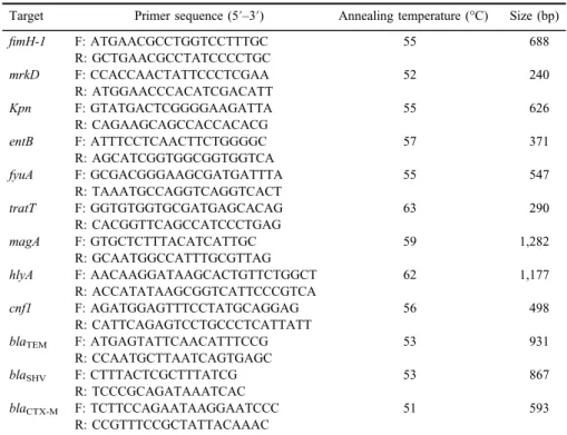
DNA extraction and polymerase chain reaction (PCR) detection of virulence and ESBL genes. PCR was applied to detect the virulence genes encoding adhesins (fimH and mrkD), fimH-like adhesin (Kpn), entrobactin biosynthesis (entB), yersiniabactin iron receptor (fyuA), serum resistance-associated outer membrane lipoprotein (traT), magA, α-hemolysin (hlyA), and cnf-1. Furthermore, the pres- ence of blaTEM, blaSHV, and blaCTX-M in the ESBL-producing isolates was confirmed by PCR. All specific primers (Genfanavaran, Iran) are listed in Table I. PCR reactions contained 1× buffer, 0.1 mM dNTPs, 2.5 mM MgCl2,
50 pmol of each primer, and 0.5 U Taq polymerase (CinnaGen, Iran) in a total volume of 25μl. The used programs were an initial denaturation (94 °C for 5 min), followed by 30 cycles at denaturation (94 °C for 1 min), annealing (specific temperature for 1 min; TableI) and extension (72 °C for 1 min), and afinal step (72 °C for 5 min).
PFGE analysis. PFGE was performed on 45 selected isolates to determine genomic diversity of the isolates. Briefly, these isolates were cultured on trypticase soy agar and then prepared bacterial plugs lysed with lysis buffer. The plugs were digested with restriction enzyme XbaI (Fermentas, Lithuania) for about 150 min at 37 °C. Then, electrophoresis of the plugs was performed in agarose 1% (Ultrapure, Invitrogen, USA) with CHEF-DR III system (Bio-Rad, USA). The stained gels with ethidium bromide were observed with Gel Doc. The obtained patterns were analyzed using GelCompare II, version 6.5 software (Applied Maths, St Martens- Latem, Belgium). Dendrogram drawing and clonal associations between the genotypes were performed with average linkage method and analyzed with unweighted pair group method analysis [15].
Table I.Primers used to amplify virulence and ESBL genes inK. pneumoniaeisolates Target Primer sequence (5′–3′) Annealing temperature (°C) Size (bp)
fimH-1 F: ATGAACGCCTGGTCCTTTGC 55 688
R: GCTGAACGCCTATCCCCTGC
mrkD F: CCACCAACTATTCCCTCGAA 52 240
R: ATGGAACCCACATCGACATT
Kpn F: GTATGACTCGGGGAAGATTA 55 626
R: CAGAAGCAGCCACCACACG
entB F: ATTTCCTCAACTTCTGGGGC 57 371
R: AGCATCGGTGGCGGTGGTCA
fyuA F: GCGACGGGAAGCGATGATTTA 55 547
R: TAAATGCCAGGTCAGGTCACT
tratT F: GGTGTGGTGCGATGAGCACAG 63 290
R: CACGGTTCAGCCATCCCTGAG
magA F: GTGCTCTTTACATCATTGC 59 1,282
R: GCAATGGCCATTTGCGTTAG
hlyA F: AACAAGGATAAGCACTGTTCTGGCT 62 1,177
R: ACCATATAAGCGGTCATTCCCGTCA
cnf1 F: AGATGGAGTTTCCTATGCAGGAG 56 498
R: CATTCAGAGTCCTGCCCTCATTATT
blaTEM F: ATGAGTATTCAACATTTCCG 53 931
R: CCAATGCTTAATCAGTGAGC
blaSHV F: CTTTACTCGCTTTATCG 53 867
R: TCCCGCAGATAAATCAC
blaCTX-M F: TCTTCCAGAATAAGGAATCCC 51 593
R: CCGTTTCCGCTATTACAAAC
Note:ESBL: extended-spectrumβ-lactamase; F: forward; R: reverse.
Statistical analysis
Statistical data analysis was performed using SPSS software version 19.0 for Windows (IBM, Chicago, USA). Among the statistical tests,χ2, two-tailed Fisher’s exact, and analysis of variance tests were used to assess and compare the relationships between various virulence factors inK. pneumoniae. Moreover, pair gene relationships were assessed using Phi coefficients. Thepvalue<0.05 was considered for all statistical accounts.
Results
Prevalence of UTI in relationship with profile of patients
Among the UTI patients included in this study, the rate of UTI in females (78.6%) was higher than males (21.4%). The age range of patients was 18–85 years. The frequency of K. pneumoniae in different age groups was 18–40 (44.3%), 41–60 (37.8%), and 61–85 (17.9%) years. Distribution of the inpatient isolates according to the hospital settings was urology (31.6%), intensive care unit (ICU; 25%), gynecology (18.4%), coronary care unit (CCU; 15%), and surgery (10%).
Phenotypic characteristics ofK. pneumoniaeisolates
Phenotypic virulence characteristics.It was found that 87.1% of the isolates had the ability to produce hemolysin. The hemolysin activity was found in 93.3%
and 82.5% of isolates collected from inpatients and outpatients, respectively.
Furthermore, the distribution of hemolysin-positive isolates in different hospital settings was ICU (100%), CCU (100%), surgery (100%), urology (89.5%), and gynecology (81.8%). Furthermore, 22.2% and 77.8% of the non-hemolysin producer isolates were recovered from inpatients and outpatients, respectively.
According to our results, 86.4% of K. pneumoniaeproduced biofilm that were classified into strong (24%), moderate (54.5%), and weak biofilm producers (21.5%). In addition, 91.7% and 82.5% of biofilm producer isolates were collected from inpatients and outpatients, respectively. All isolates that were collected from ICU, CCU, and gynecology wards had the ability to form strong to moderate biofilm formation. The isolates that had no ability in biofilm production were collected from age group 18–40 (79%), 41–60 (10.5%), and 61–85 (10.5%) years.
It was found that among 122 hemolysis-positive isolates, altogether 110 (90.2%) had the ability for biofilm formation (p=0.004).
The HMV phenotype was found in 17 (12.1%) of isolates in which 15 (88.2%) and 2 (11.8%) of them were isolated from inpatients and outpatients, respectively (p=0.001). Based on our investigations, allK. pneumoniaeisolates showed the presence of type 1 (MSHA) and type 3 fimbriae (MRHA).
Antibiotic susceptibility of K. pneumoniae isolates. According to the disk diffusion results, the susceptibility of K. pneumoniaeisolates to antibiotics was meropenem (87.9%), norfloxacin (77.1%), gentamicin (75.7%), tobramycin (75.7%), nalidixic acid (75%), ampicillin–sulbactam (66.4%), ceftazidime (65%), cefotaxime (62.1%), SXT (61.4%), cephalotin (60.7%), cephalexin (60.7%), and ticarcillin (12.9%). Furthermore, all isolates were resistant to amoxicillin. As shown in Table II, 53 of the isolates with co-resistance to amoxicillin and ticarcillin were the most frequent resistance pattern (R1), whereas 12 isolates showed single patterns (data not shown).
The distribution of antibiotic resistance according to the outpatients and inpatients is also shown in Table III. Among the antibiotics tested, there was a significant difference between the resistance to all antibiotics in inpatients and outpatients groups (p<0.05; TableIII).
According to the results, 33 (23.6%) of K. pneumoniae isolates were classified as MDR isolates. Out of 33 MDR isolates, 14 (42.4%) were completely resistant to all antibiotics investigated. MDR in females was higher than males (26 vs. 7), but these differences were not statistically significant (p>0.05).
Table II.Antibiotic resistance patterns ofK. pneumoniaeisolates
Pattern name Resistance patterns
No. of isolates
R1 AMX, TC 52
R2 AMX 18
R3 AMX, TC, CL, KF, CTX, CAZ, SAM, SXT, NOR, MEM, GEN, TB, NAL 14
R4 AMX, TC, SXT 10
R5 AMX, TC, CL, KF, CTX, CAZ, SAM, SXT 6
R6 AMX, TC, CL, KF, CTX, CAZ, SXT 6
R7 AMX, TC, CL, KF, CTX, CAZ, SAM, SXT, NOR, GEN, TB, NAL 6
R8 AMX, TC, CL, KF, CTX, CAZ, SXT, NOR, GEN, TB, NAL 3
R9 AMX, TC, CL, KF, CTX, CAZ, SAM, GEN, TB 3
R10 AMX, TC, SAM, SXT, GEN, TB, NAL 2
R11 AMX, TC, CL, KF, CTX, CAZ, SAM, NOR, NAL 2
R12 AMX, TC, CL, KF, CTX, CAZ, SAM, SXT, NOR, NAL 2
R13 AMX, TC, SAM 2
R14 AMX, TC, CTX, CAZ, CL, KF, SAM, NOR, MEM, GEN, TB, NAL 2 Note: AMX: amoxicillin; TC: ticarcillin; CRO: ceftriaxone; CAZ: ceftazidime; CL: cephalexin;
KF: cephalotin; SAM: ampicillin–sulbactam; SXT: trimethoprim–sulfamethoxazole; NOR: norfloxacin;
GEN: gentamicin; TB: tobramycin; NAL: nalidixic acid; MEM: meropenem.
Furthermore, 51.5% of MDR isolates were in the age groups of 41–60, 30.3% in 18–40, and 18.2% in 61–85 years. Twenty-nine isolates of MDR isolates were collected from inpatients and four isolates from outpatients. In addition, the distribution of MDR isolated from inpatients in different hospital wards was ICU (31%), urology (24.2%), gynecology (20.7%), CCU (13.8%), and surgery (10.3%).
The ESBL phenotypic screening by combined disk test showed that among 140 isolates, 48 isolates (34.3%) were ESBL producers. Among the ESBL producers, 35 (72.9%) belonged to inpatients and 13 (27.1%) to outpatients (p=0.0001). TableIVdemonstrates the antimicrobial resistance pattern of ESBL- producing isolates for applied antibiotics. All ESBL-positive isolates exhibited resistance to ticarcillin, cephalotin, cephalexin, and cefotaxime. Furthermore, the ESBL-positive isolates exhibited the lowest resistance to meropenem (33.3%;
Table IV).
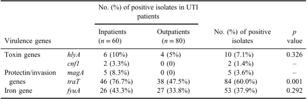
Distribution of virulence genes amongK. pneumoniaeisolates.According to our results,fimHandmrkDadhesins, siderophore receptor geneentB, and fimH-like adhesin (kpn) were detected in 100% of the isolates. The prevalence rate oftraT, iron receptor gene fyuA, and magAcapsular gene was 60%, 37.9%, and 3.6%, respectively. Toxinsα-hemolysin (hlyA) andcnf-1were present in 7.1% and 1.4%
of isolates, respectively. The prevalence and distribution of different virulence genes according to the inpatients and outpatients is shown in TableV. It was found that isolates from inpatients had more virulence genes as compared to the isolates
Table III.Distribution of antibiotic resistance according to the outpatients and inpatients
Antibiotic list
No. (%) of antibiotic resistance
Total no. (%) of positive isolates
(n=140)
p value Inpatients
(n=60)
Outpatients (n=80)
β-lactams Ticarcillin 57 (95.0%) 65 (81.2%) 122 0.020
Cephalotin 37 (61.7%) 18 (22.5%) 55 0.000
Cephalexin 37 (61.7%) 18 (22.5%) 55 0.000
Ceftazidime 36 (60.0%) 15 (18.8%) 51 0.000
Cefotaxime 38 (63.3%) 15 (18.8%) 53 0.000
β-lactam/
β-lactamase inhibitors
Ampicillin– sulbactam
38 (63.3%) 9 (11.2%) 47 0.000
Carbapenem Meropenem 15 (25.0%) 2 (2.5%) 17 0.000
Quinolones Nalidixic acid 31 (51.7%) 4 (5.0%) 35 0.000
Norfloxacin 28 (46.7%) 4 (5.0%) 32 0.000
Aminoglycosides Tobramycin 28 (46.7%) 6 (7.5%) 34 0.000
Gentamicin 28 (46.7%) 6 (7.5%) 34 0.000
Sulfonamides Trimethoprim– sulfamethoxazole
31 (51.7%) 22 (27.5%) 53 0.005
from outpatients and this difference was significant aboutcnf-1, magA, and traT genes (p<0.05; Table V). Furthermore, it was observed that magA gene was statistically more frequent among HMV-producing isolates compared with non- HMV producer isolates (p=0.001).
Correlation between virulence characteristics and antimicrobial resistance. The correlation of virulence characteristics with ESBL production and antibiotic resis- tance is shown in TablesIVandVI, respectively. The correlation between biofilm
Table IV.Prevalence of antibiotic resistance and virulence genes among ESBL-positive and -negative isolates
Antibiotic list
% (n)
pvalue ESBL+(n=48) ESBL−(n=92) Total (N=140)
Ticarcillin 48 (100) 73 (80.4) 122 (87.1) 0.000
Cephalotin 48 (100) 7 (7.6) 55 (39.3) 0.000
Cephalexin 48 (100) 7 (7.6) 55 (39.3) 0.000
Cefotaxime 48 (100) 5 (5.4) 53 (37.9) 0.000
Ceftazidime 47 (97.9) 2 (2.2) 49 (35.0) 0.000
SXT 38 (79.2) 16 (17.6) 54 (38.6) 0.000
SAM 38 (79.2) 9 (9.8) 47 (33.6) 0.000
Meropenem 16 (33.3) 1 (1.1) 17 (12.1) 0.000
Tobramycin 31 (64.6) 3 (3.3) 34 (24.3) 0.000
Gentamicin 31 (64.6) 3 (3.3) 34 (24.3) 0.000
Nalidixic acid 31 (64.6) 4 (4.3) 35 (25.0) 0.000
Norfloxacin 30 (62.5) 2 (2.2) 32 (22.9) 0.000
Virulence genes
traT 32 (66.7) 52 (56.5) 84 (60.0) 0.279
fyuA 25 (52.1) 28 (30.4) 53 (37.9) 0.010
magA 2 (4.2) 3 (3.3) 5 (3.6) 1.000
hlyA 7 (14.6) 3 (3.3) 10 (7.1) 0.032
cnf-1 2 (4.2) 0 (0) 2 (1.4) –
Table V.Distribution of virulence genes according to the inpatients and outpatients
Virulence genes
No. (%) of positive isolates in UTI patients
No. (%) of positive isolates
p value Inpatients
(n=60)
Outpatients (n=80)
Toxin genes hlyA 6 (10%) 4 (5%) 10 (7.1%) 0.326
cnf1 2 (3.3%) 0 (0) 2 (1.4%) –
Protectin/invasion genes
magA 5 (8.3%) 0 (0) 5 (3.6%) –
traT 46 (76.7%) 38 (47.5%) 84 (60.0%) 0.001
Iron gene fyuA 26 (43.3%) 27 (33.8%) 53 (37.9%) 0.292
Note:UTI: urinary tract infection.
TableVI.Correlationofvirulencecharacteristicswithantibioticresistanceintheisolates Virulencefactors
Antibioticresistance(number) TCKFCLCTXCAZMEMSAMNALNORSXTGENTB Hemolysis1064848484517423330445231 Biofilm106515150*47*17423231472929 HMV1513*13*13*13*5*11*17*10*12*8*8* hlyA98*8*8*8*0512244 cnf-1222220222222 magA322221222222 traT73383864321336*26*233226*26* fyuA4728*28*28*25*4201313211313 Note:TC:ticarcillin;CTX:cefotaxime;CAZ:ceftazidime;CL:cephalexin;KF:cephalotin;SAM:ampicillin–sulbactam;SXT:trimethoprim–sulfamethoxazole; NOR:norfloxacin;GEN:gentamicin;TB:tobramycin;NAL:nalidixicacid;MEM:meropenem;HMV:hypermucoviscosity. *p<0.05(significantcorrelationbetweenthevirulencefactorandantibioticresistance).
production and antimicrobial resistance was found to be statistically significant for cefotaxime and ceftazidime antibiotics (p<0.05). In addition, isolates harboring HMV were more resistant to all of the antibiotics, except ticarcillin (p<0.05). Our results showed that there was no significant difference between resistance to the antibiotics and hemolysin production (p>0.05; TableIV). According to TableIV, isolates harboring traTgene were more resistant to aminoglycosides (p<0.05), nalidixic acid (p=0.049), and ampicillin–sulbactam (p=0.006). Furthermore, isolates harboringfyuA andhlyA genes showed higher resistance to thefirst- and third-generation cephalosporins (p<0.05).
Genotypic detection of ESBLs. PCR amplification on phenotypically ESBL- producing isolates (n=48) showed that 44 (91.7%), 16 (33.3%), and 2 (4.2%) isolates were positive forblaCTX-M,blaTEM, andblaSHVgenes, respectively. Of the 48 phenotypically ESBL-producing isolates, 22.9% and 4.2% of the isolates were shown to have simultaneouslyblaTEM+blaCTX-MandblaSHV+blaCTX-Mgenes, respectively. The prevalence ofblaCTX-MandblaTEMalone in the phenotypically ESBL-positive isolates was 64.5% and 4.2%, respectively. There were only two ESBL-producing isolates negative for ESBL genes. It was found that the preva- lence ofblaCTX-MandblaTEMwas significantly higher in inpatients as compared to the outpatient isolates (p<0.05).
Genotypic virulence patterns in K. pneumoniae isolates. In this study, 12 virulence patterns (KP patterns) of K. pneumoniae were obtained by different combinations of virulence genes (Table VII). All isolates indicated multiple virulence markers and contained four to seven virulence genes. Three isolates showed unique patterns, whereas KP1 with the presence offimH,mrkD,kpn,entB, andtraTwas the most prevalent profile among the isolates (37.8%; Table VII).
Table VII.Common virulence patterns identified among theK. pneumoniaeisolates
Pattern fimH mrkD kpn entB traT fyuA magA hlyA cnf-1
No. (%) of isolates
Kp1 + + + + + − − − − 53 (37.8%)
Kp2 + + + + − − − − − 26 (18.6%)
Kp3 + + + + − + − − − 24 (17.1%)
Kp4 + + + + + + − − − 21 (15.0%)
Kp5 + + + + − − − + − 3 (2.2%)
Kp6 + + + + + − + − − 3 (2.2%)
Kp7 + + + + − + − + − 3 (2.2%)
Kp8 + + + + + + + − − 2 (1.4%)
Kp9 + + + + + + − + − 2 (1.4%)
Kp10 + + + + + − − + + 1 (0.7%)
Kp11 + + + + + + − − + 1 (0.7%)
Kp12 + + + + + − − + − 1 (0.7%)
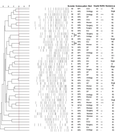
Genomic analysis ofK. pneumoniaeisolates by PFGE.Totally, 45K. pneumoniae isolates were considered for PFGE analysis, based on the virulence patterns, antibiotic resistance, and epidemiological data. These isolates were clustered into 41 clonal groups, based on the depicted dendrogram (C1–C41). The isolates showed 9–21 bands in PFGE that most of them had 16 bands (Figure1). In accordance with
Figure 1.XbaI-digested DNA from 45K. pneumoniaeisolates in PFGE. Dendrogram was constructed based on UPGMA using Dice coefficient with a 1.0% band position tolerance, and the scales above the
dendrogram indicate the percentage of similarity. The dotted line indicates 80% similarity
the similarity coefficient of 80%, only two clone types (C1 and C2) with two isolates in each were observed. Isolates in C1 were collected from the hospitalized patients in urology and surgery wards of two different hospitals. Isolates in C2 belonged to women hospitalized in ICU and gynecological ward and both of them were recovered in one hospital.
Discussion
Clinicians need to know about virulence properties and antimicrobial resistance patterns of pathogens causing UTI for a better understanding of these infections. In this study, we evaluated correlations between virulence properties and antimicrobial resistance of the K. pneumoniae isolates in UTI patients.
In agreement with the other reports, a high prevalence of virulence genes fimH,mrkD,kpn, andentBwas observed inK. pneumoniaeisolates [2,16] that indicated their important role in pathogenesis of these isolates. It was found that all isolates harboring magA were isolated from inpatients that in accordance with other studies suggest significant role of MagA in pathogenesis ofK. pneumoniae in complicated or severe UTIs [17]. To the best of our knowledge, there is no study reporting the presence of CNF1 inK. pneumoniaeisolated from UTIs, whereas in this study,cnf1was detected in two isolates that were recovered from hospitalized patients. The cnf1-positive isolates were associated with ESBL, strong biofilm production, and harbored fimH, mrkD, kpn, entB, hlyA, fyuA, and traT genes;
moreover, these isolates were sensitive only to meropenem. These characteristics suggest that K. pneumoniae harboring cnf1 toxin genes have the potential to become high-risk isolates in hospitals as they are capable to develop MDR phenotype. Similar to our findings, a high frequency rate of biofilm formation was reported inK. pneumoniaeisolated from different countries [2,18]. This may be related to the significant prevalence of factors implicating in biofilm formation such as type 1 or type 3fimbriae and EntB [2,19]. In this study, biofilm formation was similar inK. pneumoniaeisolated from inpatient and outpatient cases, which suggests role of biofilm formation in bacterial colonization in both inpatients and outpatients [20]. In this study, the isolates collected from inpatients were capable to form a moderate and strong biofilm compared to outpatients that can be associated with long hospitalization and challenges for treatment of inpatients.
This study in accordance with the results of Soto et al. [21] showed that isolates containing hemolysin produced biofilm in moderate to strong that suggests that colonization with hemolytic and biofilm producer isolates is more likely to develop into UTIs. Furthermore, all isolates that had ability to forming HMV were positive for hemolytic and biofilm production and it has been suggested that
this ability increases colonization of K. pneumoniae. Other recent studies also identified that the HMV-producing isolates had higher biofilm than non-HMV isolates [22,23].
β-lactams, cephalosporins, and SXT are among the recommended antibiotics for treatment of UTIs [24]. However, according to this study and others, it is recommended that the use of these antibiotics, especially β-lactams, should be limited [25, 26]. The frequent and misuse of these antibiotics has led to the high resistance levels. On the contrary, our results and others showed the high effective- ness of carbapenems, quinolones, and aminoglycosides againstK. pneumoniaeto be considered as effective treatment agents, especially in complicated UTIs [24,25].
Despite this, there are some studies that reported significant rate of resistance to these antibiotics inK. pneumoniae[27,28]. The variable results could be related to the differences in geographical area, UTI type, sampling, and social habits of the communities [29].
To date, infections caused by MDRK. pneumoniaehave become a major challenge for treatment, resulting in increased mortality rates and excessive costs for patients [30]. MDR was observed in about 24% of the isolates that was lower than studies in Nepal [31], Ethiopia [29], and China [32]. It is possible the excessive use of antibiotics and lack of appropriate strategies for control of UTIs increase the rate of MDR isolates in the societies [29].
In this study, 34.5% of isolates produced ESBL that was lower than the rate of ESBL in other geographical regions of Iran (70% and 97%) [33,34]. The studies showed that the prevalence of ESBL inK. pneumoniaewas higher than 10% for half of the countries in different geographical regions. These frequency rates were variable from 0% in Iceland to 91% in Romania [35]. In this study, the frequency rate of ESBLs among inpatient isolates was significantly higher than among outpatient isolates. This could be due to the extensive use of different classes of antibiotics in inpatients that increases the possibility of the ESBL acquisition as compared to the outpatients [36]. Furthermore, high antibiotic resistance especially MDR phenotype was found among ESBL-positive isolates as compared to non- ESBL isolates, which can lead to serious difficulties in treatment of these patients.
In this study, 46 (95.8%) of phenotypically ESBL-positive isolates indicated the presence of the ESBL genes. The absence of the ESBL genes in two of the phenotypically ESBL-positive isolates suggests that ESBL production in these isolates might be related to other classes of ESBLs, which requires further studies [37]. In accordance with ourfindings, a high prevalence of CTX-M is reported among the ESBL-positive strains from Asia, America, and Africa [35, 38,39].
Furthermore, in agreement with the present study, an increased shift from TEM or SHV to CTX-M genotype was shown in Europe [35,38]; whereas in addition to CTX-M, the high frequency of TEM and SHV genotypes was reported in Isfahan,
Iran [39]. Different concurrent presence of ESBL genes is also reported from Iran and other countries [35]. For example, on the contrary to our findings, the simultaneous presence of blaSHV, blaCTX-M, andblaTEMgenes was observed in a study in Iran (37%) [33].
Some associations were found between the presence of virulence character- istics and antibiotic resistance. In this regard, a correlation between the presence of traT,fyuA, orcnf-1 genes with antibiotic resistance was observed. It was also found that all resistant isolates to meropenem produced biofilm or isolates with HMV property showed higher tendency to antibiotic resistance. Furthermore, all ESBL- positive isolates were biofilm producers that moderate and strong biofilm production was detected in 58.3% and 33.3% of ESBL producers, respectively. The prevalence of HMV phenotype was higher among ESBL-producing isolates, where 12 (25%) of ESBLs exhibited HMV compared to the non-ESBLs (5.4%) (p=0.002). ESBL- positive isolates tended to harbor some of the virulence genes such asfyuAandhlyA more than ESBL-negative isolates (p<0.05). It was also found that the isolates with blaTEMgenotype significantly harboredfyuA(p=0.048) andhlyAvirulence genes.
These correlations suggest that the co-presence of the virulence and resistance genes on same mobile genetic elements may enhance their spread by horizontal gene transfer [40].
In this study, 41 patterns in PFGE were observed that demonstrated the high genetic variability of K. pneumoniae isolates in Tehran, Iran. This diversity suggests that the isolates are obtained from different clones and may indicate the good hygienic conditions in these hospitals. There were only two pulsotypes that isolates in clone type C2 were collected from one hospital and with same antibiotic resistance patterns (R8). This suggests that the isolates in C2 may originate from one clone in this hospital and there is a risk of spreading the clone in the hospital environments.
In conclusion, a significant rate of virulence factors and antibiotic resistance was observed in K. pneumoniae isolates. Furthermore, the correlations were observed between the antibiotic resistance and virulence characteristics in these isolates that could increase their ability in pathogenicity in the urinary tract. These findings and others indicate that improvement in strategies of antibiotic therapy and control of UTIs caused by K. pneumoniae should be performed before the emerging of MDR strains for which there are no effective antibiotics.
Acknowledgements
This work wasfinancially supported by Pasteur Institute of Iran. FE and MH contributed equally to this work and should be considered as co-first authors.
Conflict of Interest The authors declare no conflict of interest.
References
1. Mann, R., Mediati, D. G., Duggin, I. G., Harry, E. J., Bottomley, A. L.: Metabolic adaptations of uropathogenicE. coliin the urinary tract. Front Cell Infect Microbiol7, 241 (2017).
2. El Fertas-Aissani, R., Messai, Y., Alouache, S., Bakour, R.: Virulence profiles and antibiotic susceptibility patterns ofKlebsiella pneumoniaestrains isolated from different clinical specimens. Pathol Biol (Paris)61, 209–216 (2013).
3. Paczosa, M. K., Mecsas, J.:Klebsiella pneumoniae: Going on the offense with a strong defense. Microbiol Mol Biol Rev80, 629–661 (2016).
4. Schroll, C., Barken, K. B., Krogfelt, K. A., Struve, C.: Role of type 1 and type 3fimbriae in Klebsiella pneumoniae biofilm formation. BMC Microbiol10, 179 (2010).
5. Chang, L., Bastian, I., Warner, M.: Survey ofKlebsiella pneumoniaebacteraemia in two South Australian hospitals and detection of hypermucoviscous phenotype and magA/rmpA genotypes in K. pneumoniaeisolates. Infection41, 559–563 (2013).
6. Blum, G., Falbo, V., Caprioli, A., Hacker, J.: Gene clusters encoding the cytotoxic necrotizing factor type 1, Prs-fimbriae andα-hemolysin form the pathogenicity island II of the uropathogenicEscherichia colistrain J96. FEMS Microbiol Lett 126, 189–195 (1995).
7. Paterson, D. L., Bonomo, R. A.: Extended-spectrumβ-lactamases: A clinical update. Clin Microbiol Rev18, 657–686 (2005).
8. Kumar, V., Sun, P., Vamathevan, J., Li, Y., Ingraham, K., Palmer, L., Huang, J., Brown, J. R.: Comparative genomics of Klebsiella pneumoniae strains with different antibiotic resistance profiles. Antimicrob Agents Chemother55, 4267–4276 (2011).
9. Derakhshan, S., Najar Peerayeh, S., Bakhshi, B.: Association between presence of virulence genes and antibiotic resistance in clinicalKlebsiella pneumoniae isolates. Lab Med47, 306–311 (2016).
10. Lina, T. T., Khajanchi, B. K., Azmi, I. J., Islam, M. A., Mahmood, B., Akter, M., Banik, A., Alim, R., Navarro, A., Perez, G., Cravioto, A., Talukder, K. A.: Phenotypic and molecular characterization of extended-spectrum beta-lactamase-producing Escherichia coli in Bangladesh. PLoS One 9, e108735 (2014).
11. Barguigua, A., El Otmani, F., Talmi, M., Bourjilat, F., Haouzane, F., Zerouali, K., Timinouni, M.: Characterization of extended-spectrumβ-lactamase-producingEscherichia coliandKlebsiella pneumoniaeisolates from the community in Morocco. J Med Microbiol 60, 1344–1352 (2011).
12. Dromigny, J., Nabeth, P., Claude, J. P. G.: Distribution and susceptibility of bacterial urinary tract infections in Dakar, Senegal. Int J Antimicrob Agents20, 339–347 (2002).
13. Yu, W. L., Ko, W. C., Cheng, K. C., Lee, H. C., Ke, D. S., Lee, C. C., Fung, C. P., Chuang, Y. C.: Association between rmpA and magA genes and clinical syndromes caused by Klebsiella pneumoniae in Taiwan. Clin Infect Dis42, 1351–1358 (2006).
14. Clinical and Laboratory Standards Institute: Performance Standards for Antimicrobial Susceptibility Testing; Twenty-Fifth Informational Supplement. CLSI Document M100-S25. Clinical and Laboratory Standards Institute, Wayne, PA, 2015, pp. 1–236.
15. Solgi, H., Badmasti, F., Giske, C. G., Aghamohammad, S., Shahcheraghi, F.: Molecular epidemiology of NDM-1- and OXA-48-producingKlebsiella pneumoniae in an Iranian hospital: Clonal dissemination of ST11 and ST893. J Antimicrob Chemother 73, 1517–1524 (2018).
16. Ranjbar, R., Memariani, H., Sorouri, R., Memariani, M.: Distribution of virulence genes and genotyping of CTX-M-15-producing Klebsiella pneumoniaeisolated from patients with community-acquired urinary tract infection (CA-UTI). Microb Pathog100, 244–249 (2016).
17. Cubero, M., Grau, I., Tubau, F., Pallarés, R., Dominguez, M., Linares, J., Ardanuy, C.:˜ HypervirulentKlebsiella pneumoniaeclones causing bacteraemia in adults in a teaching hospital in Barcelona, Spain (2007–2013). Clin Microbiol Infect22, 154–160 (2016).
18. Barreto, S., Zambrano, M., Araque, M.: Phenotypic variations of susceptibility inKlebsiella pneumoniaestrains of nosocomial origin and their association with biofilm formation.
Investigacion Clinica50, 221–229 (2009).
19. Vuotto, C., Longo, F., Balice, M. P., Donelli, G., Varaldo, P. E.: Antibiotic resistance related to biofilm formation inKlebsiella pneumoniae. Pathogens3, 743–758 (2014).
20. Jacobsen, S. A., Stickler, D., Mobley, H., Shirtliff, M.: Complicated catheter-associated urinary tract infections due toEscherichia coliandProteus mirabilis. Clin Microbiol Rev 21, 26–59 (2008).
21. Soto, S., Smithson, A., Martinez, J., Horcajada, J., Mensa, J., Vila, J.: Biofilm formation in uropathogenicEscherichia colistrains: Relationship with prostatitis, urovirulence factors and antimicrobial resistance. J Urol177, 365–368 (2007).
22. Shah, R. K., Ni, Z. H., Sun, X. Y., Wang, G. Q., Li, F.: The determination and correlation of various virulence genes, ESBL, serum bactericidal effect and biofilm formation of clinical isolated classicalKlebsiella pneumoniaeand hypervirulentKlebsiella pneumoniaefrom respiratory tract infected patients. Polish J Microbiol66, 501–508 (2017).
23. Wacharotayankun, R., Arakawa, Y., Ohta, M., Tanaka, K., Akashi, T., Mori, M., Kato, N.:
Enhancement of extracapsular polysaccharide synthesis in Klebsiella pneumoniae by RmpA2, which shows homology to NtrC and FixJ. Infect Immun61, 3164–3174 (1993).
24. Yang, Q., Zhang, H., Wang, Y., Xu, Z., Zhang, G., Chen, X., Xu, Y., Cao, B., Kong, H., Ni, Y., Yu, Y., Sun, Z., Hu, B., Huang, W., Wu, A., Feng, X., Liao, K., Luo, Y., Hu, Z., Chu, Y., Lu, J., Su, J., Gui, B., Duan, Q., Zhang, S., Shao, H., Badal, R. E.: Antimicrobial susceptibilities of aerobic and facultative Gram-negative bacilli isolated from Chinese patients with urinary tract infections between 2010 and 2014. BMC Infect Dis17, 192 (2017).
25. Al Benwan, K., Al Sweih, N., Rotimi, V. O.: Etiology and antibiotic susceptibility patterns of community- and hospital-acquired urinary tract infections in a general hospital in Kuwait. Med Princ Pract19, 440–446 (2010).
26. Mishra, M. P., Sarangi, R., Padhy, R. N.: Prevalence of multidrug resistant uropathogenic bacteria in pediatric patients of a tertiary care hospital in Eastern India. J Infect Public Health9, 308–314 (2016).
27. Navaneeth, B., Belwadi, S., Suganthi, N.: Urinary pathogens’ resistance to common antibiotics: A retrospective analysis. Tropical doctor32, 20–22 (2002).
28. Das, R., Chandrashekhar, T., Joshi, H., Gurung, M., Shrestha, N., Shivananda, P.:
Frequency and susceptibility profile of pathogens causing urinary tract infections at a tertiary care hospital in western Nepal. Singapore Med J47, 281–285 (2006).
29. Alemu, A., Moges, F., Shiferaw, Y., Tafess, K., Kassu, A., Anagaw, B., Agegn, A.: Bacterial profile and drug susceptibility pattern of urinary tract infection in pregnant women at University of Gondar Teaching Hospital, Northwest Ethiopia. BMC Res Notes5, 197 (2012).
30. Hou, X.-H., Song, X.-Y., Ma, X.-B., Zhang, S.-Y., Zhang, J.-Q.: Molecular characterization of multidrug-resistantKlebsiella pneumoniaeisolates. Braz J Microbiol46, 759–68 (2015).
31. Chander, A., Shrestha, C. D.: Prevalence of extended spectrum beta lactamase producing Escherichia coliandKlebsiella pneumoniaeurinary isolates in a tertiary care hospital in Kathmandu, Nepal. BMC Res Notes6, 487 (2013).
32. Li, B., Yi, Y., Wang, Q., Woo, P. C., Tan, L., Jing, H., Gao, G. F., Liu, C. H.: Analysis of drug resistance determinants inKlebsiella pneumoniaeisolates from a tertiary-care hospital in Beijing, China. PLoS One7, e42280 (2012).
33. Mahmoudi, S., Pourakbari, B., Rahbarimanesh, A., Abdolsalehi, M. R., Ghadiri, K., Mamishi, S.: An outbreak of ESBL-producingKlebsiella pneumoniaein an Iranian referral hospital: Epidemiology and molecular typing. Infect Disord Drug Targets (2018).
34. Feizabadi, M. M., Delfani, S., Raji, N., Majnooni, A., Aligholi, M., Shahcheraghi, F., Parvin, M., Yadegarinia, D.: Distribution ofblaTEM,blaSHV,blaCTX-Mgenes among clinical isolates of Klebsiella pneumoniae at Labbafinejad Hospital, Tehran, Iran. Microb Drug Resist16, 49–53 (2010).
35. Coque, T. M., Baquero, F., Canton, R.: Increasing prevalence of ESBL-producing Enterobacteriaceaein Europe. Euro Surveill13, 19044 (2008).
36. Al Mously, N., Al Arfaj, O., Al Fadhil, L., Mukaddam, S.: Antimicrobial susceptibility patterns of ESBLEscherichia coliisolated from community and hospital-acquired urinary tract infections. J Health Spec4, 133 (2016).
37. Ahmed, D., Wahid, S. U. H., Sadique, T., Sultana, N., Islam, M., Halim, F., Islam, N., Hossain, A.: Recurrent urinary tract infection due to co-infection with extended spectrum β-lactamase-producer uropathogenicEscherichia coliand enteroaggregativeE. coli. JMM Case Rep1, 1–5 (2014).
38. Barguigua, A., El Otmani, F., Talmi, M., Reguig, A., Jamali, L., Zerouali, K., Timinouni, M.: Prevalence and genotypic analysis of plasmid-mediated beta-lactamases among urinary Klebsiella pneumoniae isolates in Moroccan community. J Antibiot (Tokyo)66, 11–16 (2013).
39. Moghadampour, M., Rezaei, A., Faghri, J.: The emergence ofblaOXA-48andblaNDMamong ESBL-producing Klebsiella pneumoniaein clinical isolates of a tertiary hospital in Iran.
Acta Microbiol Immunol Hung65, 335–344 (2018).
40. Qin, X., Hu, F., Wu, S., Ye, X., Zhu, D., Zhang, Y., Wang, M.: Comparison of adhesin genes and antimicrobial susceptibilities between uropathogenic and intestinal commensal Escherichia coli strains. PLoS One8, e61169 (2013).