bacterial meningitis in Morocco from 2015 to 2018
YOUSSEF IKKEN
1,2p, R EDA CHAROF
2, AMINA BENAOUDA
3, FARIDA HILALI
1,
SANAE AKKAOUI
4, MOSTAFA ELOUENNASS
5and YASSINE SEKHSOKH
11Biosafety Level 3 and Research Laboratory, Mohammed V Military Teaching Hospital, Faculty of Medicine and Pharmacy, Mohammed V University in Rabat, Rabat 10 000, Morocco
2Laboratory of Medical Bacteriology, National Institute of Hygiene in Rabat, 27, Avenue Ibn Batouta, B.P. 769, Rabat 10 000, Morocco
3Laboratory of Microbiology, Cheick-Zaid University Hospital, University Internationale Abulcasis of Sciences and Health in Rabat, B.P. 6533, Avenue Allal El Fassi, Madinat Al Irfane, Rabat 10 000, Morocco
4Research Laboratory in Oral Biology and Biotechnology, Faculty of Dental Medicine, Mohammed V University in Rabat, Rabat 10 000, Morocco
5Laboratory of Bacteriology, Mohammed V Military Teaching Hospital, Faculty of Medicine and Pharmacy, Mohammed V University in Rabat, Rabat 10 000, Morocco
Received: June 2, 2020 • Accepted: September 1, 2020 Published online: November 21, 2020
ABSTRACT
Over a 4-year study period from 2015 to 2018, altogether 183 isolates of bacterial meningitis were collected from 12 hospitals covering the entire Moroccan territory.Neisseria meningitidisrepresented 58.5%,Streptococcus pneumoniae35.5%, andHaemophilus influenzae type b6%.H. influenzae type b mainly affected 5-year-olds and unvaccinated adults.N. meningitidis serogroup Brepresented 90.7%
followed byserogroup W135with 6.5%. Decreased susceptibility to penicillin G (DSPG) for all isolates accounted for 15.7%, with 11.6% being resistant to penicillin G (PG) and 4.1% decreased susceptibility.
Cumulative results of all strains showed 2.7% decreased susceptibility to amoxicillin and 3.3% resistant, 2.2% of isolates were resistant to third-generation cephalosporin and 2.2% were decreased susceptible, 5.5% were resistant to chloramphenicol and 2.7% were resistant to rifampin. The frequency of DSPG observed in our study is more common inS. pneumoniaethan in N. meningitidis(P < 0.05). These isolates have been found to be highly susceptible to antibiotics used for treatment and prophylaxis chemotherapy and the observed resistance remains rare. The impact of introduction of conjugate vaccines againstH. influenzae type bandS. pneumoniae(PCVs) is an advantage in reducing meningitis cases due to these two species.
KEYWORDS
antimicrobial resistance, co-resistance,Haemophilus influenzae, epidemiology meningitis,Neisseria meningitidis,Streptococcus pneumoniae, vaccination
INTRODUCTION
The most common causes of bacterial meningitis are Neisseria meningitidis, Streptococcus pneumoniae, and Haemophilus influenzae type b, and these are responsible for 75,000, 118,400, and 83,000 deaths worldwide, respectively and lead to severe neurological morbidity, despite advances in antimicrobial therapy [1]. In sub-Saharan Africa, the so-called“meningitis
Acta Microbiologica et Immunologica Hungarica
67 (2020) 4, 243–251 DOI:
10.1556/030.2020.01222
© 2020 Akademiai Kiado, Budapest
RESEARCH ARTICLE
*Corresponding author. National Institute of Hygiene in Rabat, 27, Avenue Ibn Batouta, B.P. 769, Rabat 10 000, Morocco.
Tel.:þ212 05 37 77 19 02 65 30;
fax:þ212 05 37 77 20 67.
E-mail:ikkenyoussef@gmail.com
belt”N. meningitidis,S. pneumoniae, andH. influenzae type b are the most common bacteria [2]. In Morocco, the cu- mulative incidence of meningitis is 2.9 per 100,000 popula- tion and 2/3 of all meningitis cases were considered meningococcal [3]. However, it is also important to diagnose and begin treatment as soon as possible to avoid any adverse consequences. The treatment of choice is the administration of amoxicillin and/or third generation cephalosporin as an empirical treatment regimen for all patients with bacterial meningitis [4]. In Morocco, the management of cases of syndromic bacterial meningitis is based on empirical anti- biotic therapy which usually includes a third generation cephalosporin (C3G), which remains a reference treatment and this treatment must be started in front of any cerebro- spinal fluid (CSF) purulent [5]. This empirical antibiotic therapy is given urgently even before the identification stage of the three isolates (N. meningitidis,S. pneumoniae, andH.
influenzae type b) and not yet known their susceptibility to first-line antibiotics. A reflection on bacteriological profile and the current situation of antibiotic resistance of three bacterial pathogens responsible for syndromic bacterial meningitis in our national context is necessary. The objective of this study is therefore to study the phenotypic profile and to determine the antimicrobial susceptibility of the three bacteria (N. meningitidis,S. pneumoniae, andH. influenzae type b) responsible for meningitis in Morocco. The treatment and chemoprophylaxis as well as the target vaccine will be highlighted in this study.
MATERIALS AND METHODS
Statement of ethics
Isolates from this study were collected as part of the national meningitis surveillance system in Morocco. The laboratory of medical bacteriology at the National Institute of Hygiene is the national laboratory of meningitis. All patient identity information was treated confidentially.
Data
The data was entered using Excel software and the statistical calculations were performed using SPSS (Statistical Package for Social Sciences version 13.0, SPSS Inc., Chicago, IL, USA).
The quantitative variables were expressed in median and quartiles. The qualitative variables were expressed in terms of number and percentage. The comparison of the qualitative variables was made by thex2test and the exact Fisher test. A P-value < 0.05was considered as statistically significant.
Design of the study
It is a case study of bacterial isolates analyzed at the level of laboratory of medical bacteriology at the National Institute of Hygiene in Morocco. These isolates were collected over a 4-year period from 2015 to 2018 from 12 hospitals covering the entire territory of the Kingdom. For this reason, 183 isolates were collected, including 107N. meningitidis, 65S.
pneumoniae, and 11 H. influenzae type bfrom the CSFs of culture-confirmed bacterial meningitis. Only one isolate per patient was included.
Microbiological identification
Samples of CSF sent to laboratories were treated according to the usual methods including: chemistry and microscopic examination. The bacterial culture was performed on poly- vitaminated chocolate agar and Columbia agar added with 5% defibrinated horse blood (Oxoid). The agar was incu- bated in jars containing 5% CO2 in an oven at 378C for a maximum of 18–72 h. Biochemical identification of these isolates was done by the API-NH and API Strep (Bio- merieux), oxidase test (Oxoid), optochine test (Oxoid), and cefinase test (Oxoid).
Serogroup and agglutination tests
Soluble antigens were tested on the CSF supernatant by latex agglutination for S. pneumoniae, H. influenzae type b, N.
meningitidis serogroup A,B,C,Y,W135,Escherichia coli K1, and Group B Streptococcus (Oxoid, Wellcogen, Remel Europe Ltd., UK). Confirmation of S. pneumoniae and H.
influenzae type b was done by the same kit from positive cultures. Subsequently and from the cultures,N. meningitidis isolates were serogrouped by agglutination test on the blade with serogroupA,B,C,29E,Y, andW135antisera (Oxoid).
Both procedures were performed in accordance with the manufacturer’s instructions.
Antibiotic susceptibility
The antibiotic susceptibility test was performed by the agar diffusion method using a standard dilution suspension of 0.5 McFarland, antibiotic discs, and MIC on Mueller–Hinton agar supplemented with 5% defibrinated horse blood (Oxoid), culture was performed under aerobic condition with increased CO2pressure. Antibiotic susceptibility testing and interpretation of results were done according to EUCAST (2019). Quality control of all antibiotics tested was examined usingStaphylococcus aureus(ATCC 25923) andE.
coli(ATCC 25922) as control strains.
The antibiotics included in this study for the three isolates are: (1) S. pneumoniae: Discs: oxacillin (1
m
g),trimethoprim-sulfamethoxazole or cotrimoxazole (1.25– 23.75
m
g), chloramphenicol (30m
g), norfloxacin (10m
g),levofloxacin (5
m
g), rifampin (5m
g), tetracycline (30m
g),erythromycin (15
m
g) (Oxoid). MIC: penicillin G, amoxi- cillin, cefotaxime, ceftriaxone, vancomycin (Liofilchem). (2) N. meningitidis: Discs: chloramphenicol (30m
g), rifampin (5m
g) (Oxoid). MIC: penicillin G, amoxicillin, cefotaxime, ceftriaxone, ciprofloxacin (Liofilchem). (3) H. influenzae type b: Discs: Penicillin G (1 unit), trimethoprim-sulfa- methoxazole (1.25–23.75m
g), chloramphenicol (30m
g),rifampin (5
m
g), tetracycline (30m
g), erythromycin (15m
g),ciprofloxacin (5
m
g), levofloxacin (5m
g). MIC: amoxicillin, ampicillin, cefotaxime, ceftriaxone, ciprofloxacin (Lio- filchem).RESULTS
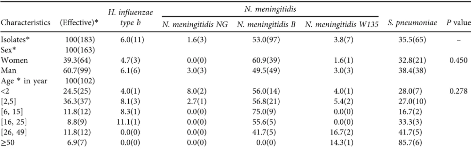
Characteristics of the isolates (Table 1)
Of the 183 isolates collected, N. meningitidis represented 58.4% including 53% (97/107) ofN. meningitidis serogroup B (N.meningitidis B), 3.8% (7/107) ofN. meningitidis serogroup W135 (N.meningitidis W135), and 1.6% (3/107) of non- serogrouped N. meningitidis (N. meningitidis NG). S. pneu- moniae and H. influenzae type b represented respectively 35.5% (65/183) and 6% (11/183). Regarding sex, 60.7% were male and 39.3% were female (Sex Ratio: 1.55). The median age of the sample studied was 3.9 [1.8; 17] years, ranging from 3 days to 65 years. In infant’s ˂2 years of age, N.
meningitidis represented 68% of meningitis cases followed by 28% ofS. pneumoniae. In the children of [2; 5] years,N.
meningitidisrepresented 64.9%, followed byS. pneumoniae with 27%. In older children [6; 15] years old,N. meningitidis represented 75% and S. pneumoniae was 16.7%. In adults [16; 25] years old,N. meningitidisrepresented 55.6% andS.
pneumoniae was 33.3%. In older adults [26; 49] years old, the N. meningitidis represented 58.4% and S. pneumoniae was at 41.7%. Beyond the age of 50, N. meningitidis was 14.3% while pneumoniae was 85.7%. However, at age ≤5 years, we have 66.7% (4/7) ofH. influenzae type bincluded in the study who reached this age.
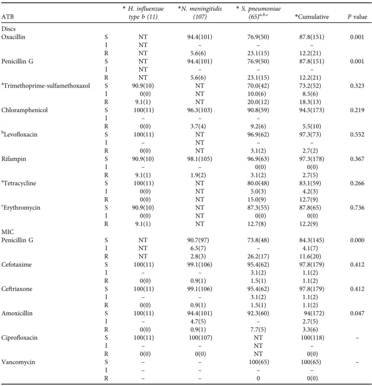
Antimicrobial resistance of isolates to antibiotics (Tables 2 and 3)
For all isolates tested ofN. meningitidis,S. pneumoniae, and H. influenzae type b; the decreased susceptibility to penicillin G (DSPG) represented 15.7% (27/183), with 11.6% (20/183) which were resistant to penicillin G (PG) and 4.1% (7/183) intermediate. Amoxicillin represents 2.7% (5/183) which were intermediate and 3.3% (6/183) resistant. 2.2% (2/183) of isolates were resistant to cefotaxime and ceftriaxone and 2.2% (2/183) were intermediate. 5.5% (10/183) were resis- tant to chloramphenicol and 2.7% (5/183) were resistant to rifampin. The frequency of DSPG observed in our study was
more common inS. pneumoniae thanN. meningitidis(P<
0.05). Of the 107 strains ofN. meningitidis, 9.3% (10/107) were DSPG of which 2.8% (3/107) were resistant, 5.6% (6/
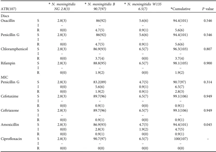
107) were reduced susceptibility to amoxicillin with a strain that was resistant 0.9% (1/107), one strain was resistant to ceftriaxone and cefotaxime 0.9% (1/107), 3.7% (4/107) strains were resistant to chloramphenicol and 1.9% (2/107) resistant to rifampin. All strains are susceptible to cipro- floxacin and no strains produced beta-lactamase. The level of DSPG and amoxicillin resistance inN. meningitidis Band N. meningitidis W135 were respectively 7.5% (8/107) vs.
1.8% (2/107) and 3.7% (4/107) vs. 1.9% (2/107). While resistance to ceftriaxone, cefotaxime, rifampin, and chlor- amphenicol was only recorded inN. meningitidis B. In our sample, resistance to amoxicillin was more common in N.
meningitidis B than N. meningitidis W135 (P < 0.05). All strains ofN. meningitidiswith DSPG have MIC of 0.064
m
g/mL at 0.25
m
g/mL and the resistant have MIC of 0.38m
g/mLat 1
m
g/mL. MIC amoxicillin in intermediate isolates range from 0.19m
g/mL to 0.75m
g/mL and the only resistant isolate was a MIC of 1m
g/mL. The MIC of ceftriaxone and cefo- taxime for the resistant strain was 0.25m
g/mL.Of the 65 strains ofS. pneumoniae, 26.2% (17/65) were of DSPG and 7.7% (5/65) were resistant to amoxicillin, 4.6%
(3/65) were of reduced susceptibility to ceftriaxone and cefotaxime with 3.1% (2/65) were intermediate and one strain 1.5% (1/65) was resistant, 9.2% (6/65) strains were resistant to chloramphenicol and 3.1% (2/65) were resistant to rifampin. Cotrimoxazole recorded 30% (18/65) of reduced susceptibility of which 20% (12/65) were resistant and 10%
(8/65) were intermediate. Levofloxacin and erythromycin represented 2.7% (2/65) and 12.7% (8/65) of resistance, respectively. Tetracycline represented 15% (9/65) of resis- tance and 5% (3/65) of intermediate resistance. All strains are susceptible to vancomycin. Strains ofS. pneumoniaethat were resistant to PG have MIC between 0.125
m
g/mL and 2m
g/mL. The MIC of resistant amoxicillin was 0.75–1m
g/mL.The intermediate MIC of cefotaxime and ceftriaxone was 0.75
m
g/mL at 1m
g/mL, and that of the resistant strain was 2m
g/mL.Table 1.Characteristics of the population of isolates studied
Characteristics (Effective)* H. influenzae type b
N. meningitidis
S. pneumoniae Pvalue N. meningitidis NG N. meningitidis B N. meningitidis W135
Isolates* 100(183) 6.0(11) 1.6(3) 53.0(97) 3.8(7) 35.5(65) –
Sex* 100(163)
Women 39.3(64) 4.7(3) 0.0(0) 60.9(39) 1.6(1) 32.8(21) 0.450
Man 60.7(99) 6.1(6) 3.0(3) 49.5(49) 3.0(3) 38.4(38)
Age*in year 100(102)
<2 24.5(25) 4.0(1) 8.0(2) 56.0(14) 4.0(1) 28.0(7) 0.278
[2,5] 36.3(37) 8.1(3) 2.7(1) 56.8(21) 5.4(2) 27.0(10)
[6, 15] 11.8(12) 8.3(1) 0.0(0) 75.0(9) 0.0(0) 16.7(2)
[16, 25] 8.8(9) 11.1(1) 0.0(0) 55.6(5) 0.0(0) 33.3(3)
[26, 49] 11.8(12) 0.0(0) 0.0(0) 41.7(5) 16.7(2) 41.7(5)
≥50 6.9(7) 0.0(0) 0.0(0) 0.0(0) 14.3(1) 85.7(6)
*Percentage % (expressed in effective).N. meningitidis:Neisseria meningitidis;S. pneumoniae:Streptococcus pneumoniae;H. influenzae type b:Haemophilus influenzae type b.
All H. influenzae type b strains in our study were sus- ceptible to the majority of antibiotics except one strain that was resistant to erythromycin, rifampin, and cotrimoxazole.
No strains produced beta-lactamase.
Co-resistance to PG and other antibiotics (Table 4)
Of the strains ofS. pneumoniae DSPG: 8.3% were resistant to cotrimoxazole, 3.1% were resistant to levofloxacin, 1.54%
were resistant to rifampin, 10% were resistant to tetracycline,
4.6% were resistant to cefotaxime and ceftriaxone, and 7.7%
were resistant to amoxicillin. This association of co-resis- tance between PG, C3G, and amoxicillin inS. pneumoniae was statistically significant (P< 0.05).
In strains of N. meningitidisDSPG: 1.9% chloramphen- icol, 0.9% rifampin, 0.9% ceftriaxone, 0.9% cefotaxime, and 3.7% amoxicillin were resistant, respectively. This associa- tion of co-resistance between PG, Beta-lactam antibiotics, rifampin, and chloramphenicol in N. meningitidis was sta- tistically significant (P< 0.05).
Table 2.Analysis of common antibiotics inNM,SP, andHibisolates
ATB
*H. influenzae type b (11)
*N. meningitidis (107)
*S. pneumoniae
(65)a,b,c *Cumulative Pvalue
Discs
Oxacillin S NT 94.4(101) 76.9(50) 87.8(151) 0.001
I NT – – –
R NT 5.6(6) 23.1(15) 12.2(21)
Penicillin G S NT 94.4(101) 76.9(50) 87.8(151) 0.001
I NT – – –
R NT 5.6(6) 23.1(15) 12.2(21)
aTrimethoprime-sulfamethoxazol S 90.9(10) NT 70.0(42) 73.2(52) 0.323
I 0(0) NT 10.0(6) 8.5(6)
R 9.1(1) NT 20.0(12) 18.3(13)
Chloramphenicol S 100(11) 96.3(103) 90.8(59) 94.5(173) 0.219
I – – –
R 0(0) 3.7(4) 9.2(6) 5.5(10)
bLevofloxacin S 100(11) NT 96.9(62) 97.3(73) 0.552
I – NT – –
R 0(0) NT 3.1(2) 2.7(2)
Rifampin S 90.9(10) 98.1(105) 96.9(63) 97.3(178) 0.367
I – – 0(0) 0(0)
R 9.1(1) 1.9(2) 3.1(2) 2.7(5)
aTetracycline S 100(11) NT 80.0(48) 83.1(59) 0.266
I 0(0) NT 5.0(3) 4.2(3)
R 0(0) NT 15.0(9) 12.7(9)
cErythromycin S 90.9(10) NT 87.3(55) 87.8(65) 0.736
I 0(0) NT 0(0) 0(0)
R 9.1(1) NT 12.7(8) 12.2(9)
MIC
Penicillin G S NT 90.7(97) 73.8(48) 84.3(145) 0.000
I NT 6.5(7) – 4.1(7)
R NT 2.8(3) 26.2(17) 11.6(20)
Cefotaxime S 100(11) 99.1(106) 95.4(62) 97.8(179) 0.412
I – – 3.1(2) 1.1(2)
R 0(0) 0.9(1) 1.5(1) 1.1(2)
Ceftriaxone S 100(11) 99.1(106) 95.4(62) 97.8(179) 0.412
I – – 3.1(2) 1.1(2)
R 0(0) 0.9(1) 1.5(1) 1.1(2)
Amoxicillin S 100(11) 94.4(101) 92.3(60) 94(172) 0.047
I – 4.7(5) – 2.7(5)
R 0(0) 0.9(1) 7.7(5) 3.3(6)
Ciprofloxacin S 100(11) 100(107) NT 100(118) –
I – – NT –
R 0(0) 0(0) NT 0(0)
Vancomycin S – – 100(65) 100(65) –
I – – – –
R – – 0 0(0)
*Percentage% (expressed in effective); a, b, c: the number tested [a(60), b(64), c(63)]; NT: Not Tested.
DISCUSSION
This national study conducted in Morocco focuses on both the epidemiological profile of the three bacteria responsible for bacterial meningitis, which were N. meningitidis, S.
pneumoniae, and H. influenzae type b, and their resistance profile. For this reason, we collected 183 isolates from different regions and cities across the country from culture- confirmed meningitis cases. Isolates are divided into 58.5%
(107/183)N. meningitidis, 35.5% (65/183)S. pneumoniae, and 6% (11/183)H. influenzae type b. The age group most affected was that ≤5 years old, which represented 60.8%. This study showed that in Moroccan children and adults, meningococcal disease causes more meningitis followed by pneumococcal disease.H. influenzae type bcome last with very low rates and mainly reaches children ≤5 years old and unvaccinated adults. The man is more affected than the woman. The technical difficulties associated with the high rates of negative culture and the demanding transport conditions of these three bacteria are technical problems limiting the collections of these isolates. As a result, the rate of positive cultivation be- tween regions of the country varies between 7 and 12%.
In the Maghreb countries of northern Africa, despite the poverty of data, pneumococcus was the one that causes more
meningitis than meningococcal [6, 7]. In Europe and the African meningitis belt, meningococcal disease and pneu- mococcus were the two most dominant bacterial species in children and adults [8, 9].
The most dominant N. meningitidis serogroup in our study wasN. meningitidis Bfollowed byN. meningitidis W135.
Serogroup commonly detected in the African meningitis belt, serogroup C,W, andXare those that cause epidemics [8]; In Europe N. meningitidis Bwas responsible for most cases of meningococcal meningitis [9, 10]; while in South Africa areN.
meningitidis W135, in Saudi Arabia were N. meningitidis A and N. meningitidis W135 [11]. The bacteriological and epidemiological profile of meningitis in Morocco was different from neighboring countries, the African meningitis belt and even countries with a tourist destination (Turkey) or spiritual (Saudi Arabia) visited by Moroccan citizens. But this profile was very similar to the profile found in Europe.
As presented by our results, there was a dramatic regression in cases of meningitis due toH. influenzae type b.
This decrease in cases ofH. influenzae type bmeningitis in children≤5 years was the result of the introduction in 2007 into the national immunization program of theH. influen- zae type bconjugate vaccine for children from the age of 2 months [12].
Table 3.Resistance Analysis inNeisseria meningitidisserogroup
ATB(107)
*N. meningitidis NG 2.8(3)
*N. meningitidis B 90.7(97)
*N. meningitidis W135
6.5(7) *Cumulative Pvalue
Discs
Oxacillin S 2.8(3) 86(92) 5.6(6) 94.4(101) 0.546
I – – – –
R 0(0) 4.7(5) 0.9(1) 5.6(6)
Penicillin G S 2.8(3) 86(92) 5.6(6) 94.4(101) 0.546
I – – – –
R 0(0) 4.7(5) 0.9(1) 5.6(6)
Chloramphenicol S 2.8(3) 86.9(93) 6.5(7) 96.3(103) 0.807
I – – – –
R 0(0) 3.7(4) 0(0) 3.7(4)
Rifampin S 2.8(3) 88.8(95) 6.5(7) 98.1(105) 0.900
I – – – –
R 0(0) 1.9(2) 0(0) 1.9(2)
MIC
Penicillin G S 2.8(3) 83.2(89) 4.7(5) 90.7(97) 0.314
I 0(0) 5.6(6) 0.9(1) 6.5(7)
R 0(0) 1.9(2) 0.9(1) 2.8(3)
Cefotaxime S 2.8(3) 89.7(96) 6.5(7) 99.1(106) 0.949
I – – – –
R 0(0) 0.9(1) 0(0) 0.9(1)
Ceftriaxone S 2.8(3) 89.7(96) 6.5(7) 99.1(106) 0.949
I – – – –
R 0(0) 0.9(1) 0(0) 0.9(1)
Amoxicillin S 2.8(3) 86.9(93) 4.7(5) 94.4(101) 0.045
I 0(0) 2.8(3) 1.9(2) 4.7(5)
R 0(0) 0.9(1) 0(0) 0.9(1)
Ciprofloxacin S 2.8(3) 90.7(97) 6.5(7) 100(107) –
I – – – –
R 0(0) 0(0) 0(0) 0(0)
*Percentage % (expressed in effective); S: Sensitive, I: Intermediate, R: Resistant.
In Morocco, the pneumococcal PCV-13 vaccine was introduced into the national immunization program in October 2010 and subsequently replaced by PCV-10.
Despite the lack of serotyping of the S. pneumoniae included in this study, there was a decrease and stability in the rate of pneumococcal meningitis disease during the periods studied after the introduction of the anti-pneu- mococcal vaccine in the immunization schedule. In a local study in Casablanca that covered the period before and after the implantation of PCV-10 and PCV-13, the inci- dence rate of invasive pneumococcal infections associated with vaccine serotypes decreased after the vaccine was introduced in children less than 2 years of age [13]. Impact of replacing PCV-13 vaccine with PCV-10 was not influ- encing and both contribute to the reduction of invasive infections in children fewer than 5 years of age, when they were implanted in national immunization programs of countries [12, 14,15].
Currently, two vaccines are developed and available for active immunization against theN. meningitidisB serogroup [16, 17], but in Morocco the meningococcal B vaccine wasn’t used yet and if this serogroup is identified, prevention will be limited to chemoprophylaxis. However, vaccination with
tetravalent vaccine ACYW135 became mandatory for all Hajjis and visitors to Mecca.
United Kingdom (UK) was the first country to introduce N. meningitidis Bvaccine in 2015 for the ages of 2, 4, and 12 months, the impact of the introduction ofN. meningitidis B vaccine in the UK infant vaccination program reduced the incidence of meningitis due toNMB[18].
Amoxicillin-resistant strains of N. meningitidis in this study were 5.6%. Whereas in previous studies at the local researches, no strains were found to be resistant to amoxi- cillin, while theN. meningitidis DSPG level is 9.3% in this study, which has changed over time in relation to these re- searches [19, 20]. Thus, the resistance reported here remained less severe than those reported elsewhere [18, 20].
In our study, we recorded resistance to cefotaxime, ceftri- axone, and rifampin, this information remained to our knowledge thefirst resistance declared forN. meningitidisin Morocco and the North African Maghreb countries [21, 22].
Resistance to chloramphenicol was similar to the rate in Tunisia [22]. And moreover resistance of rifampin was declared in a coutry of Latin America (Brazil) [23], while in the African meningitis belt the reported results are below ours [24]. The phenotypic profile ofN. meningitidis DSPG Table 4.Co-resistance of common antibiotics relative to sensitivity and resistance of PG inNeisseria meningitidis and Streptococcus
pneumoniae N. meningitidis(107)
Pvalue
S. pneumoniae(65)a, b, c
Pvalue PG
Resistant** Sensible* Resistant** Sensible*
(RþI) R 2.8(3)
90.7 (97)
(RþI) R 26.2(17)
48 (73.85)
9.4(10) I 6.5(7) 26.2(17) I 0(0)
Discs**
SXTa – – – – ––– 8.3(5) 5(3) 22(13) 15(9) 0.881
– – 3.3(2) 7(4)
CHL 1.9(2) 1.9(2) 1.9(2) 1.9(2) 0.006 0(0) 0(0) 9.2(6) 9.2(6) 0.149
0(0) 0(0) 0(0) 0(0)
LEVb – – – – ––– 3.1(2) 3.1(2) 0(0) 0(0) 0.067
– – 0(0) 0(0)
RIF 0.9(1) 0.9(1) 0.9(1) 0.9(1) 0.020 1.5(1) 1.5(1) 1.5(1) 1.5(1) 0.458
0(0) 0(0) 0(0) 0(0)
TETa – – – – ––– 10(6) 8.3(5) 10(6) 6.7(4) 0.037
– – 1.7(1) 3.3(2)
ERYc – – – – ––– 4.8(3) 4.8(3) 7.9(5) 7.9(5) 0.326
– – 0(0) 0(0)
MIC**
CTX 0.9(1) 0.9(1) 0(0) 0(0) 0.000 4.6(3) 1.5(1) 0(0) 0(0) 0.012
0(0) 0(0) 3.1(2) 0(0)
CRO 0.9(1) 0.9(1) 0(0) 0(0) 0.000 4.6(3) 1.5(1) 0(0) 0(0) 0.012
0(0) 0(0) 3.1(2) 0(0)
AMX 3.7(4) 0.9(1) 1.9(2) 0(0) 0.000 7.7(5) 7.7(5) 0(0) 0(0) 0.001
2.8(3) 1.9(2) 0(0) 0(0)
CIP 0(0) 0(0) 0(0) 0(0) ––– – – – – –––
0(0) 0(0) – –
*Expressed as percentage %(expressed in effective); ** RþI(%): R(%)/I(%); PG: Penicillin G, SXT: Trimethoprim-sulfamethoxazole; CHL:
Chloramphenicol; LEV: Levofloxacine; RIF: Rifampin; TET: Tetracycline; ERY: Erythromycin; CTX: Cefotaxime; CRO: Ceftriaxone; AMX:
Amoxicillin; CIP: Ciprofloxacin; MIC: Minimum inhibitory concentration.
a60 isolates tested forS. pneumoniae.
b64 isolates tested forS. pneumoniae.
c63 isolates tested forS. pneumoniae.
or PG-resistant is associated with mutations in the penA gene responsible for this reduced susceptibility to PG [25].
Therefore, one study has shown that increased resistance to C3G was observed in strains of decreased susceptibility to penicillin hosting a new modified penA gene allele
“penA327”. These strains containing “penA327” allele belonged to the CC11 clonal complex and belonged to serogroup B and C [26]. In our study,N. meningitidisDSPG isolates were associated with co-resistance with chloram- phenicol and rifampin (P < 0.05) and resistance to third- generation cephalosporin – C3G –(cefotaxime and ceftri- axone) and amoxicillin (P< 0.001).
Strains ofS. pneumoniaeDSPG in our study recorded a rate of 26.2%. This rate was lower than that reported in a local study and other countries, which were conducted prior to the introduction of PCV vaccine [6, 21, 27]. Iso- lates resistant to amoxicillin were 7.7%, while C3G was 4.6% reduced susceptibility in our study and one strain 1.5% (1/65) was resistant. These rates appeared a littley more than for amoxicillin and less than for C3G those reported in a other Moroccan study [27] and less than those reported elsewhere for both antibiotics [6, 21]. With the exception of cotrimoxazole (SXT), the observed resis- tance to chloramphenicol 9.2% and erythromycin 12.7%
were more or less equivocal to those of the Moroccan study [27]. The resistance to levofloxacin presented here was already described in a study in Morocco that reported a rate of resistance of 1.2% [28]. However, the resistance to rifampin was 3.1%; this resistance remained to our knowledge the only rate so far reported in the Kingdom of Morocco and in the north of Africa. The results of our study noted a co-resistance between PG and beta-lactam antibiotics inS. pneumoniae, this association was statisti- cally significant (P< 0.05).
The proportions of resistance reported in this study forS.
pneumoniaeappear generally to be less moderate and stable than the proportions exposed here later in Morocco or else- where. Thisfinding would be the result of the introduction of the pneumococcal vaccine PCVs into the national immuni- zation program in 2010, as it was an additional benefit for reducing antibiotic resistance ofS. pneumoniae[29].
Except one strain that was resistant to erythromycin, rifampin, and cotrimoxazole, the high susceptibility to anti- biotics and the rarity ofH. influenzae type bstrains observed here was the result of the introduction of anti-H. influenzae type bvaccine into the national vaccination program [12].
For the treatment of syndromic bacterial meningitis, the national meningitis program recommended the use of cephalosporin as much as empirical treatment for the management of cases of confirmed bacterial meningitis and for probable bacterial meningitis [5]. In this study, the re- sults of antibiotic susceptibility obtained for all isolates to cefotaxime, ceftriaxone, and vancomycin are very high;
despite the C3G resistance observed in this study, which remained very rare. In addition, our study recommended the continued use of this empirical treatment for children, ad- olescents, and adults in the national meningitis program with surveillance of the development of resistance.
Chemoprophylaxis in Morocco was based on taking Rifampin 200 mg twice in two days for the eradication ofN.
meningitidis oropharyngeal carried for contacts cases [5].
Oropharyngeal portage ofN. meningitidisvaries by age and geographic area, it must be monitored laboratory-based to map the distribution of serogroup of N. meningitidis and determine the current potential need for vaccination [30, 31].
The resistance to rifampin observed in this study for the three isolates may have evolved as a result of the use of this prophylaxis for contact cases ofN. meningitidis[32] and/or could be due to the use of rifampin in the treatment of tuberculosis patients in Morocco which remained a public health problem in the Kingdom [33]. Phenotypic studies and genotypic studies are useful for monitoring serogroup dis- tribution and also assist national vaccination programs in the choice of vaccine to introduce [34].
All this would result in a review of the prophylactic vision in the contacts either by an adequate vaccine intervention or moderation in chemoprophylaxis with monitoring of developments of meningitis disease in these patients.
CONCLUSION
In conclusion, syndromic bacterial meningitis ofN. menin- gitidis, S. pneumoniae, and H. influenzae type b isolates in Morocco appeared to have a high susceptibility to antibiotics used for empirical treatment and chemoprophylaxis despite the appearance of some resistance to amoxicillin, rifampin, and cephalosporin which remained very low and very rare.
The anti-H. influenzae type band PCVs vaccination intro- duced into the national immunization program reflected good efficacy. For theN. meningitidisvaccine, this needs to be revised and adapted to the current situation. In the end, monitoring the evolution of antibiotic resistance in these three isolates and monitoring the effectiveness of long-term vaccines are necessary for disease control and for the ade- quacy of treatment and prophylaxis over time.
Conflicts of interest:The authors declare no conflict of in- terest.
ACKNOWLEDGMENTS
We thank all colleagues of the hospital laboratories for the conservation of recovered strains and the information pro- vided.
We thank Prof. Mohammed Frikh for the reading of the article and the corrections.
We thank Miss Nada Ikken and Mr. Anass Ikken for the English language reading of the article and its correction.
REFERENCES
[1] Houri H, Pormohammad A, Riahi SM, Nasiri MJ, Fallah F, Dabiri H, et al. Acute bacterial meningitis in Iran: Systematic review and
meta-analysis. PLoS One 2017; 12: e0169617. https://doi.org/10.
1371/journal.pone.0169617.
[2] Agier L, Martiny N, Thiongane O, Mueller JE, Paireau J, Watkins ER, et al. Towards understanding the epidemiology of Neisseria meningitidis in the African meningitis belt: a multi-disciplinary overview. Int J Infect Dis 2017; 54: 103–12. https://doi.org/10.
1016/j.ijid.2016.10.032.
[3] Merabet M, Aouragh R, Idrissi A. Les meningites bacteriennes aigues communautaires chez les enfants de moins de 5 ansa la region Tanger-Tetouan-Al Hoceima (Maroc) 2006-2015 : Profil
epidemiologique, clinique et biologique.Community-acquired acute bacterial meningitis in children under 5. Antropo 2018; 40:
1–11.
[4] Costerus JM, Brouwer MC, Bijlsma MW, Tanck MW, van der Ende A, van de Beek D. Impact of an evidence-based guideline on the management of community-acquired bacterial meningitis: a prospective cohort study. Clin Microbiol Infect 2016; 22: 928–33.
https://doi.org/10.1016/j.cmi.2016.07.026.
[5] Barkia A, Braikat M, Charof R, Souadiya H, El Idrissi MH, Moumni H, et al. Guide de la lutte contre les meningites bacteriennes communautaires. Guideline on the Community Bacterial Meningitis Control. MoH Moro, WHO 2010; 78: 1–78.
[6] Ramdani-Bouguessa N, Ziane H, Bekhoucha S, Guechi Z, Azzam A, Touati D, et al. Evolution of antimicrobial resistance and serotype distribution of Streptococcus pneumoniae isolated from children with invasive and noninvasive pneumococcal diseases in Algeria from 2005 to 2012. New Microbes New Infect 2015; 6:
42–8.https://doi.org/10.1016/j.nmni.2015.02.008.
[7] Smaoui H, Bouafsoun A, Kechrid A. Epidemiology of bacterial meningitis in tunisian children (2000–2011). Arch Dis Child 2012; 97: A249–A249.https://doi.org/10.1136/archdischild-2012- 302724.0867.
[8] World Health Organization. Meningitis Weekly Bulletin.Weekly feedback bulletin on cerebrospinal meningitis. 2019 April 1st to 7th. 1–10 p. n.d.
[9] van Ettekoven CN, van de Beek D, Brouwer MC. Update on community-acquired bacterial meningitis: guidance and chal- lenges. Clin Microbiol Infect 2017; 23: 601–6.https://doi.org/10.
1016/j.cmi.2017.04.019.
[10] Kharkhal HN, Titov LP. Serogroup diversity and antibiotic sus- ceptibility of Neisseria meningitidis: meningococcus infection monitoring in Belarus. Acta Microbiol Immunol Hung 2019; 66:
443–57.
[11] Borrow R, Caugant DA, Ceyhan M, Christensen H, Dinleyici EC, Findlow J, et al. Meningococcal disease in the Middle East and Africa: Findings and updates from the Global Meningococcal Initiative. J Infect 2017; 75: 1–11. https://doi.org/10.1016/j.jinf.
2017.04.007.
[12] Braikat M, Barkia A, Mdaghri N El, Rainey JJ, Cohen AL, Teleb N.
Vaccination with Haemophilus influenzae type b conjugate vac- cine reduces bacterial meningitis in Morocco. Vaccine 2012; 30:
2594–9.https://doi.org/10.1016/j.vaccine.2012.01.041.
[13] Diawara I, Zerouali K, Katfy K, Zaki B, Belabbes H, Najib J, et al.
Invasive pneumococcal disease among children younger than 5 years of age before and after introduction of pneumococcal con- jugate vaccine in Casablanca, Morocco. Int J Infect Dis 2015; 40:
95–101.https://doi.org/10.1016/j.ijid.2015.09.019.
[14] de Oliveira LH, Camacho LAB, Coutinho ESF, Martinez-Silveira MS, Carvalho AF, Ruiz-Matus C, et al. Impact and Effectiveness of 10 and 13-Valent Pneumococcal Conjugate Vaccines on Hospi- talization and Mortality in Children Aged Less than 5 Years in Latin American Countries: A Systematic Review. PLoS One 2016;
11: e0166736.https://doi.org/10.1371/journal.pone.0166736.
[15] Shah N. Impact of pneumococcal conjugate vaccines (PCV) on pneumonia, the forgotten killer of children. Pediatr Infect Dis 2016; 8: 72–5.https://doi.org/10.1016/j.pid.2016.06.001.
[16] Ceyhan M, Ozsurekci Y, Lucidarme J, Borrow R, G€urler N, Emiroglu MK, et al. Characterization of invasive Neisseria men- ingitidis isolates recovered from children in Turkey during a period of increased serogroup B disease, 2013–2017. Vaccine 2020;
38: 3545–52.https://doi.org/10.1016/j.vaccine.2020.03.024.
[17] Parikh SR, Campbell H, Bettinger JA, Harrison LH, Marshall HS, Martinon-Torres F, et al. The everchanging epidemiology of meningococcal disease worldwide and the potential for prevention through vaccination, J Infect 2020; 81(4): 483–98.https://doi.org/
10.1016/j.jinf.2020.05.079.
[18] Watson PS, Turner DPJ. Clinical experience with the meningo- coccal B vaccine, Bexsero(®): prospects for reducing the burden of meningococcal serogroup B disease. Vaccine 2016; 34: 875–80.
https://doi.org/10.1016/j.vaccine.2015.11.057.
[19] Zerouali K, Elmdaghri N, Boudouma M, Benbachir M.
Serogroups, serotypes, serosubtypes and antimicrobial suscepti- bility of Neisseria meningitidis isolates in Casablanca, Morocco.
Eur J Clin Microbiol Infect Dis 2002; 21: 483–5.https://doi.org/10.
1007/s10096-002-0736-y.
[20] El Mdaghri N, Jilali N, Belabbes H, Jouhadi Z, Lahssoune M, Zaid S. Epidemiological profile of invasive bacterial diseases in children in Casablanca, Morocco: antimicrobial susceptibilities and sero- type distribution. East Mediterr Health J 2012; 18: 1097–101.
https://doi.org/10.26719/2012.18.11.1097.
[21] Ben Haj Khalifa A, Mastouri M, Ben Abdallah H, Noomen S, Kheder M. Les meningites purulentes dans la region de Monastir, Tunisie (1999–2006) : aspects bacteriologiques etetat de resistance aux antibiotiques. Acquired bacterial meningitis in Monastir re- gion, Tunisia (1999–2006): bacteriological aspects and suscepti- bility. Bull La Soc Pathol Exot 2011; 104: 42–8.https://doi.org/10.
1007/s13149-010-0077-5.
[22] Saguer A, Smaoui H, Taha M-K, Kechrid A. Characterization of invasive Neisseria meningitidis strains isolated at the Children’s Hospital of Tunis, Tunisia. East Mediterr Health J 2016; 22: 343–9.
https://doi.org/10.26719/2016.22.5.343.
[23] Gorla MC, Pinhata JMW, Dias UJ, de Moraes C, Lemos AP. Sur- veillance of antimicrobial resistance in Neisseria meningitidis strains isolated from invasive cases in Brazil from 2009 to 2016. J Med Microbiol 2018; 67: 750–6.https://doi.org/10.1099/jmm.0.000743.
[24] Hedberg ST, Fredlund H, Nicolas P, Caugant DA, Olcen P, Unemo M. Antibiotic susceptibility and characteristics of Neisseria menin- gitidis isolates from the African meningitis belt, 2000 to 2006:
phenotypic and genotypic perspectives. Antimicrob Agents Che- mother 2009; 53: 1561–6.https://doi.org/10.1128/AAC.00994-08.
[25] Harcourt BH, Anderson RD, Wu HM, Cohn AC, Macneil JR, Taylor TH, et al. Population-based surveillance of neisseria meningitidis antimicrobial resistance in the United States. Open Forum Infect Dis 2015; 2(3).https://doi.org/10.1093/ofid/ofv117.
[26] Deghmane A-E, Hong E, Taha M-K. Emergence of meningococci with reduced susceptibility to third-generation cephalosporins. J Antimicrob Chemother 2017; 72: 95–8. https://doi.org/10.1093/
jac/dkw400.
[27] Benbachir M, Elmdaghri N, Belabbes H, Haddioui G, Benzaid H, Zaki B. Eleven-year surveillance of antibiotic resistance in Strep- tococcus pneumoniae in Casablanca (Morocco). Microb Drug Resist 2012; 18: 157–60.https://doi.org/10.1089/mdr.2011.0130.
[28] Benouda A, Ben Redjeb S, Hammami A, Sibille S, Tazir M, Ramdani-Bouguessa N. Antimicrobial resistance of respiratory pathogens in North African countries. J Chemother 2009; 21:
627–32.https://doi.org/10.1179/joc.2009.21.6.627.
[29] Levy C, Varon E, Taha MK, Bechet S, Bonacorsi S, Cohen R, et al.
Evolution des meningites bacteriennes de l’enfant en France sous l’effet des vaccinations.Change in French bacterial meningitis in children resulting from vaccination. Arch Pediatr 2014; 21:
736–44.https://doi.org/10.1016/j.arcped.2014.04.025.
[30] Fekete S, Szabo D, Tamas L, Polony G. The role of the microbiome in otorhinolaryngology. Orv Hetil 2019; 160: 1533–41.https://doi.
org/10.1556/650.2019.31451.
[31] Tefera Z, Mekonnen F, Tiruneh M, Belachew T. Carriage rate of Neisseria meningitidis, antibiotic susceptibility pattern and asso- ciated risk factors among primary school children in Gondar town, Northwest Ethiopia. BMC Infect Dis 2020; 20: 358.https://
doi.org/10.1186/s12879-020-05080-w.
[32] TothA, Berta B, Tirczka T, Jekkel C, Abrah am A, Prohaszka Z, et al. First description of a rifampicin-resistant Neisseria menin- gitidis serogroup Y strain causing recurrent invasive meningo- coccal disease in Hungary. Acta Microbiol Immunol Hung 2017;
64: 1–7.https://doi.org/10.1556/030.64.2017.006.
[33] Karimi H, En-Nanai L, Oudghiri A, Chaoui I, Laglaoui A, Bour- kadi JE, et al. Performance of GenoType®MTBDRplus assay in the diagnosis of drug-resistant tuberculosis in Tangier, Morocco. J Glob Antimicrob Resist 2018; 12: 63–7.https://doi.org/10.1016/j.
jgar.2017.09.002.
[34] Brik A, Terrade A, Hong E, Deghmane A, Taha MK, Bouafsoun A, et al. Phenotypic and genotypic characterization of meningococcal isolates in Tunis, Tunisia: High diversity and impact on vaccina- tion strategies. Int J Infect Dis IJID Off Publ Int Soc Infect Dis 2020; 91: 73–8.https://doi.org/10.1016/j.ijid.2019.11.013.