GROWTH OF BHK CELLS FOR THE PRODUCTION AND PURIFICATION OF FOOT-AND-MOUTH DISEASE VIRUS
J. POLATNICK H. L. BACHRACH
Plum Island Animal Disease Center Agricultural Research Service U.S. Department of Agriculture
Greenport, New York
I. INTRODUCTION
The primary function of the Plum Island Animal Disease Center is to study animal diseases not presently endemic to the United States of America. Foot-and-mouth disease is the disease under most intensive study. To meet the needs for the production of foot-and-mouth disease virus (FMDV), we have developed and orga- nized facilities for the production of baby hamster kidney (BHK) cells in large quantities in roller bottles. These cells are in- fected with FMDV, and the crude virus harvests are subsequently processed to yield pure virus of good quality and in satisfactory quantity for biochemical and immunological studies. Previous pa- pers during the past decade (Polatnick and Bachrach, 1964? Bach-
603
rach and Polatnick, 1968; Polatnick and Bachrach, 1972) and the following discussion present details of the procedures used.
II. EQUIPMENT, CELLS, AND MEDIA
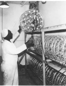
We are currently producing about 3 χ 1 0 H BHK cells weekly, distributed among some 400 2-liter round Baxter bottles. Sets of nineteen of these bottles are held in place in cylindrical wire racks and rotated on a three-tiered roller mill. The bottles re- main enclosed in the racks throughout all cycles of washing, dry heat sterilization, cell seeding, virus production, and decontam- ination. Figure 1 shows the roller mill with bottle-containing racks in place and the movable overhead electric hoist that is
FIGURE 1 Three-tiered roller mill for cylindrical wire racks each holding 19 Baxter bottle cultures.
GROWTH OF BHK CELLS 605
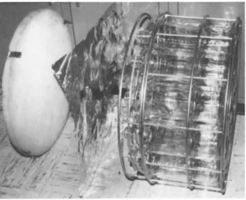
FIGURE 2 Partially exploded view of cylindrical rack for holding 19 Baxter bottles showing metal lid with polyurethane foam bonded to undersidef aluminum foil, and two-piece wire rack - upper section is joined to lower section by three wing nuts.
used to maneuver the racks. Figure 2 provides an exploded view of a rack, showing the sheet of heavy-weight aluminum foil that serves to cover the bottles instead of individual rubber stopper- ing. The aluminum foil is pressed tightly in place by polyure- thane foam bonded to the underside of the lid which in turn clips onto the upper rim of the rack.
Uncloned BHK cells were originally obtained from McPherson and Stoker. Later, BHK clone 13 from passage 21 cells were ob- tained from the American Type,Culture Collection, Rockville, Md.
Early passages of these cells were dispersed in a dimethysul- foxide-containing medium, frozen at a controlled rate, and stored in liquid nitrogen. These frozen cells are drawn upon whenever cells currently in production require replacement.
The original McPherson-Stoker (1962) growth medium called for the use of twice the usual concentration of purified amino acids in a bicarbonate-buffered modified Eagle1s basal medium, 80%,
Component Percentage in medium
Eagle's basal medium, modified to contain 0.02 M Tris and 0.5%
lactalbumin hydrolysate Tryptose phosphate broth
(Difco) Bovine serum Penicillin Streptomycin Fungizone
10
100 units/ml 100 yg/ml 2 yg/ml 80
10
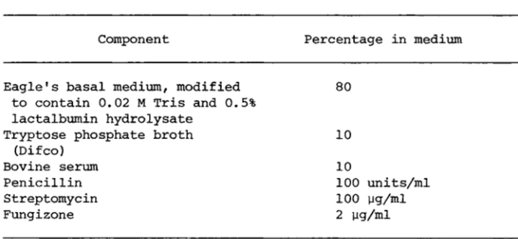
containing 10% tryptose phosphate, and 10% calf serum. For eco- nomic reasons, we replaced the costly purified amino acids with lactalbumin hydrolyzate fortified with histidine. For ease of handling and for preventing large swings in pH, the bicarbonate buffer was essentially replaced by 0.02M Tris, and an adjustment was made in the sodium chloride concentration to maintain iso- tonicity (Polatnick and Bachrach, 1964). Table I outlines the composition of the growth medium.
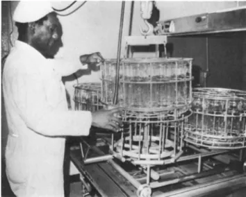
The media (minus serum, glutamine, vitamins, and antibiotics, which are prepared separately and stored frozen) is sterilized by passage through 220 millimicron Millipore filters and usually stored for at least one week at 37° and another two weeks at room temperature. Just prior to use, the serum and other tem- perature-sensitive components are added. The bottles are washed
in a specially constructed unit servicing 4 loaded racks at a time, as shown in Fig. 3, after which they are sterilized in a dry-heat room-sized oven as shown in Fig. 4 (Bachrach and Polat- nick, 1968).
TABLE I Growth Medium for Production of BHK Cells
FIGURE 3 Glassware washer with special header to accommodate four racks of Baxter bottles.
FIGURE 4 Dry heat sterilizing chamber measuring 4 feet 10 inches by 9 feet 5 inches.
607
III. CELL PASSAGE
Cells grown in the presence of serum screened for adventitious viruses and mycoplasma are reserved for use in seeding new cul- tures. One hundred ml of growth medium containing 20-25 χ 1 06 of these cells are delivered into each production bottle, and the cells are incubated for 4 days at 37°. The fluid is changed and incubation is continued for another 2 days. At this time, growth has reached 600-800 x 1 06 cells, which are now used for virus production.
IV. VIRUS PRODUCTION
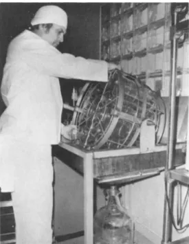
FIGURE 5 Tilting apparatus for discarding spent media direct- ly to a waste line and also for collecting virus fluids into a chilled vessel. The figure shows equipment for the latter opera- tion.
GROWTH OF BHK CELLS 609
The spent growth medium from the 6-day-old BHK cells is dis- carded by means of the tilting apparatus shown in Fig. 5, and the drained cultures are infected with virus in 60 ml of an equi- volume mixture of the modified Eagle's salts and 0.16 M Tris buf- fer in the absence of any serum. Seed virus is maintained by passage only through primary calf kidney culture cells. For vi- rus production, the seed virus is used to infect a few 5-day-old cultures of BHK cells to obtain virus for use in infecting all the major virus-production cells on the following day. The mul- tiplicity of infection is of the order of several tenths of a plaque-forming unit (PFU) per cell, and the infectious fluid is harvested when the cells start sloughing off the glass. The time required for maximum virus yield varies with the virus type, but it is usually 20-24 hours.
V. VIRUS PURIFICATION
The crude virus harvests, with titers averaging 2-3 x 1 08 PFU per ml, are freed of cell debris by passage through a Sharpies centrifuge. Polyethylene glycol (PEG) of 20,000 daltons is added to a clarified harvest to make a 6% solution (Wagner et al., 1970). After standing for a minimum of one hour, or over a week- end at 4° if the work schedule so requires, the PEG precipitate is collected in Spinco 15 rotor tubes by spinning for 20 minutes at 15,000 rpm. The precipitate is resuspended in 0.16 M Tris buffer containing 1.0% EDTA (Tris-EDTA) to one-tenth of the orig- inal harvest volume. The PEG precipitation cycle is repeated once more, yielding a hundred-fold concentrate of partially puri- fied virus with titers ranging from 1-3 * 1 01 0 PFU per ml.
This virus concentrate is then layered over CsCl and centri- fugea in a Spinco SW 25.2 rotor for 200 minutes at 25,000 rpm at 20°. The procedure, modified from Bachrach et al. (1964), has in each centrifuge tube, from the bottom up, 0.5 ml of pi.6 CsCl in
Tris-EDTA, 7 ml of pi.42 CsCl in Tris-EDTA, 7 ml of 80% solution of pi.42 CsCl in Tris-EDTA, and 42 ml of the twice PEG-precipi- tated viral concentrate. The viral zone is detectable after cen- trifugation at pi.43 by its strong light-scattering properties.
The tubes are punctured, and the viral zone in each tube is col- lected dropwise directly in a dialysis bag. The purified virus is dialyzed against several changes of 0.05 M potassium phos- phate, pH 7.5, containing 0.2 M potassium chloride, and is then stored in plastic vials at 4° for future investigative work. The virus preparations, with titers ranging from 5 χ 1 0 ^ to 5 χ 10"^
PFU/ml, meet several different standards of purity which will be discussed in the following section. There may be occasions when additional purification is required, and this is accomplished by an additional centrifugation of the virus through an organic liquid into CsCl (Bachrach and Polatnick, 1968).
The concentration of virus is determined from optical density measurements at 259 my, at which wavelength FMDV has an extinction coefficient of 76.0 (Bachrach et al., 1964). Yields of pure virus have reached as high as 18 mg per rack of BHK cells, or approxi- mately 1 mg per bottle, depending on virus type. A more usual yield is from 5 to 6 mg per rack.
VI. PARAMETERS OF VIRUS QUALITY
Pure FMDV has icosahedral symmetry with an estimated 32 cap- someric subunits (Breese et al., 1965). Figure 6 shows a nega- tively stained electron micrograph in which some details of the fine structure of the virus can be seen.
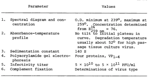
The procedures in standard use to establish virus purity and integrity are the absorbance-temperature (A-T) profile (Bachrach, 1964), and SDS disc Polyacrylamide gel electrophoresis (PAGE).
In determining the A-T profile, virus is first diluted in 0.01 M potassium phosphate, pH 7.5, to an optical density value of about
GROWTH OF BHK CELLS 611
FIGURE 6 Negatively stained (phosphotungstic acid) electron micrograph of foot-and-mouth disease virus, type A; magnification 450,000 X. Picture courtesy of S. S. Breese, Jr. of this labora- tory.
0.3. Readings are taken in a Beckman DK2 ratio-recording spec- trophotometer, using electrically heated sealed cuvettes. As seen in Fig. 7, the relative absorbance of intact purified virus remains unchanged from 0 to about 52° on curve 1, after which the viral RNA is released and undergoes reversible heat denaturation
(hyperchromic section of curve 1 and all of curve 2 ) . The exact temperature of virus degradation (T^) is a function of the con- centration and valence of the cation, the virus type and its pas- sage history. Virus from animals, which has had only a few pas- sages in tissue culture, degrades at temperatures of 40° or lower, producing a transient hypochromic dip in the A-T profile of the released RNA. The RNA has a melting temperature (Tm = midpoint of O.D. rise) of about 56° and a hyperchromicity of 1.24. Partially degraded virus manifests itself by the appear-
ftW 6 S i I V 3 D N V 9 U O S a V 3 A I 1 V 1 3 M
*M CO 10 ,0 0
Ö MH •H
*H 3 •U
0 0 +J CO -ς "H
Cu fa cr»
0 • en •H
U tJ -u fa Ö
ϋ Ö -CJ Ö Ö 3 - M QJ 0 å 3ï
Q) Í
0 co M 4J + J Ö
bï 00 3 0 fO Ö c; Ö
¹ Ö • H
"ä CO
å Ή —ν "M fa fO Ö cq CO
4J *H CJ fO ¹ rH 0 Ö 0
ϋ 0 th
fa CJ
CO 1 •H
M Ö CO 0 Ö eu C* eu CU rH 3i fO rH s. M 00 LT) co
• c; 3ï CM "H c; rQ
S Ö ra
D 4J en
CJ 0 eu H 0 *H
CO eu CJ co QJ 0 3 3 rH
eu
l
tJ
ROM ·
ΜÇ ö ftî 3ïs. CM r-H Ö «Ë
¹ ·
¼
ΜÇ ffj
Ο tes
M C U r-H
eu ^ CO CO
Ö M Ö Ö Ö
¹ 4J ά Ö
CU
rH 3
4J Ο
fO ^ ΜÇ Ö Ο eu Ö -õ 4J t n ÎH
u
Ï co
3 • ¹ CJ>q
CO -U 3 fO
- CJ
Ö Ö
rH
Ö
^ - Ñ Ö fa co co
ï _ ï
c;CO ·<Η rC|
fa û . §
ö Ö
co to 3 -c; ra
ï ¼
MHra -M -õ é ς
ra -w Ö Ε,Ο Ö
O
C/)EH ΜÇ M
612
GROWTH OF BHK CELLS 613 TABLE II Parameters Used in the Routine Assessment
of the Quality of Production Runs of Purified and Concentrated Foot-and-Mouth Disease Virus, Types A, 0, and C
Parameter Values
1. Spectral diagram and con- O.D. minimum at 239°, maximum at centration 259°. Concentration determined
from E^L = 76. 259 nm
No tilt to initial plateau in 259°. Concentration determined from E^L = 76. 259 nm
No tilt to initial plateau in 2. Absorbance-temperature
259°. Concentration determined from E^L = 76. 259 nm
No tilt to initial plateau in profile curve. Degradation temperature
usually about 52° for high pas- sage tissue culture virus.
3. Sedimentation constant 140 S
4. Polyacrylamide gel electro- Four proteins, VPi_4 phoresis
5 x 1 01 0 to 5 x 1 01 1 PFU/ml 5. Infectivity titer 5 x 1 01 0 to 5 x 1 01 1 PFU/ml 6. Complement fixation Determinations of virus type
ance at low temperatures of a positively sloped line in place of the absorbance plateau shown by intact virus.
PAGE runs on 12.5% gels containing 8 M urea reveal three major proteins of FMDV, VP-^, VP2/ ar*d VP3 of molecular weights in thou-
sand daltons of about 33, 30, and 27, respectively. A fourth vi- rus protein, V P4, is frequently seen at about the 13,000 dalton zone. Virus protein 3 is sensitive to tryptic digestion, yield- ing a V P 3a^ 3k doublet difficult to resolve photographically. Vi- rus preparations from BHK cells sometimes contain V P 3a 3^, pre- sumably produced by cellular or serum trypsin or by other en- zymes with similar specificity. Figure 8 shows typical PAGE pat- terns of virus containing small (A) and large (B) amounts of
v p3 a , 3b-
When considered necessary, analytical ultracentrifugation and complement fixation tests are conducted on purified virus for con- firmation of homogeneity and immunological type, respectively.
Table II shows the parameters used in assessing the quality of production runs of purified virus.
VII. DISCUSSION AND CONCLUSION
The current weekly output of the virus production unit from 400 Baxter bottle cultures of BHK cells is 100 to 200 mg of puri- fied virus. This production represents only a portion of our capacity of 2,052 cultures. All phases of production at the pres- ent level, including cleaning and decontaminating the working area, are carried out by three technicians. It is estimated that full production could be attained without an appreciable increase in staff, but such production would necessitate more serum for cell growth and zonal centrifuges for purifying the virus.
As in most cell production facilities, our system is not with- out its problems. Difficulties are at times encountered with the cells, virus, serum and other components of the system even when these components are pretested. Problems appear to occur most frequently during the humid weeks of late summer and have been attributed in the past to mold contamination or to the seemingly poor growth-promoting properties of serum collected from our cat- tle during that season. For purposes of decontamination, the whole working area can be fumigated by the introduction of form- aldehyde through the air inlet ducts.
The use of bottle cultures rather than large suspension tanks provides flexibility for the concommitant preparation of differ- ent cell types. Of perhaps more importance, cells used in the roller cultures can be and have been of primary tissue origin, in contrast to the heteroploid or tumorigenic cells required for growth in suspension. A further advantage of the roller bottle system lies in its adaptability to researchers with various needs. Racks of cells are delivered routinely to several labora- tory research sections where they are infected with any one of the 7 different types (at least 61 subtypes) of FMDV or other viruses being investigated.
GROWTH OF BHK CELLS 615 REFERENCES
Bachrach, H. L., C1964), J. Mol. Biol. 8, 348-358.
Bachrach, H. L., Trautman, R., Breese, S. S., (1964), Am. J. Vet.
Res. 25, 333-342.
Bachrach, H. L., Polatnick, J., (1968), Biotech, and Bioeng. 10, 589-599.
Breese, S. S., Jr., Trautman, R., Bachrach, H. L*, (1965), Science 150, 1303-1305.
MacPherson, I., Stoker, M., C1962), Virology 16, 147-151.
Polatnick, J., Bachrach, H. L., (1964), Appl. Microbiol. 12, 368- 373.
Polatnick, J., Bachrach, H. L., (1972), Growth 36, 247-253.
Wagner, G. G., Card, J. L., Cowan, Ê., (1970), Archiv, ges.
Virusforsch 30, 343-352.
