MICROBIAL DYSBIOSIS AND MICROBIOTA – GUT – RETINA AXIS: THE LESSON FROM BRAIN NEURODEGENERATIVE DISEASES TO PRIMARY
OPEN-ANGLE GLAUCOMA PATHOGENESIS OF AUTOIMMUNITY
N
ARTTAYAC
HAIWIANGand T
EERAP
OYOMTIP*
Faculty of Optometry, Ramkhamhaeng University, Bangkok, Thailand(Received: 23 March 2019; accepted: 23 April 2019)
In recent years, microbiota-associated neurodegenerative diseases have been exploited and provided new insight into disease pathogenesis. However, primary open-angle glaucoma (POAG), known as a complex neurodegenerative disease resulting from retinal ganglion cell death and optic nerve damage, can cause irreversible blindness and visualfield loss. POAG, which shares several similarities with Parkinson’s disease (PD) and Alzheimer’s disease (AD), has limited studies and slow progression in the understanding of pathogenesis when compared to PD and AD.
In this review, we summarized the current knowledge of POAG and commensal microbiota, combined with several lines of evidence in PD and AD to propose a possible hypothesis for POAG pathogenesis: microorganisms cause glaucoma via gut–retina axis, resulting in autoantibodies and autoreactive T cells that lead to autoimmunity. Furthermore, dual-hit hypothesis, an example of a commensal pathogen that causes PD, was partially exported in POAG. Finally, future perspectives are suggested to expand understanding of POAG.
Keywords: primary open-angle glaucoma, microbiota, gut–retina axis, pathogenesis, autoimmunity, neurodegenerative diseases
Introduction
Primary open-angle glaucoma (POAG) is recognized as an ocular neurodegenerative disease via retinal ganglion cells (RGCs) death and causes irreversible blindness as well as vision defects globally, leading to decreased quality of life [1]. Glaucoma will affect approximately 79.6 million people by 2020 and increase to 111 million by 2040 [2,
3]. The disease is classified into
*Corresponding author; E-mails:tpteera075@gmail.com;teera.p@ru.ac.th
elevated intraocular pressure (IOP) resulting from abnormal aqueous humor
flowing via the development of trabecular meshwork cell
fibrosis [4] and normal tension glaucoma (NTG; IOP
<22 mmHg). The end results of both types are RGCs death via innate and adaptive immunity. In the elevated IOP type, current treatments are IOP reduction composed of surgical intervention or medication.
Surgeries may be associated with potential vision-threatening consequences, while the failure rate is approximately 11% [5
–7]. For this reason, medication therapiesare considered the primary care option, which is a signi
ficant step in minimizing the disease progression in the early stages of glaucomatous eye development.
However, currently available pharmacological treatments may cause adverse effects and drug interactions. Long-term treatment can cause subclinical in
flam- mation and conjunctiva
fibrosis development leading to increasing failure rates of trabeculectomy [8
–11]. The long-term treatment initially requires two or moreantiglaucoma drugs, which may predict failure in the present medication [5,
6].Importantly, combination drug treatments are widely used for lowering IOP, leading to low patient compliance with multiple daily administrations of eye drops. Consequently, understanding of cellular and molecular pathogenesis is an essential pathway to the development of novel interventions and therapies to overcome glaucoma.
Besides the elevated IOP, NTG patients are also treated with IOP-lowering medication. However, 12% of patients
’treatments are not successful in terms of disease control. Moreover, the
“Collaborative Normal-Tension Glaucoma Study
”showed that 30% of IOP reduction was not able to suppress visual loss.
These studies indicated that IOP management is not a prominent target for medica- tion [12]. At present, Trivli et al. [13] suggest that this type of glaucoma is closely associated with multifactorial components. The observational epidemiological study shows that the ocular morphology of NTG is associated with visual
field defects, suggesting that ocular morphology dysregulation alters the mechanical stress and axonal damage to destroy optic nerve heads and RGCs.
However, these
findings remain controversial [13,
14]. In addition, vascular disorderscomposed of vascular dysregulation, neovasculization, and ischemia have been
proposed as a pathogenesis of NTG [15]. Gherghel et al. [16] showed that ocular
blood
flow of a patient, as measured using the mean of a peripheral laser Doppler,
was altered depending on the temperature. A cold provocation test illustrates that the
instability of both mean ocular perfusion pressure and blood pressure (BP) occurred
in patients compared to healthy controls [17]. In a prospective clinical validation
study, the NTG patients impaired autonomic cardiovascular regulation, analyzed by
an alteration in BP variability and heart rate during resting conditions [18]. In a rabbit
experimental model, the instability, induced endothelin-1 injection of the optic nerve,
and blood
flow resulted in RGC loss [19]. The current model suggests that it is theeffect of chronic oxidative stress, known as ischemia-reperfusion injury [20].
However, these models do not offer a complete understanding of POAG pathogene- sis as there are several factors associated with POAG, such as genetic predisposition and iron-de
ficiency anemia [21,
22]. An initiation of POAG is still ambiguous andpathogenesis has not been clearly elucidated.
Previously, the pathophysiological features of glaucoma were compared with other neurodegenerative diseases, especially Parkinson
’s diseases (PD) and Alzheimer
’s disease (AD), leading to the reclassi
fication of glaucoma as optic neuropathy with effects in the central nervous system (CNS). The optic nerve is a part of the CNS. Several similarities in clinical manifestations, such as age, presymptomatic stage, clinical progression, genetic predisposition, and cellular pathogenesis, have been found to be shared between glaucoma and neurodegen- erative diseases [23,
24]. For this reason, the study of PD and AD may providenew insight into glaucoma disease.
In the past decade, human microbiota has been rapidly exploited into neurodegenerative diseases and autoimmunity. The alteration of intestinal microbiome into pathogenic bacteria or other groups of bacteria has been broadly recognized as microbial dysbiosis and plays a role as a contributing factor in disease pathogenesis [25,
26]. Intestinal gut microbiota is well characterized as aconfounding factor in several diseases such as amyotrophic lateral sclerosis (ALS), multiple sclerosis (MS), AD, and PD, since the gut microbiome is facile and changed via diet and exercise [27
–30]. A microbiota–gut
–brain axis is a cross-talk between gut and microbiota, in which enteric bacteria are able to enter the circulation and pass the blood
–brain barrier (BBB), known as the humoral pathway, to penetrate the CNS via the vagus nerve, which is a neural pathway [31,
32]. Inaddition, a dual-hit hypothesis was applied in PD, where a pathogen invades the brain through an immunoprivileged site like the eye of the host by gut and nose [33].
A population-based follow-up study covering 8 years showed that POAG was signi
ficantly associated with AD and saved as an AD predictor [34], suggesting that there is a potential link between brain degenerative diseases and glaucoma and retinal degenerative disease. Therefore, this review will summarize and propose the microbiota
–gut
–retina axis and dual-hit hypothesis as potential pathogenesis in POAG based on current knowledge of brain neurodegenerative diseases and glaucoma studies, which may be applied for the development of a novel therapy.
Commensal Microbiota: From Neurodegenerative Diseases to POAG
Microbial dysbiosis is a crucial player in the onset of neurological disorders
and progressive neuronal loss, including ischemic reperfusion injury [25,
35].In a mouse model, the gut microbiota was strongly associated with AD. For example, intensity-dependent chronic noise exposure reduced gut microbiota diversity and caused the increase of in
flammatory mediators, resulting in AD-like effects in the brain [36]. Signi
ficant differences in abundance of gut microbiota were shown when comparing between senescence-accelerated mouse prone 8 (SAMP8), recognized as well established deterministic of AD, and senescence-accelerated mouse resistance 1; as such, Lachnospiraceae, Alistipes species, Akkermansia species, and Odoribactor species were enriched in SAMP8 [37]. To con
firm this relationship, the Drosophila model with AD pathology showed various groups of commensal bacteria [38]. Recent human study revealed that the bacterial diversity of gut microbiota in AD patients is distinctive from normal controls, such as Actinobacteria, Bacteroides species, Ruminococcus species, Selenomonadales, and Lachnospiraceae [39]. Therefore, these are able to imply that the perturbation of commensal microbiota is involved in the pathogenesis of AD. Xu and Wang [40] suggested that the gut microbial metabolites should be considered as biomarkers because there is a positive correlation with disease progression in AD.
Regarding AD, several studies of the PD model also found a correlation between gut microbiota and disease pathogenesis, in which dopaminergic neurons were devastated by the aggregation and accumulation of
α-synuclein (
αSyn) [26,
41]. In dual-hit hypothesis (Braak’s hypothesis), the antigens may enter via the nasal and gastric pathways. The enteric nervous system is the early site of
αSyn, which subsequently locates in the brain via the vagus nerve [33,
42]. In theclinical study, the decreasing of Lachnospiraceae and increasing of Lactobacil- laceae as well as Christensenellaceae are associated with the severity of idiopathic PD [43]. Furthermore, PD patients were commonly found to have extraordinary increases in coliform bacteria in intestinal microbiome overgrowth [44,
45].Importantly, there is bias in anti-in
flammatory bacteria and pro-in
flammatory
bacterial microbiome between PD patients and healthy controls. Butyrate-
producing bacteria, normally genera Blautia, Coprococcus, and Roseburia, were
reduced in the feces of PD. On the contrary, Ralstonia genus, pro-in
flammatory
bacteria, was predominant in healthy controls compared to PD patients [46]. As a
result, the balancing of in
flammatory response might regulate gut microbiota,
which is associated with PD pathogenesis. A colonic biopsy sample of PD
patients, in which dysbiosis was detected by the decreasing of short-chain fatty
acids compared to healthy controls, increases Toll-like receptor 4 (TLR4),
cytokine expression, and CD3
+T cells. Moreover, TLR4 knockout mouse,
induced using oral rotenone, indicated that lower intestinal and motor dysfunction
including neurodegenerative and neuroin
flammation suggested that innate
immunity may play a role in microbial dysbiosis in PD [47].
Gastrointestinal permeability saves as an anatomic barrier and one explana- tion of microbiota and brain damage [25]. Increased permeability is able to disseminate bacterial components or pathogens to the blood and brain [48,
49]. Byway of illustration, the level of lipopolysaccharides (LPS) in serum roughly increases in AD and sporadic ALS patients. It is capable of activating monocytes and decreasing IL-10 [50]. In a rat model, the disruption of gut-barrier function and dysbiosis results in bacterial translocation [51]. Importantly, the BBB integrity of AD is decreased, which leads to immune activation [52]. Another example is a cohort observation in PD patients, where the
αSyn, and serum LPS-binding protein, endotoxin marker were increased and associated with intestinal hyper- permeability [53]. Among these studies, it was suggested that gastrointestinal permeability has a connection to the dissemination of antigens or in
flammatory factors to the brain.
At present, several diets have the ability to increase epithelial permeability [54] and alter microbiota. As a case in point, unsaturated fatty acid diets affect microbial varieties and increase eightfold in intestinal permeability in a mouse model [55] as well as chicken feeding with different patterns, ad libitum versus restrictive feeding, represented cecal microbiota deviation and may potentially disturb intestinal physiology including morphology and permeability [56].
Furthermore, there is evidence to support that commensal bacteria microorganisms interface with permeability. Chen et al. [57] elucidated that microbial treatment, Lactobacillus rhamnosus GG, improves intestinal permeability and modulates dysbiosis results in a protective role in a sepsis mouse model. Other treatment examples are Puerariae Lobatae Radix and Chuanxiong Rhizoma, which showed that this combination is able to rebalance gut microbiota dysbiosis and revive gut
–brain barrier disruption [58].
The roles of commensal microbiota in ocular disease are slowly being
explored, but are signi
ficantly important [59]. Previously, Horai et al. performed
spontaneous uveitis on a mouse model and showed that commensal bacteria activate
T cells by non-cognate interaction and subsequently promote autoreactive T cells
in
filtration across blood
–retinal barriers. The autoreactive T cells respond to
autoantigen in the retina and result in in
flammation [60]. In experimental
autoimmune uveitis, the pathology was induced by interphotoreceptor-binding
peptides, indicating the alteration of gut microbiome via oral antibiotics adminis-
tration associated with uveitis severity [61]. A recent study in animal models
coupled with adoptive transfer experiments that activated immune response by
antigen injection through the anterior chamber, which should activate T cells in the
spleen or thymus via anterior chamber-associated immune deviation (ACAID),
showed retinal neurodegeneration resulting from heat shock proteins (HSPs)-
specific CD4
+Th1, which requires priming from commensal bacteria [62,
63].Altogether, dysbiosis is also involved in basic glaucoma pathogenesis, similar to other neurodegenerative diseases. Therefore, the microbiota
–gut
–retinal axis is possible.
A common example of commensal pathogenic bacteria is Helicobacter pylori, which was reclassi
fied as extragastroduodenal diseases such as immune thrombocytopenic purpura and iron-de
ficiency anemia [64]. Accordingly, H. pylori cause other diseases. Many studies have supported that infected patients signi
ficantly increase the risk of developing several neurodegenerative diseases including PD, AD, MS, and POAG [65
–68]. In meta-analysis and case–control study, PD patients infected with H. pylori showed higher severity of PD and H. pylori eradication therapy to improve disease severity, suggesting that com- mensal pathogens may contribute to disease deterioration [66]. This is in accor- dance with glaucoma studies. Kountouras et al. [69] showed that H. pylori eradication decreases IOP and improves visual
field. In a study by Atilgan et al. [70], the H. pylori infection decreased temporal quadrant retinal nerve
fiber layer thickness when compared between pretreatment and posttreatment to eradicate H. pylori infection from patients, indicating the early signs of glaucoma.
Surprisingly, H. pylori can be detected in trabeculectomy specimens of patients, suggesting that bacteria can successfully colonize trabecular meshwork cells [71].
This evidence shows that the microbiota
–gut
–retina axis is available in our body, although the translocation route remains unknown.
In addition, the speci
fic H. pylori IgG antibody is able to be found in the serum and aqueous humor of POAG patients [72,
73]. Increasing the IgGantibody is possible for cross-reactivity with ocular tissue [74]. Previous researches showed that H. pylori activate autoantibodies via molecular mimicry in autoimmune disorders such as cardiovascular and autoimmune thyroid disease [75,
76].H. pylori-seropositive PD patients upregulated eight autoantibodies when compared with H. pylori-seronegative PD patients [77].
In glaucomatous tissue, HSPs are increased and play a role in neurodegenera- tion [78,
79], which is a potential target of autoantibodies due to the high levelof sequence homology with microbial HSPs [80]. In human studies, HSPs- speci
fic autoantibodies and other autoantibodies increase in the serum and aqueous humor of glaucoma patients [81
–83].Ex vivo stimulation by HSP60 in glaucoma peripheral blood monocyte showed Th2 bias [84]. This study in blood samples of glaucomatous patients compared with non-glaucomatous controls exhibited a trend toward decreased frequency of regulatory T cell. Ex vivo stimulation by a speci
fic antibody to
εchain of human CD3, CD28, and CD137 revealed CD4
+T response in glaucomatous samples [85]. In an animal model, a glaucomatous formation and RGCs loss were induced by HSPs [86,
87].Adoptive transfer of T and B cells from glaucomatous mice provokes
detrimental outcome in normal recipient mice [88]. For this reason, it might suggest that T-helper cell and B cell can circulate to the eye and destroy the retina cells resulting in the progression of visual
field loss.
Finally, the other innate immunity may collaborate to augment the pathology of glaucoma. For example, pathogen-associated molecular pattern recognitions are involved in glaucoma pathogenesis. For instance, the TLR4 may be the critical player in recognizing HSPs as damage-associated molecular patterns in glaucoma [89]. This is consistent with H. pylori, producing miscellaneous antigens that activate the immune system by TLRs and increase TLR4 expression in gastric epithelial cells [90,
91]. Taken together, a commen-sal bacterium such as H. pylori may be one actor in glaucoma pathogenesis by causing autoimmunity.
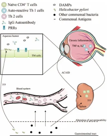
In summary, we proposed a hypothesis for glaucoma, neurodegenerative disease, and autoimmunity resulting from orchestrations of T and B cells, in which bacteria or endotoxins are able to migrate to the ocular region using the microbiota
–gut
–retina axis via alteration of gastrointestinal permeability and increased in
flammatory cytokines and autoantibodies in the blood and aqueous humor. H. pylori are able to enter the eye through unknown routes of transloca- tion. Commensal bacteria can shed antigens to blood and is subsequently captured in the spleen, an important organ of ocular immunity, which causes ACAID, resulting in immune cell activation as well as homing to the ocular region. Furthermore, the antigens may appear in the anterior chamber resulting in Th1 development, which requires the commensal gut microbiota. Therefore, dysbiosis may initiate the step of POAG development by priming immunity (Figure
1).Oral Microbiota: A Possible Source of Antigens That May Cause Glaucoma
Other examples of microbiota in neurodegenerative diseases are oral
microbiota dysbiosis. Oral pathogens such as Porphyromonas gingivalis and
Treponema forsythia are associated with AD and able to be found in the brain of
AD patients [92
–94]. Recently, the reduction ofP. gingivalis toxin, gingipains,
was able to decrease the bacterial load in the brain and abate neuroin
flammation
[95]. However, no direct evidence has shown that oral pathogens are associated
with glaucoma. In this
field, Astafur et al. carried out the
first study that showed
oral bacteria load signi
ficantly increases in mouthwash specimens from glaucoma
patients using 16s RNA and pyrosequencing. However, there are confounding
factors appearing in this study. The results showed signi
ficant differences in age,
gender, and diabetes status [96]. Consequently, the data were strati
fied and showed
Streptococcus spp. in glaucoma cases when compared with control [97]. Recently, dental health identi
fied by the number of natural teeth was proposed as a marker for glaucoma. However, there are some controversies that should be addressed and validated in a large sample group. Moreover, classi
fied periodontal status with other factors may affect the study [98,
99].Figure 1.The possible model of glaucoma pathogenesis is autoimmunity disease. The gut microbiota is able to shed the antigens or translocate the bacterium to the outside of the gastrointestinal tract via the alteration of epithelial permeability. The antigens may activate Th2 development and increase antibodies by unknown mechanisms. Moreover, the routes of bacteria migration are undercharacterized. The trabecular meshwork (TM) cells are able to recognize both autoantigens, the bacteria antigens, and damage-associated molecular pattern (DAMPs) using pathogen-associated molecular pattern (PAMPs), such as TLR4, and autoantibodies to generate imbalanced signaling of inflammation, which causes retinal damage as a consequence. In addition, the activation of autoreactive T cells by antigen induction in anterior chamber results in anterior chamber-associated immune deviation (ACAID). This mechanism is not fully understood, although it requires the commensal microbiota, resulting in Th1 development and inflammation in the eye.
This should be further investigated in the future
Nutrition Behavior May be Considered as Supplement for Glaucoma Patients
As previously mentioned, gastrointestinal permeability is altered depend- ing on diet and microbiota. Supplementation of high fat and protein diets was an increase of bacteria in the gut such as Alistipes, Bacteroides, and Bilophila organisms, whereas the diet enriched with a high sugar caused a decrease of the bene
ficial gut microbiome (Lactobacillus, Ruminococcaceae, and Lachnospir- aceaeae) [100
–102]. These may directly or indirectly affect the cause ofglaucoma. Several lines of evidence suggested that omega fatty acids, caffeine, and ketogenic diet have been reported to provide a neuroprotective role in glaucoma [103,
104]. In the clinical study, carbohydrate ingestion in POAGpatients showed systemic autonomic dysregulation [105]. Ketogenic diets, modi
fied gut microbiota, and permeability may play a role in the neuroprotection of glaucoma [106
–108]. A population-based study in Japan suggested theconsumption of meat, which is positively associated with open-angle glaucoma [109]. In Canada, a multicenter cross-sectional study reported that approximately one in nine glaucoma patients apply complementary and alternative medicine, including diets, for their disease [110]. Accumulation of studies showed that these diets convert gut microbiome [107].
Nowadays, the current research suggested that an individual
’s diet might have an impact on IOP and progression of the disease [111]. It may help people to improve and maintain their eyesight. A study found that salt-enriched diet intake is associated with a decreasing IOP in the eyes [112]. Thus, proper salt consumption may be the bene
fit for glaucoma patients. In addition, it is also found that combination of retinol and vitamin B1 seems to involve in glaucoma progression especially in the high dose [113]. Moreover, high-antioxidant foods, including leafy green vegetables,
flavonoid-rich fruits, and red wine, propose a low risk of glaucoma [114
–116]. In this regard, we speculated that there is some relationbetween diets, commensal microbiota, and immunity in the eye, which may cause in
flammation and retina damage, resulting in glaucoma. Therefore, we suggested that to improve the effectiveness of lowering IOP medication, nutrition manage- ment should not be neglected.
Further Perspective
POAG is a disease that signi
ficantly affects the quality of life. Current
medication therapies are able to slow disease progressions but may be associ-
ated with drug resistance during prolonged treatment. Therefore, understanding
of glaucoma pathogenesis is a signi
ficantly pivotal approach. At present, innate
lymphoid cells play a key role in autoimmunity, although glaucoma has a gap in this area. Moreover, the microbiome has rapidly progressed. The gut
–retina axis is possible, as mentioned earlier. The route of migration should be identi
fied in the future. How the gut
–microbiota activates T and B cells to produce the autoantigens, which are able to react with ocular antigens? How many cell types are associated with this mechanism? In addition, how the ACAID involved in glaucoma and autoimmunity should be linked with gut microbiota? These uninvestigated questions were shown in Figure
1. Finally,oral microbiota is also important due to the association between oral infection and neurodegenerative diseases. To answer this question, a cohort study of periodontal disease and commensal infection should not be neglected in glaucoma study. Finally, diet may be adopted as a contributing factor for glaucoma pathogenesis and may be applied in combinatorial treatment for glaucoma patients. These aspects should be con
firmed in a laboratory with epidemiological effort.
Conclusions
POAG concepts have been modi
fied in the past decade. Advances in science and technology have been exploring and explaining glaucoma patho- genesis from several aspects, especially autoimmunity. Our review summa- rized the relationship between glaucoma and commensal bacteria, which resulted in one possible hypothesis: microorganisms prime the immune cells to breakdown self-tolerance and cause autoimmunity. The commensal dys- biosis locates to the eye by gut
–retina axis, although the route of translocation is underinvestigated. For this reason, a suf
ficient-component cause model should be revised or reconsidered. Finally, we suggested a potential way to slow the progression of the disease using diet coupled with medication treatment. However, the pathogenesis has multidisciplinary factors that affect and cause the disease.
Acknowledgements
Dr. TP and NC are grateful for the support provided by the Faculty of
Optometry, Ramkhamhaeng University, Thailand. This review was not a part of
the project, which obtained a funding from the university or other companies. This
article did not receive any specific grant from funding agencies in the public,
commercial, or not-for-pro
fit sectors. Therefore, the funding source had no such
involvement in the body and detail of the article.
Conflict of Interest
The authors declare no con
flict of interest regarding the publication of this paper.
References
1. Chun, Y. S., Sung, K. R., Park, C. K., Kim, H. K., Yoo, C., Kim, Y. Y., Park, K. H., Kim, C. Y., Choi, K. R., Lee, K. W., Han, S., Kim, C. S., LIGHT (Life Quality of Glaucoma Patients Who Underwent Treatment) Study of the Korean Glaucoma Society:
Vision-related quality of life according to location of visualfield loss in patients with glaucoma. Acta Ophthalmol97, e772–e779 (2019).
2. Tham, Y. C., Li, X., Wong, T. Y., Quigley, H. A., Aung, T., Cheng, C. Y.: Global prevalence of glaucoma and projections of glaucoma burden through 2040: A systematic review and meta-analysis. Ophthalmology121, 2081–2090 (2014).
3. Quigley, H. A., Broman, A. T.: The number of people with glaucoma worldwide in 2010 and 2020. Br J Ophthalmol90, 262–267 (2006).
4. Wei, X., Cho, K. S., Thee, E. F., Jager, M. J., Chen, D. F.: Neuroinflammation and microglia in glaucoma: Time for a paradigm shift. J Neurosci Res97, 70–76 (2019).
5. Lu, L. J., Tsai, J. C., Liu, J.: Novel pharmacologic candidates for treatment of primary open-angle glaucoma. Yale J Biol Med90, 111–118 (2017).
6. Motlagh, B. F.: Medical therapy versus trabeculectomy in patients with open-angle glaucoma. Arq Bras Oftalmol79, 233–237 (2016).
7. Burr, J., Azuara-Blanco, A., Avenell, A., Tuulonen, A.: Medical versus surgical interventions for open angle glaucoma. Cochrane Database Syst Rev9, CD004399 (2012).
8. Skuta, G. L., Parrish, R. K., 2nd: Wound healing in glaucomafiltering surgery. Surv Ophthalmol32, 149–170 (1987).
9. Schwab, I. R., Linberg, J. V., Gioia, V. M., Benson, W. H., Chao, G. M.: Foreshortening of the inferior conjunctival fornix associated with chronic glaucoma medications.
Ophthalmology99, 197–202 (1992).
10. Broadway, D. C., Grierson, I., O’Brien, C., Hitchings, R. A.: Adverse effects of topical antiglaucoma medication. I. The conjunctival cell profile. Arch Ophthalmol 112, 1437–1445 (1994).
11. Broadway, D. C., Grierson, I., O’Brien, C., Hitchings, R. A.: Adverse effects of topical antiglaucoma medication. II. The outcome of filtration surgery. Arch Ophthalmol112, 1446–1454 (1994).
12. Collaborative Normal-Tension Glaucoma Study Group: The effectiveness of intraocular pressure reduction in the treatment of normal-tension glaucoma. Collaborative Normal- Tension Glaucoma Study Group. Am J Ophthalmol126, 498–505 (1998).
13. Trivli, A., Koliarakis, I., Terzidou, C., Goulielmos, G. N., Siganos, C. S., Spandidos, D. A., Dalianis, G., Detorakis, E. T.: Normal-tension glaucoma: Pathogenesis and genetics. Exp Ther Med17, 563–574 (2019).
14. Lee, J. W., Wong, R. L., Chan, J. C., Wong, I. Y., Lai, J. S.: Differences in corneal parameters between normal tension glaucoma and primary open-angle glaucoma. Int Ophthalmol35, 67–72 (2015).
15. Xu, H., Zhai, R., Zong, Y., Kong, X., Jiang, C., Sun, X., He, Y., Li, X.: Comparison of retinal microvascular changes in eyes with high-tension glaucoma or normal-tension glaucoma: A quantitative optic coherence tomography angiographic study. Graefes Arch Clin Exp Ophthalmol256, 1179–1186 (2018).
16. Gherghel, D., Hosking, S. L., Cunliffe, I. A.: Abnormal systemic and ocular vascular response to temperature provocation in primary open-angle glaucoma patients: A case for autonomic failure? Invest Ophthalmol Vis Sci45, 3546–3554 (2004).
17. Kurysheva, N. I., Ryabova, T. Y., Shlapak, V. N.: Heart rate variability: The comparison between high tension and normal tension glaucoma. EPMA J 9, 35–45 (2018).
18. Lindemann, F., Kuerten, D., Koch, E., Fuest, M., Fischer, C., Voss, A., Plange, N.: Blood pressure and heart rate variability in primary open-angle glaucoma and normal tension glaucoma. Curr Eye Res43, 1507–1513 (2018).
19. Cioffi, G. A., Sullivan, P.: The effect of chronic ischemia on the primate optic nerve. Eur J Ophthalmol9, S34–S36 (1999).
20. Mozaffarieh, M., Flammer, J.: New insights in the pathogenesis and treatment of normal tension glaucoma. Curr Opin Pharmacol13, 43–49 (2013).
21. Firat, P. G., Demirel, E. E., Dikci, S., Kuku, I., Genc, O.: Evaluation of iron deficiency anemia frequency as a risk factor in glaucoma. Anemia2018, 1456323 (2018).
22. Chen, Y. T., Chen, S. N., Liu, C. S.: The relationship between optic atrophy 1 polymorphism and normal tension glaucoma in Taiwan. Taiwan J Ophthalmol 8, 82–86 (2018).
23. Danesh-Meyer, H. V., Levin, L. A.: Glaucoma as a neurodegenerative disease. J Neuroophthalmol35, S22–S28 (2015).
24. Gupta, N., Yucel, Y. H.: Glaucoma as a neurodegenerative disease. Curr Opin Ophthalmol 18, 110–114 (2007).
25. Spielman, L. J., Gibson, D. L., Klegeris, A.: Unhealthy gut, unhealthy brain: The role of the intestinal microbiota in neurodegenerative diseases. Neurochem Int 120, 149–163 (2018).
26. Dehhaghi, M., Kazemi Shariat Panahi, H., Guillemin, G. J.: Microorganisms’footprint in neurodegenerative diseases. Front Cell Neurosci12, 466 (2018).
27. Baxter, N. T., Schmidt, A. W., Venkataraman, A., Kim, K. S., Waldron, C., Schmidt, T. M.: Dynamics of human gut microbiota and short-chain fatty acids in response to dietary interventions with three fermentablefibers. MBio10, e02566-18 (2019).
28. Thirumangalakudi, L., Prakasam, A., Zhang, R., Bimonte-Nelson, H., Sambamurti, K., Kindy, M. S., Bhat, N. R.: High cholesterol-induced neuroinflammation and amyloid precursor protein processing correlate with loss of working memory in mice. J Neurochem 106, 475–485 (2008).
29. Agarwal, P., Wang, Y., Buchman, A. S., Holland, T. M., Bennett, D. A., Morris, M. C.:
MIND diet associated with reduced incidence and delayed progression of Parkinsonism A in old age. J Nutr Health Aging22, 1211–1215 (2018).
30. Wlodarek, D.: Role of ketogenic diets in neurodegenerative diseases (Alzheimer’s disease and Parkinson’s disease). Nutrients11, 169 (2019).
31. Sandhu, K. V., Sherwin, E., Schellekens, H., Stanton, C., Dinan, T. G., Cryan, J. F.:
Feeding the microbiota-gut-brain axis: Diet, microbiome, and neuropsychiatry. Transl Res 179, 223–244 (2017).
32. Dumitrescu, L., Popescu-Olaru, I., Cozma, L., Tulba, D., Hinescu, M. E., Ceafalan, L. C., Gherghiceanu, M., Popescu, B. O.: Oxidative stress and the microbiota-gut-brain axis.
Oxid Med Cell Longev2018, 2406594 (2018).
33. Hawkes, C. H., Del Tredici, K., Braak, H.: Parkinson’s disease: A dual-hit hypothesis.
Neuropathol Appl Neurobiol33, 599–614 (2007).
34. Lin, I. C., Wang, Y. H., Wang, T. J., Wang, I. J., Shen, Y. D., Chi, N. F., Chien, L. N.:
Glaucoma, Alzheimer’s disease, and Parkinson’s disease: An 8-year population-based follow-up study. PLoS One9, e108938 (2014).
35. Giau, V. V., Wu, S. Y., Jamerlan, A., An, S. S. A., Kim, S. Y., Hulme, J.: Gut microbiota and their neuroinflammatory implications in Alzheimer’s disease. Nutrients10, E1765 (2018).
36. Cui, B., Su, D., Li, W., She, X., Zhang, M., Wang, R., Zhai, Q.: Effects of chronic noise exposure on the microbiome-gut-brain axis in senescence-accelerated prone mice:
Implications for Alzheimer’s disease. J Neuroinflammation15, 190 (2018).
37. Peng, W., Yi, P., Yang, J., Xu, P., Wang, Y., Zhang, Z., Huang, S., Wang, Z., Zhang, C.:
Association of gut microbiota composition and function with a senescence-accelerated mouse model of Alzheimer’s disease using 16S rRNA gene and metagenomic sequencing analysis. Aging (Albany NY)10, 4054–4065 (2018).
38. Kong, Y., Jiang, B., Luo, X.: Gut microbiota influences Alzheimer’s disease pathogenesis by regulating acetate in Drosophila model. Future Microbiol13, 1117–1128 (2018).
39. Zhuang, Z. Q., Shen, L. L., Li, W. W., Fu, X., Zeng, F., Gui, L., Lu, Y., Cai, M., Zhu, C., Tan, Y. L., Zheng, P., Li, H. Y., Zhu, J., Zhou, H. D., Bu, X. L., Wang, Y. J.: Gut microbiota is altered in patients with Alzheimer’s disease. J Alzheimers Dis 63, 1337–1346 (2018).
40. Xu, R., Wang, Q.: Towards understanding brain-gut-microbiome connections in Alzhei- mer’s disease. BMC Syst Biol10, 63 (2016).
41. Brettschneider, J., Del Tredici, K., Lee, V. M., Trojanowski, J. Q.: Spreading of pathology in neurodegenerative diseases: A focus on human studies. Nat Rev Neurosci16, 109–120 (2015).
42. Del Tredici, K., Braak, H.: A not entirely benign procedure: Progression of Parkinson’s disease. Acta Neuropathol115, 379–384 (2008).
43. Barichella, M., Severgnini, M., Cilia, R., Cassani, E., Bolliri, C., Caronni, S., Ferri, V., Cancello, R., Ceccarani, C., Faierman, S., Pinelli, G., De Bellis, G., Zecca, L., Cereda, E., Consolandi, C., Pezzoli, G.: Unraveling gut microbiota in Parkinson’s disease and atypical parkinsonism. Mov Disord34(3), 396–405 (2018).
44. Tan, A. H., Mahadeva, S., Thalha, A. M., Gibson, P. R., Kiew, C. K., Yeat, C. M., Ng, S. W., Ang, S. P., Chow, S. K., Tan, C. T., Yong, H. S., Marras, C., Fox, S. H., Lim, S. Y.:
Small intestinal bacterial overgrowth in Parkinson’s disease. Parkinsonism Relat Disord 20, 535–540 (2014).
45. Gabrielli, M., Bonazzi, P., Scarpellini, E., Bendia, E., Lauritano, E. C., Fasano, A., Ceravolo, M. G., Capecci, M., Rita Bentivoglio, A., Provinciali, L., Tonali, P. A., Gasbarrini, A.: Prevalence of small intestinal bacterial overgrowth in Parkinson’s disease.
Mov Disord26, 889–892 (2011).
46. Keshavarzian, A., Green, S. J., Engen, P. A., Voigt, R. M., Naqib, A., Forsyth, C. B., Mutlu, E., Shannon, K. M.: Colonic bacterial composition in Parkinson’s disease. Mov Disord30, 1351–1360 (2015).
47. Perez-Pardo, P., Dodiya, H. B., Engen, P. A., Forsyth, C. B., Huschens, A. M., Shaikh, M., Voigt, R. M., Naqib, A., Green, S. J., Kordower, J. H., Shannon, K. M., Garssen, J., Kraneveld, A. D., Keshavarzian, A.: Role of TLR4 in the gut-brain axis in Parkinson’s disease: A translational study from men to mice. Gut68, 829–843 (2018).
48. Obrenovich, M. E. M.: Leaky gut, leaky brain? Microorganisms6, 107 (2018).
49. Yacyshyn, B., Meddings, J., Sadowski, D., Bowen-Yacyshyn, M. B.: Multiple sclerosis patients have peripheral blood CD45RO+B cells and increased intestinal permeability.
Dig Dis Sci41, 2493–2498 (1996).
50. Zhang, R., Miller, R. G., Gascon, R., Champion, S., Katz, J., Lancero, M., Narvaez, A., Honrada, R., Ruvalcaba, D., McGrath, M. S.: Circulating endotoxin and systemic immune activation in sporadic amyotrophic lateral sclerosis (sALS). J Neuroimmunol 206, 121–124 (2009).
51. Munoz, L., Borrero, M. J., Ubeda, M., Conde, E., Del Campo, R., Rodriguez-Serrano, M., Lario, M., Sanchez-Diaz, A. M., Pastor, O., Diaz, D., Garcia-Bermejo, L., Monserrat, J., Alvarez-Mon, M., Albillos, A.: Intestinal immune dysregulation driven by dysbiosis promotes barrier disruption and bacterial translocation in rats with cirrhosis. Hepatology 70, 925–938 (2019).
52. Wada, H.: Blood-brain barrier permeability of the demented elderly as studied by cerebrospinalfluid-serum albumin ratio. Intern Med37, 509–513 (1998).
53. Berdel, W. E., Okamoto, S.: Ether lipids in cancer chemotherapy. Keio J Med39, 75–78 (1990).
54. Shen, Q. X., Xu, G. X., Shen, M. H.: Effect of early enteral nutrition (EN) on endotoxin in serum and intestinal permeability in patients with severe acute pancreatitis. Eur Rev Med Pharmacol Sci21, 2764–2768 (2017).
55. Cani, P. D., Bibiloni, R., Knauf, C., Waget, A., Neyrinck, A. M., Delzenne, N. M., Burcelin, R.: Changes in gut microbiota control metabolic endotoxemia-induced inflammation in high-fat diet-induced obesity and diabetes in mice. Diabetes 57, 1470–1481 (2008).
56. Metzler-Zebeli, B. U., Siegerstetter, S. C., Magowan, E., Lawlor, P. G., Petri, R. M., Ne, O. C., Zebeli, Q.: Feed restriction modifies intestinal microbiota-host mucosal networking in chickens divergent in residual feed intake. mSystems4(2019).
57. Chen, L., Li, H., Li, J., Chen, Y., Yang, Y.:Lactobacillus rhamnosus GG treatment improves intestinal permeability and modulates microbiota dysbiosis in an experimental model of sepsis. Int J Mol Med43, 1139–1148 (2019).
58. Chen, R., Wu, P., Cai, Z., Fang, Y., Zhou, H., Lasanajak, Y., Tang, L., Ye, L., Hou, C., Zhao, J.: Puerariae Lobatae Radix with chuanxiong Rhizoma for treatment of cerebral ischemic stroke by remodeling gut microbiota to regulate the brain-gut barriers of dietary capsaicin against chronic low-grade inflammation. J Nutr Biochem65, 101–114 (2018).
59. Wen, X., Hu, X., Miao, L., Ge, X., Deng, Y., Bible, P. W., Wei, L.: Epigenetics, microbiota, and intraocular inflammation: New paradigms of immune regulation in the eye. Prog Retin Eye Res64, 84–95 (2018).
60. Horai, R., Zarate-Blades, C. R., Dillenburg-Pilla, P., Chen, J., Kielczewski, J. L., Silver, P. B., Jittayasothorn, Y., Chan, C. C., Yamane, H., Honda, K., Caspi, R. R.: Microbiota- dependent activation of an autoreactive T cell receptor provokes autoimmunity in an immunologically privileged site. Immunity43, 343–353 (2015).
61. Nakamura, Y. K., Metea, C., Karstens, L., Asquith, M., Gruner, H., Moscibrocki, C., Lee, I., Brislawn, C. J., Jansson, J. K., Rosenbaum, J. T., Lin, P.: Gut microbial alterations associated with protection from autoimmune Uveitis. Invest Ophthalmol Vis Sci 57, 3747–3758 (2016).
62. Chen, H., Cho, K. S., Vu, T. H. K., Shen, C. H., Kaur, M., Chen, G., Mathew, R., McHam, M. L., Fazelat, A., Lashkari, K., Au, N. P. B., Tse, J. K. Y., Li, Y., Yu, H., Yang, L., Stein- Streilein, J., Ma, C. H. E., Woolf, C. J., Whary, M. T., Jager, M. J., Fox, J. G., Chen, J., Chen, D. F.: Commensal microflora-induced T cell responses mediate progressive neurodegeneration in glaucoma. Nat Commun9, 3209 (2018).
63. Benhar, I., London, A., Schwartz, M.: The privileged immunity of immune privileged organs: The case of the eye. Front Immunol3, 296 (2012).
64. Tsay, F. W., Hsu, P. I.:H. pyloriinfection and extra-gastroduodenal diseases. J Biomed Sci25, 65 (2018).
65. Jaruvongvanich, V., Sanguankeo, A., Jaruvongvanich, S., Upala, S.: Association between Helicobacter pylori infection and multiple sclerosis: A systematic review and meta-analysis. Mult Scler Relat Disord7, 92–97 (2016).
66. Dardiotis, E., Tsouris, Z., Mentis, A. A., Siokas, V., Michalopoulou, A., Sokratous, M., Dastamani, M., Bogdanos, D. P., Deretzi, G., Kountouras, J.:H. pyloriand Parkinson’s disease: Meta-analyses including clinical severity. Clin Neurol Neurosurg 175, 16–24 (2018).
67. Fani, L., Wolters, F. J., Ikram, M. K., Bruno, M. J., Hofman, A., Koudstaal, P. J., Darwish Murad, S., Ikram, M. A.:Helicobacter pyloriand the risk of dementia: A population-based study. Alzheimers Dement14, 1377–1382 (2018).
68. Zeng, J., Liu, H., Liu, X., Ding, C.: The relationship betweenHelicobacter pyloriinfection and open-angle glaucoma: A meta-analysis. Invest Ophthalmol Vis Sci56, 5238–5245 (2015).
69. Kountouras, J., Mylopoulos, N., Chatzopoulos, D., Zavos, C., Boura, P., Konstas, A. G., Venizelos, J.: Eradication ofHelicobacter pylorimay be beneficial in the management of chronic open-angle glaucoma. Arch Intern Med162, 1237–1244 (2002).
70. Atilgan, C. U., Kosekahya, P., Yozgat, A., Sen, E., Berker, N., Caglayan, M., Sendul, S. Y., Altiparmak, E., Yilmazbas, P.: Are optic nerve heads of patients withHelicobacter pyloriinfection more susceptible to glaucomatous damage? Helicobacter22, 1–6 (2017).
71. Zavos, C., Kountouras, J., Sakkias, G., Venizelos, I., Deretzi, G., Arapoglou, S.:
Histological presence ofHelicobacter pyloribacteria in the trabeculum and iris of patients with primary open-angle glaucoma. Ophthalmic Res47, 150–156 (2012).
72. Kountouras, J., Mylopoulos, N., Boura, P., Bessas, C., Chatzopoulos, D., Venizelos, J., Zavos, C.: Relationship betweenHelicobacter pyloriinfection and glaucoma. Ophthal- mology108, 599–604 (2001).
73. Deshpande, N., Lalitha, P., Krishna das, S. R., Jethani, J., Pillai, R. M., Robin, A., Karthik:
Helicobacter pyloriIgG antibodies in aqueous humor and serum of subjects with primary open angle and pseudo-exfoliation glaucoma in a South Indian population. J Glaucoma17, 605–610 (2008).
74. Kountouras, J., Mylopoulos, N., Konstas, A. G., Zavos, C., Chatzopoulos, D., Boukla, A.:
Increased levels ofHelicobacter pyloriIgG antibodies in aqueous humor of patients with primary open-angle and exfoliation glaucoma. Graefes Arch Clin Exp Ophthalmol241, 884–890 (2003).
75. Chmiela, M., Gonciarz, W.: Molecular mimicry inHelicobacter pyloriinfections. World J Gastroenterol23, 3964–3977 (2017).
76. Choi, Y. M., Kim, T. Y., Kim, E. Y., Jang, E. K., Jeon, M. J., Kim, W. G., Shong, Y. K., Kim, W. B.: Association between thyroid autoimmunity and Helicobacter pylori infection. Korean J Intern Med32, 309–313 (2017).
77. Suwarnalata, G., Tan, A. H., Isa, H., Gudimella, R., Anwar, A., Loke, M. F., Mahadeva, S., Lim, S. Y., Vadivelu, J.: Augmentation of autoantibodies byHelicobacter pyloriin Parkinson’s disease patients may be linked to greater severity. PLoS One11, e0153725 (2016).
78. Chen, S., Brown, I. R.: Neuronal expression of constitutive heat shock proteins:
Implications for neurodegenerative diseases. Cell Stress Chaperones12, 51–58 (2007).
79. Cao, Y., Gao, L., Tang, R., Zhang, W.: Hsp70 protects human trabecular meshwork cells injury induced by UVB through Smad pathway. Pharmazie72, 334–337 (2017).
80. Magen, E., Delgado, J. S.:Helicobacter pyloriand skin autoimmune diseases. World J Gastroenterol20, 1510–1516 (2014).
81. Tezel, G., Seigel, G. M., Wax, M. B.: Autoantibodies to small heat shock proteins in glaucoma. Invest Ophthalmol Vis Sci39, 2277–2287 (1998).
82. Wax, M. B., Tezel, G., Kawase, K., Kitazawa, Y.: Serum autoantibodies to heat shock proteins in glaucoma patients from Japan and the United States. Ophthalmology108, 296–302 (2001).
83. Joachim, S. C., Bruns, K., Lackner, K. J., Pfeiffer, N., Grus, F. H.: Antibodies to alpha B-crystallin, vimentin, and heat shock protein 70 in aqueous humor of patients with normal tension glaucoma and IgG antibody patterns against retinal antigen in aqueous humor. Curr Eye Res32, 501–509 (2007).
84. Guo, C., Wu, N., Niu, X., Wu, Y., Chen, D., Guo, W.: Comparison of T helper cell patterns in primary open-angle glaucoma and normal-pressure glaucoma. Med Sci Monit 24, 1988–1996 (2018).
85. Yang, X., Zeng, Q., Goktas, E., Gopal, K., Al-Aswad, L., Blumberg, D. M., Cioffi, G. A., Liebmann, J. M., Tezel, G.: T-lymphocyte subset distribution and activity in patients with glaucoma. Invest Ophthalmol Vis Sci60, 877–888 (2019).
86. Wax, M. B., Tezel, G., Yang, J., Peng, G., Patil, R. V., Agarwal, N., Sappington, R. M., Calkins, D. J.: Induced autoimmunity to heat shock proteins elicits glaucomatous loss of retinal ganglion cell neurons via activated T-cell-derived fas-ligand. J Neurosci 28, 12085–12096 (2008).
87. Joachim, S. C., Wax, M. B., Seidel, P., Pfeiffer, N., Grus, F. H.: Enhanced characterization of serum autoantibody reactivity following HSP 60 immunization in a rat model of experimental autoimmune glaucoma. Curr Eye Res35, 900–908 (2010).
88. Gramlich, O. W., Ding, Q. J., Zhu, W., Cook, A., Anderson, M. G., Kuehn, M. H.:
Adoptive transfer of immune cells from glaucomatous mice provokes retinal ganglion cell loss in recipients. Acta Neuropathol Commun3, 56 (2015).
89. Poyomtip, T.: Roles of toll-like receptor 4 for cellular pathogenesis in primary open-angle glaucoma: A potential therapeutic strategy. J Microbiol Immunol Infect 52, 201–206 (2018).
90. Pachathundikandi, S. K., Lind, J., Tegtmeyer, N., El-Omar, E. M., Backert, S.: Interplay of the gastric pathogenHelicobacter pyloriwith toll-like receptors. Biomed Res Int2015, 192420 (2015).
91. Su, B., Ceponis, P. J., Lebel, S., Huynh, H., Sherman, P. M.:Helicobacter pyloriactivates Toll-like receptor 4 expression in gastrointestinal epithelial cells. Infect Immun 71, 3496–3502 (2003).
92. Bell, J. S., Spencer, J. I., Yates, R. L., Yee, S. A., Jacobs, B. M., DeLuca, G. C.: Invited review: From nose to gut – The role of the microbiome in neurological disease.
Neuropathol Appl Neurobiol45, 195–215 (2019).
93. Aguayo, S., Schuh, C., Vicente, B., Aguayo, L. G.: Association between Alzheimer’s disease and oral and gut microbiota: Are pore forming proteins the missing link? J Alzheimers Dis65, 29–46 (2018).
94. Ranjan, R., Abhinay, A., Mishra, M.: Can oral microbial infections be a risk factor for neurodegeneration? A review of the literature. Neurol India66, 344–351 (2018).
95. Dominy, S. S., Lynch, C., Ermini, F., Benedyk, M., Marczyk, A., Konradi, A., Nguyen, M., Haditsch, U., Raha, D., Griffin, C., Holsinger, L. J., Arastu-Kapur, S., Kaba, S., Lee, A., Ryder, M. I., Potempa, B., Mydel, P., Hellvard, A., Adamowicz, K., Hasturk, H., Walker, G. D., Reynolds, E. C., Faull, R. L. M., Curtis, M. A., Dragunow, M., Potempa, J.:
Porphyromonas gingivalisin Alzheimer’s disease brains: Evidence for disease causation and treatment with small-molecule inhibitors. Sci Adv5, eaau3333 (2019).
96. Astafurov, K., Elhawy, E., Ren, L., Dong, C. Q., Igboin, C., Hyman, L., Griffen, A., Mittag, T., Danias, J.: Oral microbiome link to neurodegeneration in glaucoma. PLoS One 9, e104416 (2014).
97. Polla, D., Astafurov, K., Hawy, E., Hyman, L., Hou, W., Danias, J.: A pilot study to evaluate the oral microbiome and dental health in primary open-angle glaucoma.
J Glaucoma26, 320–327 (2017).
98. Agrawal, K., Agrawal, R.: Re: Pasquale et al.: Prospective study of oral health and risk of primary open-angle glaucoma in men: Data from the health professionals follow-up study (Ophthalmology. 2016;123:2318-2327). Ophthalmology124, e49–e50 (2017).
99. Pasquale, L. R., Hyman, L., Wiggs, J. L., Rosner, B. A., Joshipura, K., McEvoy, M., McPherson, Z. E., Danias, J., Kang, J. H.: Prospective study of oral health and risk of primary open-angle glaucoma in men: Data from the health professionals follow-up study.
Ophthalmology123, 2318–2327 (2016).
100. David, L. A., Maurice, C. F., Carmody, R. N., Gootenberg, D. B., Button, J. E., Wolfe, B. E., Ling, A. V., Devlin, A. S., Varma, Y., Fischbach, M. A., Biddinger, S. B., Dutton, R. J., Turnbaugh, P. J.: Diet rapidly and reproducibly alters the human gut microbiome.
Nature505, 559–563 (2014).
101. Beilharz, J. E., Kaakoush, N. O., Maniam, J., Morris, M. J.: The effect of short-term exposure to energy-matched diets enriched in fat or sugar on memory, gut microbiota and markers of brain inflammation and plasticity. Brain Behav Immun57, 304–313 (2016).
102. Daniel, H., Gholami, A. M., Berry, D., Desmarchelier, C., Hahne, H., Loh, G., Mondot, S., Lepage, P., Rothballer, M., Walker, A., Bohm, C., Wenning, M., Wagner, M., Blaut, M., Schmitt-Kopplin, P., Kuster, B., Haller, D., Clavel, T.: High-fat diet alters gut microbiota physiology in mice. ISME J8, 295–308 (2014).
103. Zarnowski, T., Tulidowicz-Bielak, M., Zarnowska, I., Mitosek-Szewczyk, K., Wnorowski, A., Jozwiak, K., Gasior, M., Turski, W. A.: Kynurenic acid and neuropro- tective activity of the ketogenic diet in the eye. Curr Med Chem24, 3547–3558 (2017).
104. Perez, C. I., Singh, K., Lin, S.: Relationship of lifestyle, exercise, and nutrition with glaucoma. Curr Opin Ophthalmol30, 82–88 (2019).
105. Cao, L., Graham, S. L., Pilowsky, P. M.: Carbohydrate ingestion induces differential autonomic dysregulation in normal-tension glaucoma and primary open angle glaucoma.
PLoS One13, e0198432 (2018).
106. Ma, D., Wang, A. C., Parikh, I., Green, S. J., Hoffman, J. D., Chlipala, G., Murphy, M. P., Sokola, B. S., Bauer, B., Hartz, A. M. S., Lin, A. L.: Ketogenic diet enhances neurovascular function with altered gut microbiome in young healthy mice. Sci Rep 8, 6670 (2018).
107. Newell, C., Bomhof, M. R., Reimer, R. A., Hittel, D. S., Rho, J. M., Shearer, J.: Ketogenic diet modifies the gut microbiota in a murine model of autism spectrum disorder. Mol Autism7, 37 (2016).
108. Zarnowski, T., Tulidowicz-Bielak, M., Kosior-Jarecka, E., Zarnowska, I., Turski, W. A., Gasior, M.: A ketogenic diet may offer neuroprotection in glaucoma and mitochondrial diseases of the optic nerve. Med Hypothesis Discov Innov Ophthalmol1, 45–49 (2012).
109. Kinouchi, R., Ishiko, S., Hanada, K., Hayashi, H., Mikami, D., Tani, T., Zenimaru, T., Kawai, M., Nakabayashi, S., Kinouchi, M., Yoshida, A.: A low meat diet increases the risk of open-angle glaucoma in women –The results of population-based, cross-sectional study in Japan. PLoS One13, e0204955 (2018).
110. Wan, M. J., Daniel, S., Kassam, F., Mutti, G., Butty, Z., Kasner, O., Trope, G. E., Buys, Y. M.: Survey of complementary and alternative medicine use in glaucoma patients.
J Glaucoma21, 79–82 (2012).
111. Al Owaifeer, A. M., Al Taisan, A. A.: The role of diet in glaucoma: A review of the current evidence. Ophthalmol Ther7, 19–31 (2018).
112. Shapiro, A., Shapiro, Y., Udassin, R., Shoenfeld, Y., Konikoff, F.: The effect of salt loading diet on the intraocular pressure. Acta Ophthalmol (Copenh)60, 35–40 (1982).
113. Ramdas, W. D., Wolfs, R. C., Kiefte-de Jong, J. C., Hofman, A., de Jong, P. T., Vingerling, J. R., Jansonius, N. M.: Nutrient intake and risk of open-angle glaucoma:
The Rotterdam Study. Eur J Epidemiol27, 385–393 (2012).
114. Giaconi, J. A., Yu, F., Stone, K. L., Pedula, K. L., Ensrud, K. E., Cauley, J. A., Hochberg, M. C., Coleman, A. L., Study of Osteoporotic Fractures Research Group: The association of consumption of fruits/vegetables with decreased risk of glaucoma among older African-American women in the study of osteoporotic fractures. Am J Ophthalmol 154, 635–644 (2012).
115. Coleman, A. L., Stone, K. L., Kodjebacheva, G., Yu, F., Pedula, K. L., Ensrud, K. E., Cauley, J. A., Hochberg, M. C., Topouzis, F., Badala, F., Mangione, C. M., Study of Osteoporotic Fractures Research Group: Glaucoma risk and the consumption of fruits and vegetables among older women in the study of osteoporotic fractures. Am J Ophthalmol 145, 1081–1089 (2008).
116. Kang, J. H., Willett, W. C., Rosner, B. A., Buys, E., Wiggs, J. L., Pasquale, L. R.:
Association of dietary nitrate intake with primary open-angle glaucoma: A prospective analysis from the nurses’health study and health professionals follow-up study. JAMA Ophthalmol134, 294–303 (2016).