EDITORIAL
DOI: 10.3748/wjg.v20.i36.12713 © 2014 Baishideng Publishing Group Inc. All rights reserved.
Contribution of TLR signaling to the pathogenesis of colitis- associated cancer in inflammatory bowel disease
Ferenc Sipos, István Fűri, Miklós Constantinovits, Zsolt Tulassay, Györgyi Műzes
Ferenc Sipos, István Fűri, Miklós Constantinovits, Györgyi Műzes, 2nd Department of Internal Medicine, Semmelweis Uni- versity, 1088 Budapest, Hungary
Zsolt Tulassay, Molecular Medicine Research Unit, Hungarian Academy of Sciences, 1088 Budapest, Hungary
Author contributions: Sipos F, Fűri I, Constantinovits M and Műzes G contributed to the writing and editing of this paper; Tul- assay Z revised the manuscript.
Correspondence to: Györgyi Műzes, MD, CSc, PhD, 2nd Department of Internal Medicine, Semmelweis University, Szent- királyi Street 46, 1088 Budapest,
Hungary. muzes.gyorgyi@med.semmelweis-univ.hu Telephone: +36-1-2660926 Fax: +36-1-2660816 Received: February 28, 2014 Revised: April 28, 2014 Accepted: May 23, 2014
Published online: September 28, 2014
Abstract
In the intestine a balance between proinflammatory and repair signals of the immune system is essential for the maintenance of intestinal homeostasis. The innate immunity ensures a primary host response to microbial invasion, which induces an inflammatory process to lo- calize the infection and prevent systemic dissemination of pathogens. The key elements of this process are the germline encoded pattern recognition receptors inclu- ding Toll-like receptors (TLRs). If pathogens cannot be eliminated, they may elicit chronic inflammation, which may be partly mediated via TLRs. Additionally, chronic inflammation has long been suggested to trig- ger tissue tumorous transformation. Inflammation, the seventh hallmark of cancer, may affect all phases of tumor development, and evade the immune system.
Inflammation acts as a cellular stressor and may trigger DNA damage or genetic instability. Furthermore, chro- nic inflammation can provoke genetic mutations and epigenetic mechanisms that promote malignant cell transformation. Colorectal cancers in inflammatory bo- wel disease patients are considered typical examples of inflammation-related cancers. Although data regarding
the role of TLRs in the pathomechanism of cancer-as- sociated colitis are rather conflicting, functionally these molecules can be classified as ”largely antitumorigenic”
and ”largely pro-tumorigenic” with the caveat that the underlying signaling pathways are mainly context (i.e., organ-, tissue-, cell-) and ligand-dependent.
© 2014 Baishideng Publishing Group Inc. All rights reserved.
Key words: Inflammation; Tissue repair; Immunore- gulation; Colitis-associated cancer; Toll-like receptor;
Inflammatory bowel disease; Carcinogenesis
Core tip: Colorectal cancers arising in inflammatory bo- wel disease patients are considered typical examples of inflammation-associated cancers. The exact role of Toll- like receptor (TLR)-signaling in colitis-associated can- cer initiation and development is conflicting. Here we aimed to summarize recent data on the contribution of TLR-mediated immune responses to inflamation-related colonic carcinogenesis.
Sipos F, Fűri I, Constantinovits M, Tulassay Z, Műzes G. Contri- bution of TLR signaling to the pathogenesis of colitis-associated cancer in inflammatory bowel disease. World J Gastroenterol 2014; 20(36): 12713-12721 Available from: URL: http://www.
wjgnet.com/1007-9327/full/v20/i36/12713.htm DOI: http://
dx.doi.org/10.3748/wjg.v20.i36.12713
INTRODUCTION
Crohn’s disease (CD) and ulcerative colitis (UC), the main clinical phenotypes of idiopathic, relapsing-remitting in- flammatory bowel disease (IBD) are systemic disorders affecting the GI-tract with frequent extraintestinal mani- festations and other associated autoimmune conditions.
IBD is considered a polygenic autoimmune disorder with a complex multifactor etiology. Generally, IBD arises in
susceptible individuals in whom upon an environmental trigger a sustained disturbed, deleterious mucosal im- mune reaction is provoked towards commensal micro- biota[1].
A balance between proinflammatory and repair sig- nals of the immune system is essential for the mainte- nance of intestinal homeostasis. The interplay of genes regulating immune functions is strongly affected by the environment, especially gut resident microbiota. On the basis of genetic alterations in CD, impaired sensing and handling of intracellular bacteria by the innate immunity seem to be one of the most relevant pathophysiologic features[1].
The innate immunity ensures a primary host response to microbial invasion, which induces an inflammatory process to localize the infection and prevent systemic dis- semination of pathogens. The key elements of this pro- cess are the germline encoded pattern recognition recep- tors (PRRs) including Toll-like receptors (TLRs), NOD- like receptors (NLRs), ribonucleic-acid (RNA) helicases, C-type lectin receptors, and cytosolic deoxyribonucleic- acid (DNA) sensors, which sense evolutionarily con- served pathogen-associated molecular patterns (PAMPs) of microbiota. The detection of PAMPs by PRRs trig- gers sequential activation of intracellular signaling path- ways resulting in induction of a wide range of cytokines and chemokines that unite the early host response to infection[2]. If pathogens cannot be eliminated, they may elicit chronic inflammation, which may be partly medi- ated via TLRs. Additionally, chronic inflammation has long been suggested to trigger tissue tumorous transfor- mation. Indeed, a higher incidence of intestinal cancers has been observed in IBD patients. However, the exact role of TLR-signaling in colitis-associated cancer (CAC) initiation and development is still unknown, therefore, we aimed to summarize the currently available information on the contribution of TLR-mediated immune responses to inflammation-related colonic carcinogenesis.
RELATION OF TOLL-LIKE RECEPTORS TO COLONIC MUCOSA
The highly conserved TLRs represent sentinels of the innate immune system. TLRs belong to the type 1 trans- membrane glycoproteins, which contain extracellular leucin-rich repeated sequences and Toll/interleukin-1 receptor (TIR) signaling domains. TLRs have five TIR- containing adaptor proteins, Myeloid differentiation factor 88 (MyD88), MyD88 adaptor-like (TIRAP), TIR domain containing adaptor inducing interferon-β (TRIF), TRIF-related adaptor molecule (TRAM)[3], and sterile α and heat-armadillo motifs[4]. TLR4 was the first recep- tor to be identified, and currently 10 TLRs have been identified in humans, and 13 in mice[5]. TLRs are mainly expressed in the cells of the innate and adaptive immune system [i.e., monocytes, macrophages, lymphocytes, mast cells, dendritic cells (DCs)], however, all TLR1-9 have also been identified as being expressed in human intesti-
nal epithelial cells (IECs)[6-8].
TLRs usually recognize microbial wall components, as well as DNA and RNA fragments. TLRs bind spe- cific motifs appearing in bacteria, fungi, protozoa, and viruses[9,10]. These motifs are mainly lipids and lipopep- tides (TLR-1, -2, -4, -6), bacterial flagellin (TLR5), and nucleic acid fragments (TLR-3,-7, -8, -9). TLR3 binds double-stranded RNA from viruses, while TLR7 and -8 can recognize single-stranded RNAs. Moreover, TLR7 recognizes immunoglobulin/self-RNA complexes within autoimmune disease conditions. Imiquimod is a specific ligand for TLR7. TLR9 is activated by bacterial and viral DNA, immunoglobulin-DNA complexes, and synthetic oligodeoxynucleotides (ODNs), which contain unmethyl- ated CpG sequences[9,10].
In vitro data have demonstrated hyporesponsiveness of IECs to TLR ligands[7,8]. Antigen-presenting cells (APCs) in the lamina propria (LP) also seem to be unre- sponsive to TLR ligands[11]. Under physiologic conditions, TLR3, -7, -8, and -9 are expressed in endosomes, or ba- solateral membrane (TLR5), where these TLRs are not exposed to pathogens unless microbiota get into the cells or invade mucosa[2]. Apical epithelial TLR9 activation by bacterial DNA fragments has been reported to take part in colonic homeostasis[12]. These findings underline a unique feature of TLRs (and other PRRs) in IECs that establishes immune tolerance to the commensal flora of the colonic mucosal interface.
In addition, epithelial TLRs contribute to balancing the composition of luminal microorganisms by regulat- ing the secretion of different antimicrobial peptides and mucosal IgA. TLR9-/- mice have impaired expression of cryptidin (α-defensin) compared to wild-type mice[12]. Sig- naling through TLR-2, -3, and -4 have all been implicated with the expression of β-defensins in IECs[13,14]. Several TLR signals in IECs induce B cell-activating factors lead- ing to immunoglobulin class switch recombination in B cells of the LP without T cell activation, resulting in IgA secretion[15]. Moreover, activation of TLR-3 and -4 has been found to induce epithelial expression of an epitheli- al immunoglobulin transporter (polymeric immunoglobu- lin receptor) that enhances luminal IgA secretion[16,17].
To date, TLR signaling can be classified into classical/
canonical and alternative/noncanonical pathways[18]. All TLRs, except TLR3, utilize the MyD88-dependent signal- ing pathway to induce the expression of proinflammatory cytokine genes[19]. TLR3 exclusively uses the TRIF path- way[19]. The classical inflammatory signaling pathway is mainly activated through MyD88, which, in turn, recruits IRAKs and TRAF6[20]. TRAF6 activates transforming growth factor-β activated kinase 1 which phosphorylates and activates the inhibitor of kappa light polypeptide gene enhancer in B-cells kinase complex, finally resulting in the release and translocation of NF-κB into the nu- cleus, thereby inducing the production of tumor necrosis factor (TNF)-α, interleukin (IL)-1, and IL-6, the key mediators of (intestinal) proinflammatory responses[21-23]. However, TLR3 and some of the TLR4 signals utilize
the TRIF adaptor molecule signaling independently of MyD88. This alternative pathway culminates in the ac- tivation of TRAF3 and IRF3, resulting in the secretion of type Ⅰ interferons (IFNs), even in the gut[24]. TLR4 is unique among the TLRs as it can activate two distinct signaling pathways: the classical pathway (through TIRAP and MyD88) and the alternative pathway (via TRIF and TRAM)[18].
ASPECTS OF COLITIS-ASSOCIATED CANCER
As early as ancient times, Hippocrates and Galenus real- ized the similarity between inflammation and cancer, and hypothesized that cancer evolved from inflammatory lesions[25]. In 1863, Rudolf Virchow observed a close etiologic relation between chronic inflammation and carcinogenesis, realizing that tumors possess a typical
“lymphoreticular infiltrate”[26,27]. The first evidence of the antitumoral effects of microbial products dates to the beginning of the 18th century, when Deider reported that infection in patients with cancer could be accompa- nied by the remission of malignancies[28]. In the 1890s, William B. Coley, a surgeon from New York, observed that repeated injections of a mixture of bacterial toxins served as an efficient antitumoral therapeutic agent[29]. Later, in 1943, lipopolysaccharide (LPS) was discovered as the “hemorrhage-producing fraction” of Coley’s ly- sate, which accounted for its antitumoral effects[28]. After the discovery of TLRs, their ligands and signaling path- ways, it was found that microbe-derived factors act by stimulating TLR signaling and activating both the innate and adaptive immune responses to enhance anti-tumor immunity[30].
In 2000, Hanahan and Weinberg[31] proposed a model to define the six hallmarks of carcinogenesis. Generally, inflammation is required to fight microbial infections, heal wounds, and maintain tissue homeostasis, however, it can lead to cancer. Inflammation, the seventh hallmark of cancer may affect all phases of tumor development, including tumor initiation, promotion, invasion and metastatic dissemination, and can evade the immune system. Inflammation acts as a cellular stressor and may trigger DNA damage or genetic instability. Furthermore, chronic inflammation can provoke genetic mutations and epigenetic mechanisms which promote malignant cell transformation. Based on these results, nowadays increas- ing evidence suggests that inflammation should also be included in this list[18,32]. In inflammation, a peculiar tissue microenvironment is induced with the capacity to tolerate tumor cell growth and metastasis by altering the immu- noregulatory mechanism, and thus making the immune system incapable of destroying tumor cells[18]. Moreover, the expression of TLRs in tumor cells may directly or in- directly contribute to tumorigenesis in several tissues and organs. Activation of TLR signaling pathways may pro- mote tumor invasion, apoptosis resistance, chemoresis- tance, tumor progression and metastasis development[18].
In chronic inflammatory conditions, when organs with large epithelial surfaces are affected, as in IBD, the epithelial barrier function is critical for disease onset. As the epithelium is densely inhabited by resident microbial flora, the role of native immunity is particularly impor- tant in recognising and distinguishing commensal enteric bacteria from invading bacteria, and thus, in maintaining tolerance and homeostasis. Subsequently, the chronic un- restrained inflammatory response which occurs in IBD is mainly driven by a disintegrated host immune regulatory network, and is further responsible for the increased sus- ceptibility to colorectal cancer (CRC).
TLRs are involved in the maintenance and function- ing of the epithelial barrier integrity in the gut and regu- lating the MyD88 adaptor protein. Therefore, TLRs may display a protective function in the control of intestinal inflammation and inflammation-associated cancer[33]. Colorectal cancers in IBD patients are considered typi- cal examples of inflammation-related cancers. However, tumors usually appear after several years of active dis- esase, with a cumulative lifetime risk of 18%-20% in UC, and up to 8% in CD[34-36]. Indeed, recent epidemiological data indicate that over 25% of all cancers are related to chronic infection and other unresolved inflammation[37]. Current results indicate that TLRs have a potential role in microbiota-associated gastrointestinal cancer metastasis through the recognition of microbiota ligands, initiating inflammation, and promoting tumorigenesis[38].
Most colorectal cancers are sporadic without any obvious connection to intestinal inflammation. Interest- ingly, IBD patients also have an increased susceptibility to other malignancies, such as lymphomas/leukemias and hepatocellular carcinoma suggesting that local inflam- mation could not only have intestinal, but also systemic tumor-promoting effects, or the genetic alterations that affect inflammatory and immune homeostasis in IBD also predispose patients to cancer in other tissues[39,40]. In IBD, the increased susceptibility to extraintestinal tumors could also be related to immunosuppressive treatment.
However, the types of tumors increasingly found in IBD patients are different from those observed in transplant patients under immunosuppression[41,42].
Both intrinsic and extrinsic inflammatory pathways are linked to carcinogenesis. Intrinsic inflammation is mainly initiated by mutations leading to oncogene activa- tion as well as to inactivation of tumor suppressors. The extrinsic pathway in terms of infection or inflammation increases cancer risk. Although in IBD patients inflamed intestinal cells already have CRC-related genetic abnor- malities before developing dysplasia, in CAC, genetic alterations seem only to be a secondary cause rather than a primary cause of carcinogenesis[43]. It is likely that ab- normalities in PRR signaling lead to dysregulated expres- sion of genes and enzymes involved in cell proliferation, apoptosis, and DNA repair prior to gene alterations. Fre- quent alternative cycles of mucosal injury and repair in the presence of tumorigenic cytokines, chemokines, and prostaglandins may also predispose to genetic mutations,
ma cell lines, HCT15, SW620 or HT29[55-57]. TLR expres- sion in tumor cells appears to promote tumorigenesis by facilitating survival and migration in a tumorous microen- vironment that is characterized by chronic inflammation and PAMPs[58]. On the other hand, the complicated inter- actions among tumor cells, immune cells, and PAMPs/
DAMPs in the tumorous microenvironment may support an inappropriate immune response or antitumor immune tolerance through TLR signaling. With regard to tumori- genesis, a typical dual role of TLR signaling pathways has been proposed, as they may be critical for cancer cell survival and progression, however they may also elicit tumor death signaling. TLR-mediated signaling is directed toward cytoprotection or tumor cell suppression, thus the pro-survival or pro-death function is context-dependent, and influenced by many intra- and extracellular factors, such as the involved tissues, surrounding microenviron- ment, genetic background, and stage of tumor develop- ment, nevertheless its precise relation to cancer networks has not yet been fully elucidated[18].
Toll-like receptor-mediated anti-tumorigenic effects During the past two decades, studies have established that boosting TLRs and downstream mediators such as type Ⅰ IFNs, can be used therapeutically to shift the bal- ance from immunotolerance to antitumoral effects[59].
The antigen-presenting capacity of tumor cells is poor, therefore, antitumoral immune responses usually depend on professional APCs like DCs[60]. DCs have been a focus of cancer research due to their ability to ini- tiate potent antitumoral immune responses. A lack of DC activation, often resulting from inhibitory signals from cancer cells, may also induce immune tolerance via T cell deletion or regulatory T cells (Tregs)[60], which favors tumor progression. TLR-activated DCs can mediate an- titumoral responses through antigen presentation, T cell activation, and direct cytotoxic effects on tumor cells[61,62]. which increase cancer risk[44,45].
The definite similarities between tumor stroma and chronic wounds have led to the suggestion that cancers are wounds that do not heal, leading finally to uncon- trolled tissue repair processes[46] (Figure 1). CRC arises from the intestinal epithelium, a highly proliferative tis- sue which renews itself every several days under steady- state conditions. Repeated or prolonged wound repair responses to tissue injury may provoke malignant cell transformation[47]. Epithelial regeneration and myofibro- blast activation, two major events in wound-healing, are strongly influenced by TLR signaling. The contribution of TLR signals to regeneration can be found in the intes- tine, where a TLR2/TLR4/MyD88 cascade mediates mu- cosal healing in the regenerative phase of DSS-colitis[48,49]. It has also been reported that TLR-mediated MyD88 sig- naling in macrophages of the LP regulate crypt stem cell differentiation and epithelial proliferation through cyclo- oxygenase-2 and prostaglandin (PG)E2 expression[49,50]. TLR4 activation has also been shown to induce IEC pro- liferation via induction of EGFR ligands[51,52].Moreover, in inflammatory circumstances the surface expression of TLR-2 and -4 may be enhanced leading to IECs respon- siveness to their ligands[53,54]. Based on these results, it seems that abnormal TLR signaling may induce enhanced epithelial proliferation, and thus may contribute to colitis- associated carcinogenesis.
TOLL-LIKE RECEPTORS IN COLITIS- ASSOCIATED CANCER
Previous studies have shown that certain TLRs are ex- pressed in colon cancers and colon cancer cell lines[18]. In colorectal cancers, TLR3, -4, -5, -7, and -8 have been found to be expressed[18], while several TLRs (including TLR7-9) are also expressed in the human colon carcino-
Acute Tissue
regeneration
Mucosal repair Inflammation Regeneration Fibrogenesis PRRs (including TLRs)
Cytokines and IFNs Growth factors (e.g., TGFβ, PDGF) Mucosal
damage Injury
Chronic, prolonged
Dysregulated wound healing Fibrosis Carcinogenesis
Figure 1 Relationship between pattern recognition receptors and tissue regeneration. In case of acute tissue injury, TLRs acts as mucosal healing factors. In the case of chronic, sustained inflammation, TLRs may promote dysregulated wound healing leading to cancer development. PRRs: Pattern recognition receptors;
TLRs: Toll-like receptors; IFNs: Interferons; TGF: Transforming growth factor; PDGF: Platelet-derived growth factor.
TLR5 activation on DCs as well as TLR9-stimulated plas- mocytoid DCs promote antitumoral immunity[63,64]. It is hypothesized that DC-mediated tumor cell killing triggers a more efficient antigen presentation to cytotoxic T cells, thus amplifying antitumoral responses.
Activation of TLRs on DCs regulates T cell activa- tion not only via the class Ⅱ major histocompatibility complex and co-stimulatory molecules, but also through TLR-induced signals in DCs that block the suppressive effect of regulatory T cells in an IL-6-dependent man- ner[65]. Moreover, TLR8 activation can directly inhibit Treg function, hence support antitumoral immunity[66]. TLR9 agonists as well as TLR-induced IFNα also have an important role since both are known to reduce tumor growth by blocking angiogenesis[67,68].
In a recent study, the colonic tumor development modulatory effect of TLR5-dependent signaling was assayed in a mouse xenograft model of human colon cancer[69]. The lack of MyD88 or TLR5 expression was found to enhance tumor growth and inhibit tumor necro- sis. In contrast, TLR5 activation by peritumoral flagellin treatment substantially increased tumor necrosis, leading to significant tumor regression.
Within the TLR family, TLR9 is specifically stimulated upon sequence- and methylation-dependent DNA signal- ing. Self-DNA and oligonucleotides containing unmeth- ylated CpG motifs are also sensed by and activate TLR9.
Modifications in the structure of nucleic acids influence their immunomodulatory, i.e., agonistic or suppressive, as well as pro- or anti-tumorigenic capacity[57,70]. TLR9 acti- vation by synthetic oligodeoxynucleotide agonists (CpG- ODN) has also demonstrated antitumor activity in xeno- graft models of murine colon cancer[71]. Moreover, TLR9 agonists induce type Ⅰ IFN secretion in DCs finally resulting in cytotoxic DCs, activated NK cells and cyto- toxic T cells, all of which possess a remarkable antitumor immune response[72,73].
Toll-like receptor-mediated pro-tumorigenic effects TLRs may act as tumor promoting factors by transmit- ting proinflammatory, anti-apoptotic, proliferative or pro- fibrogenic signals in either the tumor cells or the tumor- ous microenvironment.
TLRs are key elements of inflammatory signaling which can be mediated by MyD88-dependent and MyD88- independent pathways. Enhancement of the signaling pathway of transcription factor nuclear factor (NF)-κB is one of the major tumor-promoting effects of TLRs.
TLR activation upregulates several tumorigenic inflam- matory cytokines (e.g., IL-1β, TNFα, IL-6) in a NF-κB- dependent manner[59,74,75]. which transcriptionally controls a large set of target genes that play important roles in cell survival, inflammation, and immune responses[76].
TLR signaling is also involved in the inhibition of apoptosis. NF-κB is considered an important anti- apoptotic pathway controlling the expression of anti- apoptotic genes and restricting the activation of pro- apoptotic pathways[77,78]. TLR signaling can activate NF-
κB through MyD88-dependent and MyD88-independent pathways, moreover, the TLR-mediated release of IL- 1β and TNFα promotes NF-κB activation. In colorectal cancer, TLR-induced NF-κB activation has been found to facilitate tumor cell survival[48]. Furthermore, in the MC26 mouse colon cancer cell line TLR4 activation was found to mediate resistance of tumor cells to cytotoxic T cell-mediated cell death and favor tumor growth[55].
The TLR-mediated promotion of wound healing may also lead to cancer development. After DSS-mediated in- jury, TLR2 and TLR4 activation facilitates epithelial repair via the MyD88-dependent pathway[48], and TLR-MyD88 signaling also regulates the expression of epiregulin, which may contribute to colon cancer development[78].
In mice, Faubion et al[79] demonstrated that chronic inflammation arising from the bowel may induce thymic involution and Treg cell suppression. These events are suggested to lead to the enhancement of inflammation- mediated processes, and worsen IBD. Restoration of ho- meostasis through suppression of TNFα production and fortification of Treg cells were proposed for the treatment of human IBD[80]. In the existing connection between TLR-signaling and Treg cells[65,81], these data on IBD may support the concept that uncontrolled inflammation weakens the Treg-mediated inhibition and increases the risk for inflammation-associated carcinogenesis.
Controversial data exist regarding the role of TLR2 in CAC. In one study, increased tumor development and higher IL-6, IL-17A and phospho-STAT3 levels were re- ported in a TLR2-deficient azoxymethane (AOM)-dextran sodium sulfate (DSS) murine model[82], while there were no differences in CAC between wild-type and TLR2- deficient AOM-DSS colitic animals[83].
The pro-tumorigenic role of TLR4 in CAC is well established. The intestinal microbiota, which normally colonize mucosal surfaces in symbiotic mutualism with the host is unique and quite stable over time[84]. The basic challenge for intestinal immune recognition is the requirement of a simultaneous delicate balance between tolerance and responsiveness towards microbes[85]. Sev- eral data suggest the existence of immune tolerance to antigens in an individual’s bacterial flora, whereas its breakdown definitely contributes to IBD pathogenesis[86]. In the colon, where there is a constant interaction be- tween microbiota and IECs, TLR4 deletion significantly reduces inflammation and tumor size in the AOM-DSS induced CAC-model in mice[87]. Additionally, overexpres- sion of the constitutively actived TLR4 exhibits a higher sensitivity to CAC in a transgenic mouse model[88]. Other studies[89,90] also support the results that both the deletion of the TLR4 adaptor MyD88 molecule and the depletion of TLR4 activating gut microbiome reduce colon cancer development.
CONCLUSION
Inflammation affects many aspects of tumorigenesis.
One of the important discoveries in the field of TLR sig-
naling and inflammation-associated cancer biology is the realization that TLRs may promote or suppress tumor formation in certain organs, including the intestine.
Although recent data regarding the role of TLRs in the pathomechanism of CAC are rather conflicting, functionally they can be classified as “largely antitumoral”
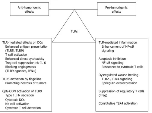
and “largely tumorigenic” molecules with the caveat that the underlying signaling pathways are mainly context, (i.e., organ-, tissue-, cell-) and ligand-dependent (Figure 2). Advanced exploration and better understanding of the relationship amongst TLR-signaling, inflammatory microenvironment and colitis-associated carcinogenesis should provide further insight into cancer development in IBD patients.
Further detailed functional analyses of TLRs in in- flammation-related tumor biology are needed to explore and define more precisely the subcellular and molecular mechanisms, hopefully allowing the introduction of se- lective new therapeutic approaches into daily practice.
REFERENCES
1 Zhang YZ, Li YY. Inflammatory bowel disease: pathogen- esis. World J Gastroenterol 2014; 20: 91-99 [PMID: 24415861 DOI: 10.3748/wjg.v20.i1.91]
2 Fukata M, Arditi M. The role of pattern recognition recep- tors in intestinal inflammation. Mucosal Immunol 2013; 6:
451-463 [PMID: 23515136 DOI: 10.1038/mi.2013.13]
3 Chang ZL. Important aspects of Toll-like receptors, ligands and their signaling pathways. Inflamm Res 2010; 59: 791-808 [PMID: 20593217 DOI: 10.1007/s00011-010-0208-2]
4 Jenkins KA, Mansell A. TIR-containing adaptors in Toll- like receptor signalling. Cytokine 2010; 49: 237-244 [PMID:
19264502 DOI: 10.1016/j.cyto.2009.01.009]
5 O’Neill LA. The interleukin-1 receptor/Toll-like receptor su- perfamily: 10 years of progress. Immunol Rev 2008; 226: 10-18
[PMID: 19161412 DOI: 10.1111/j.1600-065X.2008.00701.x]
6 Fischer M, Ehlers M. Toll-like receptors in autoimmunity.
Ann N Y Acad Sci 2008; 1143: 21-34 [PMID: 19076342 DOI:
10.1196/annals.1443.012]
7 Otte JM, Cario E, Podolsky DK. Mechanisms of cross hypo- responsiveness to Toll-like receptor bacterial ligands in in- testinal epithelial cells. Gastroenterology 2004; 126: 1054-1070 [PMID: 15057745]
8 Melmed G, Thomas LS, Lee N, Tesfay SY, Lukasek K, Mi- chelsen KS, Zhou Y, Hu B, Arditi M, Abreu MT. Human intestinal epithelial cells are broadly unresponsive to Toll- like receptor 2-dependent bacterial ligands: implications for host-microbial interactions in the gut. J Immunol 2003; 170:
1406-1415 [PMID: 12538701]
9 Akira S, Hemmi H. Recognition of pathogen-associated mo- lecular patterns by TLR family. Immunol Lett 2003; 85: 85-95 [PMID: 12527213 DOI: 10.1016/S0165-2478(02)00228-6]
10 Kanzler H, Barrat FJ, Hessel EM, Coffman RL. Therapeutic targeting of innate immunity with Toll-like receptor agonists and antagonists. Nat Med 2007; 13: 552-559 [PMID: 17479101 DOI: 10.1038/nm1589]
11 Smythies LE, Sellers M, Clements RH, Mosteller-Barnum M, Meng G, Benjamin WH, Orenstein JM, Smith PD. Hu- man intestinal macrophages display profound inflammatory anergy despite avid phagocytic and bacteriocidal activity. J Clin Invest 2005; 115: 66-75 [PMID: 15630445]
12 Lee J, Mo JH, Katakura K, Alkalay I, Rucker AN, Liu YT, Lee HK, Shen C, Cojocaru G, Shenouda S, Kagnoff M, Eckmann L, Ben-Neriah Y, Raz E. Maintenance of colonic homeostasis by distinctive apical TLR9 signalling in intestinal epithelial cells. Nat Cell Biol 2006; 8: 1327-1336 [PMID: 17128265 DOI:
10.1038/ncb1500]
13 Uehara A, Fujimoto Y, Fukase K, Takada H. Various human epithelial cells express functional Toll-like receptors, NOD1 and NOD2 to produce anti-microbial peptides, but not pro- inflammatory cytokines. Mol Immunol 2007; 44: 3100-3111 [PMID: 17403538]
14 Vora P, Youdim A, Thomas LS, Fukata M, Tesfay SY, Lu- kasek K, Michelsen KS, Wada A, Hirayama T, Arditi M, Abreu MT. Beta-defensin-2 expression is regulated by TLR Anti-tumorigenic
effects Pro-tumorigenic
effects
TLRs
TLR-mediated effects on DCs Enhanced antigen presentation (TLR5, TLR9)
T cell activation
Enhanced direct cytotoxicity Treg cell suppression via IL-6 Blocking angiogenesis (TLR9 agonists, IFNα) TLR5 activation by flagellins Promoting necrosis of tumors CpG-ODN activation of TLR9 Type Ⅰ IFN secretion Cytotoxic DCs NK cell activation Cytotoxic T cell activation
TLR-mediated inflammation Enhancement of NF-κB signaling
Apoptosis inhibition NF-κB signaling
Resistance to cytotoxic T cells Dysregulated wound healing TLR2-, TLR4-signaling Epiregulin overexpression Suppression of regulatory T cells (Treg)
Constitutive TLR4 activation
Figure 2 Dual role of Toll-like receptor signaling in colitis-associated carcinogenesis. While some direct and indirect effects of TLR-signaling act largely as an anti-tumorigenic factors, other effects may promote cancer development. TLR: Toll-like receptor; DC: Dendritic cell; IL: Interleukin; IFN: Interferon; ODN: Oligodeoxy- nucleotide; NF-κB: Nuclear factor-κB.
signaling in intestinal epithelial cells. J Immunol 2004; 173:
5398-5405 [PMID: 15494486]
15 Shang L, Fukata M, Thirunarayanan N, Martin AP, Arn- aboldi P, Maussang D, Berin C, Unkeless JC, Mayer L, Abreu MT, Lira SA. Toll-like receptor signaling in small intestinal epithelium promotes B-cell recruitment and IgA production in lamina propria. Gastroenterology 2008; 135: 529-538 [PMID:
18522803 DOI: 10.1053/j.gastro.2008.04.020]
16 Schneeman TA, Bruno ME, Schjerven H, Johansen FE, Chady L, Kaetzel CS. Regulation of the polymeric Ig receptor by signaling through TLRs 3 and 4: linking innate and adap- tive immune responses. J Immunol 2005; 175: 376-384 [PMID:
15972671]
17 Bruno ME, Rogier EW, Frantz AL, Stefka AT, Thompson SN, Kaetzel CS. Regulation of the polymeric immunoglobulin re- ceptor in intestinal epithelial cells by Enterobacteriaceae: im- plications for mucosal homeostasis. Immunol Invest 2010; 39:
356-382 [PMID: 20450283 DOI: 10.3109/08820131003622809]
18 Basith S, Manavalan B, Yoo TH, Kim SG, Choi S. Roles of toll-like receptors in cancer: a double-edged sword for de- fense and offense. Arch Pharm Res 2012; 35: 1297-1316 [PMID:
22941474 DOI: 10.1007/s12272-012-0802-7]
19 Medzhitov R, Preston-Hurlburt P, Kopp E, Stadlen A, Chen C, Ghosh S, Janeway CA. MyD88 is an adaptor protein in the hToll/IL-1 receptor family signaling pathways. Mol Cell 1998; 2: 253-258 [PMID: 9734363]
20 Takeda K, Akira S. TLR signaling pathways. Semin Immunol 2004; 16: 3-9 [PMID: 14751757]
21 Akira S, Uematsu S, Takeuchi O. Pathogen recognition and innate immunity. Cell 2006; 124: 783-801 [PMID: 16497588]
22 Kawai T, Akira S. Pathogen recognition with Toll-like recep- tors. Curr Opin Immunol 2005; 17: 338-344 [PMID: 15950447]
23 O’Neill LA. How Toll-like receptors signal: what we know and what we don’t know. Curr Opin Immunol 2006; 18: 3-9 [PMID: 16343886]
24 Pandey S, Agrawal DK. Immunobiology of Toll-like recep- tors: emerging trends. Immunol Cell Biol 2006; 84: 333-341 [PMID: 16834572]
25 Trinchieri G. Cancer and inflammation: an old intuition with rapidly evolving new concepts. Annu Rev Immunol 2012; 30: 677-706 [PMID: 22224761 DOI: 10.1146/annurev- immunol-020711-075008]
26 Balkwill F, Mantovani A. Inflammation and cancer: back to Virchow? Lancet 2001; 357: 539-545 [PMID: 11229684]
27 Kim EH, Hong KS, Hong H, Hahm KB. Detouring the Unde- sired Route of Helicobacter pylori-Induced Gastric Carcino- genesis. Cancers (Basel) 2011; 3: 3018-3028 [PMID: 24212943 DOI: 10.3390/cancers3033018]
28 Garay RP, Viens P, Bauer J, Normier G, Bardou M, Jeannin JF, Chiavaroli C. Cancer relapse under chemotherapy: why TLR2/4 receptor agonists can help. Eur J Pharmacol 2007; 563:
1-17 [PMID: 17383632]
29 Wiemann B, Starnes CO. Coley’s toxins, tumor necrosis fac- tor and cancer research: a historical perspective. Pharmacol Ther 1994; 64: 529-564 [PMID: 7724661 DOI: 10.1016/0163-72 58(94)90023-X]
30 de Kivit S, Tobin MC, Forsyth CB, Keshavarzian A, Landay AL. Regulation of Intestinal Immune Responses through TLR Activation: Implications for Pro- and Prebiotics. Front Immunol 2014; 5: 60 [PMID: 24600450]
31 Hanahan D, Weinberg RA. The hallmarks of cancer. Cell 2000; 100: 57-70 [PMID: 10647931]
32 Lazebnik Y. What are the hallmarks of cancer? Nat Rev Can- cer 2010; 10: 232-233 [PMID: 20355252]
33 Aviello G, Corr SC, Johnston DG, O’Neill LA, Fallon PG.
MyD88 adaptor-like (Mal) regulates intestinal homeostasis and colitis-associated colorectal cancer in mice. Am J Physiol Gastrointest Liver Physiol 2014; 306: G769-G778 [PMID:
24603458]
34 Rubin DC, Shaker A, Levin MS. Chronic intestinal inflam-
mation: inflammatory bowel disease and colitis-associated colon cancer. Front Immunol 2012; 3: 107 [PMID: 22586430 DOI: 10.3389/fimmu.2012.00107]
35 Eaden JA, Abrams KR, Mayberry JF. The risk of colorectal cancer in ulcerative colitis: a meta-analysis. Gut 2001; 48:
526-535 [PMID: 11247898]
36 Canavan C, Abrams KR, Mayberry J. Meta-analysis: colorec- tal and small bowel cancer risk in patients with Crohn’s disease. Aliment Pharmacol Ther 2006; 23: 1097-1104 [PMID:
16611269]
37 Vendramini-Costa DB, Carvalho JE. Molecular link mecha- nisms between inflammation and cancer. Curr Pharm Des 2012; 18: 3831-3852 [PMID: 22632748]
38 Lu Q, Ding H, Li W. Role of Toll-like receptors in microbio- ta-associated gastrointestinal cancer metastasis. J Cancer Res Ther 2013; 9 Suppl: S142-S149 [PMID: 24516050 DOI: 10.4103 /0973-1482.122509]
39 Farrell RJ, Ang Y, Kileen P, O’Briain DS, Kelleher D, Keel- ing PW, Weir DG. Increased incidence of non-Hodgkin’s lymphoma in inflammatory bowel disease patients on im- munosuppressive therapy but overall risk is low. Gut 2000;
47: 514-519 [PMID: 10986211]
40 Castro FA, Liu X, Försti A, Ji J, Sundquist J, Sundquist K, Koshiol J, Hemminki K. Increased risk of hepatobiliary can- cers after hospitalization for autoimmune disease. Clin Gas- troenterol Hepatol 2014; 12: 1038-45.e7 [PMID: 24246767 DOI:
10.1016/j.cgh.2013.11.007]
41 Sodemann U, Bistrup C, Marckmann P. Cancer rates after kidney transplantation. Dan Med Bull 2011; 58: A4342 [PMID:
22142571]
42 AlBugami M, Kiberd B. Malignancies: pre and post trans- plantation strategies. Transplant Rev (Orlando) 2014; 28: 76-83 [PMID: 24439783 DOI: 10.1016/j.trre.2013.12.002]
43 Ullman TA, Itzkowitz SH. Intestinal inflammation and can- cer. Gastroenterology 2011; 140: 1807-1816 [PMID: 21530747 DOI: 10.1053/j.gastro.2011.01.057]
44 Kundu JK, Surh YJ. Inflammation: gearing the journey to cancer. Mutat Res 2008; 659: 15-30 [PMID: 18485806 DOI:
10.1016/j.mrrev.2008.03.002]
45 Ono M. Molecular links between tumor angiogenesis and inflammation: inflammatory stimuli of macrophages and cancer cells as targets for therapeutic strategy. Cancer Sci 2008; 99: 1501-1506 [PMID: 18754859 DOI: 10.1111/
j.1349-7006.2008.00853.x]
46 Dvorak HF. Tumors: wounds that do not heal. Similarities between tumor stroma generation and wound healing. N Engl J Med 1986; 315: 1650-1659 [PMID: 3537791]
47 Dolberg DS, Hollingsworth R, Hertle M, Bissell MJ. Wound- ing and its role in RSV-mediated tumor formation. Science 1985; 230: 676-678 [PMID: 2996144]
48 Fukata M, Chen A, Klepper A, Krishnareddy S, Vamadevan AS, Thomas LS, Xu R, Inoue H, Arditi M, Dannenberg AJ, Abreu MT. Cox-2 is regulated by Toll-like receptor-4 (TLR4) signaling: Role in proliferation and apoptosis in the intestine.
Gastroenterology 2006; 131: 862-877 [PMID: 16952555]
49 Pull SL, Doherty JM, Mills JC, Gordon JI, Stappenbeck TS.
Activated macrophages are an adaptive element of the co- lonic epithelial progenitor niche necessary for regenerative responses to injury. Proc Natl Acad Sci USA 2005; 102: 99-104 [PMID: 15615857]
50 Brown SL, Riehl TE, Walker MR, Geske MJ, Doherty JM, Stenson WF, Stappenbeck TS. Myd88-dependent positioning of Ptgs2-expressing stromal cells maintains colonic epithelial proliferation during injury. J Clin Invest 2007; 117: 258-269 [PMID: 17200722]
51 Brandl K, Sun L, Neppl C, Siggs OM, Le Gall SM, Tomisato W, Li X, Du X, Maennel DN, Blobel CP, Beutler B. MyD88 signaling in nonhematopoietic cells protects mice against induced colitis by regulating specific EGF receptor ligands.
Proc Natl Acad Sci USA 2010; 107: 19967-19972 [PMID:
21041656 DOI: 10.1073/pnas.1014669107]
52 Hsu D, Fukata M, Hernandez YG, Sotolongo JP, Goo T, Maki J, Hayes LA, Ungaro RC, Chen A, Breglio KJ, Xu R, Abreu MT. Toll-like receptor 4 differentially regulates epidermal growth factor-related growth factors in response to intes- tinal mucosal injury. Lab Invest 2010; 90: 1295-1305 [PMID:
20498653 DOI: 10.1038/labinvest.2010.100]
53 Lin Y, Lee H, Berg AH, Lisanti MP, Shapiro L, Scherer PE.
The lipopolysaccharide-activated toll-like receptor (TLR)-4 induces synthesis of the closely related receptor TLR-2 in adipocytes. J Biol Chem 2000; 275: 24255-24263 [PMID:
10823826]
54 Rehli M, Poltorak A, Schwarzfischer L, Krause SW, An- dreesen R, Beutler B. PU.1 and interferon consensus se- quence-binding protein regulate the myeloid expression of the human Toll-like receptor 4 gene. J Biol Chem 2000; 275:
9773-9781 [PMID: 10734131]
55 Huang B, Zhao J, Li H, He KL, Chen Y, Chen SH, Mayer L, Unkeless JC, Xiong H. Toll-like receptors on tumor cells fa- cilitate evasion of immune surveillance. Cancer Res 2005; 65:
5009-5014 [PMID: 15958541]
56 Fűri I, Sipos F, Germann TM, Kalmár A, Tulassay Z, Mol- nár B, Műzes G. Epithelial toll-like receptor 9 signaling in colorectal inflammation and cancer: clinico-pathogenic aspects. World J Gastroenterol 2013; 19: 4119-4126 [PMID:
23864774 DOI: 10.3748/wjg.v19.i26.4119]
57 Fűri I, Sipos F, Spisák S, Kiszner G, Wichmann B, Schöller A, Tulassay Z, Műzes G, Molnár B. Association of self-DNA mediated TLR9-related gene, DNA methyltransferase, and cytokeratin protein expression alterations in HT29-cells to DNA fragment length and methylation status. Scienti- ficWorldJournal 2013; 2013: 293296 [PMID: 24459426 DOI:
10.1155/2013/293296]
58 Pikarsky E, Porat RM, Stein I, Abramovitch R, Amit S, Kasem S, Gutkovich-Pyest E, Urieli-Shoval S, Galun E, Ben- Neriah Y. NF-kappaB functions as a tumour promoter in inflammation-associated cancer. Nature 2004; 431: 461-466 [PMID: 15329734]
59 Pradere JP, Dapito DH, Schwabe RF. The Yin and Yang of Toll-like receptors in cancer. Oncogene 2014; 33: 3485-3495 [PMID: 23934186 DOI: 10.1038/onc.2013.302]
60 Palucka K, Banchereau J. Cancer immunotherapy via den- dritic cells. Nat Rev Cancer 2012; 12: 265-277 [PMID: 22437871 DOI: 10.1038/nrc3258]
61 Garaude J, Kent A, van Rooijen N, Blander JM. Simultane- ous targeting of toll- and nod-like receptors induces ef- fective tumor-specific immune responses. Sci Transl Med 2012; 4: 120ra16 [PMID: 22323829 DOI: 10.1126/scitrans- lmed.3002868]
62 Drobits B, Holcmann M, Amberg N, Swiecki M, Grundtner R, Hammer M, Colonna M, Sibilia M. Imiquimod clears tu- mors in mice independent of adaptive immunity by convert- ing pDCs into tumor-killing effector cells. J Clin Invest 2012;
122: 575-585 [PMID: 22251703 DOI: 10.1172/JCI61034]
63 Cubillos-Ruiz JR, Engle X, Scarlett UK, Martinez D, Barber A, Elgueta R, Wang L, Nesbeth Y, Durant Y, Gewirtz AT, Sentman CL, Kedl R, Conejo-Garcia JR. Polyethylenimine- based siRNA nanocomplexes reprogram tumor-associated dendritic cells via TLR5 to elicit therapeutic antitumor im- munity. J Clin Invest 2009; 119: 2231-2244 [PMID: 19620771 DOI: 10.1172/JCI37716]
64 Nierkens S, den Brok MH, Garcia Z, Togher S, Wagenaars J, Wassink M, Boon L, Ruers TJ, Figdor CG, Schoenberger SP, Adema GJ, Janssen EM. Immune adjuvant efficacy of CpG oligonucleotide in cancer treatment is founded spe- cifically upon TLR9 function in plasmacytoid dendritic cells. Cancer Res 2011; 71: 6428-6437 [PMID: 21788345 DOI:
10.1158/0008-5472.CAN-11-2154]
65 Pasare C, Medzhitov R. Toll pathway-dependent blockade of CD4+CD25+ T cell-mediated suppression by dendritic
cells. Science 2003; 299: 1033-1036 [PMID: 12532024]
66 Peng G, Guo Z, Kiniwa Y, Voo KS, Peng W, Fu T, Wang DY, Li Y, Wang HY, Wang RF. Toll-like receptor 8-mediated re- versal of CD4+ regulatory T cell function. Science 2005; 309:
1380-1384 [PMID: 16123302]
67 Damiano V, Garofalo S, Rosa R, Bianco R, Caputo R, Gelardi T, Merola G, Racioppi L, Garbi C, Kandimalla ER, Agrawal S, Tortora G. A novel toll-like receptor 9 agonist cooperates with trastuzumab in trastuzumab-resistant breast tumors through multiple mechanisms of action. Clin Cancer Res 2009;
15: 6921-6930 [PMID: 19903791 DOI: 10.1158/1078-0432.
CCR-09-1599]
68 Pfeffer LM. Biologic activities of natural and synthetic type I interferons. Semin Oncol 1997; 24: S9-63-S9-S9-63-69 [PMID:
9208874]
69 Rhee SH, Im E, Pothoulakis C. Toll-like receptor 5 engage- ment modulates tumor development and growth in a mouse xenograft model of human colon cancer. Gastroen- terology 2008; 135: 518-528 [PMID: 18538140 DOI: 10.1053/
j.gastro.2008.04.022]
70 Sipos F, Műzes G, Fűri I, Spisák S, Wichmann B, Germann TM, Constantinovits M, Krenács T, Tulassay Z, Molnár B. In- travenous Administration of a Single-Dose Free-Circulating DNA of Colitic Origin Improves Severe Murine DSS-Colitis.
Pathol Oncol Res 2014; Epub ahead of print [PMID: 24723054]
71 Heckelsmiller K, Rall K, Beck S, Schlamp A, Seiderer J, Jahrsdörfer B, Krug A, Rothenfusser S, Endres S, Hartmann G. Peritumoral CpG DNA elicits a coordinated response of CD8 T cells and innate effectors to cure established tumors in a murine colon carcinoma model. J Immunol 2002; 169:
3892-3899 [PMID: 12244187]
72 Krieg AM. Development of TLR9 agonists for cancer thera- py. J Clin Invest 2007; 117: 1184-1194 [PMID: 17476348]
73 Kadowaki N, Antonenko S, Liu YJ. Distinct CpG DNA and polyinosinic-polycytidylic acid double-stranded RNA, re- spectively, stimulate CD11c- type 2 dendritic cell precursors and CD11c+ dendritic cells to produce type I IFN. J Immunol 2001; 166: 2291-2295 [PMID: 11160284]
74 Grivennikov S, Karin E, Terzic J, Mucida D, Yu GY, Vallab- hapurapu S, Scheller J, Rose-John S, Cheroutre H, Eckmann L, Karin M. IL-6 and Stat3 are required for survival of intestinal epithelial cells and development of colitis-associated cancer.
Cancer Cell 2009; 15: 103-113 [PMID: 19185845 DOI: 10.1016/
j.ccr.2009.01.001]
75 Popivanova BK, Kitamura K, Wu Y, Kondo T, Kagaya T, Kaneko S, Oshima M, Fujii C, Mukaida N. Blocking TNF- alpha in mice reduces colorectal carcinogenesis associated with chronic colitis. J Clin Invest 2008; 118: 560-570 [PMID:
18219394 DOI: 10.1172/JCI32453]
76 Tukhvatulin AI, Gitlin II, Shcheblyakov DV, Artemicheva NM, Burdelya LG, Shmarov MM, Naroditsky BS, Gudkov AV, Gintsburg AL, Logunov DY. Combined stimulation of Toll-like receptor 5 and NOD1 strongly potentiates activity of NF-κB, resulting in enhanced innate immune reactions and resistance to Salmonella enterica serovar Typhimurium infection. Infect Immun 2013; 81: 3855-3864 [PMID: 23897616 DOI: 10.1128/IAI.00525-13]
77 Dutta J, Fan Y, Gupta N, Fan G, Gélinas C. Current insights into the regulation of programmed cell death by NF-kappaB.
Oncogene 2006; 25: 6800-6816 [PMID: 17072329]
78 Wang CY, Mayo MW, Korneluk RG, Goeddel DV, Baldwin AS. NF-kappaB antiapoptosis: induction of TRAF1 and TRAF2 and c-IAP1 and c-IAP2 to suppress caspase-8 activa- tion. Science 1998; 281: 1680-1683 [PMID: 9733516]
79 Faubion WA, De Jong YP, Molina AA, Ji H, Clarke K, Wang B, Mizoguchi E, Simpson SJ, Bhan AK, Terhorst C.
Colitis is associated with thymic destruction attenuating CD4+25+ regulatory T cells in the periphery. Gastroenterol- ogy 2004; 126: 1759-1770 [PMID: 15188171 DOI: 10.1053/
j.gastro.2004.03.015]
80 Erdman SE, Poutahidis T. Roles for inflammation and regu- latory T cells in colon cancer. Toxicol Pathol 2010; 38: 76-87 [PMID: 20019355 DOI: 10.1177/0192623309354110]
81 Li J, Wang FP, She WM, Yang CQ, Li L, Tu CT, Wang JY, Jiang W. Enhanced high-mobility group box 1 (HMGB1) modulates regulatory T cells (Treg)/T helper 17 (Th17) bal- ance via toll-like receptor (TLR)-4-interleukin (IL)-6 pathway in patients with chronic hepatitis B. J Viral Hepat 2014; 21:
129-140 [PMID: 24383926 DOI: 10.1111/jvh.12152]
82 Lowe EL, Crother TR, Rabizadeh S, Hu B, Wang H, Chen S, Shimada K, Wong MH, Michelsen KS, Arditi M. Toll-like receptor 2 signaling protects mice from tumor development in a mouse model of colitis-induced cancer. PLoS One 2010; 5:
e13027 [PMID: 20885960 DOI: 10.1371/journal.pone.0013027]
83 Salcedo R, Worschech A, Cardone M, Jones Y, Gyulai Z, Dai RM, Wang E, Ma W, Haines D, O’hUigin C, Marincola FM, Trinchieri G. MyD88-mediated signaling prevents develop- ment of adenocarcinomas of the colon: role of interleukin 18. J Exp Med 2010; 207: 1625-1636 [PMID: 20624890 DOI:
10.1084/jem.20100199]
84 Bäckhed F, Ley RE, Sonnenburg JL, Peterson DA, Gordon JI. Host-bacterial mutualism in the human intestine. Science 2005; 307: 1915-1920 [PMID: 15790844]
85 Rakoff-Nahoum S, Paglino J, Eslami-Varzaneh F, Edberg S, Medzhitov R. Recognition of commensal microflora by toll-like receptors is required for intestinal homeostasis. Cell
2004; 118: 229-241 [PMID: 15260992]
86 Garrett WS, Gordon JI, Glimcher LH. Homeostasis and in- flammation in the intestine. Cell 2010; 140: 859-870 [PMID:
20303876 DOI: 10.1016/j.cell.2010.01.023]
87 Fukata M, Hernandez Y, Conduah D, Cohen J, Chen A, Breglio K, Goo T, Hsu D, Xu R, Abreu MT. Innate immune signaling by Toll-like receptor-4 (TLR4) shapes the inflam- matory microenvironment in colitis-associated tumors.
Inflamm Bowel Dis 2009; 15: 997-1006 [PMID: 19229991 DOI:
10.1002/ibd.20880]
88 Fukata M, Shang L, Santaolalla R, Sotolongo J, Pastorini C, España C, Ungaro R, Harpaz N, Cooper HS, Elson G, Kosco-Vilbois M, Zaias J, Perez MT, Mayer L, Vamadevan AS, Lira SA, Abreu MT. Constitutive activation of epithelial TLR4 augments inflammatory responses to mucosal injury and drives colitis-associated tumorigenesis. Inflamm Bowel Dis 2011; 17: 1464-1473 [PMID: 21674704 DOI: 10.1002/
ibd.21527]
89 Rakoff-Nahoum S, Medzhitov R. Regulation of spontane- ous intestinal tumorigenesis through the adaptor protein MyD88. Science 2007; 317: 124-127 [PMID: 17615359]
90 Li Y, Kundu P, Seow SW, de Matos CT, Aronsson L, Chin KC, Kärre K, Pettersson S, Greicius G. Gut microbiota accel- erate tumor growth via c-jun and STAT3 phosphorylation in APCMin/+ mice. Carcinogenesis 2012; 33: 1231-1238 [PMID:
22461519 DOI: 10.1093/carcin/bgs137]
P- Reviewer: Rao VS S- Editor: Ma YJ L- Editor: Webster JR E- Editor: Zhang DN
8226 Regency Drive, Pleasanton, CA 94588, USA Telephone: +1-925-223-8242
Fax: +1-925-223-8243 E-mail: bpgoffice@wjgnet.com
Help Desk: http://www.wjgnet.com/esps/helpdesk.aspx http://www.wjgnet.com
I S S N 1 0 0 7 - 9 3 2 7
9 7 7 1 0 07 9 3 2 0 45 3 6