Review
Focusing on the Cell Type Specific Regulatory Actions of NLRX1
Tünde Fekete1, Dóra Bencze1,2, Eduárd Bíró2,3, Szilvia Benk ˝o3 and Kitti Pázmándi1,*
Citation: Fekete, T.; Bencze, D.; Bíró, E.; Benk˝o, S.; Pázmándi, K. Focusing on the Cell Type Specific Regulatory Actions of NLRX1.Int. J. Mol. Sci.
2021,22, 1316. https://doi.org/
10.3390/ijms22031316
Academic Editor: Ernesto Fedele Received: 5 January 2021 Accepted: 25 January 2021 Published: 28 January 2021
Publisher’s Note:MDPI stays neutral with regard to jurisdictional claims in published maps and institutional affil- iations.
Copyright: © 2021 by the authors.
Licensee MDPI, Basel, Switzerland.
This article is an open access article distributed under the terms and conditions of the Creative Commons Attribution (CC BY) license (https://
creativecommons.org/licenses/by/
4.0/).
1 Department of Immunology, Faculty of Medicine, University of Debrecen, 1 Egyetem Square, H-4032 Debrecen, Hungary; fekete.tunde@med.unideb.hu (T.F.); bencze.dora@med.unideb.hu (D.B.)
2 Doctoral School of Molecular Cell and Immune Biology, University of Debrecen, 1 Egyetem Square, H-4032 Debrecen, Hungary; biro.eduard@med.unideb.hu
3 Department of Physiology, Faculty of Medicine, University of Debrecen, 98 Nagyerdei krt., H-4002 Debrecen, Hungary; benkosz@med.unideb.hu
* Correspondence: pazmandikitti@yahoo.de; Tel./Fax: +36-52-417-159
Abstract:Cells utilize a diverse repertoire of cell surface and intracellular receptors to detect exoge- nous or endogenous danger signals and even the changes of their microenvironment. However, some cytosolic NOD-like receptors (NLR), including NLRX1, serve more functions than just being general pattern recognition receptors. The dynamic translocation between the cytosol and the mitochondria allows NLRX1 to interact with many molecules and thereby to control multiple cellular functions.
As a regulatory NLR, NLRX1 fine-tunes inflammatory signaling cascades, regulates mitochondria- associated functions, and controls metabolism, autophagy and cell death. Nevertheless, literature data are inconsistent and often contradictory regarding its effects on individual cellular functions.
One plausible explanation might be that the regulatory effects of NLRX1 are highly cell type specific and the features of NLRX1 mediated regulation might be determined by the unique functional activity or metabolic profile of the given cell type. Here we review the cell type specific actions of NLRX1 with a special focus on cells of the immune system. NLRX1 has already emerged as a potential therapeutic target in numerous immune-related diseases, thus we aim to highlight which regulatory properties of NLRX1 are manifested in disease-associated dominant immune cells that presumably offer promising therapeutic solutions to treat these disorders.
Keywords: NLRX1; regulation; immune cells; myeloid cells; lymphoid cells; antiviral immunity;
NF-κB pathway; autophagy; mitochondria; metabolism
1. Introduction
1.1. NLRs: A Large Receptor Family with Versatile Roles
Innate immunity serves as the first line of defense against invading pathogens. Cells of the innate immune system utilize a vast repertoire of evolutionary conserved pattern recognition receptors (PRRs) such as toll-like receptors (TLRs), C-type lectin receptors (CLRs), retinoic acid-inducible gene (RIG)-I-like receptors (RLRs) and nucleotide-binding oligomerization domain (NOD)-like receptors (NLRs) to detect various microbial motifs or danger signals [1]. Therefore, they play a central role in the initiation of host defense during the early stages of infection and subsequently contribute to the generation of adaptive immune responses as well [2].
Among PRRs, NLRs comprise a large family of intracellular receptors/sensors, which play an essential role in recognition of a wide variety of pathogen-associated molecular pat- terns (PAMP) and exogenous/endogenous damage-associated molecular patterns (DAMP), and eventually lead to the induction of innate immune effector functions. Besides, NLRs have been recognized to be involved in various cellular processes as positive or negative regulators. To date, 22 NLRs have been identified in humans, and at least 34 in mice [3].
Based on their unique function, they are classified into five subgroups:
Int. J. Mol. Sci.2021,22, 1316. https://doi.org/10.3390/ijms22031316 https://www.mdpi.com/journal/ijms
Int. J. Mol. Sci.2021,22, 1316 2 of 35
(1) The most thoroughly characterized NLRs form large multiprotein complexes, termed inflammasomes, the activation of which leads to the cleavage of pro-IL-1ß and pro-IL- 18 pro-inflammatory cytokines into their mature, biologically active form. Among NLRs, NLRP1, NLRP3, NLRP6 and NLRC4 are known to form inflammasomes.
(2) Several NLRs act as positive or negative regulators of signal transduction. The so- called regulatory NLRs control various intracellular signaling cascades such as the nuclear factor-kappa B (NF-κB), the type I interferon (IFN) and the mitogen-activated protein kinase (MAPK) pathways initiated by other PRRs. This group is composed of several members such as NOD1, NOD2, NLRC5, nucleotide-binding domain and leucine-rich repeat–containing protein X1 (NLRX1), NLRP12 and NLRC3.
(3) A specific subgroup of NLRs, including NLRC5 and the class II major histocompati- bility complex transactivator (CIITA), can function as enhanceosomes and control the transcription of MHC I and II genes, respectively.
(4) Upon bacterial sensing, some NLRs, such as NOD1 and NOD2, are able to recruit the ATGL16L1 autophagy modulating protein to the cell membrane to initiate the formation of autophagosomes [4].
(5) Reproductive NLRs (NLRP2, NLRP5, NLRP7) control embryogenesis and reproduc- tion. The function of these NLRs has been extensively reviewed in reference [5].
Regarding the structure of NLRs, they all share a common C-terminal leucine-rich repeat (LRR) domain, which are responsible for ligand recognition; a central NOD, also called as NACHT domain, which upon nucleotide binding mediates the activation and oligomerization of the receptor; and an N-terminal domain, which binds to distinct adaptor proteins and exerts multiple downstream signaling. Based on their N-terminal effector domains, NLRs are divided into four subfamilies:
(1) The NLRA subfamily possesses an acidic transactivation domain (TA) and consists of only one member, the CIITA.
(2) The NLRB subfamily has a baculoviral inhibition of apoptosis protein repeat (BIR) domain. Similar to NLRA subfamily, NLRB subfamily has also only one member, the NLR family apoptosis inhibitory protein (NAIP).
(3) The NLRC subfamily owns a caspase recruitment and activation domain (CARD), and in contrast to the above mentioned two families, the NLRC subfamily consists of six receptors: NOD1 (or NLRC1), NOD2 (or NLRC2), NLRC3, NLRC4, NLRC5, and NLRX1.
(4) The NLRP subfamily has a pyrin domain (PYD), and comprising 14 members rep- resents the largest subfamily of NLRs: NLRP1, NLRP2, NLRP3, NLRP4, NLRP7, NLRP12, NLRP5, NLRP6, NLRP8, NLRP9, NLRP10, NLRP11, NLRP13, and finally NLRP14 (reviewed in reference [4]).
1.2. Beyond the Inflammasomes: Regulatory NLRs
The inflammasome forming NLRs represent without doubt the most studied subgroup of the NLR family. However, due to their diverse functions, regulatory NLRs have also landed at the forefront of research in the field of immunology. The functions of regulatory NLRs have already studied in various immune cells such as macrophages or human dendritic cells (DCs), since these receptors can modulate diverse signaling pathways initiated by other PRRs and can shape immune cell mediated responses. Inflammatory reactions must be tightly controlled to keep the immune response in balance. On one hand, the host must mount an immune response strong enough to clear the pathogen from the body, on the other hand, the immune response must be fine-tuned to prevent excessive collateral damage. Modulation of the inflammatory cascades such as NF-κB, MAPK and type I IFN pathways has been already documented by these receptors [6]. Regulatory NLRs may act synergistically or antagonistically with other PRRs, that seems to depend on the type of cell, receptor or ligand involved.
For instance, the bacterial sensors, NOD1 and NOD2 are positive regulatory NLRs, as they enhance inflammatory responses. Upon ligand sensing, NOD2 oligomerizes and
recruits distinct proteins resulting in the formation of a multiprotein complex, termed the NODosome. Receptor-interacting-serine/threonine-protein kinase 2 (RIP2), mitochondrial antiviral signaling protein (MAVS), NF-kappa-B essential modulator (NEMO) and X-linked inhibitor of apoptosis (XIAP) are some of the few recruited proteins, which can affect downstream signaling events. It has been described that the NODosome promotes type I IFN signaling via either TNF receptor-associated factor (TRAF) 3/ IFN regulatory factor (IRF) 7 or MAVS/IRF3 and modulates NF-κB signaling as well [6].
In contrast, NLRC3 and NLRP12 are negative regulatory NLRs, the major function of which is to prevent unwanted inflammation. Similar to the inflammasome and the NODosome, these NLRs are also capable of forming multiprotein complexes, termed TRAFosomes. NLRC3 interacts with TRAF6, while NLRP12 binds to TRAF3. NLRC3 inhibits NF-κB signaling by decreasing the overall ubiquitination of TRAF6. In addition, NLRC3 negatively regulates type I IFN responses by interfering with the stimulator of interferon genes (STING)/TANK-binding kinase 1 (TBK1) axis via directly binding to STING and TBK1 [7]. NLRP12 is the negative regulator of canonical and non-canonical NF-κB signaling and plays a role in inhibiting the extracellular signal-regulated kinase (ERK) pathway as well [8].
Besides the abovementioned receptors, NLRC5 has emerged as a regulatory NLR, which affects the TLR- and RLR-mediated NF-κB and type I IFN signaling negatively [9–11].
Fekete et al. also described that NLRC5 suppresses the RLR-induced type I IFN response in plasmacytoid DCs (pDC) similar to NLRX1, which is another important regulatory NLR [12]. NLRX1 has been primarily described as a negative regulator of inflammatory reactions, but soon conflicting data have been reported pertaining to its role in immune responses. The observed discrepancies might be attributed to the different cell types, and the opposing regulatory effects of NLRX1 are most striking when immune cells are compared to non-immune cells. Therefore, in this review we would like to focus on the regulatory function of NLRX1 in a cell type specific manner.
1.3. X Marks the Spot: NLRX1, a Mitochondria-Associated Regulatory NLR
NLRX1, also known as NOD5, NOD9, or CLR11.3 belongs to the subfamily of NLRC receptors. It consists of 975 amino acids and is expressed ubiquitously. “X” in algebra is often used to signify the unknown. Similarly, the X in NLRX1 refers to its enigmatic structure, more precisely to its not fully characterized N-terminal domain. Instead of the commonly shared CARD domain of NLRC receptors, only one domain was identified in the N-terminus, the mitochondrial targeting sequence (MTS), which uniquely localizes NLRX1 to the mitochondrial membrane [13]. It was initially reported, that NLRX1 is associated with the mitochondrial outer membrane [14]. Later, it was shown that NLRX1 can also localize to the inner mitochondrial membrane and within the matrix, and last but not least in the cytosol. All these data suggest, that similar to other NLRs, NLRX1 can shuttle between distinct cellular compartments, from the cytosol to the mitochondria that makes it possible to interact with a multitude of cellular pathways [6]. The C-terminal domain of NLRX1 displays a three-domain architecture [15]. It consists of an N-terminal helical domain, central seven LRRs and a C-terminal three-helix bundle, essential for structural integrity. It has been revealed that the C-terminal domain of NLRX1 can form hexamers by the trimerization of dimers both in solution and in crystal structures [16]. Hong et al. also found that NLRX1 can directly interact with RNA ligands but not DNA probably through a positively charged surface area of the C-terminal fragment. Similar to other NLRs, it contains a central NOD/NACHT domain as well [15] (Figure1).
Int. J. Mol. Sci.2021,22, 1316 4 of 35
Int. J. Mol. Sci. 2021, 22, x FOR PEER REVIEW 4 of 36
Figure 1. Schematic representation of NLRX1 structure. CTHB: C‐terminal three‐helix bundle; LRRs: leucine‐rich repeats;
MTS: mitochondrial targeting sequence; NACHT: domain conserved in NAIP, CIITA, HET‐E and TP1; NOD: nucleotide‐
binding oligomerization domain; NTHD: N‐terminal helical domain.
NLRX1 has diverse cellular functions depending on its localization and interacting partners. NLRX1 also serves as a scaffold protein and is able to take part in the assembly of large multiprotein complexes [17]. In addition, it has been documented that NLRX1 regulates the NF‐κB and type I IFN signaling, affects the MAPK pathway, modulates ROS production, influences the main metabolic pathways and impacts autophagy and cell death. Besides regulating host‐pathogen interactions, NLRX1 acts as a tumor suppressor or, on the contrary, might facilitate metastasis development. More importantly, it has a protective role in the development and progression of various autoimmune diseases such as multiple sclerosis, lupus and inflammatory bowel disease. Furthermore, NLRX1 has also been found to contribute to metabolic disorders. The importance of NLRX1 in inflammatory, metabolic and oncological diseases have been already addressed in previous and recent reviews [17,18]. Since NLRX1 contributes to a multitude of diseases, targeting NLRX1 could offer promising treatment strategies in immune cell‐, or non‐
immune cell‐mediated diseases [18].
Before discussing the cell type specific differences in NLRX1 function, in the next section we briefly review the underlying molecular mechanisms behind the complex regulatory role of NLRX1, which have been identified to date.
2. Regulatory Mechanism behind the Multiple Actions of NLRX1 2.1. Regulation of Antiviral Immunity
To detect replicating viruses, cells utilize cytosolic RLRs including RIG‐I and melanoma differentiation‐associated protein 5 (MDA5), which are specialized for sensing viral replication intermediates in the cytoplasm. RLRs use the mitochondrial adaptor MAVS to stimulate type I IFN responses. MAVS contains an N‐terminal CARD domain, through which it interacts with RIG‐I. Moore et al. elegantly proved in an in vitro system using HeLa and HEK293T cells that NLRX1 is a negative regulator of RIG‐I, and possibly MDA‐5‐induced antiviral signaling by competing with the receptors for the CARD domain of MAVS. They reported that NLRX1 is located on the mitochondrial outer membrane and similar to RIG‐I, it can bind the CARD domain of MAVS. Via this interaction NLRX1 negatively regulates Sendai virus‐induced type I IFN production by inhibiting MAVS mediated IRF3 dimer formation [14]. Under in vivo conditions, the negative regulatory role of NLRX1 in virus induced inflammation was proved by Allen et al. in a mouse model of influenza infection. However, NLRX1−/− mice cleared the virus more quickly, exhibited enhanced levels of IL‐6 and IFN‐β, and showed more severe lung injury and morbidity [19].
Apart from direct competition for MAVS binding, NLRX1 can indirectly inhibit RIG‐
I‐MAVS signaling as well. In a human hepatoma cell line, it was demonstrated that the nucleotide‐binding domain of NLRX1 attenuates hepatitis C virus (HCV)‐triggered RIG‐
I‐MAVS signaling by recruiting poly(rC) binding protein 2 (PCBP2) to MAVS. PCBP2 Figure 1.Schematic representation of NLRX1 structure. CTHB: C-terminal three-helix bundle; LRRs: leucine-rich repeats;
MTS: mitochondrial targeting sequence; NACHT: domain conserved in NAIP, CIITA, HET-E and TP1; NOD: nucleotide- binding oligomerization domain; NTHD: N-terminal helical domain.
NLRX1 has diverse cellular functions depending on its localization and interacting partners. NLRX1 also serves as a scaffold protein and is able to take part in the assembly of large multiprotein complexes [17]. In addition, it has been documented that NLRX1 regulates the NF-κB and type I IFN signaling, affects the MAPK pathway, modulates ROS production, influences the main metabolic pathways and impacts autophagy and cell death.
Besides regulating host-pathogen interactions, NLRX1 acts as a tumor suppressor or, on the contrary, might facilitate metastasis development. More importantly, it has a protective role in the development and progression of various autoimmune diseases such as multiple sclerosis, lupus and inflammatory bowel disease. Furthermore, NLRX1 has also been found to contribute to metabolic disorders. The importance of NLRX1 in inflammatory, metabolic and oncological diseases have been already addressed in previous and recent reviews [17,18]. Since NLRX1 contributes to a multitude of diseases, targeting NLRX1 could offer promising treatment strategies in immune cell-, or non-immune cell-mediated diseases [18].
Before discussing the cell type specific differences in NLRX1 function, in the next section we briefly review the underlying molecular mechanisms behind the complex regulatory role of NLRX1, which have been identified to date.
2. Regulatory Mechanism behind the Multiple Actions of NLRX1 2.1. Regulation of Antiviral Immunity
To detect replicating viruses, cells utilize cytosolic RLRs including RIG-I and melanoma differentiation-associated protein 5 (MDA5), which are specialized for sensing viral repli- cation intermediates in the cytoplasm. RLRs use the mitochondrial adaptor MAVS to stimulate type I IFN responses. MAVS contains an N-terminal CARD domain, through which it interacts with RIG-I. Moore et al. elegantly proved in an in vitro system using HeLa and HEK293T cells that NLRX1 is a negative regulator of RIG-I, and possibly MDA- 5-induced antiviral signaling by competing with the receptors for the CARD domain of MAVS. They reported that NLRX1 is located on the mitochondrial outer membrane and similar to RIG-I, it can bind the CARD domain of MAVS. Via this interaction NLRX1 nega- tively regulates Sendai virus-induced type I IFN production by inhibiting MAVS mediated IRF3 dimer formation [14]. Under in vivo conditions, the negative regulatory role of NLRX1 in virus induced inflammation was proved by Allen et al. in a mouse model of influenza infection. However, NLRX1−/−mice cleared the virus more quickly, exhibited enhanced levels of IL-6 and IFN-β, and showed more severe lung injury and morbidity [19].
Apart from direct competition for MAVS binding, NLRX1 can indirectly inhibit RIG- I-MAVS signaling as well. In a human hepatoma cell line, it was demonstrated that the nucleotide-binding domain of NLRX1 attenuates hepatitis C virus (HCV)-triggered RIG-I- MAVS signaling by recruiting poly(rC) binding protein 2 (PCBP2) to MAVS. PCBP2 induces K48-linked polyubiquitination and subsequent proteasomal degradation of MAVS, thus it limits type I IFN production [20]. Furthermore, NLRX1 can also be used as pro-viral
host factor by many other viruses to modulate the host’s antiviral response and aid virus replication and survival. For example, it was published that in Rhesus monkeys simian immunodeficiency virus (SIV) triggers NLRX1 expression in the beginning of the infection to facilitate its own replication [21]. It was also demonstrated that ORF9c protein of SARS- CoV-2 interacts with NLRX1, through which it might influence MAVS-mediated type I IFN and pro-inflammatory cytokine secretion [22].
NLRX1-mediated inhibition of the antiviral immune response can be overcome with the help of FAS-associated factor-1 (FAF1), a member of the FAS death-inducing signaling complex. Besides being involved in FAS-mediated apoptosis, FAF1 can directly bind to NRLX1 thereby freeing MAVS and enabling RIG-I-MAVS interaction. Thus FAF1 positively regulates type I IFN production of immune cells in response to RNA viruses, and its inhibitory effect on NLRX1 further proves the negative regulatory role of NLRX1 in MAVS- mediated responses [23].
Besides regulating the RIG-I-MAVS-induced type I IFN signaling, NLRX1 controls the STING and TBK1-mediated pathways as well. NLRX1, by binding the DNA sensor STING, disrupts STING-TBK1 signaling and thereby suppresses TBK1 activation, which is required for type I IFN production in response to stimuli like human immunodeficiency virus (HIV)-1, DNA viruses, cGAMP and dsDNA in myeloid cells. In line with this, NLRX1 deficient mice are more resistant to HIV-1 and DNA viruses [24]. Interestingly, in contrast to IRF3 pathway, NLRX1 positively regulates the NF-κB-driven IRF1-mediated early antiviral responses in hepatocytes suggesting the opposing regulatory role of NLRX1 in the control of IRF3 and IRF1 pathways. In hepatocytes, NLRX1 competes with protein kinase R (PKR) for viral RNA binding, which prevents the PKR induced blockade of host’s protein synthesis. Thus, NLRX1 helps to maintain IRF1 upregulation, while inhibits IRF3 dimerization upon viral infection [25].
Other studies also showed that NLRX1 can positively control the innate antiviral immune responses via interaction with viral proteins. Influenza A virus expresses a small protein, polymerase basic protein 1-frame 2 (PB1-F2), which induces apoptosis in innate immune cells by disrupting mitochondrial membrane potential. In macrophages, NLRX1 promotes type I IFN production and protects macrophages from influenza-induced apop- tosis by binding to the viral PB1-F2. Similarly, NLRX1−/−mice infected with influenza displayed impaired type I IFN response, increased viral replication in the lung, and in- creased airway hyperreactivity [26].
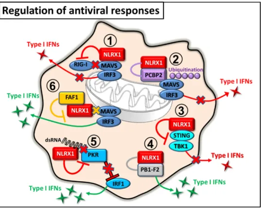
The previous results clearly indicate that the mode of NLRX1-mediated regulation in antiviral responses is cell type specific and mainly determined by the features of the invad- ing viruses, which can directly or indirectly modulate the function of NLRX1 (Figure2).
By negatively regulating antiviral immune responses, NLRX1 not only makes the host susceptible to viral infections but promotes virus-induced tumor development (e.g., Ka- posi’s sarcoma or primary effusion lymphoma) as well [27]. On the other hand, due to the potential detrimental and autoimmune promoting effects of sustained type I IFN levels, antiviral immune responses must be tightly controlled. Thus, it seems that NLRX1, at least partially, is responsible to keep this delicate balance under control.
2.2. Regulation of NF-κB Pathway
In 2011, studies from two different research groups were published in the same is- sue of Immunity stating that besides regulating RIG-I and STING signaling pathways, NLRX1 is also capable to modulate TLR signaling in vivo, and influence NF-κB-driven pro-inflammatory responses. Allen et al. studied the role of NLRX1 in the TLR signaling of mice and found that, upon lipopolysaccharide (LPS) stimulation, NLRX1−/−macrophages produced higher amounts of IFN-βand IL-6 compared to wild type macrophages. Intratra- cheal instillation with LPS significantly increased IL-6 levels and inflammation in the lung of NLRX1−/−mice compared to wild type animals. Furthermore, their results suggest that NLRX1 suppresses NF-kB activation by interacting and interfering with TRAF6, which regulates a diverse range of innate immune signaling pathways, including TLR4 [19].
Int. J. Mol. Sci.2021,22, 1316 6 of 35
Int. J. Mol. Sci. 2021, 22, x FOR PEER REVIEW 6 of 36
Figure 2. Regulation of antiviral responses by NLRX1. 1. NLRX1 inhibits RIG‐I binding to MAVS, resulting in decreased type I IFN response. 2. NLRX1 recruits PCBP2 to MAVS and thereby triggers its ubiquitination and degradation that leads to reduced type I IFN levels. 3. NLRX1 prevents TBK1‐mediated type I IFN secretion by binding to STING. 4. NLRX1 interacts with PB1‐
F2 and promotes type I IFN production. 5. NLRX1 competes with PKR for binding to double stranded RNA and prevents the PKR‐driven blockade of host’s protein synthesis, which promotes the upregulation of IRF1‐mediated early antiviral responses. 6. FAF1 interacts with NLRX1 and blocks the binding of NLRX1 to MAVS that positively effects the MAVS‐mediated type IFN responses.
2.2. Regulation of NF‐κB Pathway
In 2011, studies from two different research groups were published in the same issue of Immunity stating that besides regulating RIG‐I and STING signaling pathways, NLRX1 is also capable to modulate TLR signaling in vivo, and influence NF‐κB‐driven pro‐
inflammatory responses. Allen et al. studied the role of NLRX1 in the TLR signaling of mice and found that, upon lipopolysaccharide (LPS) stimulation, NLRX1−/− macrophages produced higher amounts of IFN‐β and IL‐6 compared to wild type macrophages.
Intratracheal instillation with LPS significantly increased IL‐6 levels and inflammation in the lung of NLRX1−/− mice compared to wild type animals. Furthermore, their results suggest that NLRX1 suppresses NF‐kB activation by interacting and interfering with TRAF6, which regulates a diverse range of innate immune signaling pathways, including TLR4 [19].
In line with these findings, Xia et al. demonstrated that NLRX1 interacts with TRAF6 in various unstimulated cells types including mouse embryonic fibroblasts (MEF), THP‐
1, RAW264.7 and 293T cells. However, upon TLR4 stimulation, both NLRX1 and TRAF6 rapidly undergo K63‐linked polyubiquitination that results in dissociation of NLRX1 from TRAF6. Once NLRX1 is free, it targets the IKK complex via its LRR domain and impairs IKKα/β phosphorylation and kinase activity for IκBα phosphorylation, resulting in attenuated NF‐κB activation and pro‐inflammatory cytokine production. Furthermore, higher levels of IL‐6 were detected in NLRX1 deficient mice, which were more susceptible to LPS induced septic shock compared to wild type animals [28].
Figure 2.Regulation of antiviral responses by NLRX1. 1. NLRX1 inhibits RIG-I binding to MAVS, resulting in decreased type I IFN response. 2. NLRX1 recruits PCBP2 to MAVS and thereby triggers its ubiquitination and degradation that leads to reduced type I IFN levels. 3. NLRX1 prevents TBK1-mediated type I IFN secretion by binding to STING. 4. NLRX1 interacts with PB1-F2 and promotes type I IFN production. 5. NLRX1 competes with PKR for binding to double stranded RNA and prevents the PKR-driven blockade of host’s protein synthesis, which promotes the upregulation of IRF1-mediated early antiviral responses. 6. FAF1 interacts with NLRX1 and blocks the binding of NLRX1 to MAVS that positively effects the MAVS-mediated type IFN responses.
In line with these findings, Xia et al. demonstrated that NLRX1 interacts with TRAF6 in various unstimulated cells types including mouse embryonic fibroblasts (MEF), THP-1, RAW264.7 and 293T cells. However, upon TLR4 stimulation, both NLRX1 and TRAF6 rapidly undergo K63-linked polyubiquitination that results in dissociation of NLRX1 from TRAF6. Once NLRX1 is free, it targets the IKK complex via its LRR domain and impairs IKKα/βphosphorylation and kinase activity for IκBαphosphorylation, resulting in attenuated NF-κB activation and pro-inflammatory cytokine production. Furthermore, higher levels of IL-6 were detected in NLRX1 deficient mice, which were more susceptible to LPS induced septic shock compared to wild type animals [28].
Another possible mechanism for hindering pro-inflammatory responses by NLRX1 is the sequestration of pro-inflammatory molecules. In an in vitro model of mitochondrial injury pro-inflammatory mediators, including MAVS, p-TBK1, p-IKK, IκB, and TRAF6 were recruited to the mitochondria, where they interacted with NLRX1. The lack of NLRX1 resulted in increased NF-κB and TBK1 mediated immune response and apoptosis in rat pulmonary microvascular endothelial cells. These data suggest that NLRX1 may have a role in the maintenance of cellular homeostasis after acute injury by sequestering pro- inflammatory molecules [29]. In addition to negatively regulating NF-κB signaling, NLRX1 also serves as a tumor suppressor [30,31].
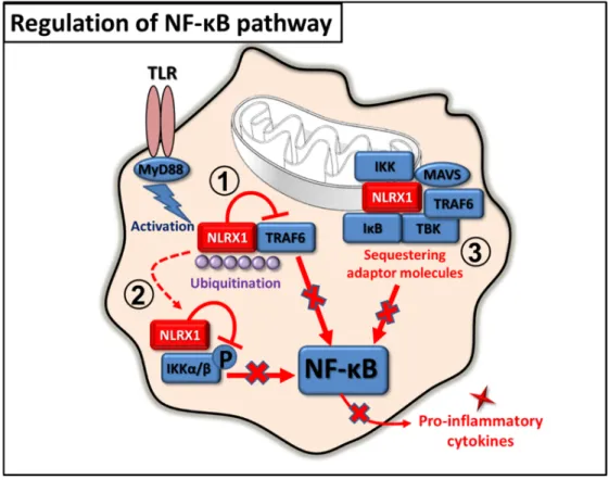
The above findings demonstrate that during pro-inflammatory immune responses the main function of NLRX1 is to prevent overzealous inflammatory responses, which can promote unwanted tissue damage in the host (Figure3).
Int. J. Mol. Sci.2021,22, 1316 7 of 35
Another possible mechanism for hindering pro‐inflammatory responses by NLRX1 is the sequestration of pro‐inflammatory molecules. In an in vitro model of mitochondrial injury pro‐inflammatory mediators, including MAVS, p‐TBK1, p‐IKK, IκB, and TRAF6 were recruited to the mitochondria, where they interacted with NLRX1. The lack of NLRX1 resulted in increased NF‐κB and TBK1 mediated immune response and apoptosis in rat pulmonary microvascular endothelial cells. These data suggest that NLRX1 may have a role in the maintenance of cellular homeostasis after acute injury by sequestering pro‐inflammatory molecules [29]. In addition to negatively regulating NF‐κB signaling, NLRX1 also serves as a tumor suppressor [30,31].
The above findings demonstrate that during pro‐inflammatory immune responses the main function of NLRX1 is to prevent overzealous inflammatory responses, which can promote unwanted tissue damage in the host (Figure 3).
Figure 3. Regulation of NF‐κB pathway by NLRX1. 1. In inactivated cells, NLRX1 interacts with TRAF6 and inhibits the NF‐κB pathway. 2. In activated cells, NLRX1 dissociates from TRAF6 via polyubiquitination and binds to IKK complex to prevent NF‐κB‐mediated pro‐inflammatory cytokine production. 3. Upon acute injury, activated MAVS, TBK1, IKK, IκB, and TRAF6 are recruited to the mitochondrion, where they interact with NLRX1, which blocks NF‐κB activity.
2.3. Regulation of Autophagy
Autophagy strongly associates with antiviral immunity since it can shape antiviral immune responses by exerting either antiviral or proviral effects depending on the type of the host cell and the invading virus. In 2012, Lei et al. identified a mitochondrial NLRX1 interacting protein, the Tu translation elongation factor (TUFM), which can also engage RIG‐I and the autophagy‐related protein (ATG) 5‐ATG12 conjugate and ATG16L1. It was described that NLRX1 and TUFM inhibits RIG‐I‐induced type IFN signaling in MEFs.
Moreover, NLRX1 and TUFM were found to be essential for vesicular stomatitis virus (VSV)‐induced autophagy in MEFs and mouse peritoneal macrophages. These findings indicate that NLRX1 and TUFM oppositely regulate type I IFN production and autophagy: their interaction is required to ensure optimal levels of autophagy and, at the
Figure 3.Regulation of NF-κB pathway by NLRX1. 1. In inactivated cells, NLRX1 interacts with TRAF6 and inhibits the NF-κB pathway. 2. In activated cells, NLRX1 dissociates from TRAF6 via polyubiquitination and binds to IKK complex to prevent NF-κB-mediated pro-inflammatory cytokine production. 3. Upon acute injury, activated MAVS, TBK1, IKK, IκB, and TRAF6 are recruited to the mitochondrion, where they interact with NLRX1, which blocks NF-κB activity.
2.3. Regulation of Autophagy
Autophagy strongly associates with antiviral immunity since it can shape antiviral immune responses by exerting either antiviral or proviral effects depending on the type of the host cell and the invading virus. In 2012, Lei et al. identified a mitochondrial NLRX1 interacting protein, the Tu translation elongation factor (TUFM), which can also engage RIG-I and the autophagy-related protein (ATG) 5-ATG12 conjugate and ATG16L1. It was described that NLRX1 and TUFM inhibits RIG-I-induced type IFN signaling in MEFs.
Moreover, NLRX1 and TUFM were found to be essential for vesicular stomatitis virus (VSV)-induced autophagy in MEFs and mouse peritoneal macrophages. These findings indicate that NLRX1 and TUFM oppositely regulate type I IFN production and autophagy:
their interaction is required to ensure optimal levels of autophagy and, at the same time, to keep IFN production in balance during viral infection [32]. The same research group described that the mitochondrial NLRX1-TUFM complex not only has a role during viral infection but also promotes autophagy and survival of tumor cells [33]. Furthermore, the oncogenic human papillomavirus 16 (HPV16) also exploits the autophagy inducing capacity of NLRX1 via its E7 oncoprotein, which can interact with the mitochondria-located NLRX1 [34].
In contrast to viral infection, it was also observed that NLRX1 negatively regulates autophagy upon bacterial infections caused byGroup A Streptococcus (GAS). After GAS invasion, the bacteria escape from the endosome to the cytosol, where they are rapidly degraded by autophagy. However, it was shown that NLRX1 interacts with the Beclin 1-UV radiation resistance-associated gene protein (UVRAG) complex via its NACHT domain to negatively regulate GAS-induced autophagy in HeLa cells [35].
Apart from canonical autophagy, NLRX1 also promotes the non-canonical form of au- tophagy, termed microtubule-associated protein 1 light chain 3 (LC3)-associated phagocyto-
Int. J. Mol. Sci.2021,22, 1316 8 of 35
sis (LAP) upon fungal infection. InHistoplasma capsulatum-activated macrophages, NLRX1 forms a complex with TUFM-ATG5-ATG12 and facilitates the incorporation of LC3-II into the phagosomal membrane that ensures its maturation. In addition, the NLRX1-TUFM interaction can also enhances the pro-inflammatory cytokine production of macrophages by the activation of MAPK signaling upon fungal stimulation [36].
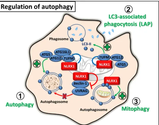
Furthermore, NLRX1 is essential for mitophagy, a selective form of autophagy, which ensures the clearance of damaged or dysfunctional mitochondria. NLRX1 contains an LC3-interacting region (LIR), through which it is able to recruit autophagosomes to the mitochondria, thus NLRX1 was described as a novel mitophagy receptor. Intracellular pathogens, for example Listeria monocytogenescan activate mitophagy via NLRX1 and promote their survival by reducing mitochondrial reactive oxygen species (mtROS) pro- duction. In bone marrow-derived macrophages (BMDM),Listeriainduces mitophagy via its listeriolysin O (LLO) component, which frees the NACHT domain of NLRX1 from the LRR-maintained autoinhibited state and allows oligomerization and subsequent binding to LC3-decorated autophagosomes [37]. Besides pathogen-induced mitophagy, NLRX1 also regulates mitophagy in mammary tumors, since NLRX1 upregulation in aggressive metastatic breast cancer cell lines is associated with higher metastatic potential. Higher level of NLRX1 expression is required for maintaining increased levels of TNF-α-induced autophagy, which ensures mitochondrial homeostasis by eliminating dysfunctional mito- chondria. In line with this, NLRX1 deficiency impairs mitochondrial metabolic function and the lysosomal turnover of mitochondria, which in turn decreases the migration and pro- liferation of aggressive triple-negative breast cancer cells [38]. Overall, these data implicate that targeting NLRX1 could offer promising strategies in the therapy of autophagy-related diseases (Figure4).
Int. J. Mol. Sci. 2021, 22, x FOR PEER REVIEW 9 of 36
Figure 4. Regulation of autophagy by NLRX1. 1. In virus‐infected or tumour cells, NLRX1 interacts with TUFM and promotes autophagy or, conversely, inhibits Beclin‐1‐UVRAG complex to reduce autophagosome formation upon bacterial infection. 2. Upon fungal infection, NLRX1‐
TUFM complex formation aids LC3‐associated phagocytosis by facilitating the incorporation of LC3‐II to the phagosomal membrane. 3. Via interaction with LC3, NLRX1 recruits
autophagosomes to the mitochondria and promotes mitophagy.
2.4. Regulation of ROS Production and Cell Death
The regulatory actions of NLRX1 are mainly associated with mitochondria. In HEK293T cells, it was observed that NLRX1 can be also localized within the mitochondrion, at the matrix side of the mitochondrial inner membrane, where it can interact with UQCRC2, a member of complex III of mitochondrial electron transport chain (ETC). Through interaction with UQCRC2, which is essential for mtROS production, NLRX1 is also capable to control mtROS‐dependent cellular functions including ROS sensitive pro‐inflammatory signaling pathways or cell death [13]. ROS have either protective or detrimental effects upon infections, and the ability of NLRX1 to modulate mtROS production can also be hijacked by many invading pathogens [39,40].
In airway epithelial cells, it was shown that NLRX1 is expressed in both the cytoplasm and at the apical surface; however, after infection it translocates to the mitochondria. Upon sensing rhinovirus RNA or synthetic polyinosinic:polycytidylic acid [poly (I:C)], the mitochondria‐associated NLRX1 promotes mtROS production, which is required for virus‐induced epithelial barrier disruption [39]. Some intracellular bacteria take advantage of the ROS producing capacity of the host cell as well. For instance, Chlamydia trachomatis utilizes ROS to activate caspase‐1 in epithelial cells [40]. Whereas in macrophages caspase‐1 initiates the secretion of high amounts of IL‐1β, in epithelial cells, which produce only negligible amounts of IL‐1β, it rather enhances lipid metabolism [41].
Chlamydia requires host‐derived lipids for intracellular growth since it replicates within a specialized membrane‐bound compartment termed the inclusion [42]. Thus, the increased lipid metabolism is a requirement for optimal growth and stability of the inclusion membrane as well as for chlamydial replication. It was found that, at first, Chlamydia
Figure 4. Regulation of autophagy by NLRX1. 1. In virus-infected or tumour cells, NLRX1 interacts with TUFM and promotes autophagy or, conversely, inhibits Beclin-1-UVRAG complex to reduce autophagosome formation upon bacterial infection. 2. Upon fungal infection, NLRX1-TUFM complex formation aids LC3-associated phagocytosis by facilitating the incorporation of LC3-II to the phagosomal membrane. 3. Via interaction with LC3, NLRX1 recruits autophagosomes to the mitochondria and promotes mitophagy.
2.4. Regulation of ROS Production and Cell Death
The regulatory actions of NLRX1 are mainly associated with mitochondria. In HEK293T cells, it was observed that NLRX1 can be also localized within the mitochondrion, at the matrix side of the mitochondrial inner membrane, where it can interact with UQCRC2, a member of complex III of mitochondrial electron transport chain (ETC). Through interac- tion with UQCRC2, which is essential for mtROS production, NLRX1 is also capable to control mtROS-dependent cellular functions including ROS sensitive pro-inflammatory signaling pathways or cell death [13]. ROS have either protective or detrimental effects upon infections, and the ability of NLRX1 to modulate mtROS production can also be hijacked by many invading pathogens [39,40].
In airway epithelial cells, it was shown that NLRX1 is expressed in both the cytoplasm and at the apical surface; however, after infection it translocates to the mitochondria.
Upon sensing rhinovirus RNA or synthetic polyinosinic:polycytidylic acid [poly (I:C)], the mitochondria-associated NLRX1 promotes mtROS production, which is required for virus- induced epithelial barrier disruption [39]. Some intracellular bacteria take advantage of the ROS producing capacity of the host cell as well. For instance,Chlamydia trachomatisutilizes ROS to activate caspase-1 in epithelial cells [40]. Whereas in macrophages caspase-1 initiates the secretion of high amounts of IL-1β, in epithelial cells, which produce only negligible amounts of IL-1β, it rather enhances lipid metabolism [41]. Chlamydia requires host- derived lipids for intracellular growth since it replicates within a specialized membrane- bound compartment termed the inclusion [42]. Thus, the increased lipid metabolism is a requirement for optimal growth and stability of the inclusion membrane as well as for chlamydial replication. It was found that, at first,Chlamydiainitiates ROS production by activating NADPH oxidases, then it also exploits the mtROS inducing capacity of NLRX1 in HeLa cells. Consequently, the increased levels of intracellular ROS stimulate the NLRP3 dependent caspase-1 activation, which ensures the survival of the pathogen [40].
Upon infections, cells often use the inflammation promoting features of ROS that can facilitate immune responses against pathogens. In HeLa cells, overexpression of NLRX1 increased the TNF-α,Shigella flexneri and poly (I:C)-induced ROS production, which potentiated the NF-κB and c-Jun N-terminal kinase (JNK) pathways. The authors speculate that the positive regulatory effect of NLRX1 on ROS production was independent of its N-terminal domain, but rather the NACHT or LRR domains of the receptor might be involved in ROS generation [43].
The regulatory role of ROS can be also observed in the context of cell death. In a model of cisplatin-induced ototoxicity, it was reported that NLRX1 promotes cisplatin-induced mitochondrial apoptosis by potentiating ROS/JNK signaling upon stimulating the auditory cells with cisplatin [44]. In addition, cisplatin insult upregulated NLRX1 expression and autophagy, which consequently triggered cell death in auditory cells [45]. Furthermore, it was also reported that NLRX1 can sensitize HEK293 cells to TNF-α-induced apoptosis through interaction with the mitochondria-localized caspase-8 and induction of mtROS production. This observation reflects the tumor suppressor feature of NLRX1 [46].
In contrast, depending on the cell type, NLRX1 can also play a protective role and can prevent cell death. It has been demonstrated that NLRX1 functions as a negative regulator of LPS-induced NF-κB signaling, and hinders inflammation and apoptosis in chondro- cytes [47]. In addition, another study revealed that NLRX1 protects tubular epithelial cells from mitochondrial injury and apoptosis by decreasing oxidative stress in a mouse model of renal ischemia-reperfusion injury [48].
Interestingly, regulation of cell death by NLRX1 is not always dependent on ROS production. It was reported that upon rotenone treatment, overexpression of NLRX1 increased the viability of mouse neuroblastoma cells in an ROS independent manner. The authors observed that NLRX1 interacts with GTPase dynamin-related protein 1 (DRP1), which has an essential role in regulating mitochondrial dynamics to promote mitochondrial fission and thus rescues cells from necrosis. In this situation, NLRX1 seems to support apoptosis rather than necrosis [49]. In addition, in non-neuronal cells, NLRX1 has another
Int. J. Mol. Sci.2021,22, 1316 10 of 35
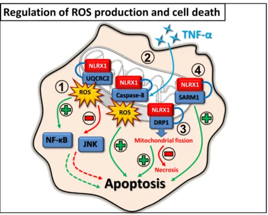
interacting partner, the sterile alpha and toll/interleukin-1 receptor (TIR) motif- containing protein 1 (SARM1), which plays a fundamental role in cell homeostasis and regulation of cell death. Via the interaction with SARM1, NLRX1 can influence the SARM1 dependent apoptosis in HEK293T [50] (Figure5).
Int. J. Mol. Sci. 2021, 22, x FOR PEER REVIEW 11 of 36
Figure 5. Regulation of ROS production and cell death by NLRX1. 1. NLRX1 interacts with UQCRC2 and can either induce or inhibit mtROS production in a cell type dependent manner.
Through regulating mtROS production, NLRX1 might have either a positive or negative effect on NF‐κB and JNK inflammatory pathways and on the induction of apoptosis. 2. NLRX1 positively regulates the TNF‐α‐induced caspase‐8 dependent apoptosis via the interaction with the mitochondrial localized caspase‐8. 3. NLRX1 binds to DRP1, and thus promotes mitochondrial fission, inhibits necrosis while supporting apoptosis. 4. NLRX1 interacts with SARM1 and promotes SARM1‐dependent apoptosis.
2.5. Regulation of Metabolism
Metabolic reprogramming of cells, and targeting the main metabolic pathways can be a promising therapeutic tool for a wide variety of diseases. NLRX1 is considered to be an emerging metabolic regulator that can control cancer cell metabolism and has the potential to alleviate the symptoms of autoimmune diseases as well.
In the literature, there are conflicting data regarding the regulatory role of NLRX1 in cell metabolism. The observed discrepancies point to the highly cell type specific nature of NLRX1 actions. Due to its mitochondrial localization and its interaction with the ETC components such as UQCRC2, a member of complex III [13], NLRX1 can be a potential regulator of oxidative phosphorylation (OXPHOS). So far, it seems that NLRX1 can promote OXPHOS in immune cells [51,52], while supports aerobic glycolysis and attenuates OXPHOS in cancer or non‐immune cells [46,53–55]. Cancer cells are characterized by high glycolytic activity and it was observed that the inhibition of glycolysis is associated with decreased NLRX1 expression [53]. Furthermore, NLRX1 impaired the activity of mitochondrial ETC components and supported aerobic glycolysis in tumour cells [46]. In addition, Sing et al. described that NLRX1 is also present in mitochondrial RNA granules, where the post‐transcriptional processing of de novo synthetized mitochondrial RNA and ribosome biogenesis occur under the control of an RNA‐binding protein, the Fas‐activated serine‐threonine kinase family protein‐5 (FASTKD5). NLRX1 can bind to FASTKD5 and mitochondrial RNA via its LRR domain, and inhibit the maturation of precursor transcripts for complexes I and IV. In the presence of NLRX1, the assembly and activity of respiratory chain complexes are attenuated resulting in decreased OXPHOS activity [54]. Moreover, NLRX1 attenuates fatty acid‐
dependent OXPHOS and enhances glycolysis in hepatocytes [55].
Figure 5.Regulation of ROS production and cell death by NLRX1. 1. NLRX1 interacts with UQCRC2 and can either induce or inhibit mtROS production in a cell type dependent manner. Through regulating mtROS production, NLRX1 might have either a positive or negative effect on NF-κB and JNK inflammatory pathways and on the induction of apoptosis. 2. NLRX1 positively regulates the TNF-α-induced caspase-8 dependent apoptosis via the interaction with the mitochondrial localized caspase-8. 3. NLRX1 binds to DRP1, and thus promotes mitochondrial fission, inhibits necrosis while supporting apoptosis. 4. NLRX1 interacts with SARM1 and promotes SARM1-dependent apoptosis.
2.5. Regulation of Metabolism
Metabolic reprogramming of cells, and targeting the main metabolic pathways can be a promising therapeutic tool for a wide variety of diseases. NLRX1 is considered to be an emerging metabolic regulator that can control cancer cell metabolism and has the potential to alleviate the symptoms of autoimmune diseases as well.
In the literature, there are conflicting data regarding the regulatory role of NLRX1 in cell metabolism. The observed discrepancies point to the highly cell type specific nature of NLRX1 actions. Due to its mitochondrial localization and its interaction with the ETC components such as UQCRC2, a member of complex III [13], NLRX1 can be a potential regulator of oxidative phosphorylation (OXPHOS). So far, it seems that NLRX1 can promote OXPHOS in immune cells [51,52], while supports aerobic glycolysis and attenuates OXPHOS in cancer or non-immune cells [46,53–55]. Cancer cells are characterized by high glycolytic activity and it was observed that the inhibition of glycolysis is associated with decreased NLRX1 expression [53]. Furthermore, NLRX1 impaired the activity of mitochondrial ETC components and supported aerobic glycolysis in tumour cells [46].
In addition, Sing et al. described that NLRX1 is also present in mitochondrial RNA granules, where the post-transcriptional processing of de novo synthetized mitochondrial RNA and ribosome biogenesis occur under the control of an RNA-binding protein, the
Fas-activated serine-threonine kinase family protein-5 (FASTKD5). NLRX1 can bind to FASTKD5 and mitochondrial RNA via its LRR domain, and inhibit the maturation of precursor transcripts for complexes I and IV. In the presence of NLRX1, the assembly and activity of respiratory chain complexes are attenuated resulting in decreased OXPHOS activity [54]. Moreover, NLRX1 attenuates fatty acid-dependent OXPHOS and enhances glycolysis in hepatocytes [55].
Contrary to the above findings, NLRX1 rather promotes OXPHOS in immune cell- related diseases. In inflammatory bowel disease (IBD) models, NLRX1 deficiency led to a metabolic switch towards aerobic glycolysis, which resulted in enhanced inflammation.
NLRX1 deficient CD4+ T cells differentiated more likely into helper T (Th) 17 cells than wild type cells, and displayed enhanced proliferation rates. This was the consequence of enhanced aerobic glycolysis caused by increased lactate dehydrogenase (LDH) activity and incomplete fatty acid oxidation due to the lack of NLRX1. Interestingly, the expression of the hypoxia-inducible factor 1-alpha (HIF-1α) transcription factor, which controls LDH activity and Th17 differentiation is also increased in the absence of NLRX1 [51]. One of the promising NLRX1 agonists in IBD is NX-13, a gut-restricted selective NLRX1 activator, which targets metabolic regulation through NLRX1. In IBD models, attenuated LDH activity, glucose uptake, NF-kB activity and ROS production were observed in the presence of NX-13, and cells were rather characterized by enhanced OXPHOS [52].
NLRX1 also has a potential to regulate the glutamine metabolism of the gut. It is a well- known fact that perturbations of intestinal glutamine level have far-reaching consequences, including IBD. NLRX1 keeps several processes in balance, such as the proliferation, tight junction function, glutamine metabolism, pro-inflammatory cytokine production of ep- ithelial cells in a sirtuin 1 (SIRT1)-dependent manner, and might prevent dysbiosis and subsequent inflammation as well. Using intestinal organoids, it was demonstrated that NLRX1 deficient epithelial cells have significantly increased glutamate dehydrogenase activity that leads to augmented proliferation, decreased tight junction function and lower expression of SIRT1. The results indicate that NLRX1 deficient mice use more glutamine, which shapes the bacterial flora of the gut in a way that is more beneficial for colitogenic bacteria [56].
Furthermore, NLRX1 controls the amino acid level of the central nervous system (CNS) as well. In the CNS, extracellular neurotransmitter and glutamate levels must be carefully balanced to avoid excitotoxicity and neuronal death. Astrocytes act as a buffer by regulating extracellular glutamate levels in CNS, however; this function might be compromised upon inflammation. Mahmoud et al. demonstrated that NLRX1 is required for both glutamate uptake and inhibition of excess glutamate release by astrocytes. NLRX1 promotes ATP production to cover the energy needs of glutamate transporters, and inhibits Ca2+release from the endoplasmic reticulum to suppress glutamate exocytosis [57] (Figure6).
Int. J. Mol. Sci.2021,22, 1316 12 of 35
Int. J. Mol. Sci. 2021, 22, x FOR PEER REVIEW 13 of 36
Figure 6. Regulation of metabolism by NLRX1. 1. In immune cells, NLRX1 blocks aerobic
glycolysis through inhibiting the activity of HIF‐1α and LDH, and glucose uptake, while inducing ETC gene expression to promote OXPHOS. 2. In non‐immune cells or cancer cells, NLRX1 impairs the production of ETC components via interaction with FASTKD5, and subsequently attenuates OXPHOS, while supporting aerobic glycolysis. 3. NLRX1 negatively regulates glutamine metabolism in a SIRT1‐dependent manner.
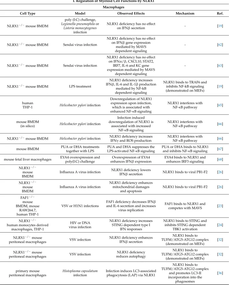
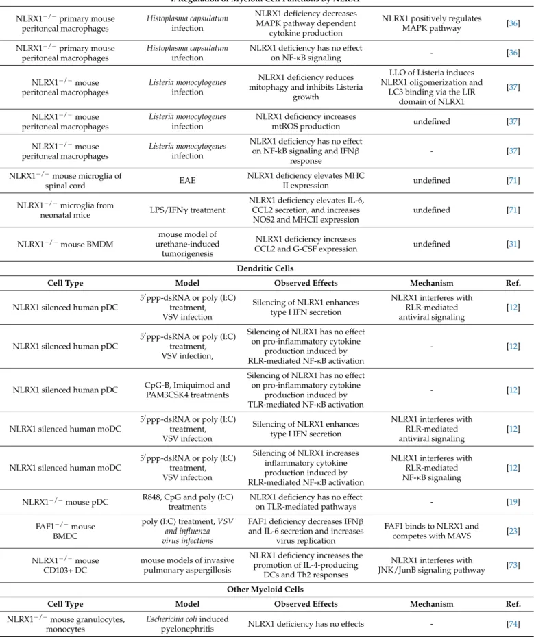
3. Immune Cell Related Actions of NLRX1 3.1. Regulation of Myeloid Cell Functions by NLRX1 3.1.1. Macrophages
Macrophages represent a highly plastic and functionally heterogeneous group of cells, which play a role in the initiation as well as in the resolution of inflammation, thus are key cellular mediators of immune responses [58,59]. The local tissue microenvironment has been reported to greatly influence the polarization of macrophages [60,61]. Interestingly, the role of NLRX1 have been examined only in the classical inflammatory M1 type of macrophages, whereas no data are available regarding the anti‐
inflammatory M2 type of macrophages that could help to understand the complexity of NLRX1 driven regulation in plastic cell populations.
As key cells in the immune response to infectious agents, macrophages act mainly against bacteria and fungi, but are also implicated in antiviral responses as well. In most of the studied cell types, NLRX1 functions as a negative regulator of RLR‐mediated type I IFN signaling by inhibiting interaction between RIG‐I and MAVS. However, interestingly, three independent research groups using differentially generated NLRX1‐
KO mice observed that NLRX1 is not involved in regulation of MAVS‐dependent type I IFN responses in macrophages. Allen et al. published that in C57BL/6 NLRX1‐deficient mice with the mixed genetic background of IC1 C57BL/6 and Balb/c, poly (I:C) triggered IFNβ production of mouse BMDMs was not affected by NLRX1 deficiency. Furthermore, no difference was observed between the BMDMs from NLRX1‐KO and wild type mice upon infection with Legionella pneumophila or Listeria monocytogenes bacteria, which are strong inducers of IFNβ secretion in macrophages [19]. Importantly, these results are in line with the observations of two other research groups. Rebsamen et al. also generated NLRX1‐deficient mice from a C57BL/6‐129/SvJ mixed background and observed normal MAVS‐dependent antiviral responses in NLRX1‐deficient BMDMs infected with Sendai Figure 6.Regulation of metabolism by NLRX1. 1. In immune cells, NLRX1 blocks aerobic glycolysis through inhibiting the activity of HIF-1αand LDH, and glucose uptake, while inducing ETC gene expression to promote OXPHOS. 2. In non-immune cells or cancer cells, NLRX1 impairs the pro- duction of ETC components via interaction with FASTKD5, and subsequently attenuates OXPHOS, while supporting aerobic glycolysis. 3. NLRX1 negatively regulates glutamine metabolism in a SIRT1-dependent manner.
3. Immune Cell Related Actions of NLRX1 3.1. Regulation of Myeloid Cell Functions by NLRX1 3.1.1. Macrophages
Macrophages represent a highly plastic and functionally heterogeneous group of cells, which play a role in the initiation as well as in the resolution of inflammation, thus are key cellular mediators of immune responses [58,59]. The local tissue microenvironment has been reported to greatly influence the polarization of macrophages [60,61]. Interestingly, the role of NLRX1 have been examined only in the classical inflammatory M1 type of macrophages, whereas no data are available regarding the anti-inflammatory M2 type of macrophages that could help to understand the complexity of NLRX1 driven regulation in plastic cell populations.
As key cells in the immune response to infectious agents, macrophages act mainly against bacteria and fungi, but are also implicated in antiviral responses as well. In most of the studied cell types, NLRX1 functions as a negative regulator of RLR-mediated type I IFN signaling by inhibiting interaction between RIG-I and MAVS. However, interestingly, three independent research groups using differentially generated NLRX1-KO mice observed that NLRX1 is not involved in regulation of MAVS-dependent type I IFN responses in macrophages. Allen et al. published that in C57BL/6 NLRX1-deficient mice with the mixed genetic background of IC1 C57BL/6 and Balb/c, poly (I:C) triggered IFNβproduction of mouse BMDMs was not affected by NLRX1 deficiency. Furthermore, no difference was observed between the BMDMs from NLRX1-KO and wild type mice upon infection with Legionella pneumophilaorListeria monocytogenesbacteria, which are strong inducers of IFNβ secretion in macrophages [19]. Importantly, these results are in line with the observations of two other research groups. Rebsamen et al. also generated NLRX1-deficient mice from a C57BL/6-129/SvJ mixed background and observed normal MAVS-dependent antiviral responses in NLRX1-deficient BMDMs infected with Sendai virus [62]. Similar data were
published by Soares et al. using C57BL/6 NLRX1-deficient mice generated from the crossing of floxed and Cre mice. The expression level of antiviral (IFNα/β, CXCL10, STAT2, IRF7) and inflammatory (IL-6, KC) genes did not differ between NLRX1-KO and wild type BMDMs following infection with Sendai virus [63]. Based on the data the authors concluded that NLRX1 does not interfere with the MAVS-dependent antiviral signaling in macrophages.
On the contrary, it seems that NLRX1 could be a potent regulator of the MAVS- independent NF-κB signaling in macrophages. Stimulation with LPS, which activates TLR4 in a MAVS-independent manner [64], resulted in significantly elevated IFNβ, IL-6 and IL-1βsecretion by NLRX1-KO BMDMs compared to wild type cells. Mechanistically it was shown that NLRX1 directly interacted with TRAF6 and inhibited TLR4/MyD88-mediated NF-κB signaling in macrophages [19].
Helicobacter pylori(H. pylori) is a pathogenic Gram-negative bacterium, which can induce chronic infection of the human gastrointestinal tract that can eventually lead to the development of ulcer or gastric cancer (GC). In a study, involving Chinese individuals, NLRX1 was one of the 51 identified gene polymorphisms, which are associated with the increased risk of GC development during aH. pyloriinfection. PCR array analysis of NLR signaling-associated and inflammasome-related molecules revealed thatH. pyloriinfection significantly downregulated NLRX1 expression and induced NF-κB signaling in THP-1 human monocyte-like cells. Based on these results the authors concluded that perturbation of NF-κB signaling due to NLRX1 gene polymorphism and/or its decreased expression followingH. pyloriinfection makes individuals more susceptible to GC development [65].
In line with these results, significant and early downregulation of NLRX1 transcript was detected by another study using time course transcriptome analysis of BMDMs upon H. pylori infection. Using in silico modeling and simulation tools, the research group found that loss of NLRX1 leads to a prompt increase in NF-κB signaling resulting in higher levels of early cytokines but not of sustained or late cytokines. Experimental validation of computational modeling predictions demonstrated that NLRX1-KO BMDMs produced significantly more IFNγand ROS compared to the wild type cells after infection [66].
Interestingly, the endogenous or pathogen-derived ligands of NLRX1 highly determine the regulatory function of NLRX1 and can modify the outcome of immune responses. Using the C-terminal region of NLRX1 as a putative ligand-binding domain, diverse compounds from three libraries were screened in an in silico molecular docking approach to identify novel endogenous ligands for NLRX1. This virtual screening identified different lipids such as punicic acid (PUA) and docosahexaenoic acid (DHA) as potential ligands for NLRX1, then binding of the molecules were experimentally verified using surface plasmon resonance spectroscopy. In vitro treatment of BMDM with PUA and DHA suppressed the LPS-activated NF-κB signaling, and the inhibitory effect of both components was abrogated in the NLRX1-KO BMDM. The anti-inflammatory effect of PUA was mediated through NLRX1, which was further confirmed in a DSS-induced colitis model, where PUA alleviated symptoms of wild type animals, but did not reduce colitis symptoms of NLRX1-KO mice. The virtual screening of a lipid library for further putative NLRX1 ligands also identified polyketides, prenol lipids, and plant sterol lipids, which possess anti-microbial, antioxidant or low-density lipoprotein (LDL) lowering effects, respectively, as well as CoA-containing fatty acids, which are important contributors of mitochondrial energy metabolism. These results suggested for the first time that NLRX1 can also act as a cytosolic lipid sensor and may function at the crossroad of metabolism and immune responses [67].
In a study to identify molecules involved in the DNA-induced innate immune re- sponse, another NLRX1 interacting protein phosphatase, the Eyes absent (EYA) 4 was identified as a regulator of IFNβsecretion in MEFs. EYA4 is a protein phosphatase with dual function that depends on its cellular localization. In the nucleus, it functions as a co-transcription factor and modulates chromatin structure via its N-terminal tyrosine- phosphatase activity. However, Okabe et al. found that in the cytoplasm, it regulates
Int. J. Mol. Sci.2021,22, 1316 14 of 35
innate immune reactions in response to undigested DNA of apoptotic cells by modulating signal transduction pathways via its C-terminal region. Following transfection of HEK293T cells with hemagglutinin (HA)-tagged NLRX1 and flag tagged EYA4 direct protein-protein interaction were confirmed by co-immunoprecipitation. To delineate its function in an im- munocompetent cell, retroviral delivery of EYA4 to fetal liver macrophages was performed, that resulted in significantly enhanced IFNβexpression upon poly (I:C) treatment. Further- more, point mutations generated in the EYA4 significantly reduced the poly (I:C)-induced IRF3-mediated IFNβresponse in macrophages, while not affected the interaction of EYA4 with MAVS. The mechanism behind the observed phenomenon is not fully understood and remained to be determined to date [68].
Besides the endogenous ligands, some pathogens are also able to provide interacting ligands for NLRX1. For example, compared to wild type mice, significantly higher viral loads and inflammation were detected in the lungs of Nlrx1−/−mice following influenza A virus infection. This was accompanied with a delayed and severely impaired type I IFN response, while the number of the alveolar macrophages was not different from that of the wild type. Upon stimulation with the synthetic RIG-I agonists 50pppRNA or poly (I:C), there was no difference in the IFNβsecretion between BMDMs from NLRX1-KO and wild type mice. On the contrary, in NLRX1-KO macrophages significantly lower IFNβsecretion, enhanced mitochondrial damage and highly elevated apoptosis were detected following influenza A virus infection when compared to their wild type counterparts. It has been described previously that in the course of influenza A virus infection, viral PB1-F2 protein triggers the disruption of mitochondrial membrane potential that ultimately leads to the apoptosis of infected host cells. Jaworska et al. showed that NLRX1 directly binds PB1-F2 impairing mitochondria-mediated early apoptosis, thus NLRX1 indirectly allows type I IFN secretion by macrophages [26].
In another study, a member of the FAS death-inducing signaling complex, the FAF1 was identified as a positive regulator of IFNβsignaling via a direct interaction of NLRX1.
FAF1 deficient mice showed lower resistance to VSV infection and produced less IFNβ and IL-6 compared to the wild type mice. Similar results were obtained using FAF1-KO BMDMs following H1N1 influenza virus infection, while there was no difference in the cytokine production following HSV infection indicating that FAF1 positively regulates IFNβ response to RNA viruses, without affecting that to DNA viruses. Furthermore, downregulation of FAF1 in BMDMs, RAW264.7 macrophages and THP-1 cells resulted in reduced IFNβand IL-6 secretion. At the molecular level it was shown that the binding site for FAF1 overlaps with the binding site for MAVS on NLRX1 suggesting that FAF1 and MAVS compete for NLRX1 binding [23].
Upon HIV or DNA virus infection, NLRX1 was found to interact with the cytosolic DNA sensor STING to negatively regulate the TBK1-dependent type I IFN responses.
Consequently, due to the reduced STING-dependent TBK1 activation, increased virus replication was observed in NLRX1 deficient human monocyte-derived macrophages and THP-1 cells [24].
It was also shown that NLRX1 directly binds TUFM, which then binds to ATG5-ATG12 and ATG16L1 to promote autophagy in cells. Importantly, decreased autophagy was de- tected in NLRX1-KO peritoneal macrophages following VSV infection compared to wild type cells. Based on these data, it was suggested that NLRX1 is a negative regulator of antiviral immune responses, since NLRX1 deficiency reduced autophagy and enhanced IFNβsecretion and eventually led to a better control of VSV replication [32]. ATG5-ATG12 are important components not only of the canonical but also the non-canonical autophagy pathway as well. Several fungal infections were reported to induce the non-canonical LAP in macrophages to kill fungi and to control ongoing infection [69,70]. For example, LAP formation was described in macrophages infected withHistoplasma capsulatum,which is a pathogenic fungus that survives phagocytosis and replicates in macrophages by interfering lysosomal acidification of phagolysosomes. Following fungal infection of mouse peritoneal macrophages, it was found that NLRX1 bound to TUFM and induced ATG5-ATG12 com-
plex formation and promoted LC3-II incorporation into the fungus containing phagosomes.
Besides autophagy, NLRX1-TUFM complex positively affected cytokine production of macrophages, thus reduced MAPK pathway activity in NLRX1 deficient cells. Most impor- tantly, NF-κB signaling pathway was not modified in NLRX1-KO macrophages following Histoplasma capsulatuminfection [36].
During mitophagy, which is a modified form of autophagy, damaged or senescent mitochondria are eliminated by an autophagosome formed around the mitochondria. It was shown that the virulence factor (LLO) of intracellularListeria monocytogenesbacteria induced mitophagy in mouse peritoneal macrophages to promote bacterial survival. This mitophagic process was mediated by direct binding of LC3 to NLRX1 through its LIR motif indicating that NLRX1 can also function as a mitophagy receptor. NLRX1 deficiency of mouse peritoneal macrophages completely abrogated Listeria-induced degradation of mitochondrial proteins and DNA, and increased the number of damaged mitochondria and the production of mtROS. Furthermore, in NLRX1-KO mouse macrophages the Listeria titer was lower compared to the wild type macrophages, suggesting that Listeria hijacks host NLRX1 dependent mitophagy to support its own survival. As a mechanism the authors identified LLO triggered conformational changes in NLRX1 that resulted in the release of LRR domain from the NACHT domain. This change unleashed NLRX1 from its repressed state and allowed oligomerization and subsequent binding of LC3 through its LIR domain to induce mitophagy. Interestingly, basal and starvation-induced autophagy was not altered in NLRX1-KO mouse peritoneal macrophages, and neither NF-κB signaling nor IFNβresponse were influenced by the absence of NLRX1 [37].
Besides pathogen-induced immune responses, functions of NLRX1 in macrophages were also investigated in association with a variety of diseases and trauma. Multiple sclerosis (MS) is a T cell-mediated autoimmune disease characterized by the inflammation of the CNS. The inflammatory condition is mainly generated by resident brain macrophages (microglia) and infiltrating monocytes/macrophages that eventually leads to neuronal demyelination and axonal damage. In experimental autoimmune encephalomyelitis (EAE), which is a widely accepted murine model of MS, a higher expression of MHC class II molecule was detected from CD45low CD11b+ resident microglia cells of the spinal cord of NLRX1-KO mice compared to the wild type animals. Furthermore, following LPS/IFNγ treatment, elevated IL-6 and CCL2 secretion, and increased nitric oxide synthase (NOS) 2 and MHCII expression was detected in cultured microglia isolated from NLRX1-KO neonatal mice compared to wild type animals, indicating that NLRX1 attenuates microglia activation and potentially represses disease progression [71].
In a mouse model of urethane-induced tumorigenesis, NLRX1-KO mice developed histiocytic sarcoma in the spleen and increased number of macrophages were detected compared to wild type animals. In serum-free conditions, BMDMs from NLRX1-KO mice showed significantly increased proliferation and expression of CCL2 and granulocyte colony-stimulating factor (G-CSF), which are involved in macrophage recruitment and proliferation, respectively. In contrast to previous reports, the authors did not find differ- ences in cell death between KO and wild type BMDMs that might be due to the serum-free conditions suggesting stimulation specific mechanisms. Interestingly, they found that NLRX1 mediates cell death in neoplastic but not in normal cells and suggest that NLRX1 mediates tumor suppression through the induction of apoptosis [31] (Table1).
The above data indicate that numerous contradictory results were reported concerning the activity of NLRX1 in macrophages. Often, similar stimulations or infection models applied on macrophages with different origin resulted in diverse regulatory mechanisms.
Therefore, there might be differences in the regulatory action of NLRX1 between bone marrow derived and peritoneal macrophages since their functions are adapted to divergent tissue environment. Furthermore, the results also clearly show that stimulation of similar type of macrophages with different pathogenic microbes might lead to opposing results since fungi, intracellular and extracellular bacteria, RNA and DNA viruses can activate various signaling pathways depending on the sensing of their PAMPs by the host. Besides,